Achieve Mastery of Medical Concepts

Study for medical school and boards with Lecturio

Respiratory Alkalosis

The respiratory system is responsible for eliminating the volatile acid carbon dioxide (CO2), which is produced via aerobic metabolism. The body produces approximately 15,000 mmol of CO2 daily, which is the majority of daily acid production; the remainder of the daily acid load (only about 70 mmol of nonvolatile acids Nonvolatile acids Acid-Base Balance) is excreted through the kidneys Kidneys The kidneys are a pair of bean-shaped organs located retroperitoneally against the posterior wall of the abdomen on either side of the spine. As part of the urinary tract, the kidneys are responsible for blood filtration and excretion of water-soluble waste in the urine. Kidneys: Anatomy. When hyperventilation occurs, excess carbon dioxide is blown off and respiratory alkalosis develops. The kidneys Kidneys The kidneys are a pair of bean-shaped organs located retroperitoneally against the posterior wall of the abdomen on either side of the spine. As part of the urinary tract, the kidneys are responsible for blood filtration and excretion of water-soluble waste in the urine. Kidneys: Anatomy respond by decreasing serum bicarbonate Bicarbonate Inorganic salts that contain the -HCO3 radical. They are an important factor in determining the ph of the blood and the concentration of bicarbonate ions is regulated by the kidney. Levels in the blood are an index of the alkali reserve or buffering capacity. Electrolytes (HCO3) through increased HCO3 excretion or decreased excretion of H+. Patients Patients Individuals participating in the health care system for the purpose of receiving therapeutic, diagnostic, or preventive procedures. Clinician–Patient Relationship present with an increased respiratory rate Respiratory rate The number of times an organism breathes with the lungs (respiration) per unit time, usually per minute. Pulmonary Examination, dyspnea Dyspnea Dyspnea is the subjective sensation of breathing discomfort. Dyspnea is a normal manifestation of heavy physical or psychological exertion, but also may be caused by underlying conditions (both pulmonary and extrapulmonary). Dyspnea, light-headedness and potentially psychologic symptoms. Diagnosis involves a thorough history, an exam, and an arterial blood gas measurement. Management focuses on addressing the underlying abnormalities, stabilizing patients Patients Individuals participating in the health care system for the purpose of receiving therapeutic, diagnostic, or preventive procedures. Clinician–Patient Relationship in acute distress, and potentially a small dose of short-acting benzodiazepines Benzodiazepines Benzodiazepines work on the gamma-aminobutyric acid type A (GABAA) receptor to produce inhibitory effects on the CNS. Benzodiazepines do not mimic GABA, the main inhibitory neurotransmitter in humans, but instead potentiate GABA activity. Benzodiazepines.  

Last updated: Feb 17, 2023

Editorial responsibility: Stanley Oiseth, Lindsay Jones, Evelin Maza

Overview

Definition

Respiratory alkalosis refers to the process that results in a decreased level of carbon dioxide (CO2) within the blood.

Epidemiology

  • Gender Gender Gender Dysphoria bias Bias Epidemiological studies are designed to evaluate a hypothesized relationship between an exposure and an outcome; however, the existence and/or magnitude of these relationships may be erroneously affected by the design and execution of the study itself or by conscious or unconscious errors perpetrated by the investigators or the subjects. These systematic errors are called biases. Types of Biases: male = female
  • Incidence Incidence The number of new cases of a given disease during a given period in a specified population. It also is used for the rate at which new events occur in a defined population. It is differentiated from prevalence, which refers to all cases in the population at a given time. Measures of Disease Frequency: dependent on the etiology
  • Incidence Incidence The number of new cases of a given disease during a given period in a specified population. It also is used for the rate at which new events occur in a defined population. It is differentiated from prevalence, which refers to all cases in the population at a given time. Measures of Disease Frequency of hyperventilation syndrome: 25%–80% in adults with anxiety Anxiety Feelings or emotions of dread, apprehension, and impending disaster but not disabling as with anxiety disorders. Generalized Anxiety Disorder

Etiology

Table: Etiologies of respiratory alkalosis
Etiology Examples
Physiologic (not pathologic)
Hypoxia-induced
  • Pulmonary embolism Pulmonary Embolism Pulmonary embolism (PE) is a potentially fatal condition that occurs as a result of intraluminal obstruction of the main pulmonary artery or its branches. The causative factors include thrombi, air, amniotic fluid, and fat. In PE, gas exchange is impaired due to the decreased return of deoxygenated blood to the lungs. Pulmonary Embolism
  • Pulmonary edema Pulmonary edema Pulmonary edema is a condition caused by excess fluid within the lung parenchyma and alveoli as a consequence of a disease process. Based on etiology, pulmonary edema is classified as cardiogenic or noncardiogenic. Patients may present with progressive dyspnea, orthopnea, cough, or respiratory failure. Pulmonary Edema
  • COPD COPD Chronic obstructive pulmonary disease (COPD) is a lung disease characterized by progressive, largely irreversible airflow obstruction. The condition usually presents in middle-aged or elderly persons with a history of cigarette smoking. Signs and symptoms include prolonged expiration, wheezing, diminished breath sounds, progressive dyspnea, and chronic cough. Chronic Obstructive Pulmonary Disease (COPD) or asthma exacerbations Asthma Exacerbations Asthma in Children
Medications
  • Aspirin Aspirin The prototypical analgesic used in the treatment of mild to moderate pain. It has anti-inflammatory and antipyretic properties and acts as an inhibitor of cyclooxygenase which results in the inhibition of the biosynthesis of prostaglandins. Aspirin also inhibits platelet aggregation and is used in the prevention of arterial and venous thrombosis. Nonsteroidal Antiinflammatory Drugs (NSAIDs) overdose
  • Nicotine overdose Nicotine Overdose Stimulants
  • Progesterone Progesterone The major progestational steroid that is secreted primarily by the corpus luteum and the placenta. Progesterone acts on the uterus, the mammary glands and the brain. It is required in embryo implantation; pregnancy maintenance, and the development of mammary tissue for milk production. Progesterone, converted from pregnenolone, also serves as an intermediate in the biosynthesis of gonadal steroid hormones and adrenal corticosteroids. Gonadal Hormones
Intracranial processes
  • Stroke
  • Encephalitis Encephalitis Encephalitis is inflammation of the brain parenchyma caused by an infection, usually viral. Encephalitis may present with mild symptoms such as headache, fever, fatigue, and muscle and joint pain or with severe symptoms such as seizures, altered consciousness, and paralysis. Encephalitis
  • Traumatic brain injury Traumatic brain injury A form of acquired brain injury which occurs when a sudden trauma causes damage to the brain. Le Fort Fractures
Psychologic etiologies
Other processes
  • Pain Pain An unpleasant sensation induced by noxious stimuli which are detected by nerve endings of nociceptive neurons. Pain: Types and Pathways
  • Fever Fever Fever is defined as a measured body temperature of at least 38°C (100.4°F). Fever is caused by circulating endogenous and/or exogenous pyrogens that increase levels of prostaglandin E2 in the hypothalamus. Fever is commonly associated with chills, rigors, sweating, and flushing of the skin. Fever

Acid–Base Review

Acid–base disorders are classified according to the primary disturbance (respiratory or metabolic) and the presence or absence of compensation Compensation Respiratory Acidosis.

Identifying the primary disturbance

Look at the pH pH The quantitative measurement of the acidity or basicity of a solution. Acid-Base Balance, PCO2 ( partial pressure Partial pressure The pressure that would be exerted by one component of a mixture of gases if it were present alone in a container. Gas Exchange of CO2), and HCO3 ( bicarbonate Bicarbonate Inorganic salts that contain the -HCO3 radical. They are an important factor in determining the ph of the blood and the concentration of bicarbonate ions is regulated by the kidney. Levels in the blood are an index of the alkali reserve or buffering capacity. Electrolytes) to determine the primary disturbance. 

  • Normal values:
    • pH pH The quantitative measurement of the acidity or basicity of a solution. Acid-Base Balance: 7.35–7.45
    • PCO2:  35–45 mm MM Multiple myeloma (MM) is a malignant condition of plasma cells (activated B lymphocytes) primarily seen in the elderly. Monoclonal proliferation of plasma cells results in cytokine-driven osteoclastic activity and excessive secretion of IgG antibodies. Multiple Myeloma Hg
    • HCO3: 22–28 mEq/L
  • “-emia” versus “-osis”:
    • “-emia” refers to “in the blood”:
      • Acidemia Acidemia Respiratory Acidosis: more hydrogen ions (H+) in the blood = pH pH The quantitative measurement of the acidity or basicity of a solution. Acid-Base Balance < 7.35
      • Alkalemia: more hydroxide ions (OH) in the blood = pH pH The quantitative measurement of the acidity or basicity of a solution. Acid-Base Balance > 7.45 
    • “-osis” refers to a process:
      • Acidosis Acidosis A pathologic condition of acid accumulation or depletion of base in the body. The two main types are respiratory acidosis and metabolic acidosis, due to metabolic acid build up. Respiratory Acidosis and alkalosis refer the processes that cause acidemia Acidemia Respiratory Acidosis and alkalemia. 
      • pH pH The quantitative measurement of the acidity or basicity of a solution. Acid-Base Balance may be normal in acidosis Acidosis A pathologic condition of acid accumulation or depletion of base in the body. The two main types are respiratory acidosis and metabolic acidosis, due to metabolic acid build up. Respiratory Acidosis and alkalosis.
  • Primary (uncompensated) respiratory disorders:
    • Disorders caused by abnormalities in PCO2
    • Both the pH pH The quantitative measurement of the acidity or basicity of a solution. Acid-Base Balance and PCO2 are abnormal, in opposite directions.
    • Primary respiratory acidosis Acidosis A pathologic condition of acid accumulation or depletion of base in the body. The two main types are respiratory acidosis and metabolic acidosis, due to metabolic acid build up. Respiratory Acidosis pH pH The quantitative measurement of the acidity or basicity of a solution. Acid-Base Balance < 7.35 and PCO2 > 45 
    • Primary respiratory alkalosis: pH pH The quantitative measurement of the acidity or basicity of a solution. Acid-Base Balance > 7.45 and PCO2 < 35
  • Primary (uncompensated) metabolic disorders:
    • Disorders caused by abnormalities in HCO3 
    • Both the pH pH The quantitative measurement of the acidity or basicity of a solution. Acid-Base Balance and PCO2 are abnormal, in the same direction. 
    • Primary uncompensated metabolic acidosis Primary uncompensated metabolic acidosis Metabolic Acidosis:  
      • pH pH The quantitative measurement of the acidity or basicity of a solution. Acid-Base Balance < 7.35 and PCO2 < 40 
      • Think: “So the acidosis Acidosis A pathologic condition of acid accumulation or depletion of base in the body. The two main types are respiratory acidosis and metabolic acidosis, due to metabolic acid build up. Respiratory Acidosis is not due to ↑ CO2 … it must be due to ↓ serum HCO3 → metabolic acidosis Acidosis A pathologic condition of acid accumulation or depletion of base in the body. The two main types are respiratory acidosis and metabolic acidosis, due to metabolic acid build up. Respiratory Acidosis
      • Confirm by looking at HCO3: will be low (< 22 mEq/L)
    • Primary uncompensated metabolic alkalosis Primary uncompensated metabolic alkalosis Metabolic Alkalosis:
      • pH pH The quantitative measurement of the acidity or basicity of a solution. Acid-Base Balance > 7.45 and PCO2 > 40
      • Think: “So the alkalosis is not due to ↓ CO2… it must be due to ↑ serum HCO3 metabolic alkalosis Metabolic alkalosis The renal system is responsible for eliminating the daily load of non-volatile acids, which is approximately 70 millimoles per day. Metabolic alkalosis also occurs when there is an increased loss of acid, either renally or through the upper GI tract (e.g., vomiting), increased intake of HCO3-, or a reduced ability to secrete HCO3- when needed. Metabolic Alkalosis
      • Confirm by looking at HCO3: will be high (> 28 mEq/L)
  • Simple disorders:
    • The presence of one of the above disorders with appropriate compensation Compensation Respiratory Acidosis
    • Respiratory disorders are compensated by renal mechanisms.
    • Metabolic disorders are compensated by respiratory mechanisms
  • Mixed disorders: two primary disorders present

Compensation Compensation Respiratory Acidosis

When acidosis Acidosis A pathologic condition of acid accumulation or depletion of base in the body. The two main types are respiratory acidosis and metabolic acidosis, due to metabolic acid build up. Respiratory Acidosis or alkalosis develops, the body will try to compensate. Often, compensation Compensation Respiratory Acidosis will result in a normal pH pH The quantitative measurement of the acidity or basicity of a solution. Acid-Base Balance.

  • In primary respiratory acid–base disorders, the kidney may try to compensate in an attempt to normalize the pH pH The quantitative measurement of the acidity or basicity of a solution. Acid-Base Balance.
    • Kidneys Kidneys The kidneys are a pair of bean-shaped organs located retroperitoneally against the posterior wall of the abdomen on either side of the spine. As part of the urinary tract, the kidneys are responsible for blood filtration and excretion of water-soluble waste in the urine. Kidneys: Anatomy respond to respiratory acidosis Acidosis A pathologic condition of acid accumulation or depletion of base in the body. The two main types are respiratory acidosis and metabolic acidosis, due to metabolic acid build up. Respiratory Acidosis by increasing serum HCO3 through ↑ secretion Secretion Coagulation Studies of H+.
    • Kidneys Kidneys The kidneys are a pair of bean-shaped organs located retroperitoneally against the posterior wall of the abdomen on either side of the spine. As part of the urinary tract, the kidneys are responsible for blood filtration and excretion of water-soluble waste in the urine. Kidneys: Anatomy respond to respiratory alkalosis by decreasing serum HCO3 through:
      • Secretion Secretion Coagulation Studies of H+
      • Urinary excretion of HCO3 (normally bicarbonate Bicarbonate Inorganic salts that contain the -HCO3 radical. They are an important factor in determining the ph of the blood and the concentration of bicarbonate ions is regulated by the kidney. Levels in the blood are an index of the alkali reserve or buffering capacity. Electrolytes is 100% absorbed)
  • In primary metabolic acid–base disorders, the lungs Lungs Lungs are the main organs of the respiratory system. Lungs are paired viscera located in the thoracic cavity and are composed of spongy tissue. The primary function of the lungs is to oxygenate blood and eliminate CO2. Lungs: Anatomy may try to compensate in an attempt to normalize the pH pH The quantitative measurement of the acidity or basicity of a solution. Acid-Base Balance.
    • Lungs Lungs Lungs are the main organs of the respiratory system. Lungs are paired viscera located in the thoracic cavity and are composed of spongy tissue. The primary function of the lungs is to oxygenate blood and eliminate CO2. Lungs: Anatomy respond to metabolic acidosis Acidosis A pathologic condition of acid accumulation or depletion of base in the body. The two main types are respiratory acidosis and metabolic acidosis, due to metabolic acid build up. Respiratory Acidosis by ↑ ventilation Ventilation The total volume of gas inspired or expired per unit of time, usually measured in liters per minute. Ventilation: Mechanics of Breathing.
    • Lungs Lungs Lungs are the main organs of the respiratory system. Lungs are paired viscera located in the thoracic cavity and are composed of spongy tissue. The primary function of the lungs is to oxygenate blood and eliminate CO2. Lungs: Anatomy respond to metabolic alkalosis Metabolic alkalosis The renal system is responsible for eliminating the daily load of non-volatile acids, which is approximately 70 millimoles per day. Metabolic alkalosis also occurs when there is an increased loss of acid, either renally or through the upper GI tract (e.g., vomiting), increased intake of HCO3-, or a reduced ability to secrete HCO3- when needed. Metabolic Alkalosis by ↓ ventilation Ventilation The total volume of gas inspired or expired per unit of time, usually measured in liters per minute. Ventilation: Mechanics of Breathing.
  • Interpreting the serum HCO3:
    • Normal range: 22–28 mEq/L
    • ↑ HCO3 is due to either:
      • Metabolic alkalosis Metabolic alkalosis The renal system is responsible for eliminating the daily load of non-volatile acids, which is approximately 70 millimoles per day. Metabolic alkalosis also occurs when there is an increased loss of acid, either renally or through the upper GI tract (e.g., vomiting), increased intake of HCO3-, or a reduced ability to secrete HCO3- when needed. Metabolic Alkalosis, or
      • Compensated chronic respiratory acidosis Chronic respiratory acidosis Respiratory Acidosis
    • ↓ HCO3 is due to either:
      • Metabolic acidosis Acidosis A pathologic condition of acid accumulation or depletion of base in the body. The two main types are respiratory acidosis and metabolic acidosis, due to metabolic acid build up. Respiratory Acidosis, or
      • Compensated chronic respiratory alkalosis

Pathophysiology

Review of relevant pulmonary concepts

  • Tidal volume Tidal volume The volume of air inspired or expired during each normal, quiet respiratory cycle. Common abbreviations are tv or V with subscript t. Ventilation: Mechanics of Breathing (TV): volume of air moved into and out of the lungs Lungs Lungs are the main organs of the respiratory system. Lungs are paired viscera located in the thoracic cavity and are composed of spongy tissue. The primary function of the lungs is to oxygenate blood and eliminate CO2. Lungs: Anatomy per breath
  • Hypercapnia Hypercapnia A clinical manifestation of abnormal increase in the amount of carbon dioxide in arterial blood. Neonatal Respiratory Distress Syndrome: elevated levels of CO2 in the blood
  • Dead space Dead space That part of the respiratory tract or the air within the respiratory tract that does not exchange oxygen and carbon dioxide with pulmonary capillary blood. Ventilation: Mechanics of Breathing: air in the respiratory tree that does not participate in gas exchange Gas exchange Human cells are primarily reliant on aerobic metabolism. The respiratory system is involved in pulmonary ventilation and external respiration, while the circulatory system is responsible for transport and internal respiration. Pulmonary ventilation (breathing) represents movement of air into and out of the lungs. External respiration, or gas exchange, is represented by the O2 and CO2 exchange between the lungs and the blood. Gas Exchange
  • Effects of deep versus shallow breaths:
    • Deep breath:  ↑ TV + fixed volume of dead space Dead space That part of the respiratory tract or the air within the respiratory tract that does not exchange oxygen and carbon dioxide with pulmonary capillary blood. Ventilation: Mechanics of Breathing
      • Dead space Dead space That part of the respiratory tract or the air within the respiratory tract that does not exchange oxygen and carbon dioxide with pulmonary capillary blood. Ventilation: Mechanics of Breathing is a smaller fraction of total ventilation Ventilation The total volume of gas inspired or expired per unit of time, usually measured in liters per minute. Ventilation: Mechanics of Breathing.
      • Better gas exchange Gas exchange Human cells are primarily reliant on aerobic metabolism. The respiratory system is involved in pulmonary ventilation and external respiration, while the circulatory system is responsible for transport and internal respiration. Pulmonary ventilation (breathing) represents movement of air into and out of the lungs. External respiration, or gas exchange, is represented by the O2 and CO2 exchange between the lungs and the blood. Gas Exchange → no hypercapnia Hypercapnia A clinical manifestation of abnormal increase in the amount of carbon dioxide in arterial blood. Neonatal Respiratory Distress Syndrome
    • Shallow breath = ↓ TV + fixed volume of dead space Dead space That part of the respiratory tract or the air within the respiratory tract that does not exchange oxygen and carbon dioxide with pulmonary capillary blood. Ventilation: Mechanics of Breathing
      • Dead space Dead space That part of the respiratory tract or the air within the respiratory tract that does not exchange oxygen and carbon dioxide with pulmonary capillary blood. Ventilation: Mechanics of Breathing is a higher fraction of total ventilation Ventilation The total volume of gas inspired or expired per unit of time, usually measured in liters per minute. Ventilation: Mechanics of Breathing.
      • Worse gas exchange Gas exchange Human cells are primarily reliant on aerobic metabolism. The respiratory system is involved in pulmonary ventilation and external respiration, while the circulatory system is responsible for transport and internal respiration. Pulmonary ventilation (breathing) represents movement of air into and out of the lungs. External respiration, or gas exchange, is represented by the O2 and CO2 exchange between the lungs and the blood. Gas Exchange → risk for hypercapnia Hypercapnia A clinical manifestation of abnormal increase in the amount of carbon dioxide in arterial blood. Neonatal Respiratory Distress Syndrome
  • Minute ventilation Minute ventilation Ventilation: Mechanics of Breathing:
  • Alveolar ventilation Alveolar ventilation Ventilation: Mechanics of Breathing (VA):
    • The fraction of the minute ventilation Minute ventilation Ventilation: Mechanics of Breathing that participates in gas exchange Gas exchange Human cells are primarily reliant on aerobic metabolism. The respiratory system is involved in pulmonary ventilation and external respiration, while the circulatory system is responsible for transport and internal respiration. Pulmonary ventilation (breathing) represents movement of air into and out of the lungs. External respiration, or gas exchange, is represented by the O2 and CO2 exchange between the lungs and the blood. Gas Exchange 
    • VA is inversely related to PaCO2
    • VA = ( tidal volume Tidal volume The volume of air inspired or expired during each normal, quiet respiratory cycle. Common abbreviations are tv or V with subscript t. Ventilation: Mechanics of Breathing dead space Dead space That part of the respiratory tract or the air within the respiratory tract that does not exchange oxygen and carbon dioxide with pulmonary capillary blood. Ventilation: Mechanics of Breathing) × respiratory rate Respiratory rate The number of times an organism breathes with the lungs (respiration) per unit time, usually per minute. Pulmonary Examination
Alveolar ventilation

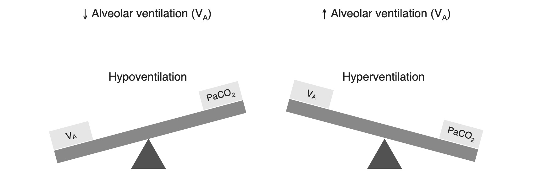
Relationship between alveolar ventilation and PCO2

Image by Lecturio.

Pathophysiology

Respiratory alkalosis is a process that results in a decreased level of carbon dioxide. 

  • Almost always due to hyperventilation (↑ respiratory rate Respiratory rate The number of times an organism breathes with the lungs (respiration) per unit time, usually per minute. Pulmonary Examination)
  • The arterial blood gas will show:
    • pH pH The quantitative measurement of the acidity or basicity of a solution. Acid-Base Balance > 7.40 
    • PCO2 < 30 mm MM Multiple myeloma (MM) is a malignant condition of plasma cells (activated B lymphocytes) primarily seen in the elderly. Monoclonal proliferation of plasma cells results in cytokine-driven osteoclastic activity and excessive secretion of IgG antibodies. Multiple Myeloma Hg 
  • The hyperventilation may be caused by:
    • ↑ Drive from the CNS (pathologic or physiologic)
    • Hypoxemia-induced causes → body attempts to correct the hypoxia Hypoxia Sub-optimal oxygen levels in the ambient air of living organisms. Ischemic Cell Damage at the expense of CO2 loss

Renal compensation Compensation Respiratory Acidosis

  • Kidneys Kidneys The kidneys are a pair of bean-shaped organs located retroperitoneally against the posterior wall of the abdomen on either side of the spine. As part of the urinary tract, the kidneys are responsible for blood filtration and excretion of water-soluble waste in the urine. Kidneys: Anatomy respond to respiratory alkalosis by decreasing serum HCO3:
  • Process takes 3–5 days to complete:
    • Cells must undergo physical changes.
    • Serum HCO3 and pH pH The quantitative measurement of the acidity or basicity of a solution. Acid-Base Balance decrease slowly during this time.
  • Degree of compensation Compensation Respiratory Acidosis defines acute versus chronic respiratory alkalosis.
Respiratory alkalosis renal compensation

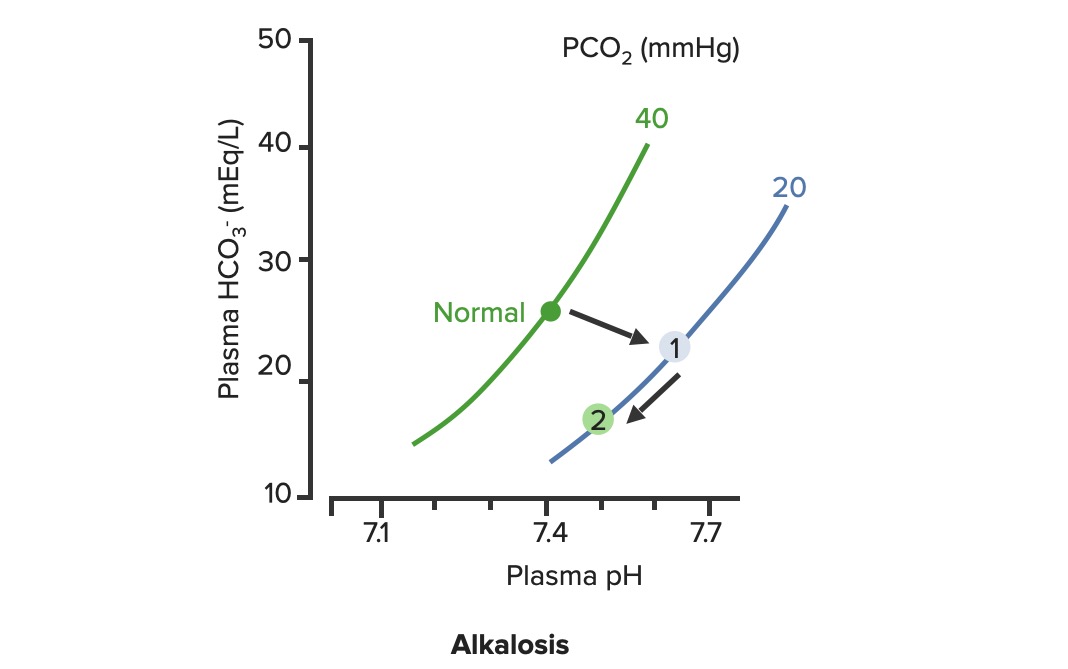
Renal compensation of respiratory alkalosis:
In respiratory alkalosis, the PCO2 is decreased, shifting the PCO2 curve to the right (1). As HCO3– levels are decreased by the kidneys, the pH improves along the PCO2 line (2)

Image by Lecturio.

Acute versus chronic respiratory alkalosis

Acute versus chronic respiratory alkalosis is defined by the degree of renal compensation Compensation Respiratory Acidosis.

  • Acute respiratory alkalosis is uncompensated:
  • Chronic respiratory alkalosis is compensated:
    • Renal compensation Compensation Respiratory Acidosis is complete.
    • HCO3 decreases by approximately 4 mEq/L per 10-mm Hg decrease in PaCO2
    • Usually asymptomatic, despite chronic hypocapnia
    • Hypocapnia is usually mild and pH pH The quantitative measurement of the acidity or basicity of a solution. Acid-Base Balance may be very near normal.

Clinical Presentation, Diagnosis, and Management

Clinical presentation

  • Tachypnea Tachypnea Increased respiratory rate. Pulmonary Examination
  • Dyspnea Dyspnea Dyspnea is the subjective sensation of breathing discomfort. Dyspnea is a normal manifestation of heavy physical or psychological exertion, but also may be caused by underlying conditions (both pulmonary and extrapulmonary). Dyspnea
  • Dizziness Dizziness An imprecise term which may refer to a sense of spatial disorientation, motion of the environment, or lightheadedness. Lateral Medullary Syndrome (Wallenberg Syndrome)/light-headedness
  • Paresthesias Paresthesias Subjective cutaneous sensations (e.g., cold, warmth, tingling, pressure, etc.) that are experienced spontaneously in the absence of stimulation. Posterior Cord Syndrome (perioral, hands/feet) due to decreased ionized calcium Ionized Calcium Hypocalcemia
  • Psychologic symptoms:
    • Anxiety Anxiety Feelings or emotions of dread, apprehension, and impending disaster but not disabling as with anxiety disorders. Generalized Anxiety Disorder
    • Fear
    • Impending doom
  • Highly variable Variable Variables represent information about something that can change. The design of the measurement scales, or of the methods for obtaining information, will determine the data gathered and the characteristics of that data. As a result, a variable can be qualitative or quantitative, and may be further classified into subgroups. Types of Variables presentation, based on underlying etiology; for example:
    • High-altitude illness: findings consistent with pulmonary and/or cerebral edema Cerebral edema Increased intracellular or extracellular fluid in brain tissue. Cytotoxic brain edema (swelling due to increased intracellular fluid) is indicative of a disturbance in cell metabolism, and is commonly associated with hypoxic or ischemic injuries. An increase in extracellular fluid may be caused by increased brain capillary permeability (vasogenic edema), an osmotic gradient, local blockages in interstitial fluid pathways, or by obstruction of CSF flow (e.g., obstructive hydrocephalus). Increased Intracranial Pressure (ICP)
    • Sepsis Sepsis Systemic inflammatory response syndrome with a proven or suspected infectious etiology. When sepsis is associated with organ dysfunction distant from the site of infection, it is called severe sepsis. When sepsis is accompanied by hypotension despite adequate fluid infusion, it is called septic shock. Sepsis and Septic Shock: fever Fever Fever is defined as a measured body temperature of at least 38°C (100.4°F). Fever is caused by circulating endogenous and/or exogenous pyrogens that increase levels of prostaglandin E2 in the hypothalamus. Fever is commonly associated with chills, rigors, sweating, and flushing of the skin. Fever, hypotension Hypotension Hypotension is defined as low blood pressure, specifically < 90/60 mm Hg, and is most commonly a physiologic response. Hypotension may be mild, serious, or life threatening, depending on the cause. Hypotension, findings consistent with originating infection (e.g., cough or dysuria Dysuria Painful urination. It is often associated with infections of the lower urinary tract. Urinary tract infections (UTIs))
    • Pulmonary embolism Pulmonary Embolism Pulmonary embolism (PE) is a potentially fatal condition that occurs as a result of intraluminal obstruction of the main pulmonary artery or its branches. The causative factors include thrombi, air, amniotic fluid, and fat. In PE, gas exchange is impaired due to the decreased return of deoxygenated blood to the lungs. Pulmonary Embolism: calf pain Pain An unpleasant sensation induced by noxious stimuli which are detected by nerve endings of nociceptive neurons. Pain: Types and Pathways, unilateral lower-extremity edema Edema Edema is a condition in which excess serous fluid accumulates in the body cavity or interstitial space of connective tissues. Edema is a symptom observed in several medical conditions. It can be categorized into 2 types, namely, peripheral (in the extremities) and internal (in an organ or body cavity). Edema

Diagnosis

Diagnosing a respiratory alkalosis typically requires a thorough history and exam and an arterial blood gas measurement.

  • Arterial blood gas:
    • Acute respiratory alkalosis (uncompensated):
      • pH pH The quantitative measurement of the acidity or basicity of a solution. Acid-Base Balance > 7.45
      • PaCO2 < 35 mm MM Multiple myeloma (MM) is a malignant condition of plasma cells (activated B lymphocytes) primarily seen in the elderly. Monoclonal proliferation of plasma cells results in cytokine-driven osteoclastic activity and excessive secretion of IgG antibodies. Multiple Myeloma Hg
      • Normal HCO3
    • Chronic respiratory alkalosis (compensated):
      • pH pH The quantitative measurement of the acidity or basicity of a solution. Acid-Base Balance > 7.4 (slightly high or near-normal)
      • PaCO2 < 35 mm MM Multiple myeloma (MM) is a malignant condition of plasma cells (activated B lymphocytes) primarily seen in the elderly. Monoclonal proliferation of plasma cells results in cytokine-driven osteoclastic activity and excessive secretion of IgG antibodies. Multiple Myeloma Hg
      • HCO3 decreased
  • Electrolytes Electrolytes Electrolytes are mineral salts that dissolve in water and dissociate into charged particles called ions, which can be either be positively (cations) or negatively (anions) charged. Electrolytes are distributed in the extracellular and intracellular compartments in different concentrations. Electrolytes are essential for various basic life-sustaining functions. Electrolytes: Abnormalities are common and may lead to complications.
  • Chest X-ray X-ray Penetrating electromagnetic radiation emitted when the inner orbital electrons of an atom are excited and release radiant energy. X-ray wavelengths range from 1 pm to 10 nm. Hard x-rays are the higher energy, shorter wavelength x-rays. Soft x-rays or grenz rays are less energetic and longer in wavelength. The short wavelength end of the x-ray spectrum overlaps the gamma rays wavelength range. The distinction between gamma rays and x-rays is based on their radiation source. Pulmonary Function Tests: to rule out other causes of tachypnea Tachypnea Increased respiratory rate. Pulmonary Examination

Management

  • Assess and address the ABCs ( airway Airway ABCDE Assessment, breathing, and circulation Circulation The movement of the blood as it is pumped through the cardiovascular system. ABCDE Assessment) if patient is in acute distress.
  • Attempt to correct the underlying abnormality.
  • Small dose of short-acting benzodiazepine

Clinical Relevance

  • Hyperventilation syndrome: an inappropriate increase in minute ventilation Minute ventilation Ventilation: Mechanics of Breathing beyond basic needs, typically associated with panic disorder Panic disorder Panic disorder is a condition marked by recurrent and episodic panic attacks that occur abruptly and without a trigger. These episodes are time-limited and present with cardiorespiratory (palpitations, shortness of breath, choking), GI (nausea, abdominal distress), and neurologic (paresthesias, lightheadedness) symptoms. Panic Disorder: Patients Patients Individuals participating in the health care system for the purpose of receiving therapeutic, diagnostic, or preventive procedures. Clinician–Patient Relationship will typically present with intermittent episodes of spontaneously resolving hyperventilation, often with mild dyspnea Dyspnea Dyspnea is the subjective sensation of breathing discomfort. Dyspnea is a normal manifestation of heavy physical or psychological exertion, but also may be caused by underlying conditions (both pulmonary and extrapulmonary). Dyspnea, dizziness Dizziness An imprecise term which may refer to a sense of spatial disorientation, motion of the environment, or lightheadedness. Lateral Medullary Syndrome (Wallenberg Syndrome), and chest tightness. Metabolic alkalosis Metabolic alkalosis The renal system is responsible for eliminating the daily load of non-volatile acids, which is approximately 70 millimoles per day. Metabolic alkalosis also occurs when there is an increased loss of acid, either renally or through the upper GI tract (e.g., vomiting), increased intake of HCO3-, or a reduced ability to secrete HCO3- when needed. Metabolic Alkalosis may develop. Diagnosis involves excluding physiologic causes of tachypnea Tachypnea Increased respiratory rate. Pulmonary Examination. Management involves reassurance Reassurance Clinician–Patient Relationship and possibly short-acting benzodiazepines Benzodiazepines Benzodiazepines work on the gamma-aminobutyric acid type A (GABAA) receptor to produce inhibitory effects on the CNS. Benzodiazepines do not mimic GABA, the main inhibitory neurotransmitter in humans, but instead potentiate GABA activity. Benzodiazepines.
  • High-altitude illness: a collective term for conditions involving cerebral and pulmonary symptoms that occur initially at high altitude: Hypoxia Hypoxia Sub-optimal oxygen levels in the ambient air of living organisms. Ischemic Cell Damage at higher altitudes stimulates hyperventilation, which can lead to respiratory alkalosis. These conditions are characterized by fluid shifts out of the vessels into the extravascular fluid space, leading to cerebral and pulmonary edema Pulmonary edema Pulmonary edema is a condition caused by excess fluid within the lung parenchyma and alveoli as a consequence of a disease process. Based on etiology, pulmonary edema is classified as cardiogenic or noncardiogenic. Patients may present with progressive dyspnea, orthopnea, cough, or respiratory failure. Pulmonary Edema. Management involves descent to a lower elevation and oxygen therapy.
  • Salicylate toxicity Toxicity Dosage Calculation: a complex mechanism that results in a mixed acid–base disorder, involving both respiratory alkalosis and metabolic acidosis Acidosis A pathologic condition of acid accumulation or depletion of base in the body. The two main types are respiratory acidosis and metabolic acidosis, due to metabolic acid build up. Respiratory Acidosis: Activation of the respiratory center in the medulla results in hyperventilation, while interference with cellular metabolism Cellular metabolism The chemical reactions in living organisms by which energy is provided for vital processes and activities and new material is assimilated. Thyroid Hormones can lead to metabolic acidosis Acidosis A pathologic condition of acid accumulation or depletion of base in the body. The two main types are respiratory acidosis and metabolic acidosis, due to metabolic acid build up. Respiratory Acidosis. The patient can also have significant vomiting Vomiting The forcible expulsion of the contents of the stomach through the mouth. Hypokalemia, and gastric ulcers and platelet dysfunction may develop. After initial stabilization, patients Patients Individuals participating in the health care system for the purpose of receiving therapeutic, diagnostic, or preventive procedures. Clinician–Patient Relationship can be treated with activated charcoal Charcoal An amorphous form of carbon prepared from the incomplete combustion of animal or vegetable matter, e.g., wood. The activated form of charcoal is used in the treatment of poisoning. Antidotes of Common Poisonings to absorb the aspirin Aspirin The prototypical analgesic used in the treatment of mild to moderate pain. It has anti-inflammatory and antipyretic properties and acts as an inhibitor of cyclooxygenase which results in the inhibition of the biosynthesis of prostaglandins. Aspirin also inhibits platelet aggregation and is used in the prevention of arterial and venous thrombosis. Nonsteroidal Antiinflammatory Drugs (NSAIDs).

References

  1. Emmett, M. & Palmer B.F. (2020). Simple and mixed acid-base disorders. UpToDate. Retrieved April 1, 2021, from https://www.uptodate.com/contents/simple-and-mixed-acid-base-disorders 
  2. Feller-Kopman, D.J. & Schwartzstein, R.M. (2021). Mechanisms, causes, and effects of hypercapnia. UpToDate. Retrieved April 1, 2021, from https://www.uptodate.com/contents/mechanisms-causes-and-effects-of-hypercapnia
  3. Theodore, A.C. (2020). Arterial blood gases. UpToDate. Retrieved April 1, 2021, from https://www.uptodate.com/contents/arterial-blood-gases
  4. Feller-Kopman, D.J. & Schwartzstein, R.M. (2021). The evaluation, diagnosis, and treatment of the adult patient with acute hypercapnic respiratory failure. UpToDate. Retrieved April 8, 2021, from https://www.uptodate.com/contents/the-evaluation-diagnosis-and-treatment-of-the-adult-patient-with-acute-hypercapnic-respiratory-failure

Create your free account or log in to continue reading!

Sign up now and get free access to Lecturio with concept pages, medical videos, and questions for your medical education.

User Reviews

Details