Skeletal muscle is striated muscleStriated muscleOne of two types of muscle in the body, characterized by the array of bands observed under microscope. Striated muscles can be divided into two subtypes: the cardiac muscle and the skeletal muscle.Muscle Tissue: Histology containing organized contractile structures known as sarcomeres that are made up of overlapping myofilamentsMyofilamentsRefers to individual proteins that together cause muscle contraction.Muscle Tissue: Histology: actin and myosin. When a nerve impulse arrives from a motorMotorNeurons which send impulses peripherally to activate muscles or secretory cells.Nervous System: Histology neuron, the signal triggers an action potentialAction PotentialAbrupt changes in the membrane potential that sweep along the cell membrane of excitable cells in response to excitation stimuli.Membrane Potential (AP) in the sarcolemmaSarcolemmaThe excitable plasma membrane of a muscle cell.Muscle Tissue: Histology (muscle cell membraneCell MembraneA cell membrane (also known as the plasma membrane or plasmalemma) is a biological membrane that separates the cell contents from the outside environment. A cell membrane is composed of a phospholipid bilayer and proteins that function to protect cellular DNA and mediate the exchange of ions and molecules. The Cell: Cell Membrane), resulting in the release of CaCACondylomata acuminata are a clinical manifestation of genital HPV infection. Condylomata acuminata are described as raised, pearly, flesh-colored, papular, cauliflower-like lesions seen in the anogenital region that may cause itching, pain, or bleeding.Condylomata Acuminata (Genital Warts) ions from the sarcoplasmic reticulumSarcoplasmic ReticulumA network of tubules and sacs in the cytoplasm of skeletal muscle fibers that assist with muscle contraction and relaxation by releasing and storing calcium ions.Muscle Tissue: Histology (SR) within the muscle cell. The CaCACondylomata acuminata are a clinical manifestation of genital HPV infection. Condylomata acuminata are described as raised, pearly, flesh-colored, papular, cauliflower-like lesions seen in the anogenital region that may cause itching, pain, or bleeding.Condylomata Acuminata (Genital Warts) causes a conformational change in regulator proteinsProteinsLinear polypeptides that are synthesized on ribosomes and may be further modified, crosslinked, cleaved, or assembled into complex proteins with several subunits. The specific sequence of amino acids determines the shape the polypeptide will take, during protein folding, and the function of the protein.Energy Homeostasis (troponin and tropomyosin), exposing myosin-binding sites on the actin filamentsActin filamentsFibers composed of microfilament proteins, which are predominately actin. They are the smallest of the cytoskeletal filaments.The Cell: Cytosol and Cytoskeleton. Using ATP energy, the myosin heads pull the myosin along the actin, shortening the sarcomereSarcomereThe repeating contractile units of the myofibril, delimited by z bands along its length.Muscle Tissue: Histology and resulting in muscle contraction. The ATP can be produced via anaerobic and aerobic mechanisms, and the primary source of ATP energy in a muscle fiber determines its functional characteristics.
Contractibility: ability to contract/shorten its length
Excitability: responds to stimulus (including electrical, hormonal, and mechanical)
Extensibility: ability to extend/stretch
Elasticity: ability to recoilRecoilVessels can stretch and return to their original shape after receiving the stroke volume of blood ejected by the left ventricle during systole.Arteries: Histology/return to normal shape when tension is released
Contains high amounts of myoglobinMyoglobinA conjugated protein which is the oxygen-transporting pigment of muscle. It is made up of one globin polypeptide chain and one heme group.Rhabdomyolysis and glycogen
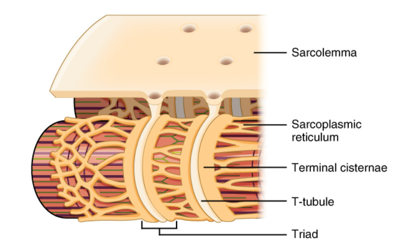
Muscle cell membraneCell MembraneA cell membrane (also known as the plasma membrane or plasmalemma) is a biological membrane that separates the cell contents from the outside environment. A cell membrane is composed of a phospholipid bilayer and proteins that function to protect cellular DNA and mediate the exchange of ions and molecules. The Cell: Cell Membrane
Allow action potentials to quickly spread to the myofibrilsMyofibrilsThe long cylindrical contractile organelles of striated muscle cells composed of actin filaments; myosin filaments; and other proteins organized in arrays of repeating units called sarcomeres.Muscle Tissue: Histology
Sarcoplasmic reticulumSarcoplasmic ReticulumA network of tubules and sacs in the cytoplasm of skeletal muscle fibers that assist with muscle contraction and relaxation by releasing and storing calcium ions.Muscle Tissue: Histology (SR):
Specialized ER containing high levels of CaCACondylomata acuminata are a clinical manifestation of genital HPV infection. Condylomata acuminata are described as raised, pearly, flesh-colored, papular, cauliflower-like lesions seen in the anogenital region that may cause itching, pain, or bleeding.Condylomata Acuminata (Genital Warts)2+
Terminal cisternae: part of the SR that lines the T-tubules → when action potentials arrive, SR is immediately stimulated to release CaCACondylomata acuminata are a clinical manifestation of genital HPV infection. Condylomata acuminata are described as raised, pearly, flesh-colored, papular, cauliflower-like lesions seen in the anogenital region that may cause itching, pain, or bleeding.Condylomata Acuminata (Genital Warts)2+ via receptorsReceptorsReceptors are proteins located either on the surface of or within a cell that can bind to signaling molecules known as ligands (e.g., hormones) and cause some type of response within the cell.Receptors in the terminal cisternae
Longitudinal SR: runs longitudinally along the myofilamentsMyofilamentsRefers to individual proteins that together cause muscle contraction.Muscle Tissue: Histology
Transverse tubules (T-tubules) and their associations with the sarcoplasmic reticulum (SR): Note how the T-tubules are channels through the sarcolemma that come in direct contact with the terminal cisternae of the SR.
Image: “Narrow T-tubules permit the conduction of electrical impulses” by Phil Schatz. License: CC BY 4.0
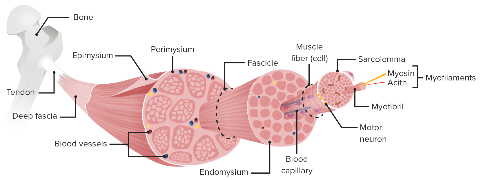
Structure of skeletal muscle: Clusters of myofilaments (actin and myosin) create myofibrils. There are multiple myofibrils within a single muscle fiber (which is a single muscle cell). The muscle fiber is surrounded by a specialized cell membrane called the sarcolemma. Multiple muscle fibers make up a muscle fascicle, and multiple muscle fascicles make up a full skeletal muscle.
MyofilamentsMyofilamentsRefers to individual proteins that together cause muscle contraction.Muscle Tissue: Histology are individual proteinsProteinsLinear polypeptides that are synthesized on ribosomes and may be further modified, crosslinked, cleaved, or assembled into complex proteins with several subunits. The specific sequence of amino acids determines the shape the polypeptide will take, during protein folding, and the function of the protein.Energy Homeostasis that together cause muscle contraction.
Sarcomeres: contractile structures formed by overlapping actin and myosin myofilamentsMyofilamentsRefers to individual proteins that together cause muscle contraction.Muscle Tissue: Histology
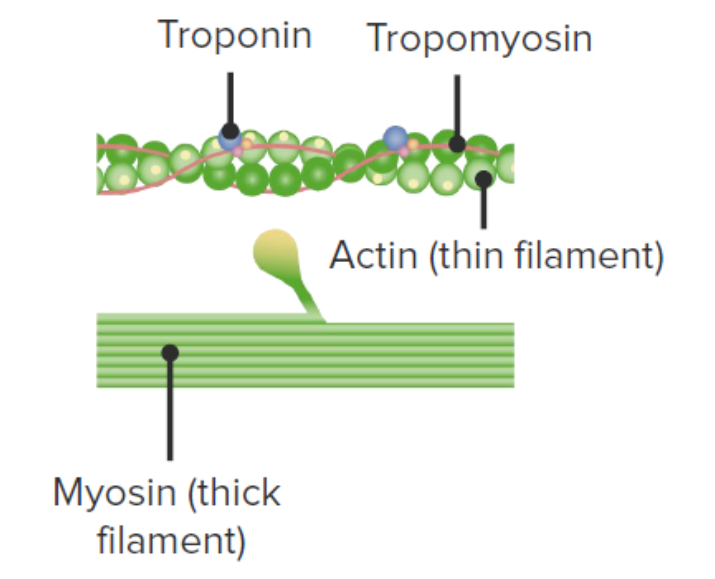
Myosin:
Thick, straight filaments arranged in parallel
Have a main shaft and a globular head on each end
Actin:
Thin filaments made of 2 long-coiling protein strands
Connected to each other at the Z line of sarcomeres
Tropomyosin: a ropelike protein covering the myosin-binding sites on actin
Troponin:
Troponin C (TnC): contains binding sites for CaCACondylomata acuminata are a clinical manifestation of genital HPV infection. Condylomata acuminata are described as raised, pearly, flesh-colored, papular, cauliflower-like lesions seen in the anogenital region that may cause itching, pain, or bleeding.Condylomata Acuminata (Genital Warts)2+
Troponin ITroponin IA troponin complex subunit that inhibits actomyosin ATPase activity thereby disrupting actin and myosin interaction. There are three troponin I subtypes: troponin i1, i2 and i3. Troponin i3 is cardiac-specific whereas troponin i1 and i2 are skeletal subtypes. Troponin i3 is a biomarker for damaged or injured cardiac myocytes and mutations in troponin i3 gene are associated with familial hypertrophic cardiomyopathy.Myocardial Infarction (TnI): inhibits actin and myosin binding
Troponin T (TnT): connects the other troponins to tropomyosin
Structure of actin (thin filament) and myosin (thick filament): Note the globular head on myosin. The yellow dots on the actin represent the myosin-binding sites, which are covered by tropomyosin in a resting state. Troponins contain the Ca-binding sites and, when Ca is present, induce a conformational change in the troponin–tropomyosin complex, exposing the myosin-binding sites on actin. When myosin can bind actin and ATP energy is present, muscle contraction occurs.
Image by Lecturio.
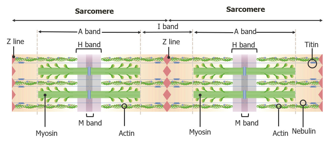
Review of sarcomereSarcomereThe repeating contractile units of the myofibril, delimited by z bands along its length.Muscle Tissue: Histology structure
The myofibrilsMyofibrilsThe long cylindrical contractile organelles of striated muscle cells composed of actin filaments; myosin filaments; and other proteins organized in arrays of repeating units called sarcomeres.Muscle Tissue: Histology are organized in a pattern that creates different bands and zones when viewed under microscopy. These bands are created by overlapping actin and myosin strands.
Z line (also called the Z band or Z disc):
Anchors and separates 1 sarcomereSarcomereThe repeating contractile units of the myofibril, delimited by z bands along its length.Muscle Tissue: Histology from another
A sarcomereSarcomereThe repeating contractile units of the myofibril, delimited by z bands along its length.Muscle Tissue: Histologyis defined as the region between 2 Z lines
Anisotropic bands (A bands):
Dark bands on microscopy → memory trick: “dark” has an “A” in it
Formed by entire length of thick myosin filaments, which includes overlapping actin filamentsActin filamentsFibers composed of microfilament proteins, which are predominately actin. They are the smallest of the cytoskeletal filaments.The Cell: Cytosol and Cytoskeleton at the ends
Isotropic bands (I bands):
Light bands on microscopy → memory trick: “light” has an “I” in it
Consist of only thin actin filamentsActin filamentsFibers composed of microfilament proteins, which are predominately actin. They are the smallest of the cytoskeletal filaments.The Cell: Cytosol and Cytoskeleton
I bands are between the A bands and include the Z line.
H zone:
Lighter zone in the middle of the A Band
Consists of only myosin filaments → excludes the ends of the myosin which are overlapping with actin
M bands:
Fine, dark line in the center of the H zone
Myosin-binding proteinsProteinsLinear polypeptides that are synthesized on ribosomes and may be further modified, crosslinked, cleaved, or assembled into complex proteins with several subunits. The specific sequence of amino acids determines the shape the polypeptide will take, during protein folding, and the function of the protein.Energy Homeostasis attach here
Diagram depicting the microscopic structure of two adjacent sarcomeres: a sarcomere is the area between Z-lines. A band: anisotropic band I band: isotropic band
Skeletal muscle cell contraction requires stimulation by an action potentialAction PotentialAbrupt changes in the membrane potential that sweep along the cell membrane of excitable cells in response to excitation stimuli.Membrane Potential from somatic motorMotorNeurons which send impulses peripherally to activate muscles or secretory cells.Nervous System: HistologyneuronsNeuronsThe basic cellular units of nervous tissue. Each neuron consists of a body, an axon, and dendrites. Their purpose is to receive, conduct, and transmit impulses in the nervous system.Nervous System: Histology.
The neuromuscular junction (NMJ)
Also called an end plate
A synapseSynapseThe junction between 2 neurons is called a synapse. The synapse allows a neuron to pass an electrical or chemical signal to another neuron or target effector cell. Synapses and Neurotransmission(i.e., connection)between a skeletal muscle cell and motorMotorNeurons which send impulses peripherally to activate muscles or secretory cells.Nervous System: Histology neuron
Each skeletal muscle cell (i.e., muscle fiber) has 1 NMJ around the midpoint of the cell.
MotorMotorNeurons which send impulses peripherally to activate muscles or secretory cells.Nervous System: Histology end plate: depression in the sarcolemmaSarcolemmaThe excitable plasma membrane of a muscle cell.Muscle Tissue: Histology of the adjacent muscle fiber, in close association with the synaptic knob
Schwann cellSchwann CellNeuroglial cells of the peripheral nervous system which form the insulating myelin sheaths of peripheral axons.Nervous System: Histology: specialized cell that surrounds and protects the NMJ
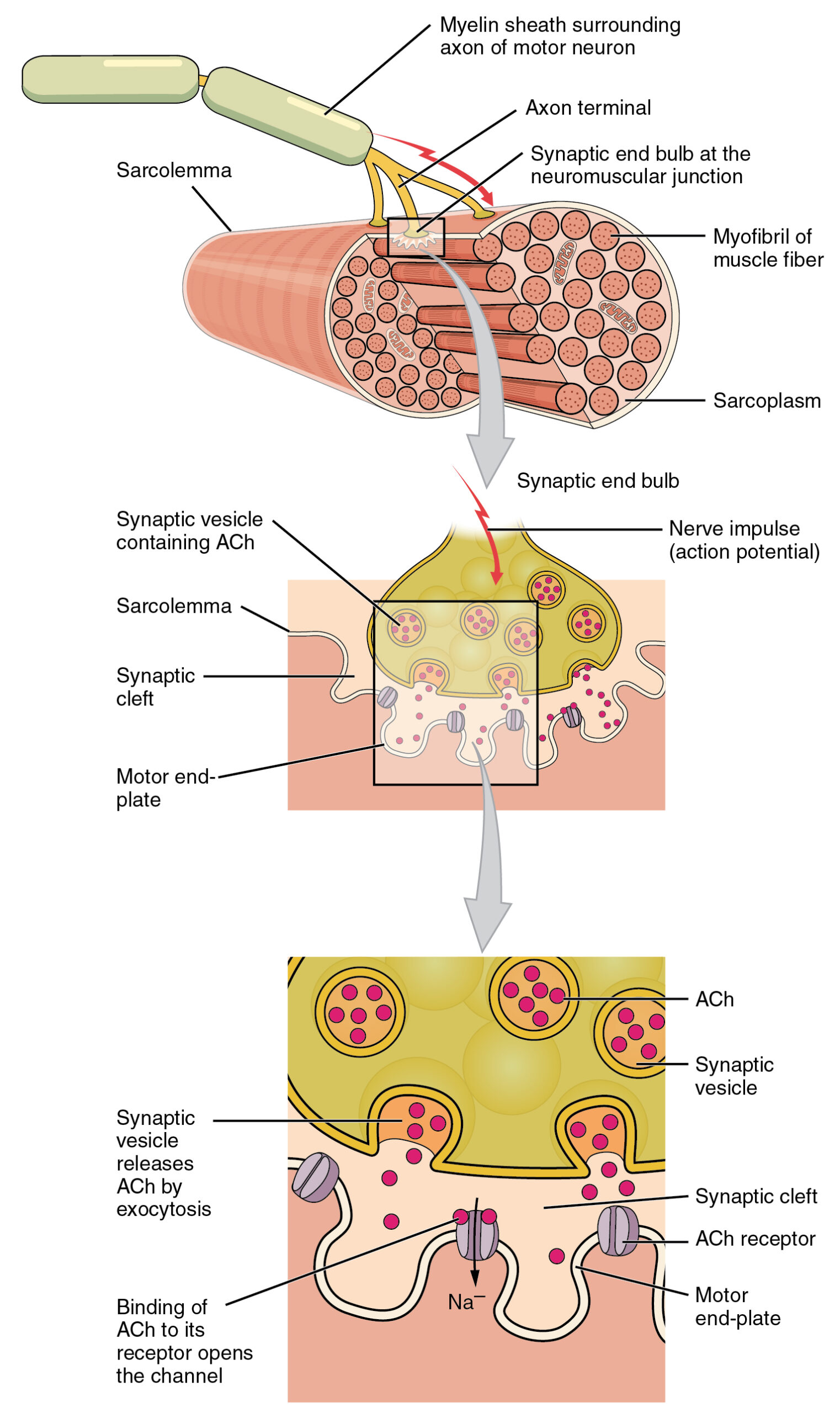
Process of transmitting a neuronal signal to the muscle cell
AcetylcholineAcetylcholineA neurotransmitter found at neuromuscular junctions, autonomic ganglia, parasympathetic effector junctions, a subset of sympathetic effector junctions, and at many sites in the central nervous system.Receptors and Neurotransmitters of the CNS (AChAChA neurotransmitter found at neuromuscular junctions, autonomic ganglia, parasympathetic effector junctions, a subset of sympathetic effector junctions, and at many sites in the central nervous system.Receptors and Neurotransmitters of the CNS) is released from synaptic vesiclesVesiclesFemale Genitourinary Examination in the synaptic knob.
AChAChA neurotransmitter found at neuromuscular junctions, autonomic ganglia, parasympathetic effector junctions, a subset of sympathetic effector junctions, and at many sites in the central nervous system.Receptors and Neurotransmitters of the CNS binds to and activates receptorsReceptorsReceptors are proteins located either on the surface of or within a cell that can bind to signaling molecules known as ligands (e.g., hormones) and cause some type of response within the cell.Receptors on the motorMotorNeurons which send impulses peripherally to activate muscles or secretory cells.Nervous System: Histology end plate (there are approximately 50 million AChAChA neurotransmitter found at neuromuscular junctions, autonomic ganglia, parasympathetic effector junctions, a subset of sympathetic effector junctions, and at many sites in the central nervous system.Receptors and Neurotransmitters of the CNSreceptorsReceptorsReceptors are proteins located either on the surface of or within a cell that can bind to signaling molecules known as ligands (e.g., hormones) and cause some type of response within the cell.Receptors per NMJ)
Acetylcholinesterase (AChE): breaks down AChAChA neurotransmitter found at neuromuscular junctions, autonomic ganglia, parasympathetic effector junctions, a subset of sympathetic effector junctions, and at many sites in the central nervous system.Receptors and Neurotransmitters of the CNS left in the synaptic cleftSynaptic cleftSynapses and Neurotransmission to “turn off” the signal
Motor end-plate and innervation ACh: acetylcholine
Image: “Motor end-plate and innervation” by Phil Schatz. License: CC BY 4.0
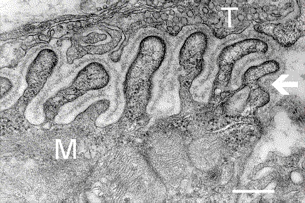
Electron micrograph showing a cross section through the neuromuscular junction: “T” is the axon terminal, and “M” is the muscle fiber. The arrow shows junctional folds with basal lamina. Postsynaptic densities are visible on the tips between the folds. The scale is 0.3 µm.
Image: “Electron micrograph showing a cross-section through the neuromuscular junction” by National Institute of Mental Health. License: Public DomainNote: This animation does not have sound.
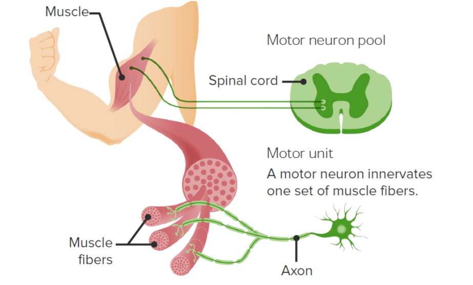
MotorMotorNeurons which send impulses peripherally to activate muscles or secretory cells.Nervous System: Histology units
A group of muscle fibers working together that are controlled by a singlemotorMotorNeurons which send impulses peripherally to activate muscles or secretory cells.Nervous System: Histology neuron
Small motorMotorNeurons which send impulses peripherally to activate muscles or secretory cells.Nervous System: Histology units:
Only a few muscle fibers per neuron
Allows for fine muscle control
Example: eye muscles
Large motorMotorNeurons which send impulses peripherally to activate muscles or secretory cells.Nervous System: Histology units:
Up to several hundred muscle fibers innervated by a single neuron
Example: large postural muscles
Depiction of a motor unit: A single motor neuron innervates multiple different muscle fibers (i.e., individual muscle cells). The group of muscle fibers innervated by the same motor neuron are called a motor unit.
Image by Lecturio.
How an Individual Muscle Fiber Contracts
Excitation
A nerve signal arrives at the synaptic knob.
Voltage-gated CaCACondylomata acuminata are a clinical manifestation of genital HPV infection. Condylomata acuminata are described as raised, pearly, flesh-colored, papular, cauliflower-like lesions seen in the anogenital region that may cause itching, pain, or bleeding.Condylomata Acuminata (Genital Warts)channelsChannelsThe Cell: Cell Membrane open, stimulating the release of AChAChA neurotransmitter found at neuromuscular junctions, autonomic ganglia, parasympathetic effector junctions, a subset of sympathetic effector junctions, and at many sites in the central nervous system.Receptors and Neurotransmitters of the CNS into the synaptic cleftSynaptic cleftSynapses and Neurotransmission.
AChAChA neurotransmitter found at neuromuscular junctions, autonomic ganglia, parasympathetic effector junctions, a subset of sympathetic effector junctions, and at many sites in the central nervous system.Receptors and Neurotransmitters of the CNS binds to and activates ligand-gated ion channelsChannelsThe Cell: Cell Membrane on the motorMotorNeurons which send impulses peripherally to activate muscles or secretory cells.Nervous System: Histology end plate of the muscle fiber.
Allows Na+ into the muscle cell
Allows K+ out of the cell
This flowFlowBlood flows through the heart, arteries, capillaries, and veins in a closed, continuous circuit. Flow is the movement of volume per unit of time. Flow is affected by the pressure gradient and the resistance fluid encounters between 2 points. Vascular resistance is the opposition to flow, which is caused primarily by blood friction against vessel walls.Vascular Resistance, Flow, and Mean Arterial Pressure of ions reverses the polarity of the sarcolemmaSarcolemmaThe excitable plasma membrane of a muscle cell.Muscle Tissue: Histology = depolarizationDepolarizationMembrane Potential
The AP propagates in all directions throughout the sarcolemmaSarcolemmaThe excitable plasma membrane of a muscle cell.Muscle Tissue: Histology, including down the T-tubules.
Excitation-contraction coupling
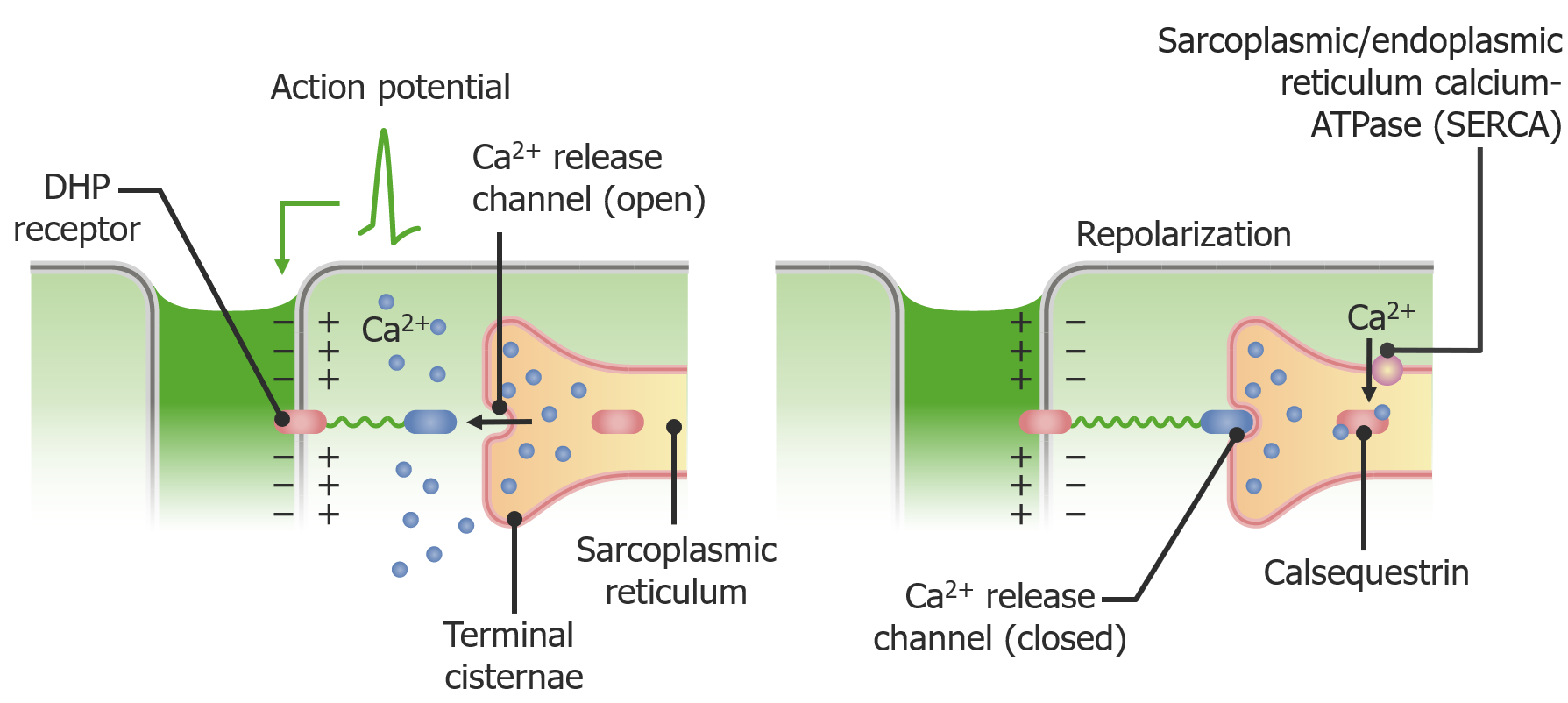
The AP stimulates voltage-dependent dihydropyridineDihydropyridinePyridine moieties which are partially saturated by the addition of two hydrogen atoms in any position.Class 4 Antiarrhythmic Drugs (Calcium Channel Blockers) (DHP) receptorsReceptorsReceptors are proteins located either on the surface of or within a cell that can bind to signaling molecules known as ligands (e.g., hormones) and cause some type of response within the cell.Receptors:
Membrane-bound receptorsReceptorsReceptors are proteins located either on the surface of or within a cell that can bind to signaling molecules known as ligands (e.g., hormones) and cause some type of response within the cell.Receptors lining the T-tubules
CaCACondylomata acuminata are a clinical manifestation of genital HPV infection. Condylomata acuminata are described as raised, pearly, flesh-colored, papular, cauliflower-like lesions seen in the anogenital region that may cause itching, pain, or bleeding.Condylomata Acuminata (Genital Warts)2+ ions flood out of the SR into the sarcoplasmSarcoplasmMuscle Tissue: Histology → bindBINDHyperbilirubinemia of the Newborn to troponins on the thin filaments (actin)
Physiology of Ca2+ release from the sarcoplasmic reticulum in response to an action potential:
A wave of depolarization (i.e., the action potential) travels down the T-tubules and triggers the voltage-dependent dihydropyridine (DHP) receptors. These DHP receptors are mechanically tethered to ryanodine receptors, which normally keep the Ca2+-release channels closed. When DHP receptors are stimulated by an action potential, they remove the ryanodine receptors from the Ca2+-release channels, allowing Ca2+ to spill out of the sarcoplasmic reticulum and into the sarcoplasm, where they can bind to troponin and stimulate muscle contraction. Dantrolene binds to ryanodine receptors, preventing Ca2+ release and muscle contraction.
Moves the myosin head into a high-energy “cocked” position
This movement is known as the recovery stroke.
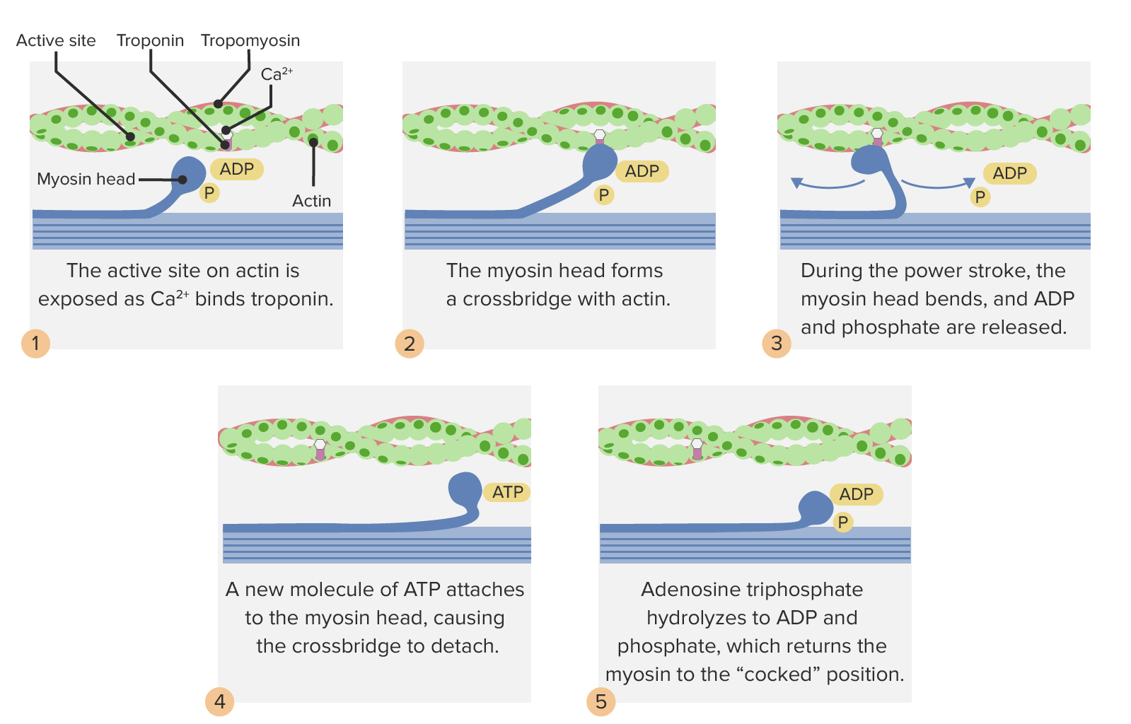
The cocked myosin head binds an exposed binding site on actin, forming a crossbridge. Note: CaCACondylomata acuminata are a clinical manifestation of genital HPV infection. Condylomata acuminata are described as raised, pearly, flesh-colored, papular, cauliflower-like lesions seen in the anogenital region that may cause itching, pain, or bleeding.Condylomata Acuminata (Genital Warts) must be present and bound to troponin in order for the myosin-binding sites on actin to be uncovered and available.
Power stroke:
Myosin releases the ADP and phosphatePhosphateInorganic salts of phosphoric acid.Electrolytes.
Myosin head expels the energy → returns to the flexed position, pulling the thin filament with it
Since many myosin heads are bound simultaneously, the thin filament remains in its new position rather than “slipping back” to its original position.
Power strokes shorten the I band and moves Z lines closer together:
→ Sarcomeres shorten and move closer together
→ Muscle fibers shorten
→ Entire muscle shortens, generating movement
Note that the myofilamentsMyofilamentsRefers to individual proteins that together cause muscle contraction.Muscle Tissue: Histology themselves do not shorten; they simply overlap more.
Note that the A band also does not shorten, though A bands do move closer together.
Myosin binds a new ATP, causing it to release from the actin.
The cycle starts over.
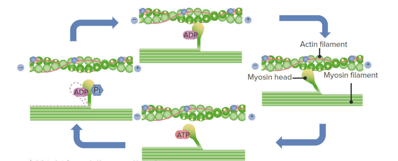
The process of cross-bridge cycling
Image by Lecturio.
Crossbridge cycling: The myosin-binding site on actin is exposed when Ca2+ binds to troponin. Adenosine triphosphate binds to the myosin head. Myosin ATPase hydrolyzes the ATP to ADP and phosphate, and this moves the myosin head into a cocked position. With ADP and phosphate still bound and the head in a cocked position, myosin is able to bind to the active sites on actin, forming a crossbridge. The ADP and phosphate are released, and the stored potential energy is released, generating the power stroke: the myosin head returns to its flexed position, pulling the actin filament with it. Adenosine triphosphate binds the myosin head, causing it to release from the actin and begin the cycle over again. This process allows the myosin to “walk” along the actin filament, shortening the sarcomere.
Image by Lecturio.
Relaxation
The motorMotorNeurons which send impulses peripherally to activate muscles or secretory cells.Nervous System: Histology neuron ceases, sending its chemical signal, AChAChA neurotransmitter found at neuromuscular junctions, autonomic ganglia, parasympathetic effector junctions, a subset of sympathetic effector junctions, and at many sites in the central nervous system.Receptors and Neurotransmitters of the CNS, into the synapseSynapseThe junction between 2 neurons is called a synapse. The synapse allows a neuron to pass an electrical or chemical signal to another neuron or target effector cell. Synapses and Neurotransmission at the NMJ.
Ryanodine receptorsRyanodine ReceptorsMalignant Hyperthermia close the Ca-release channelsChannelsThe Cell: Cell Membrane on the SR, preventing further CaCACondylomata acuminata are a clinical manifestation of genital HPV infection. Condylomata acuminata are described as raised, pearly, flesh-colored, papular, cauliflower-like lesions seen in the anogenital region that may cause itching, pain, or bleeding.Condylomata Acuminata (Genital Warts)2+ efflux.
Sarco-/endoplasmic reticulumEndoplasmic reticulumA system of cisternae in the cytoplasm of many cells. In places the endoplasmic reticulum is continuous with the plasma membrane (cell membrane) or outer membrane of the nuclear envelope. If the outer surfaces of the endoplasmic reticulum membranes are coated with ribosomes, the endoplasmic reticulum is said to be rough-surfaced; otherwise it is said to be smooth-surfaced.The Cell: Organelles Ca-ATPase (SERCA): pumps CaCACondylomata acuminata are a clinical manifestation of genital HPV infection. Condylomata acuminata are described as raised, pearly, flesh-colored, papular, cauliflower-like lesions seen in the anogenital region that may cause itching, pain, or bleeding.Condylomata Acuminata (Genital Warts) back into the SR, removing it from the sarcoplasmSarcoplasmMuscle Tissue: Histology
Calsequestrin: binds CaCACondylomata acuminata are a clinical manifestation of genital HPV infection. Condylomata acuminata are described as raised, pearly, flesh-colored, papular, cauliflower-like lesions seen in the anogenital region that may cause itching, pain, or bleeding.Condylomata Acuminata (Genital Warts)2+ within the SR, which stores/sequesters it until a new signal for muscle contraction arrives
Without CaCACondylomata acuminata are a clinical manifestation of genital HPV infection. Condylomata acuminata are described as raised, pearly, flesh-colored, papular, cauliflower-like lesions seen in the anogenital region that may cause itching, pain, or bleeding.Condylomata Acuminata (Genital Warts)2+, the troponin–tropomyosin complex shifts, covering the binding sites on actin.
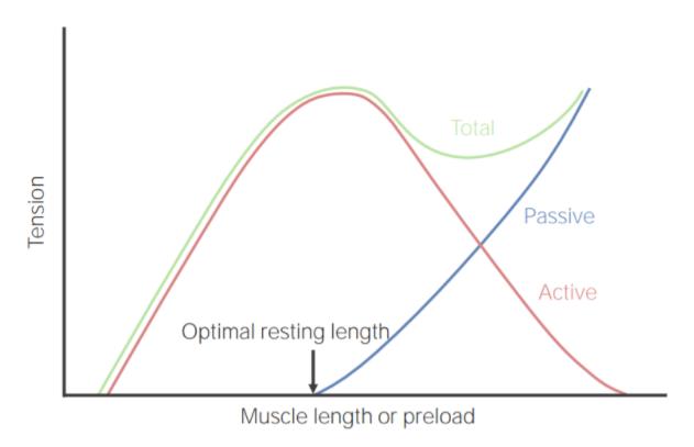
The resting length of the sarcomereSarcomereThe repeating contractile units of the myofibril, delimited by z bands along its length.Muscle Tissue: Histology has a direct influence on the force generated when the sarcomereSarcomereThe repeating contractile units of the myofibril, delimited by z bands along its length.Muscle Tissue: Histology shortens. This is called the length–tension relationship.
Active tension: the tension produced by power strokes
The amount of tension that can be actively produced is dependent on the starting length of the sarcomereSarcomereThe repeating contractile units of the myofibril, delimited by z bands along its length.Muscle Tissue: Histology.
Overcontracted at rest (i.e., shorter starting length):
The ends of the thick filaments are close to Z lines.
Minimal room for them to contract further
→ A weak contraction before the fiber runs out of room to contract
Overstretched at rest (i.e., longer starting length):
Minimal overlap between actin and myosin
Fewer myosin heads can come in contact with the actin.
→ Weaker initial contraction
Optimal resting length:
The length at which a muscle can produce the greatest force when it contracts
Controlled by the CNS
Muscle tone: state of partial contraction that is maintained by the CNS under resting conditions, generating the optimal resting length
Passive tension: tension that resists the myofilamentsMyofilamentsRefers to individual proteins that together cause muscle contraction.Muscle Tissue: Histology being pulled apart
Total muscle tension: equals active tension plus passive tension
Length–tension relationship in skeletal muscle
Image by Lecturio.
Threshold, latent periods, and twitch
Threshold: minimum voltage necessary to generate an AP (an all-or-none response)
Latent period:
The time between onset of the AP and onset of the muscle contraction (i.e., the twitch)
During this time, excitation–contraction coupling is occurring:
CaCACondylomata acuminata are a clinical manifestation of genital HPV infection. Condylomata acuminata are described as raised, pearly, flesh-colored, papular, cauliflower-like lesions seen in the anogenital region that may cause itching, pain, or bleeding.Condylomata Acuminata (Genital Warts) ions are released from the SR.
No increase in tension during the latent period
Typically lasts approximately 2 milliseconds
Twitch:
An isolated, rapid contraction followed by rapid relaxation
Tension increases throughout this phase until peak tension is reached.
Relaxation phase,
Contraction ends and tension decreases.
CaCACondylomata acuminata are a clinical manifestation of genital HPV infection. Condylomata acuminata are described as raised, pearly, flesh-colored, papular, cauliflower-like lesions seen in the anogenital region that may cause itching, pain, or bleeding.Condylomata Acuminata (Genital Warts)2+ ions are pumped back into the SR → without CaCACondylomata acuminata are a clinical manifestation of genital HPV infection. Condylomata acuminata are described as raised, pearly, flesh-colored, papular, cauliflower-like lesions seen in the anogenital region that may cause itching, pain, or bleeding.Condylomata Acuminata (Genital Warts)2+, crossbridge formation cannot occur → muscle fibers return to their resting state
Coordinating twitches so that muscles can do meaningful work
A single isolated twitch of a single muscle fiber cannot do meaningful work, and increasing the voltage of the stimulus does not increase the strength of a twitch. Ways to increase the strength of a muscle contraction include:
Recruitment (also called multiple motorMotorNeurons which send impulses peripherally to activate muscles or secretory cells.Nervous System: Histology unit summation): increasing the voltage stimulus to the motorMotorNeurons which send impulses peripherally to activate muscles or secretory cells.Nervous System: Histology neuron itself excites more nerve fibersNerve FibersSlender processes of neurons, including the axons and their glial envelopes (myelin sheath). Nerve fibers conduct nerve impulses to and from the central nervous system.Nervous System: Histology → excites more motorMotorNeurons which send impulses peripherally to activate muscles or secretory cells.Nervous System: Histology units
↑ Frequency of stimulation:
Repetitive stimulation → increases tension with each twitch because:
The SR cannot fully recover all of the CaCACondylomata acuminata are a clinical manifestation of genital HPV infection. Condylomata acuminata are described as raised, pearly, flesh-colored, papular, cauliflower-like lesions seen in the anogenital region that may cause itching, pain, or bleeding.Condylomata Acuminata (Genital Warts)2+ between twitches
If twitches cannot fully recover before the next twitch starts, tension increases (known as temporal summation or wave summation)
At > 40 stimuli per second:
Muscle has no time to relax at all.
Muscle goes into a sustained prolonged contraction known as tetanusTetanusTetanus is a bacterial infection caused by Clostridium tetani, a gram-positive obligate anaerobic bacterium commonly found in soil that enters the body through a contaminated wound. C. tetani produces a neurotoxin that blocks the release of inhibitory neurotransmitters and causes prolonged tonic muscle contractions. Tetanus.
TetanusTetanusTetanus is a bacterial infection caused by Clostridium tetani, a gram-positive obligate anaerobic bacterium commonly found in soil that enters the body through a contaminated wound. C. tetani produces a neurotoxin that blocks the release of inhibitory neurotransmitters and causes prolonged tonic muscle contractions. Tetanus does not occur in the body under normal physiologic conditions.
MotorMotorNeurons which send impulses peripherally to activate muscles or secretory cells.Nervous System: Histology units function asynchronously:
When 1 motorMotorNeurons which send impulses peripherally to activate muscles or secretory cells.Nervous System: Histology unit relaxes, another takes over.
Allows for “smooth” muscle contractions in which the muscle as a whole does not lose tension
Principles of muscle stimulation: Increasing the frequency of stimulation increases the strength of the muscle contraction.
Image by Lecturio.
Types of skeletal muscle contraction
There are multiple types of muscle contraction based on how the muscle fiber changes length during the contraction:
Isometric:
A muscular contraction in which the length of the muscle does not change
Maintain constant tension in the muscle as the muscle changes length
Example: bicep curls
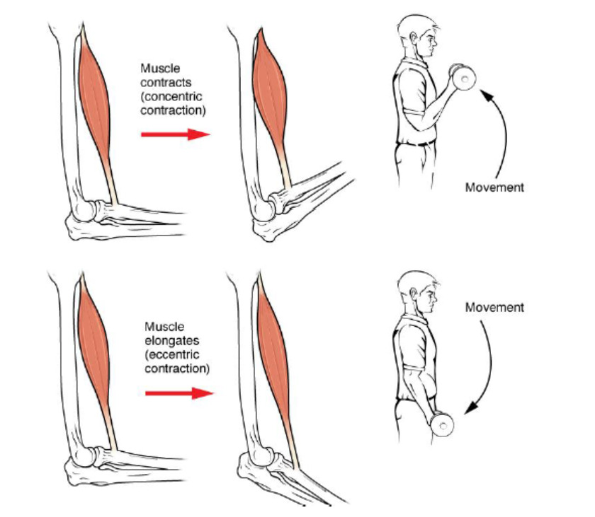
Have concentric and eccentric phases
Concentric:
Shortening of the sarcomereSarcomereThe repeating contractile units of the myofibril, delimited by z bands along its length.Muscle Tissue: Histology, muscle fiber, and muscle, generating limb movement
E.g., lifting a weight during a bicep curl
Eccentric:
Lengthening the muscle while still contracting (i.e., generating force)
Occurs when the resistanceResistancePhysiologically, the opposition to flow of air caused by the forces of friction. As a part of pulmonary function testing, it is the ratio of driving pressure to the rate of air flow.Ventilation: Mechanics of Breathing against the muscle is greater than the force generated
E.g., lowering a bicep curl
Auxotonic contraction:
Simultaneous changes in both muscle tension and length
I.e., a combination of isometric and isotonicIsotonicSolutions having the same osmotic pressure as blood serum, or another solution with which they are compared.Renal Sodium and Water Regulation contractions
AdenosineAdenosineA nucleoside that is composed of adenine and d-ribose. Adenosine or adenosine derivatives play many important biological roles in addition to being components of DNA and RNA. Adenosine itself is a neurotransmitter.Class 5 Antiarrhythmic Drugs triphosphate is the primary energy source required to generate the power strokes causing muscle contraction. There are several different ways this ATP energy is generated, and there are several different types of muscle fibers based on their capacity to use different energy sources.
Energy sources
AdenosineAdenosineA nucleoside that is composed of adenine and d-ribose. Adenosine or adenosine derivatives play many important biological roles in addition to being components of DNA and RNA. Adenosine itself is a neurotransmitter.Class 5 Antiarrhythmic Drugs triphosphate concentration in the muscle fiber is only enough to sustain full contraction for 1 to 2 seconds. Therefore, ADP must be rephosphorylated to generate new ATP, allowing the muscle to continue contracting, which requires energy.
For immediate energy:
Phosphagen system:
CreatineCreatineAn amino acid that occurs in vertebrate tissues and in urine. In muscle tissue, creatine generally occurs as phosphocreatine. Creatine is excreted as creatinine in the urine.Acute Kidney InjuryphosphatePhosphateInorganic salts of phosphoric acid.Electrolytes: an energy-storage molecule that can donate a phosphate groupPhosphate groupNucleic Acids to ADP
CK: transfers the phosphate groupPhosphate groupNucleic Acids from creatineCreatineAn amino acid that occurs in vertebrate tissues and in urine. In muscle tissue, creatine generally occurs as phosphocreatine. Creatine is excreted as creatinine in the urine.Acute Kidney InjuryphosphatePhosphateInorganic salts of phosphoric acid.Electrolytes to ADP → ATP
The phosphagen system provides nearly all the energy used in short bursts of intense activity.
GlycolysisGlycolysisGlycolysis is a central metabolic pathway responsible for the breakdown of glucose and plays a vital role in generating free energy for the cell and metabolites for further oxidative degradation. Glucose primarily becomes available in the blood as a result of glycogen breakdown or from its synthesis from noncarbohydrate precursors (gluconeogenesis) and is imported into cells by specific transport proteins. Glycolysis: converts glycogen → lactate, generating ATP in the process
Produces enough ATP to sustain activity for about 30–40 seconds
Lactate and metabolites (e.g., H+, inorganic phosphatePhosphateInorganic salts of phosphoric acid.Electrolytes) build up → major factor in muscle fatigueFatigueThe state of weariness following a period of exertion, mental or physical, characterized by a decreased capacity for work and reduced efficiency to respond to stimuli.Fibromyalgia
For long-term energy: aerobic respirationRespirationThe act of breathing with the lungs, consisting of inhalation, or the taking into the lungs of the ambient air, and of exhalation, or the expelling of the modified air which contains more carbon dioxide than the air taken in.Nose Anatomy (External & Internal)
The major source of energy for activity lasting longer than approximately 30‒40 seconds
Requires O2
Occurs once cardiovascular changes have “caught up” with the increase in activity level and blood flowBlood flowBlood flow refers to the movement of a certain volume of blood through the vasculature over a given unit of time (e.g., mL per minute).Vascular Resistance, Flow, and Mean Arterial Pressure is now delivering enough O2 for aerobic respirationRespirationThe act of breathing with the lungs, consisting of inhalation, or the taking into the lungs of the ambient air, and of exhalation, or the expelling of the modified air which contains more carbon dioxide than the air taken in.Nose Anatomy (External & Internal) to occur
Fatty acidsAcidsChemical compounds which yield hydrogen ions or protons when dissolved in water, whose hydrogen can be replaced by metals or basic radicals, or which react with bases to form salts and water (neutralization). An extension of the term includes substances dissolved in media other than water.Acid-Base Balance and glucoseGlucoseA primary source of energy for living organisms. It is naturally occurring and is found in fruits and other parts of plants in its free state. It is used therapeutically in fluid and nutrient replacement.Lactose Intolerance are used to generate ATP through the Krebs cycleKrebs cycleThe citric acid cycle, also known as the tricarboxylic acid (TCA) cycle or the krebs cycle, is a cyclic set of reactions that occurs in the mitochondrial matrix. The TCA cycle is the continuation of any metabolic pathway that produces pyruvate, which is converted into its main substrate, acetyl-CoA.Citric Acid Cycle and oxidative phosphorylationPhosphorylationThe introduction of a phosphoryl group into a compound through the formation of an ester bond between the compound and a phosphorus moiety.Post-translational Protein Processing (i.e., the electron transport chainElectron transport chainThe electron transport chain (ETC) sends electrons through a series of proteins, which generate an electrochemical proton gradient that produces energy in the form of adenosine triphosphate (ATP).Electron Transport Chain (ETC) (ETCETCThe electron transport chain (ETC) sends electrons through a series of proteins, which generate an electrochemical proton gradient that produces energy in the form of adenosine triphosphate (ATP).Electron Transport Chain (ETC)))
Aerobic respirationRespirationThe act of breathing with the lungs, consisting of inhalation, or the taking into the lungs of the ambient air, and of exhalation, or the expelling of the modified air which contains more carbon dioxide than the air taken in.Nose Anatomy (External & Internal) continues until endurance is depleted via:
↓ Glycogen and blood glucoseGlucoseA primary source of energy for living organisms. It is naturally occurring and is found in fruits and other parts of plants in its free state. It is used therapeutically in fluid and nutrient replacement.Lactose Intolerance (BG)
Loss of fluid and electrolytesElectrolytesElectrolytes are mineral salts that dissolve in water and dissociate into charged particles called ions, which can be either be positively (cations) or negatively (anions) charged. Electrolytes are distributed in the extracellular and intracellular compartments in different concentrations. Electrolytes are essential for various basic life-sustaining functions.Electrolytes through sweating
These energy sources are not used one at a time. Mechanisms blend as exercise continues.
Types of skeletal muscle fibers
There are 3 primary types of skeletal muscle fibers, found in different muscles throughout the body based on their function.
Type I fibersType I fibersSkeletal muscle fibers characterized by their expression of the type I myosin heavy chain isoforms which have low ATPase activity and effect several other functional properties – shortening velocity, power output, rate of tension redevelopment.Muscle Tissue: Histology: slow-twitch muscle fibersSlow-twitch muscle fibersSkeletal muscle fibers characterized by their expression of the type I myosin heavy chain isoforms which have low ATPase activity and effect several other functional properties – shortening velocity, power output, rate of tension redevelopment.Energy Homeostasis
Slow oxidative fibers
Fatigue-resistant motorMotorNeurons which send impulses peripherally to activate muscles or secretory cells.Nervous System: Histology units
Examples of activities that require the use of slow oxidative fibers:
Back musclesBack musclesThe back is composed of several muscles of varying sizes and functions, which are grouped into intrinsic (or primary) back muscles and extrinsic (or secondary) back muscles. The extrinsic muscles comprise the superficial and intermediate muscle groups, while the intrinsic muscles comprise the deep muscles.Muscles of the Back: Anatomy used to maintain posture
Running a marathon
Type II fibersType II fibersSkeletal muscle fibers characterized by their expression of the type II myosin heavy chain isoforms which have high ATPase activity and effect several other functional properties – shortening velocity, power output, rate of tension redevelopment. Several fast types have been identified.Muscle Tissue: Histology: fast-twitch muscle fibersFast-twitch muscle fibersSkeletal muscle fibers characterized by their expression of the type II myosin heavy chain isoforms which have high ATPase activity and effect several other functional properties – shortening velocity, power output, rate of tension redevelopment. Several fast types have been identified.Energy Homeostasis
Type IIA:
Fast oxidative glycolytic fibers
FatigueFatigueThe state of weariness following a period of exertion, mental or physical, characterized by a decreased capacity for work and reduced efficiency to respond to stimuli.Fibromyalgia resistant
Used in movement that requires higher sustained power
Example of activity using fast oxidative glycolytic fibers: 800-meter race
Type IIB:
Fast glycolytic fibers
Store large amounts of glycogen
Fatigue-prone due to buildup of lactic acid during use
Examples of activities using fast glycolytic fibers:
Shot put
Long jump
100-meter dash
Table: Muscle fiber types and properties
SO/Type I
FOG/Type IIA
FOG/Type IIB
Synonyms
Red
Red
White
Myosin ATPase activity
Slow
Fast
Fast
FatigueFatigueThe state of weariness following a period of exertion, mental or physical, characterized by a decreased capacity for work and reduced efficiency to respond to stimuli.FibromyalgiaresistanceResistancePhysiologically, the opposition to flow of air caused by the forces of friction. As a part of pulmonary function testing, it is the ratio of driving pressure to the rate of air flow.Ventilation: Mechanics of Breathing capacity
High
Moderate
Low
Oxidative capacity
High
Moderate
Low
Glycolytic capacity
Low
Moderate
High
MyoglobinMyoglobinA conjugated protein which is the oxygen-transporting pigment of muscle. It is made up of one globin polypeptide chain and one heme group.Rhabdomyolysis content
High
Moderate
Low
Mitochondrial volume
High
Moderate
Low
Capillary density
High
Moderate
Low
SO: slow oxidative
FG: fast glycolytic
FOG: fast oxidative glycolytic
Duchenne muscular dystrophyMuscular DystrophyBecker Muscular Dystrophy (DMDDMDDuchenne muscular dystrophy (DMD) is an X-linked recessive genetic disorder that is caused by a mutation in the dmd gene. The mutation leads to the production of abnormal dystrophin, resulting in muscle-fiber destruction and replacement with fatty or fibrous tissue.Duchenne Muscular Dystrophy): an X-linked recessiveX-Linked RecessiveDuchenne Muscular Dystrophy genetic disorder that is caused by a mutationMutationGenetic mutations are errors in DNA that can cause protein misfolding and dysfunction. There are various types of mutations, including chromosomal, point, frameshift, and expansion mutations. Types of Mutations in the DMD geneDMD geneDuchenne Muscular Dystrophy. The mutationMutationGenetic mutations are errors in DNA that can cause protein misfolding and dysfunction. There are various types of mutations, including chromosomal, point, frameshift, and expansion mutations. Types of Mutations leads to the production of abnormal dystrophinDystrophinA muscle protein localized in surface membranes which is the product of the Duchenne/Becker muscular dystrophy gene. Individuals with Duchenne muscular dystrophy usually lack dystrophin completely while those with Becker muscular dystrophy have dystrophin of an altered size. It shares features with other cytoskeletal proteins such as spectrin and alpha-actinin but the precise function of dystrophin is not clear. One possible role might be to preserve the integrity and alignment of the plasma membrane to the myofibrils during muscle contraction and relaxation.Blotting Techniques, resulting in muscle-fiber destruction and replacement with fatty and fibrousFibrousFibrocystic Change tissue. Affected individuals present with progressive proximal muscle weaknessProximal Muscle WeaknessLambert-Eaton Myasthenic Syndrome leading to the eventual loss of ambulation, as well as contracturesContracturesProlonged shortening of the muscle or other soft tissue around a joint, preventing movement of the joint.Wound Healing, scoliosisScoliosisScoliosis is a structural alteration of the vertebral column characterized by a lateral spinal curvature of greater than 10 degrees in the coronal plane. Scoliosis can be classified as idiopathic (in most cases) or secondary to underlying conditions. Scoliosis, cardiomyopathyCardiomyopathyCardiomyopathy refers to a group of myocardial diseases associated with structural changes of the heart muscles (myocardium) and impaired systolic and/or diastolic function in the absence of other heart disorders (coronary artery disease, hypertension, valvular disease, and congenital heart disease). Cardiomyopathy: Overview and Types (CM), and respiratory failureRespiratory failureRespiratory failure is a syndrome that develops when the respiratory system is unable to maintain oxygenation and/or ventilation. Respiratory failure may be acute or chronic and is classified as hypoxemic, hypercapnic, or a combination of the two. Respiratory Failure.
Myasthenia gravisMyasthenia GravisMyasthenia gravis (MG) is an autoimmune neuromuscular disorder characterized by weakness and fatigability of skeletal muscles caused by dysfunction/destruction of acetylcholine receptors at the neuromuscular junction. MG presents with fatigue, ptosis, diplopia, dysphagia, respiratory difficulties, and progressive weakness in the limbs, leading to difficulty in movement. Myasthenia Gravis (MG): an autoimmune neuromuscular disorder characterized by weakness and fatigability of skeletal musclesSkeletal musclesA subtype of striated muscle, attached by tendons to the skeleton. Skeletal muscles are innervated and their movement can be consciously controlled. They are also called voluntary muscles.Muscle Tissue: Histology caused by dysfunction and/or destruction of AChAChA neurotransmitter found at neuromuscular junctions, autonomic ganglia, parasympathetic effector junctions, a subset of sympathetic effector junctions, and at many sites in the central nervous system.Receptors and Neurotransmitters of the CNSreceptorsReceptorsReceptors are proteins located either on the surface of or within a cell that can bind to signaling molecules known as ligands (e.g., hormones) and cause some type of response within the cell.Receptors at the NMJ. Individuals present with fatigueFatigueThe state of weariness following a period of exertion, mental or physical, characterized by a decreased capacity for work and reduced efficiency to respond to stimuli.Fibromyalgia, ptosisPtosisCranial Nerve Palsies, diplopiaDiplopiaA visual symptom in which a single object is perceived by the visual cortex as two objects rather than one. Disorders associated with this condition include refractive errors; strabismus; oculomotor nerve diseases; trochlear nerve diseases; abducens nerve diseases; and diseases of the brain stem and occipital lobe.Myasthenia Gravis, dysphagiaDysphagiaDysphagia is the subjective sensation of difficulty swallowing. Symptoms can range from a complete inability to swallow, to the sensation of solids or liquids becoming “stuck.” Dysphagia is classified as either oropharyngeal or esophageal, with esophageal dysphagia having 2 sub-types: functional and mechanical. Dysphagia, respiratory difficulties, and progressive weakness in the limbs, leading to difficulty in movement. Myasthenia gravisMyasthenia GravisMyasthenia gravis (MG) is an autoimmune neuromuscular disorder characterized by weakness and fatigability of skeletal muscles caused by dysfunction/destruction of acetylcholine receptors at the neuromuscular junction. MG presents with fatigue, ptosis, diplopia, dysphagia, respiratory difficulties, and progressive weakness in the limbs, leading to difficulty in movement. Myasthenia Gravis can progress to a life-threatening cholinergic crisisCholinergic CrisisMyasthenia Gravis with respiratory failureRespiratory failureRespiratory failure is a syndrome that develops when the respiratory system is unable to maintain oxygenation and/or ventilation. Respiratory failure may be acute or chronic and is classified as hypoxemic, hypercapnic, or a combination of the two. Respiratory Failure, but prognosisPrognosisA prediction of the probable outcome of a disease based on a individual’s condition and the usual course of the disease as seen in similar situations.Non-Hodgkin Lymphomas is generally good with treatment.
Spastic paralysis: a state of continued contraction, which can cause suffocation if the laryngeal and/or respiratory muscles are affected, and can be caused by cholinesteraseCholinesteraseLiver Function Tests inhibitors, a toxin found in some pesticides. Such toxins block the function of AChE, the enzyme normally responsible for breaking down AChAChA neurotransmitter found at neuromuscular junctions, autonomic ganglia, parasympathetic effector junctions, a subset of sympathetic effector junctions, and at many sites in the central nervous system.Receptors and Neurotransmitters of the CNS in the NMJ. Blocking degradation of AChAChA neurotransmitter found at neuromuscular junctions, autonomic ganglia, parasympathetic effector junctions, a subset of sympathetic effector junctions, and at many sites in the central nervous system.Receptors and Neurotransmitters of the CNS leads to sustained contractions. Individuals should be kept lying down and calm.
DystoniaDystoniaDystonia is a hyperkinetic movement disorder characterized by the involuntary contraction of muscles, resulting in abnormal postures or twisting and repetitive movements. Dystonia can present in various ways as may affect many different skeletal muscle groups. Dystonia: a movement disorder characterized by sustained or intermittent muscle contractions causing involuntary movements, twisting, and/or fixed postures. The disorder may be hereditary, idiopathicIdiopathicDermatomyositis, or acquired, and it can be classified into focal, multifocalMultifocalRetinoblastoma, segmental, or generalized dystoniaGeneralized DystoniaDystonia based on anatomical involvement. Treatment involves pharmacologic management with levodopaLevodopaThe naturally occurring form of dihydroxyphenylalanine and the immediate precursor of dopamine. Unlike dopamine itself, it can be taken orally and crosses the blood-brain barrier. It is rapidly taken up by dopaminergic neurons and converted to dopamine. It is used for the treatment of parkinsonian disorders and is usually given with agents that inhibit its conversion to dopamine outside of the central nervous system.Parkinson’s Disease Drugs, anticholinergicAnticholinergicAnticholinergic drugs block the effect of the neurotransmitter acetylcholine at the muscarinic receptors in the central and peripheral nervous systems. Anticholinergic agents inhibit the parasympathetic nervous system, resulting in effects on the smooth muscle in the respiratory tract, vascular system, urinary tract, GI tract, and pupils of the eyes. Anticholinergic Drugs agents, and/or botulinum toxinBotulinum toxinToxic proteins produced from the species Clostridium botulinum. The toxins are synthesized as a single peptide chain which is processed into a mature protein consisting of a heavy chain and light chain joined via a disulfide bond. The botulinum toxin light chain is a zinc-dependent protease which is released from the heavy chain upon endocytosis into presynaptic nerve endings. Once inside the cell the botulinum toxin light chain cleaves specific snare proteins which are essential for secretion of acetylcholine by synaptic vesicles. This inhibition of acetylcholine release results in muscular paralysis.Botulism.
ElectromyographyElectromyographyRecording of the changes in electric potential of muscle by means of surface or needle electrodes.Becker Muscular Dystrophy (EMG): a diagnostic procedure that assesses muscle activation in response to neuronal activity. The procedure is employed to differentiate between neuropathic and myopathic muscle weakness, determine the extent of nerve damage, and localize neural injury.
References
Hall JE, & Hall, ME. (2021). Contraction of skeletal muscle. In Guyton and Hall Textbook of Medical Physiology, 14th Ed. pp 79–109. Elsevier.
Squire, JM. (2016). Muscle contraction: Sliding filament history, sarcomere dynamics, and the two Huxleys. Global Cardiology Science & Practice. 2016(2), e201611. https://doi.org/10.21542/gcsp.2016.11
Squire, J. (2019). The actin-myosin interaction in muscle: Background and overview. International Journal of Molecular Sciences. 20(22), 5715. https://doi.org/10.3390/ijms20225715
Create your free account or log in to continue reading!