Nerve tissue consists of 2 principal types of cells: neurons and supporting cells. The neuron is the structural and functional/electrically excitable unit of the nervous systemNervous systemThe nervous system is a small and complex system that consists of an intricate network of neural cells (or neurons) and even more glial cells (for support and insulation). It is divided according to its anatomical components as well as its functional characteristics. The brain and spinal cord are referred to as the central nervous system, and the branches of nerves from these structures are referred to as the peripheral nervous system.Nervous System: Anatomy, Structure, and Classification that receives, processes, and transmits electrical signals to and from other parts of the nervous systemNervous systemThe nervous system is a small and complex system that consists of an intricate network of neural cells (or neurons) and even more glial cells (for support and insulation). It is divided according to its anatomical components as well as its functional characteristics. The brain and spinal cord are referred to as the central nervous system, and the branches of nerves from these structures are referred to as the peripheral nervous system.Nervous System: Anatomy, Structure, and Classification via its cell processes. There are multiple types of neurons that are classified based on their anatomic structure and function as sensory neuronsSensory neuronsNeurons which conduct nerve impulses to the central nervous system.Autonomic Nervous System: Anatomy, motor neurons, and interneurons. The functional components of a neuron include dendrites (to receive signals), a cell body (to drive cellular activities), an axon (to conduct impulses to target cells), and synaptic junctions (specialized junctions between neurons that facilitate the transmission of impulses between neurons; they are also found between axons and effector/target cells, such as muscle and gland cells). Supporting cells are called neuroglial cells and are located close to the neurons; however, these cells do not conduct electrical signals. The CNS consists of 4 types of glial cells: oligodendrocytes, astrocytes, microglia, and ependymal cells, each having a different function. In the PNS, the supporting cells are called peripheral neuroglia and include Schwann cells, satellite cells, and various other cells having specific structures and functions. Schwann cells surround the processes of nerve cells and isolate them from adjacent cells and the extracellular matrixExtracellular matrixA meshwork-like substance found within the extracellular space and in association with the basement membrane of the cell surface. It promotes cellular proliferation and provides a supporting structure to which cells or cell lysates in culture dishes adhere.Hypertrophic and Keloid Scars by producing a lipid-rich myelin sheath, ensuring the rapid conduction of nerve impulses. Satellite cells are similar to Schwann cells, but they surround the nerve cell bodies. In the CNS, oligodendrocytes produce and maintain the myelin sheath. A nerve is composed of a collection of bundles (or fascicles) of nerve fibers. Within the CNS, the brainBrainThe part of central nervous system that is contained within the skull (cranium). Arising from the neural tube, the embryonic brain is comprised of three major parts including prosencephalon (the forebrain); mesencephalon (the midbrain); and rhombencephalon (the hindbrain). The developed brain consists of cerebrum; cerebellum; and other structures in the brain stem.Nervous System: Anatomy, Structure, and Classification and spinal cordSpinal cordThe spinal cord is the major conduction pathway connecting the brain to the body; it is part of the CNS. In cross section, the spinal cord is divided into an H-shaped area of gray matter (consisting of synapsing neuronal cell bodies) and a surrounding area of white matter (consisting of ascending and descending tracts of myelinated axons). Spinal Cord: Anatomy tissue can be classified as gray or white matterWhite MatterThe region of central nervous system that appears lighter in color than the other type, gray matter. It mainly consists of myelinated nerve fibers and contains few neuronal cell bodies or dendrites.Brown-Séquard Syndrome, depending on the tissue composition. White matterWhite MatterThe region of central nervous system that appears lighter in color than the other type, gray matter. It mainly consists of myelinated nerve fibers and contains few neuronal cell bodies or dendrites.Brown-Séquard Syndrome is most notably composed of myelinatedMyelinatedInternuclear Ophthalmoplegia nerve fibers, whereas gray matterGray matterRegion of central nervous system that appears darker in color than the other type, white matter. It is composed of neuronal cell bodies; neuropil; glial cells and capillaries but few myelinated nerve fibers.Cerebral Cortex: Anatomy is made up of neuronal cell bodies.
Electrically excitable cells that receive, process, and transmit signals throughout the body
Important components of the CNS and PNS
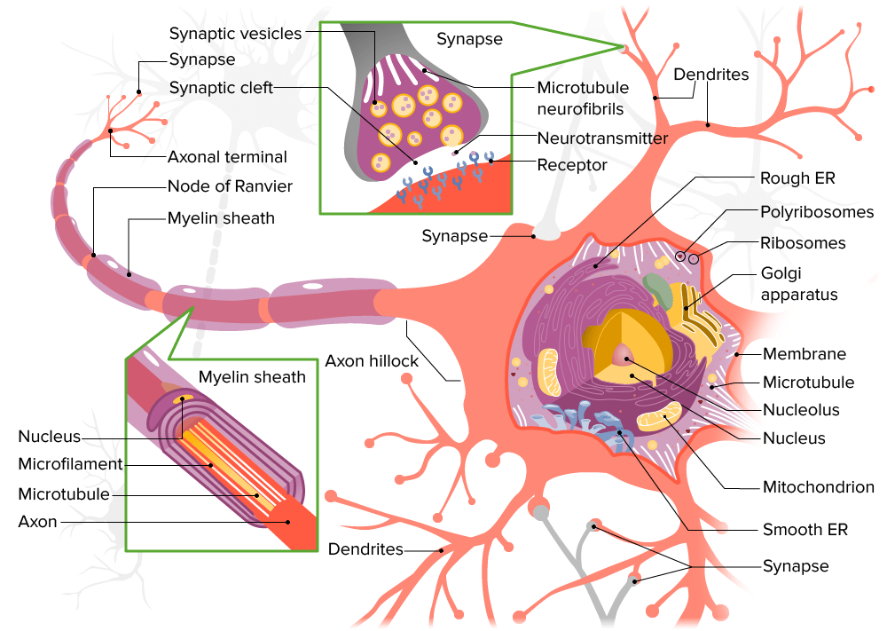
Parts of a neuron
Neurons consist of 3 main parts:
Dendrites:
Branched (tree-like) appendages (processes)
Receive signals from:
Axons of other neurons (via synapses)
Sensory epithelial cells
Environment
Cytoplasm contains:
Nissl bodies:
Basophilic granular regions
Made up of clusters of rough endoplasmic reticulumEndoplasmic reticulumA system of cisternae in the cytoplasm of many cells. In places the endoplasmic reticulum is continuous with the plasma membrane (cell membrane) or outer membrane of the nuclear envelope. If the outer surfaces of the endoplasmic reticulum membranes are coated with ribosomes, the endoplasmic reticulum is said to be rough-surfaced; otherwise it is said to be smooth-surfaced.The Cell: Organelles and ribosomesRibosomesMulticomponent ribonucleoprotein structures found in the cytoplasm of all cells, and in mitochondria, and plastids. They function in protein biosynthesis via genetic translation.The Cell: Organelles
MicrotubulesMicrotubulesSlender, cylindrical filaments found in the cytoskeleton of plant and animal cells. They are composed of the protein tubulin and are influenced by tubulin modulators.The Cell: Cytosol and Cytoskeleton and neurofilaments
Cell body:
Also called the soma or perikaryon
Contains:
NucleusNucleusWithin a eukaryotic cell, a membrane-limited body which contains chromosomes and one or more nucleoli (cell nucleolus). The nuclear membrane consists of a double unit-type membrane which is perforated by a number of pores; the outermost membrane is continuous with the endoplasmic reticulum. A cell may contain more than one nucleus.The Cell: Organelles:
Often large
Pale staining
Prominent nucleolusNucleolusWithin most types of eukaryotic cell nucleus, a distinct region, not delimited by a membrane, in which some species of rRNA are synthesized and assembled into ribonucleoprotein subunits of ribosomes. In the nucleolus rRNA is transcribed from a nucleolar organizer, i.e., a group of tandemly repeated chromosomal genes which encode rRNA and which are transcribed by RNA polymerase I.The Cell: Organelles
Some neurons are binuclear.
Rough endoplasmic reticulumEndoplasmic reticulumA system of cisternae in the cytoplasm of many cells. In places the endoplasmic reticulum is continuous with the plasma membrane (cell membrane) or outer membrane of the nuclear envelope. If the outer surfaces of the endoplasmic reticulum membranes are coated with ribosomes, the endoplasmic reticulum is said to be rough-surfaced; otherwise it is said to be smooth-surfaced.The Cell: Organelles
RibosomesRibosomesMulticomponent ribonucleoprotein structures found in the cytoplasm of all cells, and in mitochondria, and plastids. They function in protein biosynthesis via genetic translation.The Cell: Organelles and polyribosomes that synthesize:
LysosomesLysosomesA class of morphologically heterogeneous cytoplasmic particles in animal and plant tissues characterized by their content of hydrolytic enzymes and the structure-linked latency of these enzymes. The intracellular functions of lysosomes depend on their lytic potential. The single unit membrane of the lysosome acts as a barrier between the enzymes enclosed in the lysosome and the external substrate. The activity of the enzymes contained in lysosomes is limited or nil unless the vesicle in which they are enclosed is ruptured or undergoes membrane fusion.The Cell: Organelles
MitochondriaMitochondriaSemiautonomous, self-reproducing organelles that occur in the cytoplasm of all cells of most, but not all, eukaryotes. Each mitochondrion is surrounded by a double limiting membrane. The inner membrane is highly invaginated, and its projections are called cristae. Mitochondria are the sites of the reactions of oxidative phosphorylation, which result in the formation of ATP. They contain distinctive ribosomes, transfer RNAs; amino Acyl tRNA synthetases; and elongation and termination factors. Mitochondria depend upon genes within the nucleus of the cells in which they reside for many essential messenger RNAs. Mitochondria are believed to have arisen from aerobic bacteria that established a symbiotic relationship with primitive protoeukaryotes.The Cell: Organelles
MicrotubulesMicrotubulesSlender, cylindrical filaments found in the cytoskeleton of plant and animal cells. They are composed of the protein tubulin and are influenced by tubulin modulators.The Cell: Cytosol and Cytoskeleton and neurofilaments
Axon:
Cylindrical process:
Conducts nerve impulses to target cells
Usually, only 1 axon is present.
Collateral branches may be present to communicate with many target cells.
Structure:
Connected to the cell body by the axon hillock (short, pyramid-shaped area)
Initial segment:
Region between the axon hillock and point of myelination
Site where an action potentialAction PotentialAbrupt changes in the membrane potential that sweep along the cell membrane of excitable cells in response to excitation stimuli.Membrane Potential is generated (or not)
Axolemma: plasma membranePlasma membraneA cell membrane (also known as the plasma membrane or plasmalemma) is a biological membrane that separates the cell contents from the outside environment. A cell membrane is composed of a phospholipid bilayer and proteins that function to protect cellular DNA and mediate the exchange of ions and molecules.The Cell: Cell Membrane covering
Axoplasma contains cytoplasm and components such as:
Abundant mitochondriaMitochondriaSemiautonomous, self-reproducing organelles that occur in the cytoplasm of all cells of most, but not all, eukaryotes. Each mitochondrion is surrounded by a double limiting membrane. The inner membrane is highly invaginated, and its projections are called cristae. Mitochondria are the sites of the reactions of oxidative phosphorylation, which result in the formation of ATP. They contain distinctive ribosomes, transfer RNAs; amino Acyl tRNA synthetases; and elongation and termination factors. Mitochondria depend upon genes within the nucleus of the cells in which they reside for many essential messenger RNAs. Mitochondria are believed to have arisen from aerobic bacteria that established a symbiotic relationship with primitive protoeukaryotes.The Cell: Organelles → provide energy
MicrotubulesMicrotubulesSlender, cylindrical filaments found in the cytoskeleton of plant and animal cells. They are composed of the protein tubulin and are influenced by tubulin modulators.The Cell: Cytosol and Cytoskeleton → anterograde and retrograde transport between the cell body and axon
Neurofilaments → provide structural support to the cell
Note: Nissl bodies are absent.
Structure of a neuron
Image by Lecturio.
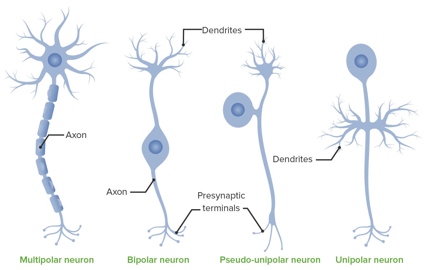
Anatomic characterization
Neurons may be classified by the number of processes (axon and dendrites) attached to the cell body.
Multipolar neurons:
Contain > 2 processes
Include:
A single axon on 1 end
Many dendrites connecting to the cell body
Most commonly found in:
BrainBrainThe part of central nervous system that is contained within the skull (cranium). Arising from the neural tube, the embryonic brain is comprised of three major parts including prosencephalon (the forebrain); mesencephalon (the midbrain); and rhombencephalon (the hindbrain). The developed brain consists of cerebrum; cerebellum; and other structures in the brain stem.Nervous System: Anatomy, Structure, and Classification
Spinal cordSpinal cordThe spinal cord is the major conduction pathway connecting the brain to the body; it is part of the CNS. In cross section, the spinal cord is divided into an H-shaped area of gray matter (consisting of synapsing neuronal cell bodies) and a surrounding area of white matter (consisting of ascending and descending tracts of myelinated axons). Spinal Cord: Anatomy
Examples:
Motor neurons
Interneurons
Pyramidal cells:
Located in the cerebral cortexCerebral cortexThe cerebral cortex is the largest and most developed part of the human brain and CNS. Occupying the upper part of the cranial cavity, the cerebral cortex has 4 lobes and is divided into 2 hemispheres that are joined centrally by the corpus callosum. Cerebral Cortex: Anatomy
Axons project down to the spinal cordSpinal cordThe spinal cord is the major conduction pathway connecting the brain to the body; it is part of the CNS. In cross section, the spinal cord is divided into an H-shaped area of gray matter (consisting of synapsing neuronal cell bodies) and a surrounding area of white matter (consisting of ascending and descending tracts of myelinated axons). Spinal Cord: Anatomy → communicate with ventral hornVentral hornOne of three central columns of the spinal cord. It is composed of gray matter spinal laminae VIII and ix.Brown-Séquard Syndrome cells → movement of skeletal musclesSkeletal musclesA subtype of striated muscle, attached by tendons to the skeleton. Skeletal muscles are innervated and their movement can be consciously controlled. They are also called voluntary muscles.Muscle Tissue: Histology
Located in the cerebellar cortexCerebellar cortexThe superficial gray matter of the cerebellum. It consists of two main layers, the stratum moleculare and the stratum granulosum.Cerebellum: Anatomy
Responsible for controlling and coordinating motor movements
RetinaRetinaThe ten-layered nervous tissue membrane of the eye. It is continuous with the optic nerve and receives images of external objects and transmits visual impulses to the brain. Its outer surface is in contact with the choroid and the inner surface with the vitreous body. The outermost layer is pigmented, whereas the inner nine layers are transparent.Eye: Anatomy
Examples of multipolar neurons: Pyramidal and Purkinje cells. Notice the multiple processes extending from the cell body.
Image by Lecturio.
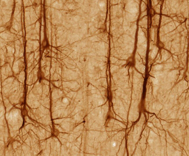
Pyramidal neurons in the cerebral cortex stained with a monoclonal antibody to neurofilament protein (SMI32): The somas (bodies) appear almost triangular with multiple dendrites attached, which are connected to long axons.
Image: “SMI32-stained pyramidal neurons in cerebral cortex” by UC Regents Davis campus. License: CC BY 3.0
Functional classification
Neurons may also be classified based on their functional role and the direction in which they transmit signals (toward or away from the CNS).
Sense touch, temperature, painPainAn unpleasant sensation induced by noxious stimuli which are detected by nerve endings of nociceptive neurons.Pain: Types and Pathways, pressure, proprioceptionProprioceptionSensory functions that transduce stimuli received by proprioceptive receptors in joints, tendons, muscles, and the inner ear into neural impulses to be transmitted to the central nervous system. Proprioception provides sense of stationary positions and movements of one’s body parts, and is important in maintaining kinesthesia and postural balance.Neurological Examination
Stimuli obtained from receptorsReceptorsReceptors are proteins located either on the surface of or within a cell that can bind to signaling molecules known as ligands (e.g., hormones) and cause some type of response within the cell.Receptors in skinSkinThe skin, also referred to as the integumentary system, is the largest organ of the body. The skin is primarily composed of the epidermis (outer layer) and dermis (deep layer). The epidermis is primarily composed of keratinocytes that undergo rapid turnover, while the dermis contains dense layers of connective tissue.Skin: Structure and Functions, skeletal muscle, tendons, and joints
Transmit visceral sensations (e.g., intestinal distension, ischemiaIschemiaA hypoperfusion of the blood through an organ or tissue caused by a pathologic constriction or obstruction of its blood vessels, or an absence of blood circulation.Ischemic Cell Damage)
Stimuli obtained from internal organs
Motor neurons:
Efferent signals: move toward the periphery
Conduct impulses to peripheral targets:
Somatic (voluntary) efferent neuronsEfferent neuronsNeurons which send impulses peripherally to activate muscles or secretory cells.Autonomic Nervous System: Anatomy innervate skeletal musclesSkeletal musclesA subtype of striated muscle, attached by tendons to the skeleton. Skeletal muscles are innervated and their movement can be consciously controlled. They are also called voluntary muscles.Muscle Tissue: Histology.
Smooth musclesSmooth musclesUnstriated and unstriped muscle, one of the muscles of the internal organs, blood vessels, hair follicles, etc. Contractile elements are elongated, usually spindle-shaped cells with centrally located nuclei. Smooth muscle fibers are bound together into sheets or bundles by reticular fibers and frequently elastic nets are also abundant.Muscle Tissue: Histology
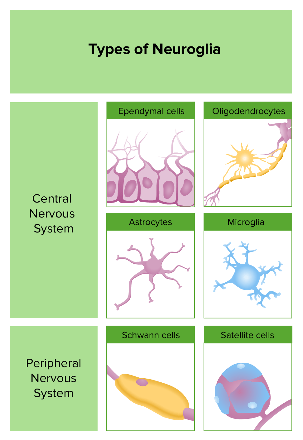
Neuroglia, also known as glial cells, are the most abundant cells in the CNS.
Have multiple functions and provide a suitable environment for neuron activity
Unlike neurons, neuroglia maintain the ability to undergo cell divisionCell DivisionA type of cell nucleus division by means of which the two daughter nuclei normally receive identical complements of the number of chromosomes of the somatic cells of the species.Cell Cycle.
Location
Neuroglia can be classified based on their location within the nervous systemNervous systemThe nervous system is a small and complex system that consists of an intricate network of neural cells (or neurons) and even more glial cells (for support and insulation). It is divided according to its anatomical components as well as its functional characteristics. The brain and spinal cord are referred to as the central nervous system, and the branches of nerves from these structures are referred to as the peripheral nervous system.Nervous System: Anatomy, Structure, and Classification.
CNS:
Astrocytes
Ependymal cells
Microglia
Oligodendrocytes
PNS:
Schwann cells
Satellite glial cells
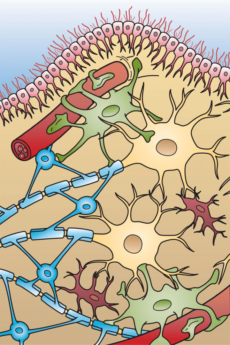
Types of neuroglia and their locations:
Ependymal cells are found only in the CNS and in small subarachnoid spaces, undertaking an epithelial-like function. Astrocytes supply neurons with nutrients and induce the formation of endothelial tight junctions, which play an important role in the
blood–brain barrier. They also fill the extracellular space of the CNS. Satellite cells are involved in the lining of the somas of neurons in the PNS. Schwann cells and microglia accelerate the rate of conduction.
Image by Lecturio.
Image showing 4 different types of glial cells in the CNS: Ependymal cells (light pink), astrocytes (green), microglial cells (red), and oligodendrocytes (light blue; functionally similar to Schwann cells in the PNS)
Image: “Glial Cell Types” by Holly Fischer. License: CC BY 3.0
Astrocytes
Location:
CNS
Subdivided into:
FibrousFibrousFibrocystic Change astrocytes: mainly in the white matterWhite MatterThe region of central nervous system that appears lighter in color than the other type, gray matter. It mainly consists of myelinated nerve fibers and contains few neuronal cell bodies or dendrites.Brown-Séquard Syndrome
Protoplasmic astrocytes: mainly in the gray matterGray matterRegion of central nervous system that appears darker in color than the other type, white matter. It is composed of neuronal cell bodies; neuropil; glial cells and capillaries but few myelinated nerve fibers.Cerebral Cortex: Anatomy
Features:
Largest of the neuroglia
Star shaped due to multiple radiating processes
Structures:
Astrocytic endfeet:
Form connections with other cells/structures
Perivascular: surround capillariesCapillariesCapillaries are the primary structures in the circulatory system that allow the exchange of gas, nutrients, and other materials between the blood and the extracellular fluid (ECF). Capillaries are the smallest of the blood vessels. Because a capillary diameter is so small, only 1 RBC may pass through at a time.Capillaries: Histology (important component of the blood–brain barrierBlood–Brain BarrierMeningitis in Children)
Perineuronal: surround neurons
Glial filaments:
Cytoplasmic components
Bundles of intermediate filamentsIntermediate filamentsCytoplasmic filaments intermediate in diameter (about 10 nanometers) between the microfilaments and the microtubules. They may be composed of any of a number of different proteins and form a ring around the cell nucleus.The Cell: Cytosol and Cytoskeleton that reinforce cell structure
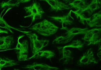
Contain glial fibrillary acid protein (GFAP) → important marker
Glycogen granules:
Cytoplasmic component
Can be broken down into glucoseGlucoseA primary source of energy for living organisms. It is naturally occurring and is found in fruits and other parts of plants in its free state. It is used therapeutically in fluid and nutrient replacement.Lactose Intolerance → energy
Functions:
Connect neurons to:
CapillariesCapillariesCapillaries are the primary structures in the circulatory system that allow the exchange of gas, nutrients, and other materials between the blood and the extracellular fluid (ECF). Capillaries are the smallest of the blood vessels. Because a capillary diameter is so small, only 1 RBC may pass through at a time.Capillaries: Histology
Pia materPia materThe innermost layer of the three meninges covering the brain and spinal cord. It is the fine vascular membrane that lies under the arachnoid and the dura mater.Meninges: Anatomy
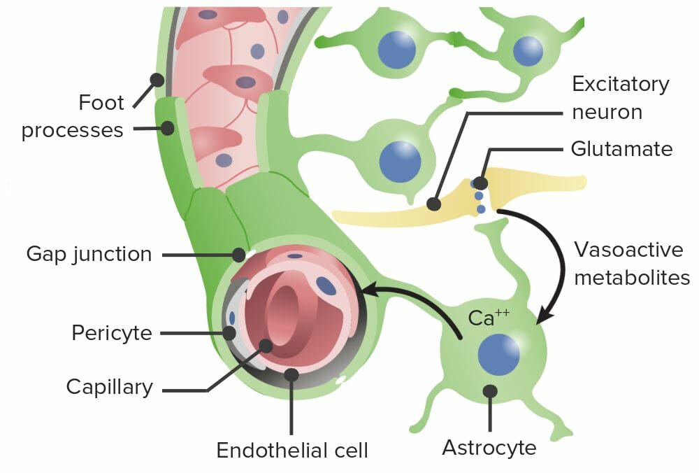
Control the environment:
Regulating cerebral blood flowBlood flowBlood flow refers to the movement of a certain volume of blood through the vasculature over a given unit of time (e.g., mL per minute).Vascular Resistance, Flow, and Mean Arterial Pressure (via CaCACondylomata acuminata are a clinical manifestation of genital HPV infection. Condylomata acuminata are described as raised, pearly, flesh-colored, papular, cauliflower-like lesions seen in the anogenital region that may cause itching, pain, or bleeding.Condylomata Acuminata (Genital Warts)2+ signaling)
Buffering extracellular ion concentrations (e.g., K+)
Clearing excess neurotransmitters
Releasing neuroactive molecules (e.g., enkephalinsEnkephalinsOne of the three major families of endogenous opioid peptides. The enkephalins are pentapeptides that are widespread in the central and peripheral nervous systems and in the adrenal medulla.Receptors and Neurotransmitters of the CNS, endothelinsEndothelins21-amino-acid peptides produced by vascular endothelial cells and functioning as potent vasoconstrictors. The endothelin family consists of three members, endothelin-1; endothelin-2; and endothelin-3. All three peptides contain 21 amino acids, but vary in amino acid composition. The three peptides produce vasoconstrictor and pressor responses in various parts of the body. However, the quantitative profiles of the pharmacological activities are considerably different among the three isopeptides.Hemostasis, somatostatinSomatostatinA 14-amino acid peptide named for its ability to inhibit pituitary growth hormone release, also called somatotropin release-inhibiting factor. It is expressed in the central and peripheral nervous systems, the gut, and other organs. SRIF can also inhibit the release of thyroid-stimulating hormone; prolactin; insulin; and glucagon besides acting as a neurotransmitter and neuromodulator. In a number of species including humans, there is an additional form of somatostatin, srif-28 with a 14-amino acid extension at the n-terminal.Gastrointestinal Secretions)
Transfer molecules to neurons:
Ions from the blood (via endfeet)
Lactate (after conversion from glucoseGlucoseA primary source of energy for living organisms. It is naturally occurring and is found in fruits and other parts of plants in its free state. It is used therapeutically in fluid and nutrient replacement.Lactose Intolerance)
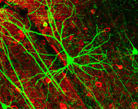
Astrocytes can be identified because, unlike other mature glia, they express glial fibrillary acidic protein (GFAP): Here, astrocytes were identified using anti-GFAP antibodies with a fluorescent label.
Central canal of the spinal cordSpinal cordThe spinal cord is the major conduction pathway connecting the brain to the body; it is part of the CNS. In cross section, the spinal cord is divided into an H-shaped area of gray matter (consisting of synapsing neuronal cell bodies) and a surrounding area of white matter (consisting of ascending and descending tracts of myelinated axons). Spinal Cord: Anatomy
Ventricles
Features:
Columnar epithelial cells
Some are ciliated
Generally have loose junctions
Specialized types connect to capillariesCapillariesCapillaries are the primary structures in the circulatory system that allow the exchange of gas, nutrients, and other materials between the blood and the extracellular fluid (ECF). Capillaries are the smallest of the blood vessels. Because a capillary diameter is so small, only 1 RBC may pass through at a time.Capillaries: Histology:
ChoroidChoroidThe thin, highly vascular membrane covering most of the posterior of the eye between the retina and sclera.Eye: Anatomy epithelial cells
Tanycytes
ChoroidChoroidThe thin, highly vascular membrane covering most of the posterior of the eye between the retina and sclera.Eye: Anatomy cells are connected together by tight junctionsTight junctionsCell-cell junctions that seal adjacent epithelial cells together, preventing the passage of most dissolved molecules from one side of the epithelial sheet to the other.The Cell: Cell Junctions → create the blood–CSF barrier
Tanycytes have:
Long processes
Large endfeet
Functions:
Cilia facilitate the movement of CSF.
ChoroidChoroidThe thin, highly vascular membrane covering most of the posterior of the eye between the retina and sclera.Eye: Anatomy epithelial cells of the choroidChoroidThe thin, highly vascular membrane covering most of the posterior of the eye between the retina and sclera.Eye: Anatomy plexus produce CSF.
Tanycytes facilitate the transport of hormonesHormonesHormones are messenger molecules that are synthesized in one part of the body and move through the bloodstream to exert specific regulatory effects on another part of the body. Hormones play critical roles in coordinating cellular activities throughout the body in response to the constant changes in both the internal and external environments. Hormones: Overview and Types.
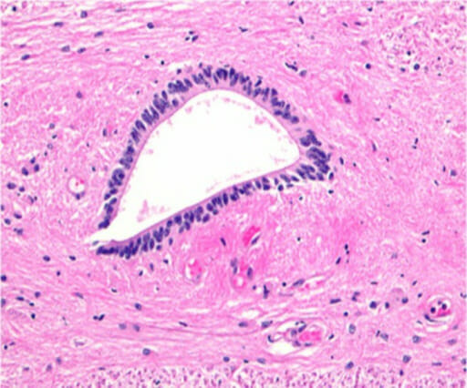
Columnar ependymal cells lining the central canal of the spinal cord
Image: “Histological image (H&E) of the human central canal” by Erfanul Saker, Brandon M Henry, Krzysztof A Tomaszewski, Marios Loukas, Joe Iwanaga, Rod J Oskouian, and R. Shane Tubbs. License: CC BY 3.0
Throughout the brainBrainThe part of central nervous system that is contained within the skull (cranium). Arising from the neural tube, the embryonic brain is comprised of three major parts including prosencephalon (the forebrain); mesencephalon (the midbrain); and rhombencephalon (the hindbrain). The developed brain consists of cerebrum; cerebellum; and other structures in the brain stem.Nervous System: Anatomy, Structure, and Classification and spinal cordSpinal cordThe spinal cord is the major conduction pathway connecting the brain to the body; it is part of the CNS. In cross section, the spinal cord is divided into an H-shaped area of gray matter (consisting of synapsing neuronal cell bodies) and a surrounding area of white matter (consisting of ascending and descending tracts of myelinated axons). Spinal Cord: Anatomy
Features:
Small
Elongated
Short processes (when activated, processes retract → the cell appears similar to a macrophage)
Dense, elongated nuclei
Functions:
Phagocytic cells that are important for:
InflammationInflammationInflammation is a complex set of responses to infection and injury involving leukocytes as the principal cellular mediators in the body’s defense against pathogenic organisms. Inflammation is also seen as a response to tissue injury in the process of wound healing. The 5 cardinal signs of inflammation are pain, heat, redness, swelling, and loss of function. Inflammation:
Release of inflammatory mediators
Act as antigen-presenting cellsAntigen-presenting cellsA heterogeneous group of immunocompetent cells that mediate the cellular immune response by processing and presenting antigens to the T-cells. Traditional antigen-presenting cells include macrophages; dendritic cells; langerhans cells; and B-lymphocytes. Follicular dendritic cells are not traditional antigen-presenting cells, but because they hold antigen on their cell surface in the form of immune complexes for b-cell recognition they are considered so by some authors.Adaptive Immune Response
Repair
Removal of cellular debris
Derived from monocytesMonocytesLarge, phagocytic mononuclear leukocytes produced in the vertebrate bone marrow and released into the blood; contain a large, oval or somewhat indented nucleus surrounded by voluminous cytoplasm and numerous organelles.Innate Immunity: Phagocytes and Antigen Presentation
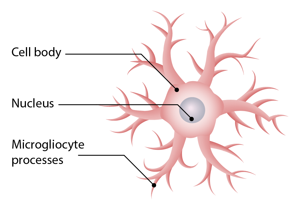
Structure of microglia
Image by Lecturio.
Oligodendrocytes and Schwann cells
The neuroglia listed below produce myelin but differ in their location within the nervous systemNervous systemThe nervous system is a small and complex system that consists of an intricate network of neural cells (or neurons) and even more glial cells (for support and insulation). It is divided according to its anatomical components as well as its functional characteristics. The brain and spinal cord are referred to as the central nervous system, and the branches of nerves from these structures are referred to as the peripheral nervous system.Nervous System: Anatomy, Structure, and Classification.
Oligodendrocytes:
Location:
CNS
Cell processes wrap around axons.
Subdivided into:
Interfascicular oligodendrocytes: mainly found in white matterWhite MatterThe region of central nervous system that appears lighter in color than the other type, gray matter. It mainly consists of myelinated nerve fibers and contains few neuronal cell bodies or dendrites.Brown-Séquard Syndrome
Satellite oligodendrocytes: mainly found in gray matterGray matterRegion of central nervous system that appears darker in color than the other type, white matter. It is composed of neuronal cell bodies; neuropil; glial cells and capillaries but few myelinated nerve fibers.Cerebral Cortex: Anatomy
Functions:
1 cell branches to myelinate many axons.
Satellite oligodendrocytes:
Not directly involved in myelination
Possibly regulate extracellular fluidExtracellular fluidThe fluid of the body that is outside of cells. It is the external environment for the cells.Body Fluid Compartments
Image of an oligodendrocyte in the process of myelinating axons
Image by Lecturio.
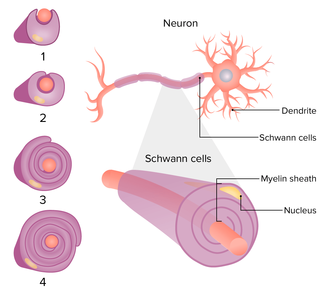
Schwann cells:
Location:
PNS
Cell wraps around axons.
Functions:
1 cell forms myelin for 1 segment of an axon.
Plays a role in the regenerationRegenerationThe physiological renewal, repair, or replacement of tissue.Wound Healing of damaged axons
Myelin sheath:
Composed of:
ProteinsProteinsLinear polypeptides that are synthesized on ribosomes and may be further modified, crosslinked, cleaved, or assembled into complex proteins with several subunits. The specific sequence of amino acids determines the shape the polypeptide will take, during protein folding, and the function of the protein.Energy Homeostasis
LipidsLipidsLipids are a diverse group of hydrophobic organic molecules, which include fats, oils, sterols, and waxes.Fatty Acids and Lipids
Insulates axons → ↑ velocity of action potentials
Separates axons from the extracellular space
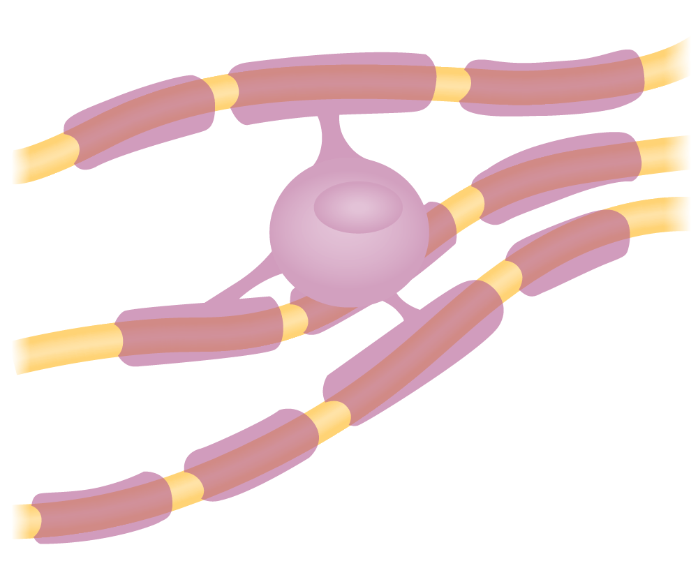
Myelination of an axon: Rotation of the Schwann cell around the axon forms an envelope of myelin sheath around the axon.
Image by Lecturio.
Satellite glial cells
Location:
PNS (ganglia)
Cover neuronal cell bodies
Functions:
Not entirely known, but likely similar to astrocytes
May include:
Structural role
Maintenance of chemical homeostasisHomeostasisThe processes whereby the internal environment of an organism tends to remain balanced and stable.Cell Injury and Death
Potential contribution to painPainAn unpleasant sensation induced by noxious stimuli which are detected by nerve endings of nociceptive neurons.Pain: Types and Pathways
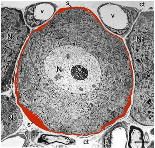
Low-magnification electron micrograph shows a satellite glial cell sheath (red) enveloping a sensory neuron.
N1: sensory neuron
s: satellite glial cell sheath
v: blood vessel
N2, N3, N4: adjacent neurons
ct: connective tissue space
Image: “Low magnification electron micrograph of mouse DRG” by Laboratory of Experimental Surgery, Hadassah-Hebrew University Medical Center, Mount Scopus Jerusalem, Israel. License: CC BY 4.0
PNS: formed when the Schwann cell wraps around axons
CNS: formed by oligodendrite processes
Nodes of Ranvier are present.
Conduction of nerve impulses is faster.
Appear white
Includes group A and B fibers:
Group A fibers are subdivided into:
A-alpha: innervate primary receptorsReceptorsReceptors are proteins located either on the surface of or within a cell that can bind to signaling molecules known as ligands (e.g., hormones) and cause some type of response within the cell.Receptors of the muscle spindle and Golgi tendon organ
A-beta: innervate secondary receptorsReceptorsReceptors are proteins located either on the surface of or within a cell that can bind to signaling molecules known as ligands (e.g., hormones) and cause some type of response within the cell.Receptors of the muscle spindle and cutaneous mechanoreceptors
A-delta: free nerve endings that transmit painPainAn unpleasant sensation induced by noxious stimuli which are detected by nerve endings of nociceptive neurons.Pain: Types and Pathways stimuli (pressure and temperature)
A-gamma: motor neurons that control intrinsic activation of the muscle spindle
Axons are separated by astrocyteAstrocyteA class of large neuroglial (macroglial) cells in the central nervous system – the largest and most numerous neuroglial cells in the brain and spinal cord. Astrocytes (from ‘star’ cells) are irregularly shaped with many long processes, including those with ‘end feet’ which form the glial (limiting) membrane and directly and indirectly contribute to the blood-brain barrier. They regulate the extracellular ionic and chemical environment, and ‘reactive astrocytes’ (along with microglia) respond to injury.Astrocytoma processes.
Nodes of Ranvier are absent.
Conduction of nerve impulses is slower.
Appear gray
Includes group C fibers: relay information from thermal, mechanical, and chemical stimuli
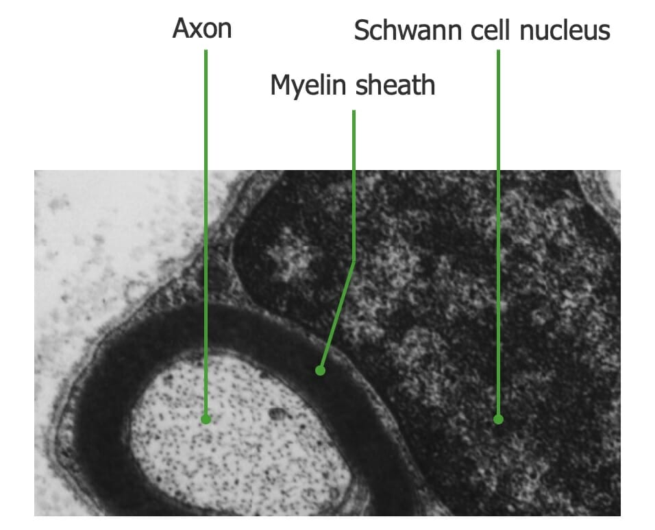
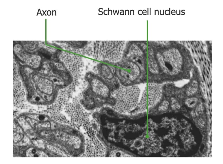
Electron microscope image of a myelinated axon: The thick, black structure surrounding the axon is the myelin sheath, which is formed by a Schwann cell.
Image by Lecturio.
Electron microscope image of an unmyelinated nerve fiber. Notice that a Schwann cell is associated with multiple axons, but these axons are not myelinated.
Image by Lecturio.
Peripheral nerves
Nerves are formed by bundles (fascicles) of sensory and motor nerve fibers.
The fascicles are held together by layers of connective tissueConnective tissueConnective tissues originate from embryonic mesenchyme and are present throughout the body except inside the brain and spinal cord. The main function of connective tissues is to provide structural support to organs. Connective tissues consist of cells and an extracellular matrix.Connective Tissue: Histology.
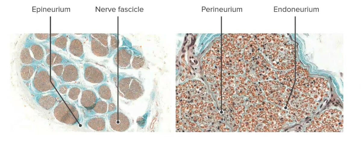
Epineurium:
Outer layer of dense, fibrousFibrousFibrocystic Changeconnective tissueConnective tissueConnective tissues originate from embryonic mesenchyme and are present throughout the body except inside the brain and spinal cord. The main function of connective tissues is to provide structural support to organs. Connective tissues consist of cells and an extracellular matrix.Connective Tissue: Histology
Histology of peripheral nerves demonstrating the different layers of connective tissue: The epineurium is the outermost layer, the perineurium surrounds the nerve fascicle, and the endoneurium (innermost layer) surrounds individual myelinated axons (or groups of unmyelinated axons).
Image by Lecturio.
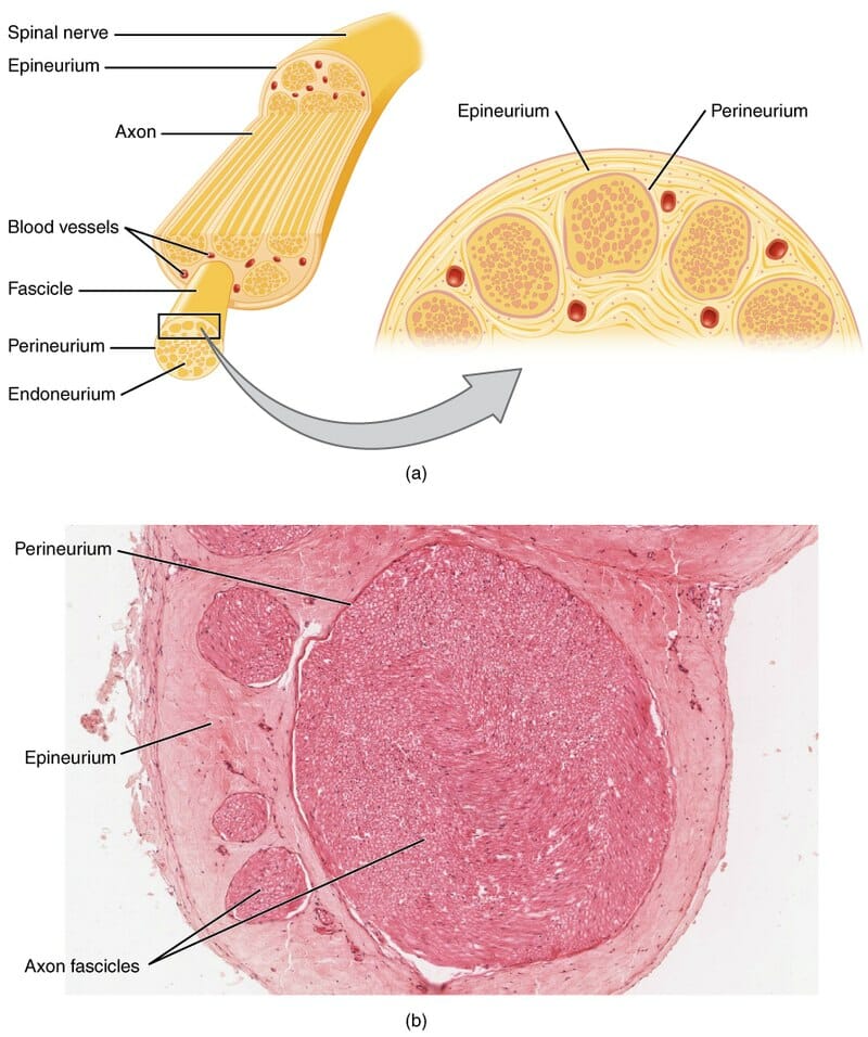
Cross-section of a nerve: The top figure (a) demonstrates the different components and layers within a peripheral nerve. These components are also visualized in a histologic specimen (b).
Image: “Cross-section of a nerve” by OpenStax College – Anatomy & Physiology. License: CC BY 3.0
Ganglia
The neuronal cell bodies of nerve fibers can reside in the CNS (brainBrainThe part of central nervous system that is contained within the skull (cranium). Arising from the neural tube, the embryonic brain is comprised of three major parts including prosencephalon (the forebrain); mesencephalon (the midbrain); and rhombencephalon (the hindbrain). The developed brain consists of cerebrum; cerebellum; and other structures in the brain stem.Nervous System: Anatomy, Structure, and Classification, spinal cordSpinal cordThe spinal cord is the major conduction pathway connecting the brain to the body; it is part of the CNS. In cross section, the spinal cord is divided into an H-shaped area of gray matter (consisting of synapsing neuronal cell bodies) and a surrounding area of white matter (consisting of ascending and descending tracts of myelinated axons). Spinal Cord: Anatomy, or cranial nerve ganglia) or in the PNS (peripheral ganglia).
General:
A ganglion is a collection of somas, which may also contain the following:
Satellite cells
Connective tissueConnective tissueConnective tissues originate from embryonic mesenchyme and are present throughout the body except inside the brain and spinal cord. The main function of connective tissues is to provide structural support to organs. Connective tissues consist of cells and an extracellular matrix.Connective Tissue: HistologycapsuleCapsuleAn envelope of loose gel surrounding a bacterial cell which is associated with the virulence of pathogenic bacteria. Some capsules have a well-defined border, whereas others form a slime layer that trails off into the medium. Most capsules consist of relatively simple polysaccharides but there are some bacteria whose capsules are made of polypeptides.Bacteroides
Basement membraneBasement membraneA darkly stained mat-like extracellular matrix (ecm) that separates cell layers, such as epithelium from endothelium or a layer of connective tissue. The ecm layer that supports an overlying epithelium or endothelium is called basal lamina. Basement membrane (bm) can be formed by the fusion of either two adjacent basal laminae or a basal lamina with an adjacent reticular lamina of connective tissue. Bm, composed mainly of type IV collagen; glycoprotein laminin; and proteoglycan, provides barriers as well as channels between interacting cell layers.Thin Basement Membrane Nephropathy (TBMN)
Sympathetic: sympathetic trunk, close to the spinal cordSpinal cordThe spinal cord is the major conduction pathway connecting the brain to the body; it is part of the CNS. In cross section, the spinal cord is divided into an H-shaped area of gray matter (consisting of synapsing neuronal cell bodies) and a surrounding area of white matter (consisting of ascending and descending tracts of myelinated axons). Spinal Cord: Anatomy
Parasympathetic: near/within visceral organs
Characteristics:
Contains neuronal cell bodies with large dendritic trees (multipolar)
Satellite cells are less prominent.
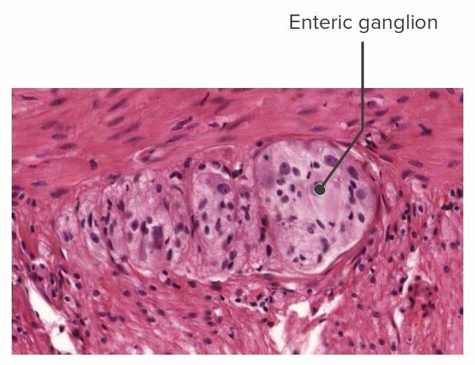
Enteric ganglia:
Location: wall of the GI tract
Characteristics:
Very small compared with other ganglia types
Lack connective tissueConnective tissueConnective tissues originate from embryonic mesenchyme and are present throughout the body except inside the brain and spinal cord. The main function of connective tissues is to provide structural support to organs. Connective tissues consist of cells and an extracellular matrix.Connective Tissue: HistologycapsuleCapsuleAn envelope of loose gel surrounding a bacterial cell which is associated with the virulence of pathogenic bacteria. Some capsules have a well-defined border, whereas others form a slime layer that trails off into the medium. Most capsules consist of relatively simple polysaccharides but there are some bacteria whose capsules are made of polypeptides.Bacteroides
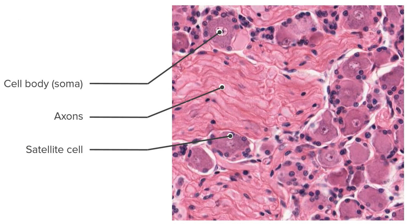
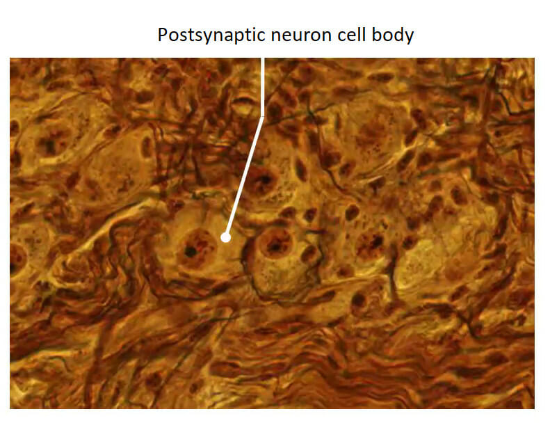
High-power photomicrograph of a histologic section of a dorsal root ganglion (hematoxylin and eosin staining)
White versus gray matterGray matterRegion of central nervous system that appears darker in color than the other type, white matter. It is composed of neuronal cell bodies; neuropil; glial cells and capillaries but few myelinated nerve fibers.Cerebral Cortex: Anatomy
The tissue of the CNS (brainBrainThe part of central nervous system that is contained within the skull (cranium). Arising from the neural tube, the embryonic brain is comprised of three major parts including prosencephalon (the forebrain); mesencephalon (the midbrain); and rhombencephalon (the hindbrain). The developed brain consists of cerebrum; cerebellum; and other structures in the brain stem.Nervous System: Anatomy, Structure, and Classification and spinal cordSpinal cordThe spinal cord is the major conduction pathway connecting the brain to the body; it is part of the CNS. In cross section, the spinal cord is divided into an H-shaped area of gray matter (consisting of synapsing neuronal cell bodies) and a surrounding area of white matter (consisting of ascending and descending tracts of myelinated axons). Spinal Cord: Anatomy) has a characteristic classification as white or gray matterGray matterRegion of central nervous system that appears darker in color than the other type, white matter. It is composed of neuronal cell bodies; neuropil; glial cells and capillaries but few myelinated nerve fibers.Cerebral Cortex: Anatomy.
White matterWhite MatterThe region of central nervous system that appears lighter in color than the other type, gray matter. It mainly consists of myelinated nerve fibers and contains few neuronal cell bodies or dendrites.Brown-Séquard Syndrome:
Gray matterGray matterRegion of central nervous system that appears darker in color than the other type, white matter. It is composed of neuronal cell bodies; neuropil; glial cells and capillaries but few myelinated nerve fibers.Cerebral Cortex: Anatomy generally contains:
Cell bodies and dendrites
Neuroglia
BrainBrainThe part of central nervous system that is contained within the skull (cranium). Arising from the neural tube, the embryonic brain is comprised of three major parts including prosencephalon (the forebrain); mesencephalon (the midbrain); and rhombencephalon (the hindbrain). The developed brain consists of cerebrum; cerebellum; and other structures in the brain stem.Nervous System: Anatomy, Structure, and Classification
Gray matterGray matterRegion of central nervous system that appears darker in color than the other type, white matter. It is composed of neuronal cell bodies; neuropil; glial cells and capillaries but few myelinated nerve fibers.Cerebral Cortex: Anatomy generally makes up the external layer of the brainBrainThe part of central nervous system that is contained within the skull (cranium). Arising from the neural tube, the embryonic brain is comprised of three major parts including prosencephalon (the forebrain); mesencephalon (the midbrain); and rhombencephalon (the hindbrain). The developed brain consists of cerebrum; cerebellum; and other structures in the brain stem.Nervous System: Anatomy, Structure, and Classification and includes:
Cerebellar cortexCerebellar cortexThe superficial gray matter of the cerebellum. It consists of two main layers, the stratum moleculare and the stratum granulosum.Cerebellum: Anatomy (outer layer) has 3 layers:
Golgi cellsGolgi cellsInhibitory interneurons embedded in the granular layer of the cerebellar cortex.Cerebellum: Anatomy (GABAergic interneurons)
Cerebral cortexCerebral cortexThe cerebral cortex is the largest and most developed part of the human brain and CNS. Occupying the upper part of the cranial cavity, the cerebral cortex has 4 lobes and is divided into 2 hemispheres that are joined centrally by the corpus callosum. Cerebral Cortex: Anatomy (outer layer) has 6 layers:
Molecular (outer) layer contains dendrites and axons from other layers.
Internal granular layer: similar to the external granular layer
Internal pyramidal layer: contains more pyramidal cell bodies
Multiform (inner) layer: contains fusiform cells
Basal nuclei/ganglia: located deep within the cerebral white matterWhite MatterThe region of central nervous system that appears lighter in color than the other type, gray matter. It mainly consists of myelinated nerve fibers and contains few neuronal cell bodies or dendrites.Brown-Séquard Syndrome
White matterWhite MatterThe region of central nervous system that appears lighter in color than the other type, gray matter. It mainly consists of myelinated nerve fibers and contains few neuronal cell bodies or dendrites.Brown-Séquard Syndrome generally makes up the internal region of the brainBrainThe part of central nervous system that is contained within the skull (cranium). Arising from the neural tube, the embryonic brain is comprised of three major parts including prosencephalon (the forebrain); mesencephalon (the midbrain); and rhombencephalon (the hindbrain). The developed brain consists of cerebrum; cerebellum; and other structures in the brain stem.Nervous System: Anatomy, Structure, and Classification and contains:
Nerve fibers
Neuroglia (mostly oligodendrites)
Blood vessels
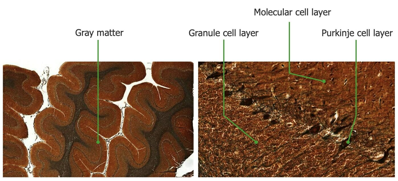
Histology of the cerebellum:
On the left, we can see the differentiation between the gray matter (lighter-colored layer) and the white matter (darker-stained inner region). On closer view of the gray matter, 3 distinct layers are seen that vary in composition, namely, molecular cell layer (made up of interneurons and neuron processes), the Purkinje cell layer (composed primarily of Purkinje cell bodies), and the granule cell layer (composed of granule and Golgi cells).
Image by Lecturio.
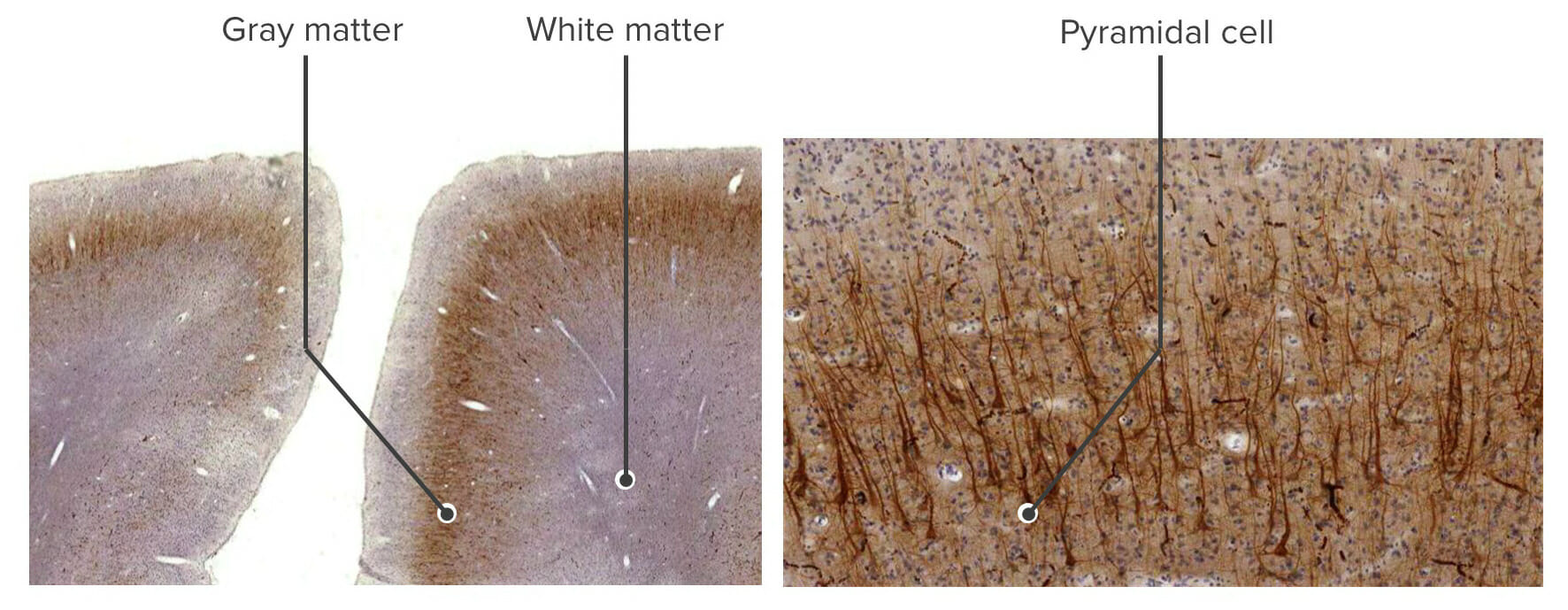
Histology of the cerebrum:
The left image shows the differentiation between gray matter and white matter. On closer examination of a portion of the gray matter (right), we can see that it is largely composed of pyramidal cell bodies.
Image by Lecturio.
Histology of the cerebrum: high-power image of a pyramidal neuron visualized using green fluorescent protein
Image: “Pyramidal neuron visualized by green fluorescent protein” by Nrets. License: CC BY 2.5
Spinal cordSpinal cordThe spinal cord is the major conduction pathway connecting the brain to the body; it is part of the CNS. In cross section, the spinal cord is divided into an H-shaped area of gray matter (consisting of synapsing neuronal cell bodies) and a surrounding area of white matter (consisting of ascending and descending tracts of myelinated axons). Spinal Cord: Anatomy
White matterWhite MatterThe region of central nervous system that appears lighter in color than the other type, gray matter. It mainly consists of myelinated nerve fibers and contains few neuronal cell bodies or dendrites.Brown-Séquard Syndrome (outermost layer):
Contains bundles of parallel ascending and descending axons (tracts)
Gray matterGray matterRegion of central nervous system that appears darker in color than the other type, white matter. It is composed of neuronal cell bodies; neuropil; glial cells and capillaries but few myelinated nerve fibers.Cerebral Cortex: Anatomy (innermost layer):
Dorsal hornDorsal hornOne of three central columns of the spinal cord. It is composed of gray matter spinal laminae i-vi.Brown-Séquard Syndrome (sensory):
Sensory neuronal axons enter the spinal cordSpinal cordThe spinal cord is the major conduction pathway connecting the brain to the body; it is part of the CNS. In cross section, the spinal cord is divided into an H-shaped area of gray matter (consisting of synapsing neuronal cell bodies) and a surrounding area of white matter (consisting of ascending and descending tracts of myelinated axons). Spinal Cord: Anatomy (cell bodies are in ganglia).
Interneurons
Ventral hornVentral hornOne of three central columns of the spinal cord. It is composed of gray matter spinal laminae VIII and ix.Brown-Séquard Syndrome (motor):
Somatic motor neuron cell bodies
Interneurons
Lateral hornLateral hornOne of three central columns of the spinal cord. It is composed of gray matter and is located laterally in lamina vii.Brown-Séquard Syndrome:
Found only in the thoracic and lumbar regions
Contains neurons of the sympathetic nervous systemNervous systemThe nervous system is a small and complex system that consists of an intricate network of neural cells (or neurons) and even more glial cells (for support and insulation). It is divided according to its anatomical components as well as its functional characteristics. The brain and spinal cord are referred to as the central nervous system, and the branches of nerves from these structures are referred to as the peripheral nervous system.Nervous System: Anatomy, Structure, and Classification
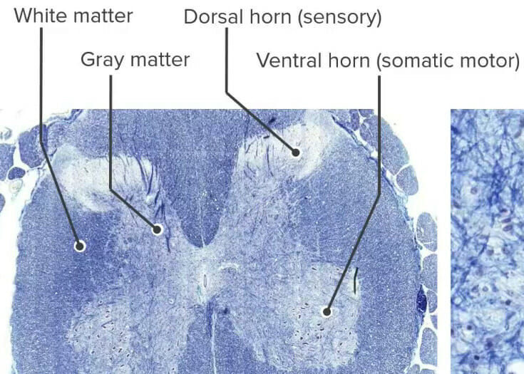
Histology of the spinal cord stained with Luxol fast blue, which stains myelinated fibers blue:
Notice that, unlike that in the brain, the white matter (containing myelinated axons) is located in the periphery and surrounding the gray matter (containing mostly neurons with scant myelinated axons). The gray matter has 3 regions containing neurons and interneurons, namely, the dorsal horn (sensory), lateral horn (sympathetic), and ventral horn (somatic motor), with each having different functions.
Tight junctionsTight junctionsCell-cell junctions that seal adjacent epithelial cells together, preventing the passage of most dissolved molecules from one side of the epithelial sheet to the other.The Cell: Cell Junctions:
Anchor capillary endothelial cells together
Create a relatively impermeable barrier to most substances and pathogens
Permeable to gases (e.g., O2, CO2)
Other solutes may require specific transporters.
PericytesPericytesUnique slender cells with multiple processes extending along the capillary vessel axis and encircling the vascular wall, also called mural cells. Pericytes are imbedded in the basement membrane shared with the endothelial cells of the vessel. Pericytes are important in maintaining vessel integrity, angiogenesis, and vascular remodeling.Capillaries: Histology:
Perivascular cells
Regulate capillary function and immune cell entry into the CNS
Podocytes from astrocytes encircle capillariesCapillariesCapillaries are the primary structures in the circulatory system that allow the exchange of gas, nutrients, and other materials between the blood and the extracellular fluid (ECF). Capillaries are the smallest of the blood vessels. Because a capillary diameter is so small, only 1 RBC may pass through at a time.Capillaries: Histology (perivascular endfeet).
Multiple sclerosisSclerosisA pathological process consisting of hardening or fibrosis of an anatomical structure, often a vessel or a nerve.Wilms Tumor: a chronic, inflammatory, autoimmune disease that leads to the destruction of oligodendrites and demyelinationDemyelinationMultiple Sclerosis of nerves in the CNS, resulting in axonal damage and degeneration that impair the transmission of action potentials. The clinical presentation varies widely but typically includes neurological symptoms that affect visionVisionOphthalmic Exam, motor functions, sensation, and autonomic function. The diagnosis is made via MRI of the entire CNS and examination of the CSF. Management involves corticosteroidsCorticosteroidsChorioretinitis for acute exacerbations and disease-modifying agents to slow disease progression.
Guillain-Barré syndromeGuillain-Barré syndromeGuillain-Barré syndrome (GBS), once thought to be a single disease process, is a family of immune-mediated polyneuropathies that occur after infections (e.g., with Campylobacter jejuni).Guillain-Barré Syndrome (GBSGBSAn acute inflammatory autoimmune neuritis caused by t cell- mediated cellular immune response directed towards peripheral myelin. Demyelination occurs in peripheral nerves and nerve roots. The process is often preceded by a viral or bacterial infection, surgery, immunization, lymphoma, or exposure to toxins. Common clinical manifestations include progressive weakness, loss of sensation, and loss of deep tendon reflexes. Weakness of respiratory muscles and autonomic dysfunction may occur.Polyneuropathy): a family of immune-mediated demyelinating polyneuropathiesPolyneuropathiesDiseases of multiple peripheral nerves simultaneously. Polyneuropathies usually are characterized by symmetrical, bilateral distal motor and sensory impairment with a graded increase in severity distally. The pathological processes affecting peripheral nerves include degeneration of the axon, myelin or both. The various forms of polyneuropathy are categorized by the type of nerve affected (e.g., sensory, motor, or autonomic), by the distribution of nerve injury (e.g., distal vs. Proximal), by nerve component primarily affected (e.g., demyelinating vs. axonal), by etiology, or by pattern of inheritance.Mononeuropathy and Plexopathy that occur after infectionsInfectionsInvasion of the host organism by microorganisms or their toxins or by parasites that can cause pathological conditions or diseases.Chronic Granulomatous Disease, in which the immune systemImmune systemThe body’s defense mechanism against foreign organisms or substances and deviant native cells. It includes the humoral immune response and the cell-mediated response and consists of a complex of interrelated cellular, molecular, and genetic components.Primary Lymphatic Organs attacks the myelin sheath and Schwann cells. Typical GBSGBSAn acute inflammatory autoimmune neuritis caused by t cell- mediated cellular immune response directed towards peripheral myelin. Demyelination occurs in peripheral nerves and nerve roots. The process is often preceded by a viral or bacterial infection, surgery, immunization, lymphoma, or exposure to toxins. Common clinical manifestations include progressive weakness, loss of sensation, and loss of deep tendon reflexes. Weakness of respiratory muscles and autonomic dysfunction may occur.Polyneuropathy is characterized by acute monophasic neuromuscular paralysisAcute Monophasic Neuromuscular ParalysisGuillain-Barré Syndrome, which is symmetric and ascending in progression. If the respiratory muscles are affected, GBSGBSAn acute inflammatory autoimmune neuritis caused by t cell- mediated cellular immune response directed towards peripheral myelin. Demyelination occurs in peripheral nerves and nerve roots. The process is often preceded by a viral or bacterial infection, surgery, immunization, lymphoma, or exposure to toxins. Common clinical manifestations include progressive weakness, loss of sensation, and loss of deep tendon reflexes. Weakness of respiratory muscles and autonomic dysfunction may occur.Polyneuropathy can progress to respiratory failureRespiratory failureRespiratory failure is a syndrome that develops when the respiratory system is unable to maintain oxygenation and/or ventilation. Respiratory failure may be acute or chronic and is classified as hypoxemic, hypercapnic, or a combination of the two. Respiratory Failure, which requires prolonged hospitalizationProlonged HospitalizationSurgical Infections. Management is mostly supportive and may require either plasma exchangePlasma exchangeRemoval of plasma and replacement with various fluids, e.g., fresh frozen plasma, plasma protein fractions (ppf), albumin preparations, dextran solutions, saline. Used in treatment of autoimmune diseases, immune complex diseases, diseases of excess plasma factors, and other conditions.Thrombotic Thrombocytopenic Purpura or IV immunoglobulinIv ImmunoglobulinDermatomyositis.
Gliomas: primary brainBrainThe part of central nervous system that is contained within the skull (cranium). Arising from the neural tube, the embryonic brain is comprised of three major parts including prosencephalon (the forebrain); mesencephalon (the midbrain); and rhombencephalon (the hindbrain). The developed brain consists of cerebrum; cerebellum; and other structures in the brain stem.Nervous System: Anatomy, Structure, and Classification tumors derived from neuroglia, which include benignBenignFibroadenoma astrocytomas, glioblastoma multiformeGlioblastoma multiformeGlioblastoma multiforme is a high-grade astrocytoma, an aggressive brain tumor arising from astrocytes, with an unknown cause and a poorly understood link to risk factors. There are two main types: primary, a more aggressive form seen more commonly in older patients, and secondary, developing from lower-grade astrocytomas and seen more commonly in younger patients.Glioblastoma Multiforme, oligodendrogliomas, and ependymomas. Astrocytomas are the most common form of glioma. Presenting symptoms vary based on the location of the tumorTumorInflammation and can manifest as focal neurologic deficitsNeurologic DeficitsHigh-Risk Headaches, encephalopathyEncephalopathyHyper-IgM Syndrome, personality changes, or seizuresSeizuresA seizure is abnormal electrical activity of the neurons in the cerebral cortex that can manifest in numerous ways depending on the region of the brain affected. Seizures consist of a sudden imbalance that occurs between the excitatory and inhibitory signals in cortical neurons, creating a net excitation. The 2 major classes of seizures are focal and generalized. Seizures. The diagnosis is based on clinical findings and is confirmed by an MRI followed by a biopsyBiopsyRemoval and pathologic examination of specimens from the living body.Ewing Sarcoma with molecular profiling. Treatment may involve surgical excision, radiationRadiationEmission or propagation of acoustic waves (sound), electromagnetic energy waves (such as light; radio waves; gamma rays; or x-rays), or a stream of subatomic particles (such as electrons; neutrons; protons; or alpha particles).Osteosarcoma therapy, and, for some tumors, chemotherapyChemotherapyOsteosarcoma.
Amyotrophic lateral sclerosisSclerosisA pathological process consisting of hardening or fibrosis of an anatomical structure, often a vessel or a nerve.Wilms Tumor: a sporadicSporadicSelective IgA Deficiency or inherited neurodegenerative disease of the upper motor neurons (UMNs) and lower motor neurons (LMNs). The exact mechanism is unknown but appears to be multifactorial. Amyotrophic lateral sclerosisSclerosisA pathological process consisting of hardening or fibrosis of an anatomical structure, often a vessel or a nerve.Wilms Tumor is characterized by signs and symptoms indicating the coexistence of UMN and LMN degeneration. The diagnosis is made clinically. Management is supportive and symptomatic, progressing to end-of-life care.
Parkinson diseaseParkinson diseaseParkinson’s disease (PD) is a chronic, progressive neurodegenerative disorder. Although the cause is unknown, several genetic and environmental risk factors are currently being studied. Individuals present clinically with resting tremor, bradykinesia, rigidity, and postural instability.Parkinson’s Disease (PDPDParkinson’s disease (PD) is a chronic, progressive neurodegenerative disorder. Although the cause is unknown, several genetic and environmental risk factors are currently being studied. Individuals present clinically with resting tremor, bradykinesia, rigidity, and postural instability.Parkinson’s Disease): a slowly progressive neurologic disorder caused by the loss of the secretionSecretionCoagulation Studies of the neurotransmitter dopamineDopamineOne of the catecholamine neurotransmitters in the brain. It is derived from tyrosine and is the precursor to norepinephrine and epinephrine. Dopamine is a major transmitter in the extrapyramidal system of the brain, and important in regulating movement.Receptors and Neurotransmitters of the CNS by dopamine-secreting cells in the substantia nigraSubstantia nigraThe black substance in the ventral midbrain or the nucleus of cells containing the black substance. These cells produce dopamine, an important neurotransmitter in regulation of the sensorimotor system and mood. The dark colored melanin is a by-product of dopamine synthesis.Basal Ganglia: Anatomy and basal gangliaBasal GangliaBasal ganglia are a group of subcortical nuclear agglomerations involved in movement, and are located deep to the cerebral hemispheres. Basal ganglia include the striatum (caudate nucleus and putamen), globus pallidus, substantia nigra, and subthalamic nucleus. Basal Ganglia: Anatomy of the brainBrainThe part of central nervous system that is contained within the skull (cranium). Arising from the neural tube, the embryonic brain is comprised of three major parts including prosencephalon (the forebrain); mesencephalon (the midbrain); and rhombencephalon (the hindbrain). The developed brain consists of cerebrum; cerebellum; and other structures in the brain stem.Nervous System: Anatomy, Structure, and Classification. DopamineDopamineOne of the catecholamine neurotransmitters in the brain. It is derived from tyrosine and is the precursor to norepinephrine and epinephrine. Dopamine is a major transmitter in the extrapyramidal system of the brain, and important in regulating movement.Receptors and Neurotransmitters of the CNS is responsible for synaptic transmissionSynaptic transmissionThe communication from a neuron to a target (neuron, muscle, or secretory cell) across a synapse. In chemical synaptic transmission, the presynaptic neuron releases a neurotransmitter that diffuses across the synaptic cleft and binds to specific synaptic receptors, activating them. The activated receptors modulate specific ion channels and/or second-messenger systems in the postsynaptic cell. In electrical synaptic transmission, electrical signals are communicated as an ionic current flow across electrical synapses.Synapses and Neurotransmission in the nerve pathways for the coordinationCoordinationCerebellar Disorders of smooth and focused activity of skeletal musclesSkeletal musclesA subtype of striated muscle, attached by tendons to the skeleton. Skeletal muscles are innervated and their movement can be consciously controlled. They are also called voluntary muscles.Muscle Tissue: Histology. Parkinson diseaseParkinson diseaseParkinson’s disease (PD) is a chronic, progressive neurodegenerative disorder. Although the cause is unknown, several genetic and environmental risk factors are currently being studied. Individuals present clinically with resting tremor, bradykinesia, rigidity, and postural instability.Parkinson’s Disease is characterized by resting tremorResting TremorParkinson’s Disease in the limbs, especially of the hands, rigidityRigidityContinuous involuntary sustained muscle contraction which is often a manifestation of basal ganglia diseases. When an affected muscle is passively stretched, the degree of resistance remains constant regardless of the rate at which the muscle is stretched. This feature helps to distinguish rigidity from muscle spasticity.Megacolon/stiffness in all muscles, slow movement (bradykinesiaBradykinesiaParkinson’s Disease), inability to initiate movement (akinesia), lack of spontaneous movements, festinating gaitGaitManner or style of walking.Neurological Examination, slurred speechSlurred SpeechCerebellar Disorders, and slowness of thought. Secondary PDPDParkinson’s disease (PD) is a chronic, progressive neurodegenerative disorder. Although the cause is unknown, several genetic and environmental risk factors are currently being studied. Individuals present clinically with resting tremor, bradykinesia, rigidity, and postural instability.Parkinson’s Disease and PD-type symptoms may also be caused by encephalitisEncephalitisEncephalitis is inflammation of the brain parenchyma caused by an infection, usually viral. Encephalitis may present with mild symptoms such as headache, fever, fatigue, and muscle and joint pain or with severe symptoms such as seizures, altered consciousness, and paralysis.Encephalitis, drugs used to treat neurologic disorders (especially the typical antipsychoticsTypical antipsychoticsAntipsychotics, also called neuroleptics, are used to treat psychotic disorders and alleviate agitation, mania, and aggression. Antipsychotics are notable for their use in treating schizophrenia and bipolar disorder and are divided into 1st-generation antipsychotics (FGAs) and atypical or 2nd-generation antipsychotics.First-Generation Antipsychotics (neuroleptics) used to treat schizophreniaSchizophreniaSchizophrenia is a chronic mental health disorder characterized by the presence of psychotic symptoms such as delusions or hallucinations. The signs and symptoms of schizophrenia are traditionally separated into 2 groups: positive (delusions, hallucinations, and disorganized speech or behavior) and negative (flat affect, avolition, anhedonia, poor attention, and alogia).Schizophrenia and other psychiatric disorders), toxins (e.g., 1-methyl-4-phenyl-1,2,3,6-tetrahydropyridine1-Methyl-4-Phenyl-1,2,3,6-TetrahydropyridineParkinson’s Disease (MPTPMPTPParkinson’s Disease)), and repetitive trauma.
RabiesRabiesAcute viral CNS infection affecting mammals, including humans. It is caused by rabies virus and usually spread by contamination with virus-laden saliva of bites inflicted by rabid animals. Important animal vectors include the dog, cat, bat, fox, raccoon, skunk, and wolf.Rabies Virus: a viral infection most often transmitted to humans through the bite of an infected animal. The rabiesRabiesAcute viral CNS infection affecting mammals, including humans. It is caused by rabies virus and usually spread by contamination with virus-laden saliva of bites inflicted by rabid animals. Important animal vectors include the dog, cat, bat, fox, raccoon, skunk, and wolf.Rabies VirusvirusVirusViruses are infectious, obligate intracellular parasites composed of a nucleic acid core surrounded by a protein capsid. Viruses can be either naked (non-enveloped) or enveloped. The classification of viruses is complex and based on many factors, including type and structure of the nucleoid and capsid, the presence of an envelope, the replication cycle, and the host range. Virology has a predilection for neural tissue and enters the peripheral motor and sensory nerves to travel to the CNS in a retrograde manner. There are 5 stages of disease in humans: incubationIncubationThe amount time between exposure to an infectious agent and becoming symptomatic.Rabies Virus, prodromeProdromeSymptoms that appear 24–48 hours prior to migraine onset.Migraine Headache, acute neurologic periodAcute neurologic periodRabies Virus, comaComaComa is defined as a deep state of unarousable unresponsiveness, characterized by a score of 3 points on the GCS. A comatose state can be caused by a multitude of conditions, making the precise epidemiology and prognosis of coma difficult to determine. Coma, and death. The diagnosis is made based on the detection of antibodiesAntibodiesImmunoglobulins (Igs), also known as antibodies, are glycoprotein molecules produced by plasma cells that act in immune responses by recognizing and binding particular antigens. The various Ig classes are IgG (the most abundant), IgM, IgE, IgD, and IgA, which differ in their biologic features, structure, target specificity, and distribution.Immunoglobulins: Types and Functions, antigens, or viral RNARNAA polynucleotide consisting essentially of chains with a repeating backbone of phosphate and ribose units to which nitrogenous bases are attached. RNA is unique among biological macromolecules in that it can encode genetic information, serve as an abundant structural component of cells, and also possesses catalytic activity.RNA Types and Structure in the biopsyBiopsyRemoval and pathologic examination of specimens from the living body.Ewing Sarcoma tissue, serum, CSF, or salivaSalivaThe clear, viscous fluid secreted by the salivary glands and mucous glands of the mouth. It contains mucins, water, organic salts, and ptyalin.Salivary Glands: Anatomy. No effective treatment for symptomatic disease exists; thus, prevention using human rabiesRabiesAcute viral CNS infection affecting mammals, including humans. It is caused by rabies virus and usually spread by contamination with virus-laden saliva of bites inflicted by rabid animals. Important animal vectors include the dog, cat, bat, fox, raccoon, skunk, and wolf.Rabies Virus immunoglobulin and vaccinationVaccinationVaccination is the administration of a substance to induce the immune system to develop protection against a disease. Unlike passive immunization, which involves the administration of pre-performed antibodies, active immunization constitutes the administration of a vaccine to stimulate the body to produce its own antibodies.Vaccination is the mainstay of management.
References
Cali, C., Agus, M., Gagnon, E., Hadwiger, M., & Magistretti, P. J. (2022). Glial cells: Architects and caretakers of the nervous system. Annual Review of Neuroscience, 45, 305-329.
Correa-Velloso, J. C., Gonçalves, M. C. B., & Fornari, R. V. (2024). Astrocyte calcium signaling in brain physiology and pathology. Frontiers in Cellular Neuroscience, 18, 1342854.
Dotiwala, A. K., & Samra, N. S. (2023). Anatomy, head and neck, blood brain barrier. In StatPearls. StatPearls Publishing.
Escartin, C., Galea, E., Lakatos, A., O’Callaghan, J. P., Petzold, G. C., Serrano-Pozo, A., Steinhäuser, C., Volterra, A., Carmignoto, G., Agarwal, A., Allen, N. J., Araque, A., Barbeito, L., Barzilai, A., Bergles, D. E., Bonvento, G., Butt, A. M., Chen, W. T., Cohen-Salmon, M., … Verkhratsky, A. (2021). Reactive astrocyte nomenclature, definitions, and future directions. Nature Neuroscience, 24(3), 312-325.
Ferreira, S., Pitres, D., Roux, F. S., & Couture, F. (2024). Blood-brain barrier: Structure, function, and role in brain homeostasis. Physiology Reviews, 104(2), 759-824.
Gibson, E. M., Geraghty, A. C., & Monje, M. (2023). Neuronal regulation of oligodendrocyte precursor cells in health and disease. Nature Reviews Neuroscience, 24(4), 209-226.
Junqueira, L. C., & Carneiro, J. (2022). Nervous tissue & the nervous system. In Basic Histology: Text and Atlas (15th ed.). McGraw-Hill.
Kidd, G. J., Ohno, N., & Trapp, B. D. (2022). The fundamental neuropathology of myelin and oligodendrocytes beyond repair. Experimental Neurology, 344, 113781.
Mescher, A. L. (2023). Nerve tissue & the nervous system. In Junqueira’s Basic Histology: Text and Atlas (16th ed., Lange Medical Books)*. McGraw-Hill Education.
Ovalle, W. K., & Nahirney, P. C. (2024). Nervous tissue. In Netter’s Essential Histology (3rd ed.)*. Elsevier.
Pawlina, W., & Ross, M. (2024). Nerve tissue. In Histology: A Text and Atlas (9th ed.)*. Wolters Kluwer Health.
Prinz, M., Masuda, T., Wheeler, M. A., & Quintana, F. J. (2021). Microglia and central nervous system-associated macrophages—from origin to disease modulation. Annual Review of Immunology, 39, 251-277.
Rollo, B. N., Zhang, D., Simkin, J. E., & Menheniott, T. R. (2021). Enteric nervous system development: Recent insights and emerging complexity. Journal of Cellular and Molecular Medicine, 25(8), 3522-3538.
Salzer, J. L., & Zalc, B. (2023). Myelination: Origin, dynamics, and evolution of a remarkable cellular assembly. Developmental Cell, 58(24), 2597-2617.
Stevens, A., & Lowe, J. S. (2023). Human Histology (5th ed.). Elsevier.
Stolt, C. C., Lommes, P., Friedrich, R. P., & Wegner, M. (2022). Transcriptional control of oligodendrocyte development and myelination in the central nervous system. Glia, 70(4), 658-673.
Voet, S., Prinz, M., & van Loo, G. (2023). Microglia in neuroinflammation: Key players in disease progression. Trends in Molecular Medicine, 29(9), 770-786.
Create your free account or log in to continue reading!