Achieve Mastery of Medical Concepts

Study for medical school and boards with Lecturio

Internuclear Ophthalmoplegia

Internuclear ophthalmoplegia Ophthalmoplegia Paralysis of one or more of the ocular muscles due to disorders of the eye muscles, neuromuscular junction, supporting soft tissue, tendons, or innervation to the muscles. Orbital and Preseptal Cellulitis (INO) is an ocular movement disorder that affects the conjugate horizontal gaze, meaning the eyes are incapable of moving in a simultaneous and coordinated manner on the horizontal plane. This disorder is usually caused by a lesion in the brain Brain The part of central nervous system that is contained within the skull (cranium). Arising from the neural tube, the embryonic brain is comprised of three major parts including prosencephalon (the forebrain); mesencephalon (the midbrain); and rhombencephalon (the hindbrain). The developed brain consists of cerebrum; cerebellum; and other structures in the brain stem. Nervous System: Anatomy, Structure, and Classification stem involving the medial longitudinal fasciculus (MLF) and is characterized by impaired adduction Adduction Examination of the Upper Limbs ipsilateral to the MLF lesion and abduction Abduction Examination of the Upper Limbs nystagmus Nystagmus Involuntary movements of the eye that are divided into two types, jerk and pendular. Jerk nystagmus has a slow phase in one direction followed by a corrective fast phase in the opposite direction, and is usually caused by central or peripheral vestibular dysfunction. Pendular nystagmus features oscillations that are of equal velocity in both directions and this condition is often associated with visual loss early in life. Albinism contralateral to the MLF lesion. Internuclear ophthalmoplegia Ophthalmoplegia Paralysis of one or more of the ocular muscles due to disorders of the eye muscles, neuromuscular junction, supporting soft tissue, tendons, or innervation to the muscles. Orbital and Preseptal Cellulitis is a clinical diagnosis. However, investigations involving neuroimaging Neuroimaging Non-invasive methods of visualizing the central nervous system, especially the brain, by various imaging modalities. Febrile Infant, especially MRI, help establish the etiology. Management of INO varies depending on the cause. The prognosis Prognosis A prediction of the probable outcome of a disease based on a individual's condition and the usual course of the disease as seen in similar situations. Non-Hodgkin Lymphomas mainly depends on the etiology. For instance, trauma patients Patients Individuals participating in the health care system for the purpose of receiving therapeutic, diagnostic, or preventive procedures. Clinician–Patient Relationship do not have a very favorable prognosis Prognosis A prediction of the probable outcome of a disease based on a individual's condition and the usual course of the disease as seen in similar situations. Non-Hodgkin Lymphomas, whereas patients Patients Individuals participating in the health care system for the purpose of receiving therapeutic, diagnostic, or preventive procedures. Clinician–Patient Relationship with ischemic and demyelinating causes have a favorable recovery.

Last updated: Oct 27, 2022

Editorial responsibility: Stanley Oiseth, Lindsay Jones, Evelin Maza

Overview

Definition

Internuclear ophthalmoplegia Ophthalmoplegia Paralysis of one or more of the ocular muscles due to disorders of the eye muscles, neuromuscular junction, supporting soft tissue, tendons, or innervation to the muscles. Orbital and Preseptal Cellulitis (INO) is an ocular movement disorder that is due to a lesion of the medial longitudinal fasciculus (MLF), mainly in the brain Brain The part of central nervous system that is contained within the skull (cranium). Arising from the neural tube, the embryonic brain is comprised of three major parts including prosencephalon (the forebrain); mesencephalon (the midbrain); and rhombencephalon (the hindbrain). The developed brain consists of cerebrum; cerebellum; and other structures in the brain stem. Nervous System: Anatomy, Structure, and Classification stem tegmentum (dorsomedial pons Pons The front part of the hindbrain (rhombencephalon) that lies between the medulla and the midbrain (mesencephalon) ventral to the cerebellum. It is composed of two parts, the dorsal and the ventral. The pons serves as a relay station for neural pathways between the cerebellum to the cerebrum. Brain Stem: Anatomy or the midbrain Midbrain The middle of the three primitive cerebral vesicles of the embryonic brain. Without further subdivision, midbrain develops into a short, constricted portion connecting the pons and the diencephalon. Midbrain contains two major parts, the dorsal tectum mesencephali and the ventral tegmentum mesencephali, housing components of auditory, visual, and other sensorimotor systems. Brain Stem: Anatomy).

Epidemiology

  • ⅓ of cases in adolescents due to multiple sclerosis Sclerosis A pathological process consisting of hardening or fibrosis of an anatomical structure, often a vessel or a nerve. Wilms Tumor
  • ⅓ of cases in old age due to stroke/infarction
  • Rare in children

Etiology

Internuclear ophthalmoplegia Ophthalmoplegia Paralysis of one or more of the ocular muscles due to disorders of the eye muscles, neuromuscular junction, supporting soft tissue, tendons, or innervation to the muscles. Orbital and Preseptal Cellulitis is mainly due to autoimmune demyelinating disorder and infarction.

  • Autoimmune INO: due to a demyelinating disorder, such as multiple sclerosis Sclerosis A pathological process consisting of hardening or fibrosis of an anatomical structure, often a vessel or a nerve. Wilms Tumor or Sjögren syndrome Sjögren Syndrome Rheumatoid Arthritis (bilateral)                                                                                                                           
  • Infarction INO: due to a brain Brain The part of central nervous system that is contained within the skull (cranium). Arising from the neural tube, the embryonic brain is comprised of three major parts including prosencephalon (the forebrain); mesencephalon (the midbrain); and rhombencephalon (the hindbrain). The developed brain consists of cerebrum; cerebellum; and other structures in the brain stem. Nervous System: Anatomy, Structure, and Classification stem infarction (unilateral)
  • Other causes:   
    • Head trauma Head trauma Head trauma occurs when external forces are directed to the skull and brain structures, resulting in damage to the skull, brain, and intracranial structures. Head injuries can be classified as open (penetrating) or closed (blunt), and primary (from the initial trauma) or secondary (indirect brain injury), and range from mild to severe and life-threatening. Head Trauma
    • Tumors:
      • Medulloblastoma Medulloblastoma Medulloblastomas are malignant primitive neuroectodermal tumors that arise in the posterior fossa in children. Medulloblastomas are the most common malignant brain tumors in children. Patients with medulloblastoma present with symptoms of increased intracranial pressure and cerebellar signs, which both evolve and worsen over weeks to a few months. Medulloblastoma
      • Glioma
      • Lymphoma Lymphoma A general term for various neoplastic diseases of the lymphoid tissue. Imaging of the Mediastinum
    • Arnold-Chiari malformation
    • Infection:
      • HIV HIV Anti-HIV Drugs
      • Syphilis Syphilis Syphilis is a bacterial infection caused by the spirochete Treponema pallidum pallidum (T. p. pallidum), which is usually spread through sexual contact. Syphilis has 4 clinical stages: primary, secondary, latent, and tertiary. Syphilis
      • Cysticercosis Cysticercosis Infection with cysticercus, the larval form of the various tapeworms of the genus taenia (usually T. solium in man). In humans they penetrate the intestinal wall and invade subcutaneous tissue, brain, eye, muscle, heart, liver, lung, and peritoneum. Brain involvement results in neurocysticercosis. Taenia/Taeniasis (caused by Taenia Taenia Taenia belong to the Cestoda class of helminths. Humans are infected with these tapeworms by eating undercooked beef (T. saginata) or pork (T. solium and T. asiatica). Taeniasis is often asymptomatic, but the ingestion of larvae can cause abdominal discomfort, nausea, and constipation or diarrhea. Taenia/Taeniasis solium)
      • Herpes zoster Herpes Zoster Varicella-zoster virus (VZV) is a linear, double-stranded DNA virus in the Herpesviridae family. Shingles (also known as herpes zoster) is more common in adults and occurs due to the reactivation of VZV. Varicella-Zoster Virus/Chickenpox
    • Hydrocephalus Hydrocephalus Excessive accumulation of cerebrospinal fluid within the cranium which may be associated with dilation of cerebral ventricles, intracranial. Subarachnoid Hemorrhage
    • Brain Brain The part of central nervous system that is contained within the skull (cranium). Arising from the neural tube, the embryonic brain is comprised of three major parts including prosencephalon (the forebrain); mesencephalon (the midbrain); and rhombencephalon (the hindbrain). The developed brain consists of cerebrum; cerebellum; and other structures in the brain stem. Nervous System: Anatomy, Structure, and Classification stem hemorrhage
    • Nutritional:
      • Wernicke encephalopathy Encephalopathy Hyper-IgM Syndrome
      • Vitamin B12 deficiency Vitamin B12 deficiency A nutritional condition produced by a deficiency of vitamin B12 in the diet, characterized by megaloblastic anemia. Since vitamin B12 is not present in plants, humans have obtained their supply from animal products, from multivitamin supplements in the form of pills, and as additives to food preparations. A wide variety of neuropsychiatric abnormalities is also seen in vitamin B12 deficiency and appears to be due to an undefined defect involving myelin synthesis. Folate and Vitamin B12

Pathophysiology

Normal physiology

The MLF has a very important role in controlling the direction of eye movements.

  • A pair of crossed fiber tracts surrounding the paramedian area of the midbrain Midbrain The middle of the three primitive cerebral vesicles of the embryonic brain. Without further subdivision, midbrain develops into a short, constricted portion connecting the pons and the diencephalon. Midbrain contains two major parts, the dorsal tectum mesencephali and the ventral tegmentum mesencephali, housing components of auditory, visual, and other sensorimotor systems. Brain Stem: Anatomy and pons Pons The front part of the hindbrain (rhombencephalon) that lies between the medulla and the midbrain (mesencephalon) ventral to the cerebellum. It is composed of two parts, the dorsal and the ventral. The pons serves as a relay station for neural pathways between the cerebellum to the cerebrum. Brain Stem: Anatomy
  • Heavily myelinated
  • Mainly controls the horizontal gaze by interconnecting the cranial nerves Cranial nerves There are 12 pairs of cranial nerves (CNs), which run from the brain to various parts of the head, neck, and trunk. The CNs can be sensory or motor or both. The CNs are named and numbered in Roman numerals according to their location, from the front to the back of the brain. The 12 Cranial Nerves: Overview and Functions III ( oculomotor nerve Oculomotor nerve The 3D cranial nerve. The oculomotor nerve sends motor fibers to the levator muscles of the eyelid and to the superior rectus, inferior rectus, and inferior oblique muscles of the eye. It also sends parasympathetic efferents (via the ciliary ganglion) to the muscles controlling pupillary constriction and accommodation. The motor fibers originate in the oculomotor nuclei of the midbrain. The 12 Cranial Nerves: Overview and Functions), IV ( trochlear nerve Trochlear nerve The 4th cranial nerve. The trochlear nerve carries the motor innervation of the superior oblique muscles of the eye. The 12 Cranial Nerves: Overview and Functions), and VI ( abducens nerve Abducens nerve The 6th cranial nerve which originates in the abducens nucleus of the pons and sends motor fibers to the lateral rectus muscles of the eye. Damage to the nerve or its nucleus disrupts horizontal eye movement control. The 12 Cranial Nerves: Overview and Functions) via interneuronal pathways:
    • The signal for saccadic eye movements (quick simultaneous movements) initiates in the frontal Frontal The bone that forms the frontal aspect of the skull. Its flat part forms the forehead, articulating inferiorly with the nasal bone and the cheek bone on each side of the face. Skull: Anatomy eye field which, in turn, activates the contralateral paramedian pontine reticular formation Reticular Formation A region extending from the pons and medulla oblongata through the mesencephalon, characterized by a diversity of neurons of various sizes and shapes, arranged in different aggregations and enmeshed in a complicated fiber network. Brain Stem: Anatomy (PPRF). 
    • The PPRF innervates the ipsilateral cranial nerve (CN) VI nucleus Nucleus Within a eukaryotic cell, a membrane-limited body which contains chromosomes and one or more nucleoli (cell nucleolus). The nuclear membrane consists of a double unit-type membrane which is perforated by a number of pores; the outermost membrane is continuous with the endoplasmic reticulum. A cell may contain more than one nucleus. The Cell: Organelles, resulting in abduction Abduction Examination of the Upper Limbs of the ipsilateral eye through the action of the lateral rectus Lateral rectus Orbit and Extraocular Muscles: Anatomy muscle. 
    • Signals from the activated abducens nucleus Nucleus Within a eukaryotic cell, a membrane-limited body which contains chromosomes and one or more nucleoli (cell nucleolus). The nuclear membrane consists of a double unit-type membrane which is perforated by a number of pores; the outermost membrane is continuous with the endoplasmic reticulum. A cell may contain more than one nucleus. The Cell: Organelles are also transmitted to the contralateral CN III nucleus Nucleus Within a eukaryotic cell, a membrane-limited body which contains chromosomes and one or more nucleoli (cell nucleolus). The nuclear membrane consists of a double unit-type membrane which is perforated by a number of pores; the outermost membrane is continuous with the endoplasmic reticulum. A cell may contain more than one nucleus. The Cell: Organelles via the MLF, resulting in adduction Adduction Examination of the Upper Limbs of the contralateral eye through the action of the medial rectus Medial rectus Orbit and Extraocular Muscles: Anatomy muscle.

Pathophysiology

Internuclear ophthalmoplegia Ophthalmoplegia Paralysis of one or more of the ocular muscles due to disorders of the eye muscles, neuromuscular junction, supporting soft tissue, tendons, or innervation to the muscles. Orbital and Preseptal Cellulitis occurs because of a lesion or dysfunction of the MLF, characterized by: 

  • Blocked signal from the MLF to the ipsilateral CN III → defective adduction Adduction Examination of the Upper Limbs on the same side as the lesion 
  • Results in a compensatory response with ↑ signal to CN VI → abducting nystagmus Nystagmus Involuntary movements of the eye that are divided into two types, jerk and pendular. Jerk nystagmus has a slow phase in one direction followed by a corrective fast phase in the opposite direction, and is usually caused by central or peripheral vestibular dysfunction. Pendular nystagmus features oscillations that are of equal velocity in both directions and this condition is often associated with visual loss early in life. Albinism on the contralateral side
  • Normal convergence Convergence Pupil: Physiology and Abnormalities of the eyes due to intact medial rectus Medial rectus Orbit and Extraocular Muscles: Anatomy innervation
Defect of horizontal gaze in internuclear ophthalmoplegia

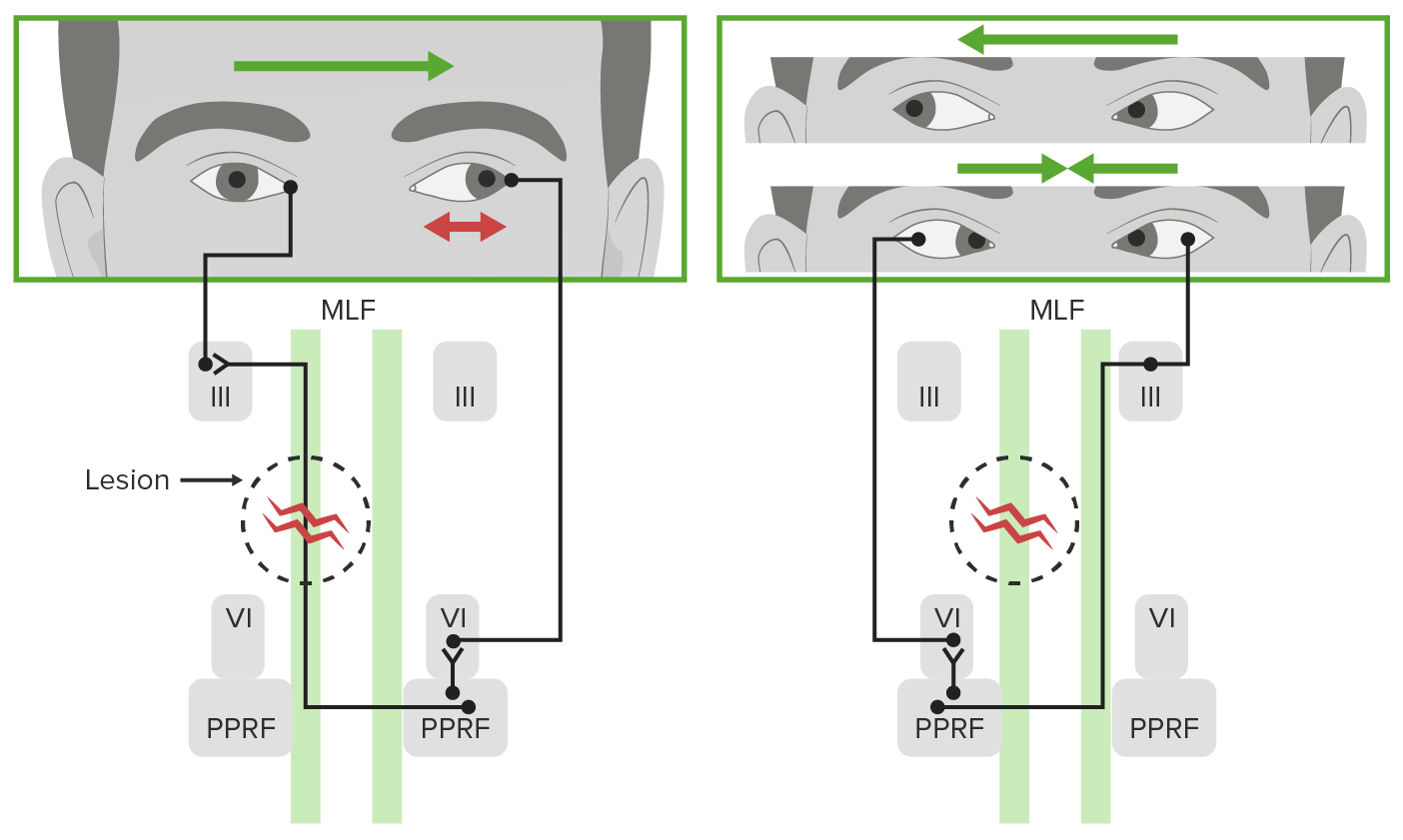
Defect of horizontal gaze in unilateral internuclear ophthalmoplegia:
Note the ipsilateral defective adduction with leftward gaze and contralateral abducting nystagmus (red double arrow in top left image). Normal rightward gaze and convergence would also be seen (right image).
MLF: medial longitudinal fasciculus
III: nucleus of the oculomotor cranial nerve III
VI: nucleus of the abducens cranial nerve VI
PPRF: paramedian pontine reticular formation

Image by Lecturio. License: CC BY-NC-SA 4.0

Clinical Presentation

The presentation of individuals with INO can vary but includes:

  • Changes in vision Vision Ophthalmic Exam and eye movements:
    • Marked limitation of adduction Adduction Examination of the Upper Limbs in eye ipsilateral to the side of MLF lesion
    • Abduction Abduction Examination of the Upper Limbs nystagmus Nystagmus Involuntary movements of the eye that are divided into two types, jerk and pendular. Jerk nystagmus has a slow phase in one direction followed by a corrective fast phase in the opposite direction, and is usually caused by central or peripheral vestibular dysfunction. Pendular nystagmus features oscillations that are of equal velocity in both directions and this condition is often associated with visual loss early in life. Albinism of contralateral eye
    • Possible vertical nystagmus Nystagmus Involuntary movements of the eye that are divided into two types, jerk and pendular. Jerk nystagmus has a slow phase in one direction followed by a corrective fast phase in the opposite direction, and is usually caused by central or peripheral vestibular dysfunction. Pendular nystagmus features oscillations that are of equal velocity in both directions and this condition is often associated with visual loss early in life. Albinism in bilateral INO
    • Blurry vision Vision Ophthalmic Exam and oscillopsia (oscillating movement of objects in the visual field Visual Field The Visual Pathway and Related Disorders)
    • Diplopia Diplopia A visual symptom in which a single object is perceived by the visual cortex as two objects rather than one. Disorders associated with this condition include refractive errors; strabismus; oculomotor nerve diseases; trochlear nerve diseases; abducens nerve diseases; and diseases of the brain stem and occipital lobe. Myasthenia Gravis
  • Others:
Internuclear ophthalmoplegia secondary to cocaine abuse

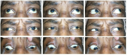
An individual with left internuclear ophtalmoplegia:
Notice that adduction of the left eye is impaired (left column of images) due to a unilateral defect of the medial longitudinal fasciculus.

Image: “fig1: Left INO with absent left eye adduction. Exotropia and left skew deviation are present.” by Richard L. Rabin et al. License: CC BY 4.0

Diagnosis and Management

Diagnosis

  • Clinical: The diagnosis is mainly clinical, based on the assessment of the patient’s ability to perform conjugate eye movements.                  
  • Radiologic:
    • MRI and CT scans may be used (MRI is preferred) to help determine an etiology.
    • Proton density Proton Density Magnetic Resonance Imaging (MRI) imaging is also helpful for diagnosing underlying demyelinating lesions. 
    • Optokinetic tape is also highly sensitive for testing for INO.

Management

Management depends on the underlying cause, and clinical improvement can vary.

  • Neurologic cause: treated after detailed neurology evaluation
  • Demyelinating/autoimmune cause: 
    • Corticosteroids Corticosteroids Chorioretinitis are the treatment of choice.
    • Dalfampridine ( potassium Potassium An element in the alkali group of metals with an atomic symbol k, atomic number 19, and atomic weight 39. 10. It is the chief cation in the intracellular fluid of muscle and other cells. Potassium ion is a strong electrolyte that plays a significant role in the regulation of fluid volume and maintenance of the water-electrolyte balance. Hyperkalemia channel blocker) has been used in patients Patients Individuals participating in the health care system for the purpose of receiving therapeutic, diagnostic, or preventive procedures. Clinician–Patient Relationship with demyelinating disease.
  • Diplopia Diplopia A visual symptom in which a single object is perceived by the visual cortex as two objects rather than one. Disorders associated with this condition include refractive errors; strabismus; oculomotor nerve diseases; trochlear nerve diseases; abducens nerve diseases; and diseases of the brain stem and occipital lobe. Myasthenia Gravis: can be treated with botulinum toxin Botulinum toxin Toxic proteins produced from the species Clostridium botulinum. The toxins are synthesized as a single peptide chain which is processed into a mature protein consisting of a heavy chain and light chain joined via a disulfide bond. The botulinum toxin light chain is a zinc-dependent protease which is released from the heavy chain upon endocytosis into presynaptic nerve endings. Once inside the cell the botulinum toxin light chain cleaves specific snare proteins which are essential for secretion of acetylcholine by synaptic vesicles. This inhibition of acetylcholine release results in muscular paralysis. Botulism injections or Fresnel prisms
  • Strabismus Strabismus Strabismus is the misalignment of the eyes while fixating the gaze on an object. Strabismus can be idiopathic, but it may also be caused by cerebral palsy, uncorrected refractive errors, and extraocular muscle or cranial nerve dysfunction. Strabismus: can be surgically treated in patients Patients Individuals participating in the health care system for the purpose of receiving therapeutic, diagnostic, or preventive procedures. Clinician–Patient Relationship with wall-eyed bilateral INO

Differential Diagnosis

  • Lateral gaze palsy Palsy paralysis of an area of the body, thus incapable of voluntary movement Cranial Nerve Palsies: due to a lesion in CN VI (abducens) resulting in impaired ipsilateral abduction Abduction Examination of the Upper Limbs. Lateral gaze palsy Palsy paralysis of an area of the body, thus incapable of voluntary movement Cranial Nerve Palsies can be caused by trauma or microvascular ischemia Ischemia A hypoperfusion of the blood through an organ or tissue caused by a pathologic constriction or obstruction of its blood vessels, or an absence of blood circulation. Ischemic Cell Damage or can be secondary to multiple other causes, including multiple sclerosis Sclerosis A pathological process consisting of hardening or fibrosis of an anatomical structure, often a vessel or a nerve. Wilms Tumor, stroke, or increased intracranial pressure Intracranial Pressure Idiopathic Intracranial Hypertension.
  • One-and-a-half syndrome: horizontal movement disorder of the eyes. One-and-a-half syndrome is characterized by a conjugate horizontal gaze palsy Palsy paralysis of an area of the body, thus incapable of voluntary movement Cranial Nerve Palsies in one direction and an internuclear ophthalmoplegia Ophthalmoplegia Paralysis of one or more of the ocular muscles due to disorders of the eye muscles, neuromuscular junction, supporting soft tissue, tendons, or innervation to the muscles. Orbital and Preseptal Cellulitis in the other. The syndrome is caused by a lesion of the PPRF and the MLF and is most commonly due to cerebrovascular diseases. 
  • Stroke: occlusion of distal penetrating arteries Arteries Arteries are tubular collections of cells that transport oxygenated blood and nutrients from the heart to the tissues of the body. The blood passes through the arteries in order of decreasing luminal diameter, starting in the largest artery (the aorta) and ending in the small arterioles. Arteries are classified into 3 types: large elastic arteries, medium muscular arteries, and small arteries and arterioles. Arteries: Histology that leads to dorsal brain Brain The part of central nervous system that is contained within the skull (cranium). Arising from the neural tube, the embryonic brain is comprised of three major parts including prosencephalon (the forebrain); mesencephalon (the midbrain); and rhombencephalon (the hindbrain). The developed brain consists of cerebrum; cerebellum; and other structures in the brain stem. Nervous System: Anatomy, Structure, and Classification stem infarction and can present as INO, diplopia Diplopia A visual symptom in which a single object is perceived by the visual cortex as two objects rather than one. Disorders associated with this condition include refractive errors; strabismus; oculomotor nerve diseases; trochlear nerve diseases; abducens nerve diseases; and diseases of the brain stem and occipital lobe. Myasthenia Gravis, or vision Vision Ophthalmic Exam changes. Brainstem stroke can also present with diverse visual symptoms, including homonymous hemianopia Hemianopia Transient Ischemic Attack (TIA) or cortical blindness Blindness The inability to see or the loss or absence of perception of visual stimuli. This condition may be the result of eye diseases; optic nerve diseases; optic chiasm diseases; or brain diseases affecting the visual pathways or occipital lobe. Retinopathy of Prematurity, skew deviation of eyes, nystagmus Nystagmus Involuntary movements of the eye that are divided into two types, jerk and pendular. Jerk nystagmus has a slow phase in one direction followed by a corrective fast phase in the opposite direction, and is usually caused by central or peripheral vestibular dysfunction. Pendular nystagmus features oscillations that are of equal velocity in both directions and this condition is often associated with visual loss early in life. Albinism, and ocular or facial pain Facial pain Pain in the facial region including orofacial pain and craniofacial pain. Associated conditions include local inflammatory and neoplastic disorders and neuralgic syndromes involving the trigeminal, facial, and glossopharyngeal nerves. Conditions which feature recurrent or persistent facial pain as the primary manifestation of disease are referred to as facial pain syndromes. Trigeminal Neuralgia.

References

  1. Rubin, M. (2020). Internuclear ophthalmoplegia. Merck Manual Professional Version. Retrieved May 28, 2021, from https://www.merckmanuals.com/professional/neurologic-disorders/neuro-ophthalmologic-and-cranial-nerve-disorders/internuclear-ophthalmoplegia
  2. Swisher, J., Kini, A., Lee, A.G. (2020). Internuclear ophthalmoplegia. American Academy of Ophthalmology. Retrieved May 28, 2021, from https://eyewiki.aao.org/Internuclear_Ophthalmoplegia
  3. Frohman, T.C., Petzold, A., and Frohman, E.M. (2020). Internuclear ophthalmoparesis. In Wilterdink, J.L. (Ed.), UpToDate. Retrieved February 6, 2022, from https://www.uptodate.com/contents/internuclear-ophthalmoparesis
  4. Toral, M., Haugsdal, J., and Wall, M. (2017). Internuclear ophthalmoplegia. University of Iowa Health Care. https://webeye.ophth.uiowa.edu/eyeforum/cases/252-internuclear-ophthalmoplegia.htm

Create your free account or log in to continue reading!

Sign up now and get free access to Lecturio with concept pages, medical videos, and questions for your medical education.

User Reviews

Details