The brain Brain The part of central nervous system that is contained within the skull (cranium). Arising from the neural tube, the embryonic brain is comprised of three major parts including prosencephalon (the forebrain); mesencephalon (the midbrain); and rhombencephalon (the hindbrain). The developed brain consists of cerebrum; cerebellum; and other structures in the brain stem. Nervous System: Anatomy, Structure, and Classification stem is a stalk-like structure that connects the cerebrum with the spinal cord Spinal cord The spinal cord is the major conduction pathway connecting the brain to the body; it is part of the CNS. In cross section, the spinal cord is divided into an H-shaped area of gray matter (consisting of synapsing neuronal cell bodies) and a surrounding area of white matter (consisting of ascending and descending tracts of myelinated axons). Spinal Cord: Anatomy and consists of the midbrain, pons, and medulla oblongata. The brain Brain The part of central nervous system that is contained within the skull (cranium). Arising from the neural tube, the embryonic brain is comprised of three major parts including prosencephalon (the forebrain); mesencephalon (the midbrain); and rhombencephalon (the hindbrain). The developed brain consists of cerebrum; cerebellum; and other structures in the brain stem. Nervous System: Anatomy, Structure, and Classification stem contains many nerves, pathways, reflex centers, and nuclei and serves as a major relay station for sensory Sensory Neurons which conduct nerve impulses to the central nervous system. Nervous System: Histology, motor Motor Neurons which send impulses peripherally to activate muscles or secretory cells. Nervous System: Histology, and autonomic information. All cranial nerves Cranial nerves There are 12 pairs of cranial nerves (CNs), which run from the brain to various parts of the head, neck, and trunk. The CNs can be sensory or motor or both. The CNs are named and numbered in Roman numerals according to their location, from the front to the back of the brain. The 12 Cranial Nerves: Overview and Functions, except I and II, originate from the brain Brain The part of central nervous system that is contained within the skull (cranium). Arising from the neural tube, the embryonic brain is comprised of three major parts including prosencephalon (the forebrain); mesencephalon (the midbrain); and rhombencephalon (the hindbrain). The developed brain consists of cerebrum; cerebellum; and other structures in the brain stem. Nervous System: Anatomy, Structure, and Classification stem. The brain Brain The part of central nervous system that is contained within the skull (cranium). Arising from the neural tube, the embryonic brain is comprised of three major parts including prosencephalon (the forebrain); mesencephalon (the midbrain); and rhombencephalon (the hindbrain). The developed brain consists of cerebrum; cerebellum; and other structures in the brain stem. Nervous System: Anatomy, Structure, and Classification stem also plays a critical role in the control of cardiovascular and respiratory function, consciousness, and the sleep Sleep A readily reversible suspension of sensorimotor interaction with the environment, usually associated with recumbency and immobility. Physiology of Sleep–wake cycle.
Last updated: Dec 15, 2025
The brain Brain The part of central nervous system that is contained within the skull (cranium). Arising from the neural tube, the embryonic brain is comprised of three major parts including prosencephalon (the forebrain); mesencephalon (the midbrain); and rhombencephalon (the hindbrain). The developed brain consists of cerebrum; cerebellum; and other structures in the brain stem. Nervous System: Anatomy, Structure, and Classification stem is located in the posterior cranial fossa Posterior cranial fossa The infratentorial compartment that contains the cerebellum and brain stem. It is formed by the posterior third of the superior surface of the body of the sphenoid (sphenoid bone), by the occipital, the petrous, and mastoid portions of the temporal bone, and the posterior inferior angle of the parietal bone. Skull: Anatomy on the dorsal aspect of the clivus. There should be CSF space between the clivus and the brain Brain The part of central nervous system that is contained within the skull (cranium). Arising from the neural tube, the embryonic brain is comprised of three major parts including prosencephalon (the forebrain); mesencephalon (the midbrain); and rhombencephalon (the hindbrain). The developed brain consists of cerebrum; cerebellum; and other structures in the brain stem. Nervous System: Anatomy, Structure, and Classification stem in normal patients Patients Individuals participating in the health care system for the purpose of receiving therapeutic, diagnostic, or preventive procedures. Clinician–Patient Relationship, and the 4th ventricle is posterior to the brain Brain The part of central nervous system that is contained within the skull (cranium). Arising from the neural tube, the embryonic brain is comprised of three major parts including prosencephalon (the forebrain); mesencephalon (the midbrain); and rhombencephalon (the hindbrain). The developed brain consists of cerebrum; cerebellum; and other structures in the brain stem. Nervous System: Anatomy, Structure, and Classification stem between it and the cerebellum Cerebellum The cerebellum, Latin for “little brain,” is located in the posterior cranial fossa, dorsal to the pons and midbrain, and its principal role is in the coordination of movements. The cerebellum consists of 3 lobes on either side of its 2 hemispheres and is connected in the middle by the vermis. Cerebellum: Anatomy. The midbrain is most superior, followed by the pons, and the medulla oblongata, which is most inferior.
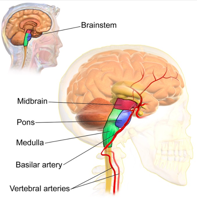
Location of the brain stem:
Note the ascending order from the medulla at the base through the pons in the middle, ending with the midbrain at the most rostral portion of the brain stem.
3 laminae:
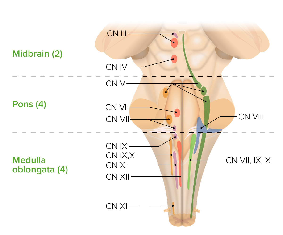
Division of cranial nerves that arise in the brainstem:
Cranial nerves (CNs) III–IV in the midbrain, V–VIII in the pons, and IX–XII in the medulla
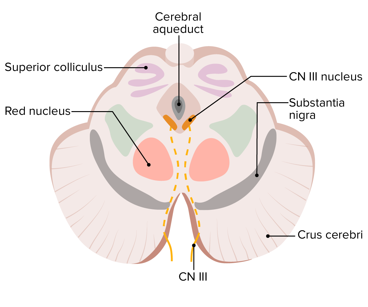
Key structures of the midbrain, including the superior colliculus, red nucleus, crus cerebri, substantia nigra, cranial nerve (CN) III nucleus and nerve, as well as the cerebral aqueduct
Image by Lecturio.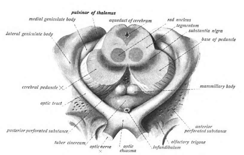
Transverse section of the midbrain at the superior colliculus level
Image: “An anatomical illustration from the 1908 edition of Sobotta’s Anatomy Atlas” by Dr. Johannes Sobotta. License: Public Domain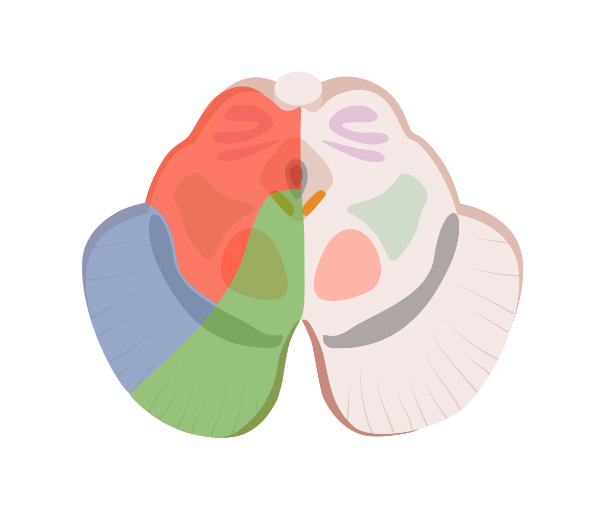
Arterial supply of the midbrain:
Note the perfusion territory of the arteries that supply the midbrain: the collicular artery in red, the thalamoperforate arteries in green, and the medial posterior choroidal artery in blue. All of these vessels are branches of the posterior cerebral artery.
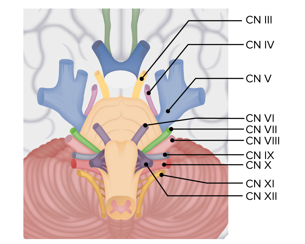
Cranial nerves throughout the brain stem:
Note the 4 cranial nerves that originate from the pons: cranial nerves (CNs) V, VI, VII, and VIII. All of these emerge from the pontine tegmentum.
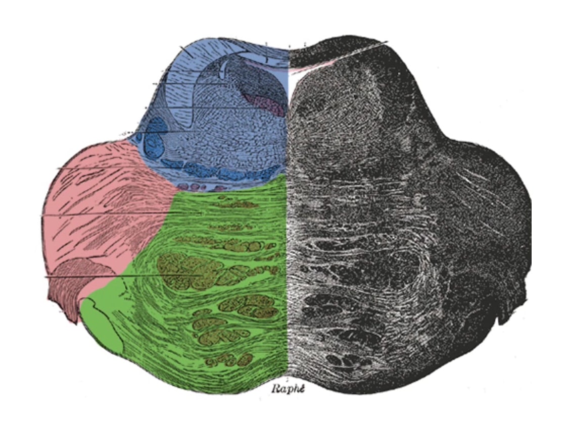
Arterial supply of the pons:
Note the perfusion territory of the arteries that supply the pons: Short circumferential branches of the basilar artery are in red, the long circumferential branches of the basilar artery as well as branches of the superior cerebellar artery are in blue, and the paramedian branches of the basilar artery are in green.
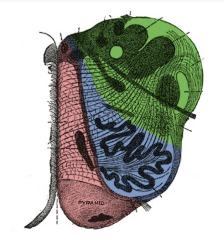
Arterial supply of the medulla oblongata:
Note the perfusion territory of the arteries that supply the medulla oblongata: The most medial portion, illustrated in red, is supplied by the anterior spinal artery. Next, in blue, is the supply from the vertebral artery. Finally, most laterally, the medulla is supplied by the posterior inferior cerebellar artery (PICA), denoted in green.