Achieve Mastery of Medical Concepts

Study for medical school and boards with Lecturio

Anterior Cord Syndrome

Anterior cord syndrome (ACS) is an incomplete cord syndrome predominantly affecting the anterior (ventral) ⅔ of the spinal cord Spinal cord The spinal cord is the major conduction pathway connecting the brain to the body; it is part of the CNS. In cross section, the spinal cord is divided into an H-shaped area of gray matter (consisting of synapsing neuronal cell bodies) and a surrounding area of white matter (consisting of ascending and descending tracts of myelinated axons). Spinal Cord: Anatomy while sparing the dorsal columns Dorsal Columns Posterior Cord Syndrome. Anterior cord syndrome can be caused by occlusion of the anterior spinal artery or by trauma, which results in disk herniation Herniation Omphalocele and bone Bone Bone is a compact type of hardened connective tissue composed of bone cells, membranes, an extracellular mineralized matrix, and central bone marrow. The 2 primary types of bone are compact and spongy. Bones: Structure and Types fragments disrupting the spinal cord Spinal cord The spinal cord is the major conduction pathway connecting the brain to the body; it is part of the CNS. In cross section, the spinal cord is divided into an H-shaped area of gray matter (consisting of synapsing neuronal cell bodies) and a surrounding area of white matter (consisting of ascending and descending tracts of myelinated axons). Spinal Cord: Anatomy. Clinical manifestations include loss of both motor Motor Neurons which send impulses peripherally to activate muscles or secretory cells. Nervous System: Histology and sensory Sensory Neurons which conduct nerve impulses to the central nervous system. Nervous System: Histology function below the level of injury. Diagnosis of ACS is by clinical exam and neuroimaging Neuroimaging Non-invasive methods of visualizing the central nervous system, especially the brain, by various imaging modalities. Febrile Infant with MRI. Management is directed at resolving the underlying cause. Preservation of motor Motor Neurons which send impulses peripherally to activate muscles or secretory cells. Nervous System: Histology function is a priority, but the prognosis Prognosis A prediction of the probable outcome of a disease based on a individual's condition and the usual course of the disease as seen in similar situations. Non-Hodgkin Lymphomas is poor.

Last updated: Mar 29, 2023

Editorial responsibility: Stanley Oiseth, Lindsay Jones, Evelin Maza

Overview

Definition

Anterior cord syndrome (ACS) is an incomplete cord syndrome affecting the anterior ⅔ of the spinal cord Spinal cord The spinal cord is the major conduction pathway connecting the brain to the body; it is part of the CNS. In cross section, the spinal cord is divided into an H-shaped area of gray matter (consisting of synapsing neuronal cell bodies) and a surrounding area of white matter (consisting of ascending and descending tracts of myelinated axons). Spinal Cord: Anatomy while sparing the dorsal columns Dorsal Columns Posterior Cord Syndrome, resulting in loss of motor Motor Neurons which send impulses peripherally to activate muscles or secretory cells. Nervous System: Histology and sensory Sensory Neurons which conduct nerve impulses to the central nervous system. Nervous System: Histology function below the level of injury.

Epidemiology

  • Estimated incidence Incidence The number of new cases of a given disease during a given period in a specified population. It also is used for the rate at which new events occur in a defined population. It is differentiated from prevalence, which refers to all cases in the population at a given time. Measures of Disease Frequency: 3.1 per 100,000
  • 2.7% of traumatic spinal cord injuries Spinal cord injuries Spinal cord injuries are complex injuries that involve damage to the neural tissue within the spinal canal. Spinal cord injuries are commonly the result of trauma. Clinical presentation varies depending on the site of injury and on whether the injury is complete or incomplete. Spinal Cord Injuries 
  • The most common pattern of spinal cord Spinal cord The spinal cord is the major conduction pathway connecting the brain to the body; it is part of the CNS. In cross section, the spinal cord is divided into an H-shaped area of gray matter (consisting of synapsing neuronal cell bodies) and a surrounding area of white matter (consisting of ascending and descending tracts of myelinated axons). Spinal Cord: Anatomy infarction

Etiology

  • Occlusion of the anterior spinal artery (ASA) and ischemia Ischemia A hypoperfusion of the blood through an organ or tissue caused by a pathologic constriction or obstruction of its blood vessels, or an absence of blood circulation. Ischemic Cell Damage to the area the ASA supplies:
    • Iatrogenic (most common): cross-clamping of the aorta Aorta The main trunk of the systemic arteries. Mediastinum and Great Vessels: Anatomy during thoracic and abdominal aortic aneurysm Aortic aneurysm An abnormal balloon- or sac-like dilatation in the wall of aorta. Thoracic Aortic Aneurysms repair 
    • Severe hypotension Hypotension Hypotension is defined as low blood pressure, specifically < 90/60 mm Hg, and is most commonly a physiologic response. Hypotension may be mild, serious, or life threatening, depending on the cause. Hypotension
    • Atherothrombotic disease
    • Vasculitis Vasculitis Inflammation of any one of the blood vessels, including the arteries; veins; and rest of the vasculature system in the body. Systemic Lupus Erythematosus
    • Cocaine Cocaine An alkaloid ester extracted from the leaves of plants including coca. It is a local anesthetic and vasoconstrictor and is clinically used for that purpose, particularly in the eye, ear, nose, and throat. It also has powerful central nervous system effects similar to the amphetamines and is a drug of abuse. Cocaine, like amphetamines, acts by multiple mechanisms on brain catecholaminergic neurons; the mechanism of its reinforcing effects is thought to involve inhibition of dopamine uptake. Local Anesthetics (rare)
  • Direct injury/trauma:
    • Crush injury Crush injury Excessive compression of parts of the body that causes muscle swelling, fracture, and/or neurological disturbances in the affected areas. Crush injury with systemic manifestations is referred to as crush syndrome. Crush Syndrome
    • Acute spinal disk herniation Herniation Omphalocele
    • Burst fracture Fracture A fracture is a disruption of the cortex of any bone and periosteum and is commonly due to mechanical stress after an injury or accident. Open fractures due to trauma can be a medical emergency. Fractures are frequently associated with automobile accidents, workplace injuries, and trauma. Overview of Bone Fractures
    • Gunshot or knife injury
  • Spinal canal Spinal Canal The cavity within the spinal column through which the spinal cord passes. Spinal Cord Injuries mass Mass Three-dimensional lesion that occupies a space within the breast Imaging of the Breast:
  • Radiation Radiation Emission or propagation of acoustic waves (sound), electromagnetic energy waves (such as light; radio waves; gamma rays; or x-rays), or a stream of subatomic particles (such as electrons; neutrons; protons; or alpha particles). Osteosarcoma myelopathy

Pathophysiology

Anterior cord syndrome starts with occlusion of the blood supply or injury to the vertebral column Vertebral column The human spine, or vertebral column, is the most important anatomical and functional axis of the human body. It consists of 7 cervical vertebrae, 12 thoracic vertebrae, and 5 lumbar vertebrae and is limited cranially by the skull and caudally by the sacrum. Vertebral Column: Anatomy. The syndrome is infrequently due to spinal cord Spinal cord The spinal cord is the major conduction pathway connecting the brain to the body; it is part of the CNS. In cross section, the spinal cord is divided into an H-shaped area of gray matter (consisting of synapsing neuronal cell bodies) and a surrounding area of white matter (consisting of ascending and descending tracts of myelinated axons). Spinal Cord: Anatomy tumors.
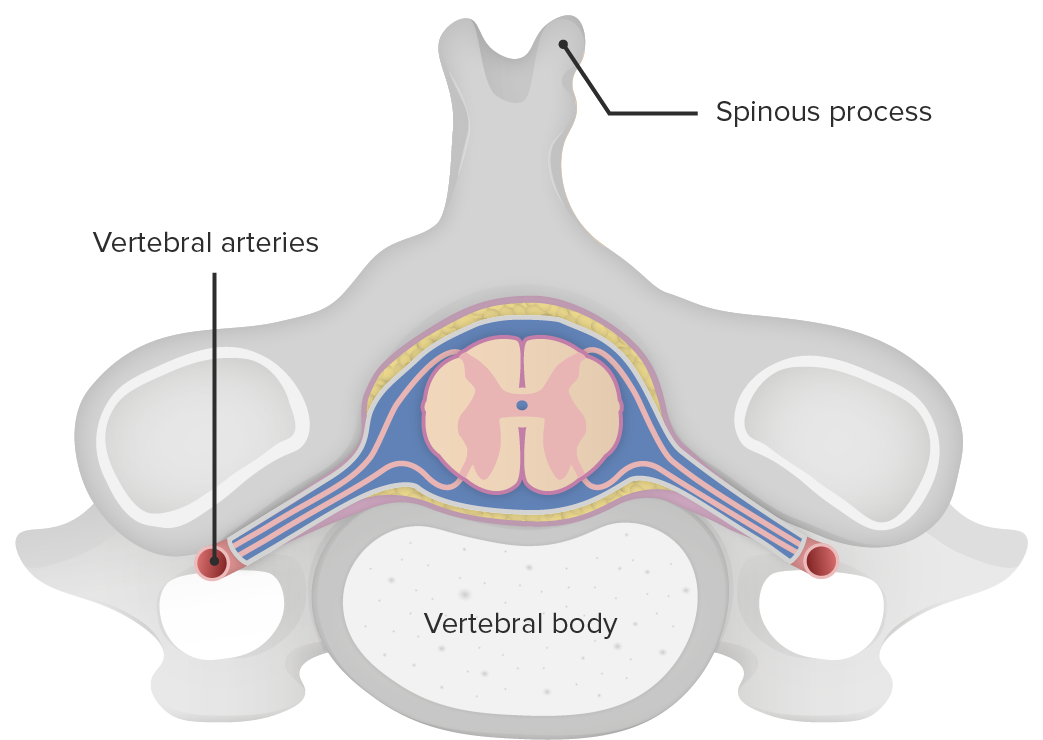
Mechanisms of injury

  • ASA occlusion:
    • Blood flow Blood flow Blood flow refers to the movement of a certain volume of blood through the vasculature over a given unit of time (e.g., mL per minute). Vascular Resistance, Flow, and Mean Arterial Pressure through the ASA is interrupted → spinal cord Spinal cord The spinal cord is the major conduction pathway connecting the brain to the body; it is part of the CNS. In cross section, the spinal cord is divided into an H-shaped area of gray matter (consisting of synapsing neuronal cell bodies) and a surrounding area of white matter (consisting of ascending and descending tracts of myelinated axons). Spinal Cord: Anatomy tissue ischemia Ischemia A hypoperfusion of the blood through an organ or tissue caused by a pathologic constriction or obstruction of its blood vessels, or an absence of blood circulation. Ischemic Cell Damage and infarction from the level of interruption of flow Flow Blood flows through the heart, arteries, capillaries, and veins in a closed, continuous circuit. Flow is the movement of volume per unit of time. Flow is affected by the pressure gradient and the resistance fluid encounters between 2 points. Vascular resistance is the opposition to flow, which is caused primarily by blood friction against vessel walls. Vascular Resistance, Flow, and Mean Arterial Pressure downwards →
    • Infarction of the corticospinal and spinothalamic pathways → ACS
  • Vertebral/burst fractures:
    • Force coming from above or below the vertebral body Vertebral body Main portion of the vertebra which bears majority of the weight. Vertebral Column: Anatomy (depending on the injury) →
    • Nucleus pulposus Nucleus Pulposus Fibrocartilage inner core of the intervertebral disc. Prolapsed or bulged nucleus pulposus leads to intervertebral disc displacement while proliferation of cells in the nucleus pulposus is associated with intervertebral disc degeneration. Spinal Disk Herniation of the intervertebral disk is forced into the vertebral body Vertebral body Main portion of the vertebra which bears majority of the weight. Vertebral Column: Anatomy → shatters outward (burst) fracture Fracture A fracture is a disruption of the cortex of any bone and periosteum and is commonly due to mechanical stress after an injury or accident. Open fractures due to trauma can be a medical emergency. Fractures are frequently associated with automobile accidents, workplace injuries, and trauma. Overview of Bone Fractures spinal cord Spinal cord The spinal cord is the major conduction pathway connecting the brain to the body; it is part of the CNS. In cross section, the spinal cord is divided into an H-shaped area of gray matter (consisting of synapsing neuronal cell bodies) and a surrounding area of white matter (consisting of ascending and descending tracts of myelinated axons). Spinal Cord: Anatomy injury related to the force and direction of the traumatic event Traumatic event An emotionally painful, shocking, stressful, and sometimes life-threatening experience. It can result from witnessing distressing events such as natural disasters, physical or sexual abuse, and terrorism or other acts of violence. Posttraumatic Stress Disorder (PTSD)
    • Posteriorly displaced fracture Fracture A fracture is a disruption of the cortex of any bone and periosteum and is commonly due to mechanical stress after an injury or accident. Open fractures due to trauma can be a medical emergency. Fractures are frequently associated with automobile accidents, workplace injuries, and trauma. Overview of Bone Fractures fragments → ACS

Neurologic sequelae

  • Anatomy: Motor Motor Neurons which send impulses peripherally to activate muscles or secretory cells. Nervous System: Histology nuclei of the spinal cord Spinal cord The spinal cord is the major conduction pathway connecting the brain to the body; it is part of the CNS. In cross section, the spinal cord is divided into an H-shaped area of gray matter (consisting of synapsing neuronal cell bodies) and a surrounding area of white matter (consisting of ascending and descending tracts of myelinated axons). Spinal Cord: Anatomy are contained within the ventral horn Ventral horn One of three central columns of the spinal cord. It is composed of gray matter spinal laminae VIII and ix. Brown-Séquard Syndrome. →
  • Interneurons mediate information from other descending tracts Descending tracts Spinal Cord: Anatomy (pyramidal and extrapyramidal motor Motor Neurons which send impulses peripherally to activate muscles or secretory cells. Nervous System: Histology systems). →
  • Synapse Synapse The junction between 2 neurons is called a synapse. The synapse allows a neuron to pass an electrical or chemical signal to another neuron or target effector cell. Synapses and Neurotransmission on the alpha and gamma motor Motor Neurons which send impulses peripherally to activate muscles or secretory cells. Nervous System: Histology neurons Neurons The basic cellular units of nervous tissue. Each neuron consists of a body, an axon, and dendrites. Their purpose is to receive, conduct, and transmit impulses in the nervous system. Nervous System: Histology
  • Leave the ventral horn Ventral horn One of three central columns of the spinal cord. It is composed of gray matter spinal laminae VIII and ix. Brown-Séquard Syndrome via the ventral nerve root →
  • Injury to the ventral horn Ventral horn One of three central columns of the spinal cord. It is composed of gray matter spinal laminae VIII and ix. Brown-Séquard Syndrome/anterior spinal cord Spinal cord The spinal cord is the major conduction pathway connecting the brain to the body; it is part of the CNS. In cross section, the spinal cord is divided into an H-shaped area of gray matter (consisting of synapsing neuronal cell bodies) and a surrounding area of white matter (consisting of ascending and descending tracts of myelinated axons). Spinal Cord: Anatomy → damage to:
    • Corticospinal tract (CST) → motor Motor Neurons which send impulses peripherally to activate muscles or secretory cells. Nervous System: Histology paralysis and absent reflexes 
    • Spinothalamic tract (STT) → bilateral loss of pain Pain An unpleasant sensation induced by noxious stimuli which are detected by nerve endings of nociceptive neurons. Pain: Types and Pathways and temperature sensation
    • Descending autonomic tracts → loss of bladder Bladder A musculomembranous sac along the urinary tract. Urine flows from the kidneys into the bladder via the ureters, and is held there until urination. Pyelonephritis and Perinephric Abscess control 
  • Tactile, position, and vibratory sensation are normal ( dorsal columns Dorsal Columns Posterior Cord Syndrome intact).
  • With acute spinal cord Spinal cord The spinal cord is the major conduction pathway connecting the brain to the body; it is part of the CNS. In cross section, the spinal cord is divided into an H-shaped area of gray matter (consisting of synapsing neuronal cell bodies) and a surrounding area of white matter (consisting of ascending and descending tracts of myelinated axons). Spinal Cord: Anatomy injury:
    • Secondary injury due to spinal cord Spinal cord The spinal cord is the major conduction pathway connecting the brain to the body; it is part of the CNS. In cross section, the spinal cord is divided into an H-shaped area of gray matter (consisting of synapsing neuronal cell bodies) and a surrounding area of white matter (consisting of ascending and descending tracts of myelinated axons). Spinal Cord: Anatomy edema Edema Edema is a condition in which excess serous fluid accumulates in the body cavity or interstitial space of connective tissues. Edema is a symptom observed in several medical conditions. It can be categorized into 2 types, namely, peripheral (in the extremities) and internal (in an organ or body cavity). Edema may manifest as neurologic deterioration over the 1st 8–12 hours.
    • Maximal 3–6 days after the injury
    • Recedes approximately 9 days after the injury
  • Gradually replaced by central hemorrhagic necrosis Hemorrhagic Necrosis Brown-Séquard Syndrome

Clinical Presentation and Diagnosis

Understanding the structures affected by an anterior cord lesion is key to correlating clinical signs and symptoms.

Clinical Presentation

History:

  • May report the acute onset of neurologic symptoms or trauma
  • Individuals with acute cervical spondylotic myelopathy Cervical Spondylotic Myelopathy Neck Pain and a history of mild or no previous deficits: neurologic emergency
  • May arrive at the emergency department as a result of trauma and unable to give a history
  • May present with symptoms in the hospital after aortic surgery

Physical exam findings: 

  • Almost always bilateral
  • Motor Motor Neurons which send impulses peripherally to activate muscles or secretory cells. Nervous System: Histology deficit: paralysis below the level of lesion 
  • Sensory Sensory Neurons which conduct nerve impulses to the central nervous system. Nervous System: Histology deficit: loss of pain Pain An unpleasant sensation induced by noxious stimuli which are detected by nerve endings of nociceptive neurons. Pain: Types and Pathways and temperature sensation
  • Preserved (posterior columns are spared): position, vibration Vibration A continuing periodic change in displacement with respect to a fixed reference. Neurological Examination sense, and light touch Light touch Neurological Examination
  • Autonomic dysfunction: urinary incontinence Urinary incontinence Urinary incontinence (UI) is involuntary loss of bladder control or unintentional voiding, which represents a hygienic or social problem to the patient. Urinary incontinence is a symptom, a sign, and a disorder. The 5 types of UI include stress, urge, mixed, overflow, and functional. Urinary Incontinence

Diagnosis

  • Imaging:
    • X-ray X-ray Penetrating electromagnetic radiation emitted when the inner orbital electrons of an atom are excited and release radiant energy. X-ray wavelengths range from 1 pm to 10 nm. Hard x-rays are the higher energy, shorter wavelength x-rays. Soft x-rays or grenz rays are less energetic and longer in wavelength. The short wavelength end of the x-ray spectrum overlaps the gamma rays wavelength range. The distinction between gamma rays and x-rays is based on their radiation source. Pulmonary Function Tests of the spine Spine The human spine, or vertebral column, is the most important anatomical and functional axis of the human body. It consists of 7 cervical vertebrae, 12 thoracic vertebrae, and 5 lumbar vertebrae and is limited cranially by the skull and caudally by the sacrum. Vertebral Column: Anatomy: to evaluate fracture Fracture A fracture is a disruption of the cortex of any bone and periosteum and is commonly due to mechanical stress after an injury or accident. Open fractures due to trauma can be a medical emergency. Fractures are frequently associated with automobile accidents, workplace injuries, and trauma. Overview of Bone Fractures
    • MRI:
      • Preferred imaging modality
      • Sagittal Sagittal Computed Tomography (CT) view: thin, “pencil-like” hyperintensities on T2 within the anterior horns
      • Axial view: hyperintense bilaterally symmetric circular to ovoid foci in the anterior horns of the spinal cord resembling “owl eyes” (also known as “snake-eyes sign” or “fried-eggs sign”)
  • Lab tests:
Sagittal view of spinal t2 mri showing “pencil-like” ischemic changes in the spinal cord, consistent with anterior cord syndrome (acs)

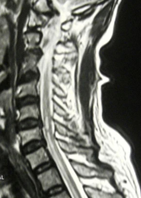
Sagittal view of spinal T2 magnetic resonance imaging showing “pencil-like” ischemic changes in the spinal cord consistent with anterior cord syndrome

Image: “T2 Weighted sagittal image shows linear hyperintensity along the lower cervical and thoracic cord without cord expansion” by Nilukshana Yogendranathan, et al. License: CC BY 4.0

Management and Prognosis

Management is directed at relieving symptoms and preserving motor Motor Neurons which send impulses peripherally to activate muscles or secretory cells. Nervous System: Histology function.

Acute emergency care

  • Medications (dependent on the cause of ACS):
    • Outcomes improve with high-dose IV steroids Steroids A group of polycyclic compounds closely related biochemically to terpenes. They include cholesterol, numerous hormones, precursors of certain vitamins, bile acids, alcohols (sterols), and certain natural drugs and poisons. Steroids have a common nucleus, a fused, reduced 17-carbon atom ring system, cyclopentanoperhydrophenanthrene. Most steroids also have two methyl groups and an aliphatic side-chain attached to the nucleus. Benign Liver Tumors within 8 hours of the injury.
    • Infuse IV bolus of methylprednisolone Methylprednisolone A prednisolone derivative with similar anti-inflammatory action. Immunosuppressants for the 1st 23 hours.
  • Thrombolysis can be considered in atherothrombotic or embolic causes.
  • Immunosuppression therapy in vasculitis Vasculitis Inflammation of any one of the blood vessels, including the arteries; veins; and rest of the vasculature system in the body. Systemic Lupus Erythematosus and other autoimmune causes
  • Bladder Bladder A musculomembranous sac along the urinary tract. Urine flows from the kidneys into the bladder via the ureters, and is held there until urination. Pyelonephritis and Perinephric Abscess catheterization for neurogenic bladder Bladder A musculomembranous sac along the urinary tract. Urine flows from the kidneys into the bladder via the ureters, and is held there until urination. Pyelonephritis and Perinephric Abscess

Surgical care

  • Laminectomy Laminectomy A surgical procedure that entails removing all (laminectomy) or part (laminotomy) of selected vertebral lamina to relieve pressure on the spinal cord and/or spinal nerve roots. Vertebral lamina is the thin flattened posterior wall of vertebral arch that forms the vertebral foramen through which pass the spinal cord and nerve roots. Neurosurgery for spinal decompression if indicated
  • Prevention of ACS during surgical procedures:
    • Protocols to avoid spinal cord Spinal cord The spinal cord is the major conduction pathway connecting the brain to the body; it is part of the CNS. In cross section, the spinal cord is divided into an H-shaped area of gray matter (consisting of synapsing neuronal cell bodies) and a surrounding area of white matter (consisting of ascending and descending tracts of myelinated axons). Spinal Cord: Anatomy ischemia Ischemia A hypoperfusion of the blood through an organ or tissue caused by a pathologic constriction or obstruction of its blood vessels, or an absence of blood circulation. Ischemic Cell Damage during aortic surgery 
    • Avoiding prolonged clamp times and IV vasopressors Vasopressors Sepsis in Children

Long-term care via rehabilitation

Complications of spinal cord Spinal cord The spinal cord is the major conduction pathway connecting the brain to the body; it is part of the CNS. In cross section, the spinal cord is divided into an H-shaped area of gray matter (consisting of synapsing neuronal cell bodies) and a surrounding area of white matter (consisting of ascending and descending tracts of myelinated axons). Spinal Cord: Anatomy injury

  • Respiratory failure Respiratory failure Respiratory failure is a syndrome that develops when the respiratory system is unable to maintain oxygenation and/or ventilation. Respiratory failure may be acute or chronic and is classified as hypoxemic, hypercapnic, or a combination of the two. Respiratory Failure (above C3–C5): loss of phrenic nerve Phrenic nerve The motor nerve of the diaphragm. The phrenic nerve fibers originate in the cervical spinal column (mostly C4) and travel through the cervical plexus to the diaphragm. Diaphragm: Anatomy function → paralysis of the diaphragm Diaphragm The diaphragm is a large, dome-shaped muscle that separates the thoracic cavity from the abdominal cavity. The diaphragm consists of muscle fibers and a large central tendon, which is divided into right and left parts. As the primary muscle of inspiration, the diaphragm contributes 75% of the total inspiratory muscle force. Diaphragm: Anatomy (may require long-term ventilator care)
  • Autonomic dysreflexia (above T6): lack of a coordinated autonomic response with heart rate Heart rate The number of times the heart ventricles contract per unit of time, usually per minute. Cardiac Physiology and blood pressure in spinal injuries
  • Spasticity Spasticity Spinal Disk Herniation
  • Urinary incontinence Urinary incontinence Urinary incontinence (UI) is involuntary loss of bladder control or unintentional voiding, which represents a hygienic or social problem to the patient. Urinary incontinence is a symptom, a sign, and a disorder. The 5 types of UI include stress, urge, mixed, overflow, and functional. Urinary Incontinence
  • Complications of immobility:
    • Deep vein thrombosis Thrombosis Formation and development of a thrombus or blood clot in the blood vessel. Epidemic Typhus ( DVT DVT Deep vein thrombosis (DVT) usually occurs in the deep veins of the lower extremities. The affected veins include the femoral, popliteal, iliofemoral, and pelvic veins. Proximal DVT is more likely to cause a pulmonary embolism (PE) and is generally considered more serious. Deep Vein Thrombosis)
    • GI stress ulcers
    • Pressure ulcers
    • Chronic pain Chronic pain Aching sensation that persists for more than a few months. It may or may not be associated with trauma or disease, and may persist after the initial injury has healed. Its localization, character, and timing are more vague than with acute pain. Pain Management

Prognosis Prognosis A prediction of the probable outcome of a disease based on a individual’s condition and the usual course of the disease as seen in similar situations. Non-Hodgkin Lymphomas

  • ACS: the worst prognosis Prognosis A prediction of the probable outcome of a disease based on a individual’s condition and the usual course of the disease as seen in similar situations. Non-Hodgkin Lymphomas of all spinal cord Spinal cord The spinal cord is the major conduction pathway connecting the brain to the body; it is part of the CNS. In cross section, the spinal cord is divided into an H-shaped area of gray matter (consisting of synapsing neuronal cell bodies) and a surrounding area of white matter (consisting of ascending and descending tracts of myelinated axons). Spinal Cord: Anatomy syndromes 
  • Recovery of function is poor:
    • Most deaths occur immediately after injury.
    • Few individuals recover the ability to walk.
    • Improvement may continue in the years following injury.
  • Women and older individuals: ↑ risk of worse outcomes 

Differential Diagnosis

  • Central cord syndrome Central Cord Syndrome Central cord syndrome (CCS) is a neurological syndrome caused by an injury to the center of the spinal cord, affecting the spinothalamic tracts ((STTs) sensory) and medial aspect of the corticospinal tracts ((CSTs) motor), most often due to trauma in patients with cervical spondylosis. Central Cord Syndrome: a neurological syndrome caused by an injury to the center of the spinal cord Spinal cord The spinal cord is the major conduction pathway connecting the brain to the body; it is part of the CNS. In cross section, the spinal cord is divided into an H-shaped area of gray matter (consisting of synapsing neuronal cell bodies) and a surrounding area of white matter (consisting of ascending and descending tracts of myelinated axons). Spinal Cord: Anatomy, which affects the spinothalamic and corticospinal tracts Corticospinal Tracts Central Cord Syndrome. Central cord syndrome Central Cord Syndrome Central cord syndrome (CCS) is a neurological syndrome caused by an injury to the center of the spinal cord, affecting the spinothalamic tracts ((STTs) sensory) and medial aspect of the corticospinal tracts ((CSTs) motor), most often due to trauma in patients with cervical spondylosis. Central Cord Syndrome is often caused by trauma in individuals with cervical spondylosis Cervical Spondylosis Neck Pain. Clinical manifestations are motor Motor Neurons which send impulses peripherally to activate muscles or secretory cells. Nervous System: Histology deficits (affects upper limbs more than lower limbs), variable Variable Variables represent information about something that can change. The design of the measurement scales, or of the methods for obtaining information, will determine the data gathered and the characteristics of that data. As a result, a variable can be qualitative or quantitative, and may be further classified into subgroups. Types of Variables sensory Sensory Neurons which conduct nerve impulses to the central nervous system. Nervous System: Histology deficits below the level of injury, and bladder Bladder A musculomembranous sac along the urinary tract. Urine flows from the kidneys into the bladder via the ureters, and is held there until urination. Pyelonephritis and Perinephric Abscess dysfunction. Diagnosis is made by exam and MRI. Management can be medical or surgical depending on the severity of the injury.
  • Posterior cord syndrome Posterior cord syndrome Posterior cord syndrome (PCS) is an incomplete spinal cord syndrome affecting the dorsal columns, the corticospinal tracts (CSTs), and descending autonomic tracts to the bladder. Posterior cord syndrome is rare but has a diverse range of etiologies, including demyelinating disorders, degenerative spinal conditions, neoplastic causes, vascular abnormalities, and hereditary neurodegenerative disorders. Posterior Cord Syndrome: an incomplete cord syndrome characterized by loss of vibration Vibration A continuing periodic change in displacement with respect to a fixed reference. Neurological Examination and sense of position below the level of injury. As a very rare condition, the status of posterior cord syndrome Posterior cord syndrome Posterior cord syndrome (PCS) is an incomplete spinal cord syndrome affecting the dorsal columns, the corticospinal tracts (CSTs), and descending autonomic tracts to the bladder. Posterior cord syndrome is rare but has a diverse range of etiologies, including demyelinating disorders, degenerative spinal conditions, neoplastic causes, vascular abnormalities, and hereditary neurodegenerative disorders. Posterior Cord Syndrome as a separate clinical entity is still under debate in the literature and may overlap with central cord syndrome Central Cord Syndrome Central cord syndrome (CCS) is a neurological syndrome caused by an injury to the center of the spinal cord, affecting the spinothalamic tracts ((STTs) sensory) and medial aspect of the corticospinal tracts ((CSTs) motor), most often due to trauma in patients with cervical spondylosis. Central Cord Syndrome. Diagnosis is made clinically and with neuroimaging Neuroimaging Non-invasive methods of visualizing the central nervous system, especially the brain, by various imaging modalities. Febrile Infant. Management includes steroids Steroids A group of polycyclic compounds closely related biochemically to terpenes. They include cholesterol, numerous hormones, precursors of certain vitamins, bile acids, alcohols (sterols), and certain natural drugs and poisons. Steroids have a common nucleus, a fused, reduced 17-carbon atom ring system, cyclopentanoperhydrophenanthrene. Most steroids also have two methyl groups and an aliphatic side-chain attached to the nucleus. Benign Liver Tumors, medication for spasticity Spasticity Spinal Disk Herniation, and/or surgery for the repair of vertebral fractures.
  • Brown-Séquard syndrome Brown-Séquard syndrome Brown-Séquard syndrome (BSS) is a rare neurologic injury that causes hemisection of the spinal cord, resulting in weakness and paralysis of one side of the body and sensory loss on the opposite side. Brown-Séquard Syndrome: hemisection Hemisection Brown-Séquard Syndrome of the spinal cord Spinal cord The spinal cord is the major conduction pathway connecting the brain to the body; it is part of the CNS. In cross section, the spinal cord is divided into an H-shaped area of gray matter (consisting of synapsing neuronal cell bodies) and a surrounding area of white matter (consisting of ascending and descending tracts of myelinated axons). Spinal Cord: Anatomy secondary to injury. Brown-Séquard syndrome Brown-Séquard syndrome Brown-Séquard syndrome (BSS) is a rare neurologic injury that causes hemisection of the spinal cord, resulting in weakness and paralysis of one side of the body and sensory loss on the opposite side. Brown-Séquard Syndrome is most commonly from a penetrating trauma (e.g., knife or bullet injury), but other etiologies include tumors, disk herniation Herniation Omphalocele, demyelination Demyelination Multiple Sclerosis, and infarction. Clinical presentation includes weakness and paralysis of 1 side of the body and sensory Sensory Neurons which conduct nerve impulses to the central nervous system. Nervous System: Histology loss on the opposite side. Diagnosis is with neurological exam and MRI. Management includes early treatment with high-dose steroids Steroids A group of polycyclic compounds closely related biochemically to terpenes. They include cholesterol, numerous hormones, precursors of certain vitamins, bile acids, alcohols (sterols), and certain natural drugs and poisons. Steroids have a common nucleus, a fused, reduced 17-carbon atom ring system, cyclopentanoperhydrophenanthrene. Most steroids also have two methyl groups and an aliphatic side-chain attached to the nucleus. Benign Liver Tumors; additional treatment is symptomatic and supportive.
  • Multiple sclerosis Sclerosis A pathological process consisting of hardening or fibrosis of an anatomical structure, often a vessel or a nerve. Wilms Tumor: a chronic inflammatory autoimmune disease leading to demyelination Demyelination Multiple Sclerosis of the CNS. The etiology of multiple sclerosis Sclerosis A pathological process consisting of hardening or fibrosis of an anatomical structure, often a vessel or a nerve. Wilms Tumor is uncertain, but both genetic and environmental factors are believed to play a role. Diagnosis is by MRI and CSF examination. Management involves corticosteroids Corticosteroids Chorioretinitis for acute exacerbations and disease-modifying agents to reduce exacerbations/slow the progression of the disease.
  • Spinal disk herniation Herniation Omphalocele (also known as herniated nucleus pulposus Nucleus Pulposus Fibrocartilage inner core of the intervertebral disc. Prolapsed or bulged nucleus pulposus leads to intervertebral disc displacement while proliferation of cells in the nucleus pulposus is associated with intervertebral disc degeneration. Spinal Disk Herniation): the expulsion of the nucleus pulposus Nucleus Pulposus Fibrocartilage inner core of the intervertebral disc. Prolapsed or bulged nucleus pulposus leads to intervertebral disc displacement while proliferation of cells in the nucleus pulposus is associated with intervertebral disc degeneration. Spinal Disk Herniation through a perforation Perforation A pathological hole in an organ, blood vessel or other soft part of the body, occurring in the absence of external force. Esophagitis in the annulus fibrosus Annulus Fibrosus Spinal Disk Herniation of the intervertebral disc Intervertebral disc Any of the 23 plates of fibrocartilage found between the bodies of adjacent vertebrae. Vertebral Column: Anatomy. Most commonly caused by degenerative disk disease Disk Disease Examination of the Lower Limbs, spinal disk herniation Herniation Omphalocele is an important pain Pain An unpleasant sensation induced by noxious stimuli which are detected by nerve endings of nociceptive neurons. Pain: Types and Pathways syndrome with the potential for neurologic impairment. Diagnosis is by clinical exam and MRI. Management ‌can be conservative with medications and physical therapy Physical Therapy Becker Muscular Dystrophy. Surgery is indicated for intractable pain Pain An unpleasant sensation induced by noxious stimuli which are detected by nerve endings of nociceptive neurons. Pain: Types and Pathways or myelopathy.

References

  1. Pearl, N.A., & Dubensky, L. (2021). Anterior cord syndrome. StatPearls. Treasure Island (FL): StatPearls Publishing. Retrieved August 23, 2021, from http://www.ncbi.nlm.nih.gov/books/NBK559117/ 
  2. Go, S. (2020). Spine Trauma. In Tintinalli, J.E., et al. (Eds.), Tintinalli’s Emergency Medicine: A Comprehensive Study Guide, 9e. McGraw-Hill Education. http://accessmedicine.mhmedical.com/content.aspx?aid=1167028097 
  3. Decker, J.E., & Hergenroeder, A.C. (2020). Overview of cervical spinal cord and cervical peripheral nerve injuries in the child or adolescent athlete. UpToDate. Retrieved August 23, 2021, from https://www.uptodate.com/contents/overview-of-cervical-spinal-cord-and-cervical-peripheral-nerve-injuries-in-the-child-or-adolescent-athlete 
  4. Yogendranathan, N., et al. (2018). A case of anterior spinal cord syndrome in a patient with unruptured thoracic aortic aneurysm with a mural thrombus. BMC cardiovascular disorders. 18(1), 48. https://dx.doi.org/10.1186%2Fs12872-018-0786-4
  5. Hansebout, R.R., & Kachur, E. (2018). Acute traumatic spinal cord injury. UpToDate. Retrieved September 1, 2021, from https://www.uptodate.com/contents/acute-traumatic-spinal-cord-injury
  6. Eisen, A. (2020). Anatomy and localization of spinal cord disorders. UpToDate. Retrieved September 1, 2021, from https://www.uptodate.com/contents/anatomy-and-localization-of-spinal-cord-disorders

Create your free account or log in to continue reading!

Sign up now and get free access to Lecturio with concept pages, medical videos, and questions for your medical education.

User Reviews

Details