Neurosurgery is a specialized field focused on the surgical management of pathologies of the brainBrainThe part of central nervous system that is contained within the skull (cranium). Arising from the neural tube, the embryonic brain is comprised of three major parts including prosencephalon (the forebrain); mesencephalon (the midbrain); and rhombencephalon (the hindbrain). The developed brain consists of cerebrum; cerebellum; and other structures in the brain stem.Nervous System: Anatomy, Structure, and Classification, spineSpineThe human spine, or vertebral column, is the most important anatomical and functional axis of the human body. It consists of 7 cervical vertebrae, 12 thoracic vertebrae, and 5 lumbar vertebrae and is limited cranially by the skull and caudally by the sacrum.Vertebral Column: Anatomy, spinal cordSpinal cordThe spinal cord is the major conduction pathway connecting the brain to the body; it is part of the CNS. In cross section, the spinal cord is divided into an H-shaped area of gray matter (consisting of synapsing neuronal cell bodies) and a surrounding area of white matter (consisting of ascending and descending tracts of myelinated axons). Spinal Cord: Anatomy, and peripheral nervesPeripheral NervesThe nerves outside of the brain and spinal cord, including the autonomic, cranial, and spinal nerves. Peripheral nerves contain non-neuronal cells and connective tissue as well as axons. The connective tissue layers include, from the outside to the inside, the epineurium, the perineurium, and the endoneurium.Nervous System: Histology. General neurosurgery includes cases of trauma and emergencies. There are a number of specialized neurosurgical practices, including oncologic neurosurgery, spinal neurosurgery, and pediatric neurosurgery. Common neurosurgery cases treat tumors, masses, herniations, various types of hemorrhages, and radicular painRadicular PainSpinal Disk Herniation. Although neurosurgery is a surgical specialty, neurosurgeons must be very competent in neurology, critical care, trauma care, and radiology.
The surgeon needs to be aware of the important structures at the site where the surgery occurs, being especially careful not to damage delicate neurovascular structures.
Bones of the cranial dome
FrontalFrontalThe bone that forms the frontal aspect of the skull. Its flat part forms the forehead, articulating inferiorly with the nasal bone and the cheek bone on each side of the face.Skull: Anatomy
ParietalParietalOne of a pair of irregularly shaped quadrilateral bones situated between the frontal bone and occipital bone, which together form the sides of the cranium.Skull: Anatomy
Temporal
OccipitalOccipitalPart of the back and base of the cranium that encloses the foramen magnum.Skull: Anatomy
Bones of the cranial floor
Sphenoid
Ethmoid
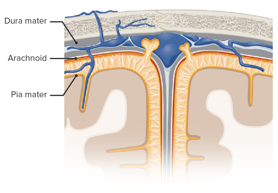
MeningesMeningesThe brain and the spinal cord are enveloped by 3 overlapping layers of connective tissue called the meninges. The layers are, from the most external layer to the most internal layer, the dura mater, arachnoid mater, and pia mater. Between these layers are 3 potential spaces called the epidural, subdural, and subarachnoid spaces. Meninges: Anatomy
Table: MeningesMeningesThe brain and the spinal cord are enveloped by 3 overlapping layers of connective tissue called the meninges. The layers are, from the most external layer to the most internal layer, the dura mater, arachnoid mater, and pia mater. Between these layers are 3 potential spaces called the epidural, subdural, and subarachnoid spaces. Meninges: Anatomy
Potential space between the dura materDura materThe outermost of the three meninges, a fibrous membrane of connective tissue that covers the brain and the spinal cord.Meninges: Anatomy and skullSkullThe skull (cranium) is the skeletal structure of the head supporting the face and forming a protective cavity for the brain. The skull consists of 22 bones divided into the viscerocranium (facial skeleton) and the neurocranium.Skull: Anatomy/vertebral columnVertebral columnThe human spine, or vertebral column, is the most important anatomical and functional axis of the human body. It consists of 7 cervical vertebrae, 12 thoracic vertebrae, and 5 lumbar vertebrae and is limited cranially by the skull and caudally by the sacrum. Vertebral Column: Anatomy
Dura materDura materThe outermost of the three meninges, a fibrous membrane of connective tissue that covers the brain and the spinal cord.Meninges: Anatomy
Divided into 2 layers:
Superficial periosteal layer
Inner meningeal layer
Grows adhered to the periosteumPeriosteumThin outer membrane that surrounds a bone. It contains connective tissue, capillaries, nerves, and a number of cell types.Bones: Structure and Types of the calvaria
Trigeminal nerveTrigeminal nerveThe 5th and largest cranial nerve. The trigeminal nerve is a mixed motor and sensory nerve. The larger sensory part forms the ophthalmic, mandibular, and maxillary nerves which carry afferents sensitive to external or internal stimuli from the skin, muscles, and joints of the face and mouth and from the teeth. Most of these fibers originate from cells of the trigeminal ganglion and project to the trigeminal nucleus of the brain stem. The smaller motor part arises from the brain stem trigeminal motor nucleus and innervates the muscles of mastication.The 12 Cranial Nerves: Overview and Functions branches: innervate supratentorial structures
Cervical nerves (C2 and C3): innervate infratentorial structures
Potential space between the arachnoid materArachnoid materA delicate membrane enveloping the brain and spinal cord. It lies between the pia mater and the dura mater. It is separated from the pia mater by the subarachnoid cavity which is filled with cerebrospinal fluid.Meninges: Anatomy and the dura materDura materThe outermost of the three meninges, a fibrous membrane of connective tissue that covers the brain and the spinal cord.Meninges: Anatomy
Site of blood collection in cases of injury to bridging veinsBridging VeinsSubdural Hemorrhage → subdural hematomaHematomaA collection of blood outside the blood vessels. Hematoma can be localized in an organ, space, or tissue.Intussusception
Arachnoid materArachnoid materA delicate membrane enveloping the brain and spinal cord. It lies between the pia mater and the dura mater. It is separated from the pia mater by the subarachnoid cavity which is filled with cerebrospinal fluid.Meninges: Anatomy (leptomeningesLeptomeningesMeninges: Anatomy)
Outer layer of the subarachnoid spaceSubarachnoid spaceThe space between the arachnoid membrane and pia mater, filled with cerebrospinal fluid. It contains large blood vessels that supply the brain and spinal cord.Subarachnoid Hemorrhage
Arachnoid trabeculae: web-like strands that separate the arachnoid and pia materPia materThe innermost layer of the three meninges covering the brain and spinal cord. It is the fine vascular membrane that lies under the arachnoid and the dura mater.Meninges: Anatomy
Subarachnoid spaceSubarachnoid spaceThe space between the arachnoid membrane and pia mater, filled with cerebrospinal fluid. It contains large blood vessels that supply the brain and spinal cord.Subarachnoid Hemorrhage
Arachnoid/pacchionian granulations: allow CSF to enter from the subarachnoid spaceSubarachnoid spaceThe space between the arachnoid membrane and pia mater, filled with cerebrospinal fluid. It contains large blood vessels that supply the brain and spinal cord.Subarachnoid Hemorrhage into the venous system
CSF: produced by the choroidChoroidThe thin, highly vascular membrane covering most of the posterior of the eye between the retina and sclera.Eye: Anatomy plexus and contained in the subarachnoid spaceSubarachnoid spaceThe space between the arachnoid membrane and pia mater, filled with cerebrospinal fluid. It contains large blood vessels that supply the brain and spinal cord.Subarachnoid Hemorrhage
Site of blood collection in cases of saccular aneurysmSaccular AneurysmSubarachnoid Hemorrhage rupture → subarachnoid hemorrhageSubarachnoid HemorrhageSubarachnoid hemorrhage (SAH) is a type of cerebrovascular accident (stroke) resulting from intracranial hemorrhage into the subarachnoid space between the arachnoid and the pia mater layers of the meninges surrounding the brain. Most SAHs originate from a saccular aneurysm in the circle of Willis but may also occur as a result of trauma, uncontrolled hypertension, vasculitis, anticoagulant use, or stimulant use. Subarachnoid Hemorrhage
Pia materPia materThe innermost layer of the three meninges covering the brain and spinal cord. It is the fine vascular membrane that lies under the arachnoid and the dura mater.Meninges: Anatomy (leptomeningesLeptomeningesMeninges: Anatomy)
Inner layer of the subarachnoid spaceSubarachnoid spaceThe space between the arachnoid membrane and pia mater, filled with cerebrospinal fluid. It contains large blood vessels that supply the brain and spinal cord.Subarachnoid Hemorrhage
Adherent to the brainBrainThe part of central nervous system that is contained within the skull (cranium). Arising from the neural tube, the embryonic brain is comprised of three major parts including prosencephalon (the forebrain); mesencephalon (the midbrain); and rhombencephalon (the hindbrain). The developed brain consists of cerebrum; cerebellum; and other structures in the brain stem.Nervous System: Anatomy, Structure, and Classification
Confers shiny appearance
Highly vascularized
Cross-sectional view of the head showcasing the meningeal layers
Image by Lecturio.
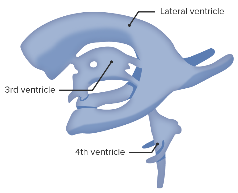
Ventricular systemVentricular SystemThe ventricular system is an extension of the subarachnoid space into the brain consisting of a series of interconnecting spaces and channels. Four chambers are filled with cerebrospinal fluid (CSF): the paired lateral ventricles, the unpaired 3rd ventricle, and the unpaired 4th ventricle. Ventricular System: Anatomy
The ventricular systemVentricular SystemThe ventricular system is an extension of the subarachnoid space into the brain consisting of a series of interconnecting spaces and channels. Four chambers are filled with cerebrospinal fluid (CSF): the paired lateral ventricles, the unpaired 3rd ventricle, and the unpaired 4th ventricle. Ventricular System: Anatomy is composed of the following structures:
Lateral ventriclesLateral ventriclesCavity in each of the cerebral hemispheres derived from the cavity of the embryonic neural tube. They are separated from each other by the septum pellucidum, and each communicates with the third ventricle by the foramen of monro, through which also the choroid plexuses (choroid plexus) of the lateral ventricles become continuous with that of the third ventricle.Ventricular System: Anatomy:
Body:
Spans the frontalFrontalThe bone that forms the frontal aspect of the skull. Its flat part forms the forehead, articulating inferiorly with the nasal bone and the cheek bone on each side of the face.Skull: Anatomy, parietalParietalOne of a pair of irregularly shaped quadrilateral bones situated between the frontal bone and occipital bone, which together form the sides of the cranium.Skull: Anatomy, temporal, and occipitalOccipitalPart of the back and base of the cranium that encloses the foramen magnum.Skull: Anatomy lobes
Anterior hornAnterior hornOne of three central columns of the spinal cord. It is composed of gray matter spinal laminae VIII and ix.Brown-Séquard Syndrome: in the frontal lobeFrontal lobeThe part of the cerebral hemisphere anterior to the central sulcus, and anterior and superior to the lateral sulcus.Cerebral Cortex: Anatomy
Posterior hornPosterior hornOne of three central columns of the spinal cord. It is composed of gray matter spinal laminae i-vi.Brown-Séquard Syndrome: curves posteromedially into the occipital lobeOccipital lobePosterior portion of the cerebral hemispheres responsible for processing visual sensory information. It is located posterior to the parieto-occipital sulcus and extends to the preoccipital notch.Cerebral Cortex: Anatomy
Inferior horn
Largest compartment of the lateral ventricle
Extends forward into the temporal lobeTemporal lobeLower lateral part of the cerebral hemisphere responsible for auditory, olfactory, and semantic processing. It is located inferior to the lateral fissure and anterior to the occipital lobe.Cerebral Cortex: Anatomy
3rd ventricle: midline, slit-like cavity
Cerebral aqueductCerebral aqueductNarrow channel in the mesencephalon that connects the third and fourth cerebral ventricles.Ventricular System: Anatomy: small tube extending throughout the dorsal quarter of the midbrainMidbrainThe middle of the three primitive cerebral vesicles of the embryonic brain. Without further subdivision, midbrain develops into a short, constricted portion connecting the pons and the diencephalon. Midbrain contains two major parts, the dorsal tectum mesencephali and the ventral tegmentum mesencephali, housing components of auditory, visual, and other sensorimotor systems.Brain Stem: Anatomy in the midline and surrounded by the periaqueductal gray
4th ventricle: between the brainBrainThe part of central nervous system that is contained within the skull (cranium). Arising from the neural tube, the embryonic brain is comprised of three major parts including prosencephalon (the forebrain); mesencephalon (the midbrain); and rhombencephalon (the hindbrain). The developed brain consists of cerebrum; cerebellum; and other structures in the brain stem.Nervous System: Anatomy, Structure, and Classification stem and the cerebellumCerebellumThe cerebellum, Latin for “little brain,” is located in the posterior cranial fossa, dorsal to the pons and midbrain, and its principal role is in the coordination of movements. The cerebellum consists of 3 lobes on either side of its 2 hemispheres and is connected in the middle by the vermis. Cerebellum: Anatomy
Ventricular system isolated from the brain: Note the lateral ventricles, the 3rd ventricle in the center, and the 4th ventricle toward the bottom.
Image by Lecturio.
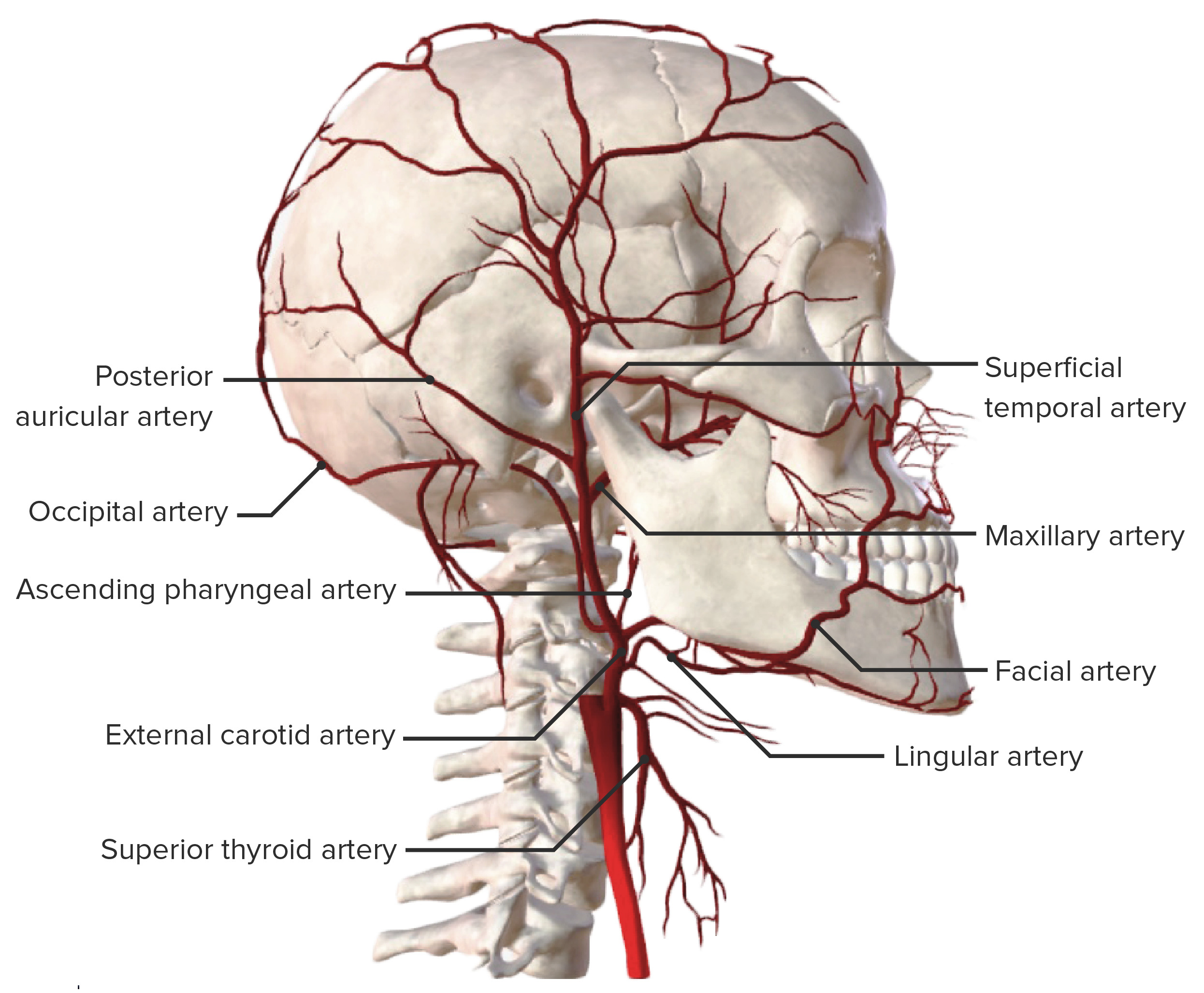
Arterial supply of the head
The arteriesArteriesArteries are tubular collections of cells that transport oxygenated blood and nutrients from the heart to the tissues of the body. The blood passes through the arteries in order of decreasing luminal diameter, starting in the largest artery (the aorta) and ending in the small arterioles. Arteries are classified into 3 types: large elastic arteries, medium muscular arteries, and small arteries and arterioles. Arteries: Histology that supply the skullSkullThe skull (cranium) is the skeletal structure of the head supporting the face and forming a protective cavity for the brain. The skull consists of 22 bones divided into the viscerocranium (facial skeleton) and the neurocranium.Skull: Anatomy and its contents can be divided into 2 large groups.
The external group is made up of the branches of the external carotid arteryExternal carotid arteryBranch of the common carotid artery which supplies the exterior of the head, the face, and the greater part of the neck.Carotid Arterial System: Anatomy:
OccipitalOccipitalPart of the back and base of the cranium that encloses the foramen magnum.Skull: Anatomy artery
The internal group is made up of branches of the circle of WillisCircle of WillisA polygonal anastomosis at the base of the brain formed by the internal carotid, proximal parts of the anterior, middle, and posterior cerebral arteries, the anterior communicating artery and the posterior communicating arteries.Subarachnoid Hemorrhage:
Anterior cerebral arteryAnterior cerebral arteryArtery formed by the bifurcation of the internal carotid artery. Branches of the anterior cerebral artery supply the caudate nucleus; internal capsule; putamen; septal nuclei; gyrus cinguli; and surfaces of the frontal lobe and parietal lobe.Cerebrovascular System: Anatomy
Middle cerebral arteryMiddle cerebral arteryThe largest of the cerebral arteries. It trifurcates into temporal, frontal, and parietal branches supplying blood to most of the parenchyma of these lobes in the cerebral cortex. These are the areas involved in motor, sensory, and speech activities.Cerebrovascular System: Anatomy
Posterior cerebral arteryPosterior cerebral arteryArtery formed by the bifurcation of the basilar artery. Branches of the posterior cerebral artery supply portions of the occipital lobe; parietal lobe; inferior temporal gyrus, brainstem, and choroid plexus.Cerebrovascular System: Anatomy
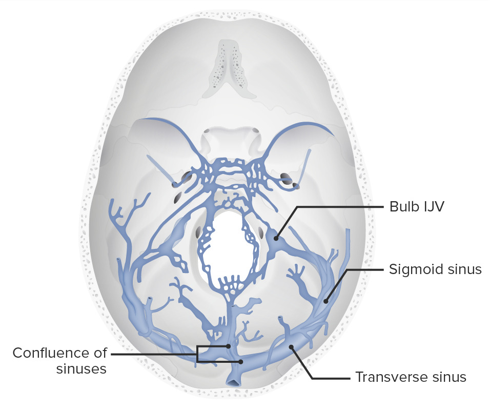
The venous drainage system of the brainBrainThe part of central nervous system that is contained within the skull (cranium). Arising from the neural tube, the embryonic brain is comprised of three major parts including prosencephalon (the forebrain); mesencephalon (the midbrain); and rhombencephalon (the hindbrain). The developed brain consists of cerebrum; cerebellum; and other structures in the brain stem.Nervous System: Anatomy, Structure, and Classification is located between the endosteal and meningeal layers of dura materDura materThe outermost of the three meninges, a fibrous membrane of connective tissue that covers the brain and the spinal cord.Meninges: Anatomy. These vessels are lined by endotheliumEndotheliumA layer of epithelium that lines the heart, blood vessels (vascular endothelium), lymph vessels (lymphatic endothelium), and the serous cavities of the body.Arteries: Histology and have no valves or smooth muscle cells in their walls.
The following sinuses are in contact with the bones of the skullSkullThe skull (cranium) is the skeletal structure of the head supporting the face and forming a protective cavity for the brain. The skull consists of 22 bones divided into the viscerocranium (facial skeleton) and the neurocranium.Skull: Anatomy:
Transverse sinusTransverse sinusThe two large endothelium-lined venous channels that begin at the internal occipital protuberance at the back and lower part of the cranium and travels laterally and forward ending in the internal jugular vein (jugular veins). One of the transverse sinuses, usually the right one, is the continuation of the superior sagittal sinus. The other transverse sinus is the continuation of the straight sinus.Cerebrovascular System: Anatomy
SigmoidSigmoidA segment of the colon between the rectum and the descending colon.Volvulus sinus
Venous sinuses of the middle and posterior fossa IJV: internal jugular vein
Image by Lecturio.
Vertebral columnVertebral columnThe human spine, or vertebral column, is the most important anatomical and functional axis of the human body. It consists of 7 cervical vertebrae, 12 thoracic vertebrae, and 5 lumbar vertebrae and is limited cranially by the skull and caudally by the sacrum. Vertebral Column: Anatomy
33 vertebrae placed in series and connected by intervertebral disks and ligaments
Vertebral columnVertebral columnThe human spine, or vertebral column, is the most important anatomical and functional axis of the human body. It consists of 7 cervical vertebrae, 12 thoracic vertebrae, and 5 lumbar vertebrae and is limited cranially by the skull and caudally by the sacrum. Vertebral Column: AnatomysegmentationSegmentationGastrointestinal Motility
Cervical: 7
Thoracic: 12
Lumbar: 5
Sacral: 5
Coccygeal: 4 (3–5)
The vertebrae form the spinal canalSpinal CanalThe cavity within the spinal column through which the spinal cord passes.Spinal Cord Injuries, which houses the spinal cordSpinal cordThe spinal cord is the major conduction pathway connecting the brain to the body; it is part of the CNS. In cross section, the spinal cord is divided into an H-shaped area of gray matter (consisting of synapsing neuronal cell bodies) and a surrounding area of white matter (consisting of ascending and descending tracts of myelinated axons). Spinal Cord: Anatomy and spinal nervesSpinal nervesThe 31 paired peripheral nerves formed by the union of the dorsal and ventral spinal roots from each spinal cord segment. The spinal nerve plexuses and the spinal roots are also included.Spinal Cord: Anatomy
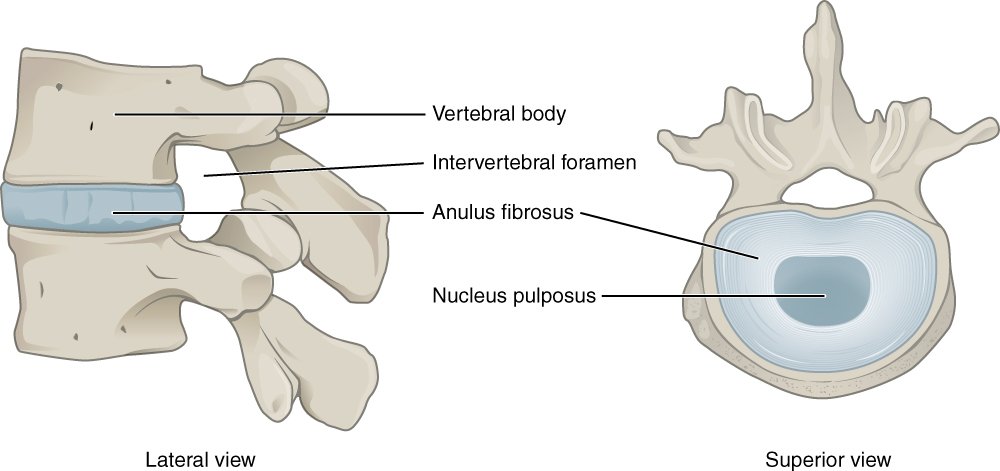
Intervertebral foramina (neuroforamen or neural foramen): foramen for the spinal nervesSpinal nervesThe 31 paired peripheral nerves formed by the union of the dorsal and ventral spinal roots from each spinal cord segment. The spinal nerve plexuses and the spinal roots are also included.Spinal Cord: Anatomy exiting the vertebral columnVertebral columnThe human spine, or vertebral column, is the most important anatomical and functional axis of the human body. It consists of 7 cervical vertebrae, 12 thoracic vertebrae, and 5 lumbar vertebrae and is limited cranially by the skull and caudally by the sacrum. Vertebral Column: Anatomy between 2 spinal vertebrae
The intervertebral disk space is highlighted in this image.
Image: “Intervertebral disc” by Phil Schatz. License: CC BY 4.0
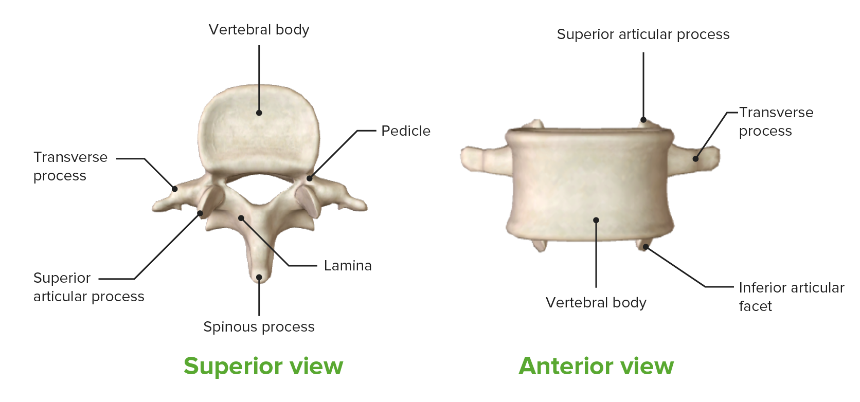
Articular (superior and inferior, form the facets)
Spinous (posterior)
Foramina:
Vertebral: Large central openings in vertebrae that collectively form the vertebral, or spinal, canal, which contains:
Spinal cordSpinal cordThe spinal cord is the major conduction pathway connecting the brain to the body; it is part of the CNS. In cross section, the spinal cord is divided into an H-shaped area of gray matter (consisting of synapsing neuronal cell bodies) and a surrounding area of white matter (consisting of ascending and descending tracts of myelinated axons). Spinal Cord: Anatomy
Nerve roots
Blood vessels
Intervertebral foramina (neuroforamen or neural foramen): opening for the spinal nervesSpinal nervesThe 31 paired peripheral nerves formed by the union of the dorsal and ventral spinal roots from each spinal cord segment. The spinal nerve plexuses and the spinal roots are also included.Spinal Cord: Anatomy
Nucleus pulposusNucleus PulposusFibrocartilage inner core of the intervertebral disc. Prolapsed or bulged nucleus pulposus leads to intervertebral disc displacement while proliferation of cells in the nucleus pulposus is associated with intervertebral disc degeneration.Spinal Disk Herniation
Cartilaginous end plates anchor the disks to the adjacent vertebrae
Craniotomy: surgical procedure that aims to access the cranial cavity and operate directly on the cerebral parenchyma by removing a boneBoneBone is a compact type of hardened connective tissue composed of bone cells, membranes, an extracellular mineralized matrix, and central bone marrow. The 2 primary types of bone are compact and spongy. Bones: Structure and Types flap from the skullSkullThe skull (cranium) is the skeletal structure of the head supporting the face and forming a protective cavity for the brain. The skull consists of 22 bones divided into the viscerocranium (facial skeleton) and the neurocranium.Skull: Anatomy.
Craniectomy: a craniotomy in which the boneBoneBone is a compact type of hardened connective tissue composed of bone cells, membranes, an extracellular mineralized matrix, and central bone marrow. The 2 primary types of bone are compact and spongy. Bones: Structure and Types flap is used as a template for a titanium or acrylic plate or is stored for later reimplantation.
Cranioplasty: surgical procedure to reconstruct the skullSkullThe skull (cranium) is the skeletal structure of the head supporting the face and forming a protective cavity for the brain. The skull consists of 22 bones divided into the viscerocranium (facial skeleton) and the neurocranium.Skull: Anatomy by placing the boneBoneBone is a compact type of hardened connective tissue composed of bone cells, membranes, an extracellular mineralized matrix, and central bone marrow. The 2 primary types of bone are compact and spongy. Bones: Structure and Types flap, or a synthetic replacement, into position during a 2nd intervention.
Classification
Hemicraniectomy: removal of boneBoneBone is a compact type of hardened connective tissue composed of bone cells, membranes, an extracellular mineralized matrix, and central bone marrow. The 2 primary types of bone are compact and spongy. Bones: Structure and Types in a single hemisphere
Bilateral craniectomy: removal of a single boneBoneBone is a compact type of hardened connective tissue composed of bone cells, membranes, an extracellular mineralized matrix, and central bone marrow. The 2 primary types of bone are compact and spongy. Bones: Structure and Types flap from both hemispheres
Depending on the location, a craniotomy is:
Supratentorial
Infratentorial
Indications
Craniotomy:
BrainBrainThe part of central nervous system that is contained within the skull (cranium). Arising from the neural tube, the embryonic brain is comprised of three major parts including prosencephalon (the forebrain); mesencephalon (the midbrain); and rhombencephalon (the hindbrain). The developed brain consists of cerebrum; cerebellum; and other structures in the brain stem.Nervous System: Anatomy, Structure, and ClassificationmassMassThree-dimensional lesion that occupies a space within the breastImaging of the Breast/tumorTumorInflammation removal
ImplantationImplantationEndometrial implantation of embryo, mammalian at the blastocyst stage.Fertilization and First Week of a device with therapeutic intent (e.g., deep brainBrainThe part of central nervous system that is contained within the skull (cranium). Arising from the neural tube, the embryonic brain is comprised of three major parts including prosencephalon (the forebrain); mesencephalon (the midbrain); and rhombencephalon (the hindbrain). The developed brain consists of cerebrum; cerebellum; and other structures in the brain stem.Nervous System: Anatomy, Structure, and Classification stimulation in the treatment of Parkinson diseaseParkinson diseaseParkinson’s disease (PD) is a chronic, progressive neurodegenerative disorder. Although the cause is unknown, several genetic and environmental risk factors are currently being studied. Individuals present clinically with resting tremor, bradykinesia, rigidity, and postural instability.Parkinson’s Disease)
Craniectomy:
Elevated intracranial pressureIntracranial PressureIdiopathic Intracranial Hypertension (ICPICPNormal intracranial pressure (ICP) is defined as < 15 mm Hg, whereas pathologically increased ICP is any pressure ≥ 20 mm Hg. Increased ICP may result from several etiologies, including trauma, intracranial hemorrhage, mass lesions, cerebral edema, increased CSF production, and decreased CSF absorption.Increased Intracranial Pressure (ICP)) refractory to medical intervention
BrainBrainThe part of central nervous system that is contained within the skull (cranium). Arising from the neural tube, the embryonic brain is comprised of three major parts including prosencephalon (the forebrain); mesencephalon (the midbrain); and rhombencephalon (the hindbrain). The developed brain consists of cerebrum; cerebellum; and other structures in the brain stem.Nervous System: Anatomy, Structure, and ClassificationherniationHerniationOmphalocele: pathologic displacementDisplacementThe process by which an emotional or behavioral response that is appropriate for one situation appears in another situation for which it is inappropriate.Defense Mechanisms of intracranial structures due to an increased pressure gradientPressure gradientVascular Resistance, Flow, and Mean Arterial Pressure between the intracranial compartments
ContraindicationsContraindicationsA condition or factor associated with a recipient that makes the use of a drug, procedure, or physical agent improper or inadvisable. Contraindications may be absolute (life threatening) or relative (higher risk of complications in which benefits may outweigh risks).Noninvasive Ventilation
Severe cardiopulmonary disease
Severe systemic collapse (e.g., sepsisSepsisSystemic inflammatory response syndrome with a proven or suspected infectious etiology. When sepsis is associated with organ dysfunction distant from the site of infection, it is called severe sepsis. When sepsis is accompanied by hypotension despite adequate fluid infusion, it is called septic shock.Sepsis and Septic Shock, adrenal crisisAdrenal crisisAdrenal crisis is the acute decompensation of adrenal function that can be triggered by another disease, surgery, stress, or increased glucocorticoid inactivation.Adrenal Insufficiency and Addison Disease, massive hemorrhage)
Abnormal coagulation parameters
Operative care
When used for decompression, the craniotomy/craniectomy procedure turns the intracranial compartment from a semirigid shape into an elasticElasticConnective Tissue: Histology container.
An incision is made on the scalp. A local anesthetic with epinephrineEpinephrineThe active sympathomimetic hormone from the adrenal medulla. It stimulates both the alpha- and beta- adrenergic systems, causes systemic vasoconstriction and gastrointestinal relaxation, stimulates the heart, and dilates bronchi and cerebral vessels.Sympathomimetic Drugs may be administered at the incision site to prevent heavy bleeding of the scalp.
Subcutaneous tissueSubcutaneous tissueLoose connective tissue lying under the dermis, which binds skin loosely to subjacent tissues. It may contain a pad of adipocytes, which vary in number according to the area of the body and vary in size according to the nutritional state.Soft Tissue Abscess and muscles are dissected to expose the skullSkullThe skull (cranium) is the skeletal structure of the head supporting the face and forming a protective cavity for the brain. The skull consists of 22 bones divided into the viscerocranium (facial skeleton) and the neurocranium.Skull: Anatomy.
Retractors are placed on the edges of the incision to expose the work area.
Burr holes are made in the skullSkullThe skull (cranium) is the skeletal structure of the head supporting the face and forming a protective cavity for the brain. The skull consists of 22 bones divided into the viscerocranium (facial skeleton) and the neurocranium.Skull: Anatomy to mark the corners of the skullSkullThe skull (cranium) is the skeletal structure of the head supporting the face and forming a protective cavity for the brain. The skull consists of 22 bones divided into the viscerocranium (facial skeleton) and the neurocranium.Skull: Anatomy segment to be removed.
The burr holes are linked using a craniotome, creating a boneBoneBone is a compact type of hardened connective tissue composed of bone cells, membranes, an extracellular mineralized matrix, and central bone marrow. The 2 primary types of bone are compact and spongy. Bones: Structure and Types flap.
The dura is separated from the boneBoneBone is a compact type of hardened connective tissue composed of bone cells, membranes, an extracellular mineralized matrix, and central bone marrow. The 2 primary types of bone are compact and spongy. Bones: Structure and Types.
After separating the boneBoneBone is a compact type of hardened connective tissue composed of bone cells, membranes, an extracellular mineralized matrix, and central bone marrow. The 2 primary types of bone are compact and spongy. Bones: Structure and Types flap from the dura, it is elevated and then removed. At this time, the boneBoneBone is a compact type of hardened connective tissue composed of bone cells, membranes, an extracellular mineralized matrix, and central bone marrow. The 2 primary types of bone are compact and spongy. Bones: Structure and Types flap can be:
Kept on the surgical table until reimplantation
Discarded (requires immediate intraoperative reconstruction with synthetic materials)
Placed within an abdominal subcutaneous incision
Preserved in a tissue bank
The dura is cut and retracted to expose the brainBrainThe part of central nervous system that is contained within the skull (cranium). Arising from the neural tube, the embryonic brain is comprised of three major parts including prosencephalon (the forebrain); mesencephalon (the midbrain); and rhombencephalon (the hindbrain). The developed brain consists of cerebrum; cerebellum; and other structures in the brain stem.Nervous System: Anatomy, Structure, and Classification to perform the specific intervention.
The intended procedure is performed on the brainBrainThe part of central nervous system that is contained within the skull (cranium). Arising from the neural tube, the embryonic brain is comprised of three major parts including prosencephalon (the forebrain); mesencephalon (the midbrain); and rhombencephalon (the hindbrain). The developed brain consists of cerebrum; cerebellum; and other structures in the brain stem.Nervous System: Anatomy, Structure, and Classification parenchyma.
The boneBoneBone is a compact type of hardened connective tissue composed of bone cells, membranes, an extracellular mineralized matrix, and central bone marrow. The 2 primary types of bone are compact and spongy. Bones: Structure and Types flap is reattached to the rest of the skullSkullThe skull (cranium) is the skeletal structure of the head supporting the face and forming a protective cavity for the brain. The skull consists of 22 bones divided into the viscerocranium (facial skeleton) and the neurocranium.Skull: Anatomy using plates and screws.
The surgeon is careful to ensure adequate homeostasisHomeostasisThe processes whereby the internal environment of an organism tends to remain balanced and stable.Cell Injury and Death before closing the scalp.
The surgeon can decide to leave a subdural or subgaleal drain in place.
The skinSkinThe skin, also referred to as the integumentary system, is the largest organ of the body. The skin is primarily composed of the epidermis (outer layer) and dermis (deep layer). The epidermis is primarily composed of keratinocytes that undergo rapid turnover, while the dermis contains dense layers of connective tissue.Skin: Structure and Functions is closed using nonabsorbable suturesNonabsorbable SuturesSurgical Instruments and Sutures.
The skinSkinThe skin, also referred to as the integumentary system, is the largest organ of the body. The skin is primarily composed of the epidermis (outer layer) and dermis (deep layer). The epidermis is primarily composed of keratinocytes that undergo rapid turnover, while the dermis contains dense layers of connective tissue.Skin: Structure and Functions is cleansed of any residue (e.g., blood, adipose tissueAdipose tissueAdipose tissue is a specialized type of connective tissue that has both structural and highly complex metabolic functions, including energy storage, glucose homeostasis, and a multitude of endocrine capabilities. There are three types of adipose tissue, white adipose tissue, brown adipose tissue, and beige or “brite” adipose tissue, which is a transitional form.Adipose Tissue: Histology)
A sterileSterileBasic Procedures gauze and skinSkinThe skin, also referred to as the integumentary system, is the largest organ of the body. The skin is primarily composed of the epidermis (outer layer) and dermis (deep layer). The epidermis is primarily composed of keratinocytes that undergo rapid turnover, while the dermis contains dense layers of connective tissue.Skin: Structure and Functions dressing are placed over the surgical wound.
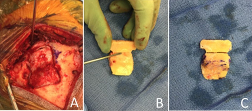
Intraoperative images of craniotomy: A: The 2-part craniotomy is elevated to reveal intact dura spanning the transverse sinus. B: The inner concavity of the 2-part bone flap is seen. The Penfield #4 instrument points to the imprint of the transverse sinus. C: The 2 bone pieces have been fixed to each other on the internal surface; here, the external convexity is restored with an excellent anatomic cosmetic result.
Image: “Intraoperative Images of Craniotomy” by Department of Neurological Surgery, University of California, San Francisco. License: CC BY 30
Cranioplasty
The timing for cranioplasty depends on the individual’s clinical status and is left to the surgeon’s discretion (at least 6 weeks to 3 months after injury).
Some surgeons wait up to 6 months, depending on the individual’s underlying medical issues.
Autologous boneBoneBone is a compact type of hardened connective tissue composed of bone cells, membranes, an extracellular mineralized matrix, and central bone marrow. The 2 primary types of bone are compact and spongy. Bones: Structure and Types should be used whenever possible (cheaper than synthetic materials), especially in young individuals owing to skullSkullThe skull (cranium) is the skeletal structure of the head supporting the face and forming a protective cavity for the brain. The skull consists of 22 bones divided into the viscerocranium (facial skeleton) and the neurocranium.Skull: Anatomy growth.
Rehabilitation can be started and continued while the individual is wearing a helmet.
PrognosisPrognosisA prediction of the probable outcome of a disease based on a individual’s condition and the usual course of the disease as seen in similar situations.Non-Hodgkin Lymphomas
Depends on the underlying condition
When performed for decompression, neurologic recovery has been reported in the weeks following the procedure (e.g., improved motorMotorNeurons which send impulses peripherally to activate muscles or secretory cells.Nervous System: Histology strength and language function)
Dural sinus perforationPerforationA pathological hole in an organ, blood vessel or other soft part of the body, occurring in the absence of external force.Esophagitis
Postcraniotomy headacheHeadacheThe symptom of pain in the cranial region. It may be an isolated benign occurrence or manifestation of a wide variety of headache disorders.Brain Abscess
Neurologic deficit
HydrocephalusHydrocephalusExcessive accumulation of cerebrospinal fluid within the cranium which may be associated with dilation of cerebral ventricles, intracranial.Subarachnoid Hemorrhage: potentially life-threatening condition caused by the excess accumulation of CSF within the ventricular systemVentricular SystemThe ventricular system is an extension of the subarachnoid space into the brain consisting of a series of interconnecting spaces and channels. Four chambers are filled with cerebrospinal fluid (CSF): the paired lateral ventricles, the unpaired 3rd ventricle, and the unpaired 4th ventricle. Ventricular System: Anatomy. HydrocephalusHydrocephalusExcessive accumulation of cerebrospinal fluid within the cranium which may be associated with dilation of cerebral ventricles, intracranial.Subarachnoid Hemorrhage can be classified as communicating, which is caused by either impaired CSF absorptionAbsorptionAbsorption involves the uptake of nutrient molecules and their transfer from the lumen of the GI tract across the enterocytes and into the interstitial space, where they can be taken up in the venous or lymphatic circulation.Digestion and Absorption or excess CSF production, or noncommunicating, which is caused by a structural blockage in CSF flowFlowBlood flows through the heart, arteries, capillaries, and veins in a closed, continuous circuit. Flow is the movement of volume per unit of time. Flow is affected by the pressure gradient and the resistance fluid encounters between 2 points. Vascular resistance is the opposition to flow, which is caused primarily by blood friction against vessel walls.Vascular Resistance, Flow, and Mean Arterial Pressure
SeizuresSeizuresA seizure is abnormal electrical activity of the neurons in the cerebral cortex that can manifest in numerous ways depending on the region of the brain affected. Seizures consist of a sudden imbalance that occurs between the excitatory and inhibitory signals in cortical neurons, creating a net excitation. The 2 major classes of seizures are focal and generalized. Seizures: abnormal, excessive and hypersynchronous firing of neuronsNeuronsThe basic cellular units of nervous tissue. Each neuron consists of a body, an axon, and dendrites. Their purpose is to receive, conduct, and transmit impulses in the nervous system.Nervous System: Histology. SeizuresSeizuresA seizure is abnormal electrical activity of the neurons in the cerebral cortex that can manifest in numerous ways depending on the region of the brain affected. Seizures consist of a sudden imbalance that occurs between the excitatory and inhibitory signals in cortical neurons, creating a net excitation. The 2 major classes of seizures are focal and generalized. Seizures can be generalized (involving both hemispheres and compromising awareness) or focal (involving a single area of the brainBrainThe part of central nervous system that is contained within the skull (cranium). Arising from the neural tube, the embryonic brain is comprised of three major parts including prosencephalon (the forebrain); mesencephalon (the midbrain); and rhombencephalon (the hindbrain). The developed brain consists of cerebrum; cerebellum; and other structures in the brain stem.Nervous System: Anatomy, Structure, and Classification and not compromising awareness)
Stroke: refers to the injury undergone by brainBrainThe part of central nervous system that is contained within the skull (cranium). Arising from the neural tube, the embryonic brain is comprised of three major parts including prosencephalon (the forebrain); mesencephalon (the midbrain); and rhombencephalon (the hindbrain). The developed brain consists of cerebrum; cerebellum; and other structures in the brain stem.Nervous System: Anatomy, Structure, and Classification tissue after interruption of blood flowBlood flowBlood flow refers to the movement of a certain volume of blood through the vasculature over a given unit of time (e.g., mL per minute).Vascular Resistance, Flow, and Mean Arterial Pressure (ischemic strokeIschemic StrokeAn ischemic stroke (also known as cerebrovascular accident) is an acute neurologic injury that occurs as a result of brain ischemia; this condition may be due to cerebral blood vessel occlusion by thrombosis or embolism, or rarely due to systemic hypoperfusion. Ischemic Stroke) or active hemorrhage (hemorrhagic strokeHemorrhagic strokeStroke due to rupture of a weakened blood vessel in the brain (e.g., cerebral hemispheres; cerebellum; subarachnoid space).Subarachnoid Hemorrhage), which has characteristic neurologic clinical features
ComaComaComa is defined as a deep state of unarousable unresponsiveness, characterized by a score of 3 points on the GCS. A comatose state can be caused by a multitude of conditions, making the precise epidemiology and prognosis of coma difficult to determine. Coma: clinical state characterized by unarousability and unresponsiveness to external stimuli
OsteomyelitisOsteomyelitisOsteomyelitis is an infection of the bone that results from the spread of microorganisms from the blood (hematogenous), nearby infected tissue, or open wounds (non-hematogenous). Infections are most commonly caused by Staphylococcus aureus.Osteomyelitis of the boneBoneBone is a compact type of hardened connective tissue composed of bone cells, membranes, an extracellular mineralized matrix, and central bone marrow. The 2 primary types of bone are compact and spongy. Bones: Structure and Types flap: inflammationInflammationInflammation is a complex set of responses to infection and injury involving leukocytes as the principal cellular mediators in the body’s defense against pathogenic organisms. Inflammation is also seen as a response to tissue injury in the process of wound healing. The 5 cardinal signs of inflammation are pain, heat, redness, swelling, and loss of function. Inflammation of the boneBoneBone is a compact type of hardened connective tissue composed of bone cells, membranes, an extracellular mineralized matrix, and central bone marrow. The 2 primary types of bone are compact and spongy. Bones: Structure and Types due to infection (most commonly by bacterial agents)
Bacterial, viral, fungal meningitisFungal meningitisMeningitis caused by fungal agents which may occur as opportunistic infections or arise in immunocompetent hosts.Meningitis: inflammationInflammationInflammation is a complex set of responses to infection and injury involving leukocytes as the principal cellular mediators in the body’s defense against pathogenic organisms. Inflammation is also seen as a response to tissue injury in the process of wound healing. The 5 cardinal signs of inflammation are pain, heat, redness, swelling, and loss of function. Inflammation of the leptomeningesLeptomeningesMeninges: Anatomy due to an infectious agent
Air embolismAir embolismBlocking of a blood vessel by air bubbles that enter the circulatory system, usually after trauma; surgical procedures, or changes in atmospheric pressure.Nonthrombotic Embolism: embolizationEmbolizationA method of hemostasis utilizing various agents such as gelfoam, silastic, metal, glass, or plastic pellets, autologous clot, fat, and muscle as emboli. It has been used in the treatment of spinal cord and intracranial arteriovenous malformations, renal arteriovenous fistulas, gastrointestinal bleeding, epistaxis, hypersplenism, certain highly vascular tumors, traumatic rupture of blood vessels, and control of operative hemorrhage.Gastrointestinal Bleeding of gas bubbles within the circulationCirculationThe movement of the blood as it is pumped through the cardiovascular system.ABCDE Assessment leading to blockage of arterial or venous blood flowBlood flowBlood flow refers to the movement of a certain volume of blood through the vasculature over a given unit of time (e.g., mL per minute).Vascular Resistance, Flow, and Mean Arterial Pressure
Ventriculostomy
Definition
A ventriculostomy is an opening created to communicate the cerebral ventricles with a sterileSterileBasic Procedures extracranial space. The therapeutic goal is drainage of CSF contained within the ventricles, decompression of intracranial spaces, and a decrease in the ICPICPNormal intracranial pressure (ICP) is defined as < 15 mm Hg, whereas pathologically increased ICP is any pressure ≥ 20 mm Hg. Increased ICP may result from several etiologies, including trauma, intracranial hemorrhage, mass lesions, cerebral edema, increased CSF production, and decreased CSF absorption.Increased Intracranial Pressure (ICP).
Indications
Acute symptomatic hydrocephalusHydrocephalusExcessive accumulation of cerebrospinal fluid within the cranium which may be associated with dilation of cerebral ventricles, intracranial.Subarachnoid Hemorrhage
ICPICPNormal intracranial pressure (ICP) is defined as < 15 mm Hg, whereas pathologically increased ICP is any pressure ≥ 20 mm Hg. Increased ICP may result from several etiologies, including trauma, intracranial hemorrhage, mass lesions, cerebral edema, increased CSF production, and decreased CSF absorption.Increased Intracranial Pressure (ICP) monitoring
Intraoperative brainBrainThe part of central nervous system that is contained within the skull (cranium). Arising from the neural tube, the embryonic brain is comprised of three major parts including prosencephalon (the forebrain); mesencephalon (the midbrain); and rhombencephalon (the hindbrain). The developed brain consists of cerebrum; cerebellum; and other structures in the brain stem.Nervous System: Anatomy, Structure, and ClassificationedemaEdemaEdema is a condition in which excess serous fluid accumulates in the body cavity or interstitial space of connective tissues. Edema is a symptom observed in several medical conditions. It can be categorized into 2 types, namely, peripheral (in the extremities) and internal (in an organ or body cavity). Edema: swellingSwellingInflammation of the brainBrainThe part of central nervous system that is contained within the skull (cranium). Arising from the neural tube, the embryonic brain is comprised of three major parts including prosencephalon (the forebrain); mesencephalon (the midbrain); and rhombencephalon (the hindbrain). The developed brain consists of cerebrum; cerebellum; and other structures in the brain stem.Nervous System: Anatomy, Structure, and Classification that occurs during another intracranial procedure
ContraindicationsContraindicationsA condition or factor associated with a recipient that makes the use of a drug, procedure, or physical agent improper or inadvisable. Contraindications may be absolute (life threatening) or relative (higher risk of complications in which benefits may outweigh risks).Noninvasive Ventilation
Use of anticoagulantsAnticoagulantsAnticoagulants are drugs that retard or interrupt the coagulation cascade. The primary classes of available anticoagulants include heparins, vitamin K-dependent antagonists (e.g., warfarin), direct thrombin inhibitors, and factor Xa inhibitors. Anticoagulants
Bleeding disordersBleeding disordersHypocoagulable conditions, also known as bleeding disorders or bleeding diathesis, are a diverse group of diseases that result in abnormal hemostasis. Physiologic hemostasis is dependent on the integrity of endothelial cells, subendothelial matrix, platelets, and coagulation factors. The hypocoagulable states result from abnormalities in one or more of these contributors, resulting in ineffective thrombosis and bleeding.Hypocoagulable Conditions
Scalp infection
BrainBrainThe part of central nervous system that is contained within the skull (cranium). Arising from the neural tube, the embryonic brain is comprised of three major parts including prosencephalon (the forebrain); mesencephalon (the midbrain); and rhombencephalon (the hindbrain). The developed brain consists of cerebrum; cerebellum; and other structures in the brain stem.Nervous System: Anatomy, Structure, and ClassificationabscessAbscessAccumulation of purulent material in tissues, organs, or circumscribed spaces, usually associated with signs of infection.Chronic Granulomatous Disease
An incision is made on the scalp. A local anesthetic with epinephrineEpinephrineThe active sympathomimetic hormone from the adrenal medulla. It stimulates both the alpha- and beta- adrenergic systems, causes systemic vasoconstriction and gastrointestinal relaxation, stimulates the heart, and dilates bronchi and cerebral vessels.Sympathomimetic Drugs may be administered at the incision site to prevent heavy bleeding of the scalp.
Subcutaneous tissueSubcutaneous tissueLoose connective tissue lying under the dermis, which binds skin loosely to subjacent tissues. It may contain a pad of adipocytes, which vary in number according to the area of the body and vary in size according to the nutritional state.Soft Tissue Abscess and muscles are dissected to expose the skullSkullThe skull (cranium) is the skeletal structure of the head supporting the face and forming a protective cavity for the brain. The skull consists of 22 bones divided into the viscerocranium (facial skeleton) and the neurocranium.Skull: Anatomy.
A burr holeBurr HoleSubdural Hemorrhage is made in the skullSkullThe skull (cranium) is the skeletal structure of the head supporting the face and forming a protective cavity for the brain. The skull consists of 22 bones divided into the viscerocranium (facial skeleton) and the neurocranium.Skull: Anatomy using a craniotome (craniostomy).
The dura is carefully perforated.
A pliable catheter with a rigid internal stylet is passed through the burr holeBurr HoleSubdural Hemorrhage and through the cerebral parenchyma into the ventricle.
The stylet is removed and adequate placement of the catheter is confirmed with CSF flowFlowBlood flows through the heart, arteries, capillaries, and veins in a closed, continuous circuit. Flow is the movement of volume per unit of time. Flow is affected by the pressure gradient and the resistance fluid encounters between 2 points. Vascular resistance is the opposition to flow, which is caused primarily by blood friction against vessel walls.Vascular Resistance, Flow, and Mean Arterial Pressure.
The catheter is directed under the skinSkinThe skin, also referred to as the integumentary system, is the largest organ of the body. The skin is primarily composed of the epidermis (outer layer) and dermis (deep layer). The epidermis is primarily composed of keratinocytes that undergo rapid turnover, while the dermis contains dense layers of connective tissue.Skin: Structure and Functions to a sterileSterileBasic Procedures exit site in the scalp, a few centimeters away from the burr holeBurr HoleSubdural Hemorrhage.
Intracranial hemorrhageIntracranial hemorrhageSubarachnoid hemorrhage (SAH) is a type of cerebrovascular accident (stroke) resulting from intracranial hemorrhage into the subarachnoid space between the arachnoid and the pia mater layers of the meninges surrounding the brain. Most sahs originate from a saccular aneurysm in the circle of willis but may also occur as a result of trauma, uncontrolled hypertension, vasculitis, anticoagulant use, or stimulant use.Subarachnoid Hemorrhage
Blockage of catheter lumen by air, blood, and/or debris
Failure to tap ventricle or catheter misplacement
Ventriculitis: inflammationInflammationInflammation is a complex set of responses to infection and injury involving leukocytes as the principal cellular mediators in the body’s defense against pathogenic organisms. Inflammation is also seen as a response to tissue injury in the process of wound healing. The 5 cardinal signs of inflammation are pain, heat, redness, swelling, and loss of function. Inflammation of the ventricles due to a pathogen
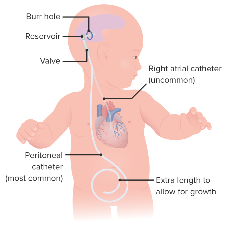
A VP shunt is a surgically created communicationCommunicationThe exchange or transmission of ideas, attitudes, or beliefs between individuals or groups.Decision-making Capacity and Legal Competence between the cerebral ventricles and the peritoneal cavityPeritoneal CavityThe space enclosed by the peritoneum. It is divided into two portions, the greater sac and the lesser sac or omental bursa, which lies behind the stomach. The two sacs are connected by the foramen of winslow, or epiploic foramen.Peritoneum: Anatomy. The aim of this treatment is to drain CSF within the ventricles and decrease ICPICPNormal intracranial pressure (ICP) is defined as < 15 mm Hg, whereas pathologically increased ICP is any pressure ≥ 20 mm Hg. Increased ICP may result from several etiologies, including trauma, intracranial hemorrhage, mass lesions, cerebral edema, increased CSF production, and decreased CSF absorption.Increased Intracranial Pressure (ICP).
CraniosynostosisCraniosynostosisCraniosynostosis is the premature fusion of 1 or more cranial sutures during the 1st year of life. Craniosynostosis is classified as simple or complex, and can be caused by environmental factors or genetic syndromes. Craniosynostosis
Dandy-Walker syndrome
IdiopathicIdiopathicDermatomyositis intracranial hypertensionHypertensionHypertension, or high blood pressure, is a common disease that manifests as elevated systemic arterial pressures. Hypertension is most often asymptomatic and is found incidentally as part of a routine physical examination or during triage for an unrelated medical encounter. Hypertension
ContraindicationsContraindicationsA condition or factor associated with a recipient that makes the use of a drug, procedure, or physical agent improper or inadvisable. Contraindications may be absolute (life threatening) or relative (higher risk of complications in which benefits may outweigh risks).Noninvasive Ventilation
Infection over the entry site
Infected CSF
AllergyAllergyAn abnormal adaptive immune response that may or may not involve antigen-specific IgEType I Hypersensitivity Reaction to shunt components (e.g., silicone)
Operative care
A U- or C-shaped incision is made on:
Kocher’s point for a frontalFrontalThe bone that forms the frontal aspect of the skull. Its flat part forms the forehead, articulating inferiorly with the nasal bone and the cheek bone on each side of the face.Skull: Anatomy approach
Keen’s point (2.5–3 cm superior and posterior to the pinna) for a parieto-occipital approach.
Subcutaneous tissueSubcutaneous tissueLoose connective tissue lying under the dermis, which binds skin loosely to subjacent tissues. It may contain a pad of adipocytes, which vary in number according to the area of the body and vary in size according to the nutritional state.Soft Tissue Abscess and muscles are dissected to expose the skullSkullThe skull (cranium) is the skeletal structure of the head supporting the face and forming a protective cavity for the brain. The skull consists of 22 bones divided into the viscerocranium (facial skeleton) and the neurocranium.Skull: Anatomy.
A burr holeBurr HoleSubdural Hemorrhage is made in the skullSkullThe skull (cranium) is the skeletal structure of the head supporting the face and forming a protective cavity for the brain. The skull consists of 22 bones divided into the viscerocranium (facial skeleton) and the neurocranium.Skull: Anatomy using a craniotome.
The catheter is cut to a premeasured length, and CSF samples are collected.
The catheter is connected to the valve and secured with silk sutures.
An incision in the abdomen and the peritoneal cavityPeritoneal CavityThe space enclosed by the peritoneum. It is divided into two portions, the greater sac and the lesser sac or omental bursa, which lies behind the stomach. The two sacs are connected by the foramen of winslow, or epiploic foramen.Peritoneum: Anatomy is accessed.
A shunt passer is used to pass the peritoneal catheter between the abdominal and cranial incisions.
The peritoneal catheter is connected to the valve and secured with silk sutures.
Patency of the peritoneal catheter is confirmed by the flowFlowBlood flows through the heart, arteries, capillaries, and veins in a closed, continuous circuit. Flow is the movement of volume per unit of time. Flow is affected by the pressure gradient and the resistance fluid encounters between 2 points. Vascular resistance is the opposition to flow, which is caused primarily by blood friction against vessel walls.Vascular Resistance, Flow, and Mean Arterial Pressure of CSF.
The distal end of the peritoneal catheter is introduced into the peritoneal cavityPeritoneal CavityThe space enclosed by the peritoneum. It is divided into two portions, the greater sac and the lesser sac or omental bursa, which lies behind the stomach. The two sacs are connected by the foramen of winslow, or epiploic foramen.Peritoneum: Anatomy.
Ventriculoperitoneal shunt in an infant with important structures, such as the valve, reservoir, and burr hole, labeled
Image by Lecturio.
Complications
Shunt infection
Intracerebral or intraventricular hemorrhageIntraventricular hemorrhageBleeding within the cerebral ventricles. It is associated with intraventricular trauma, aneurysm, vascular malformations, hypertension and in very low birth weight infants.Intracerebral Hemorrhage
Malposition of the shunt
Abdominal perforationPerforationA pathological hole in an organ, blood vessel or other soft part of the body, occurring in the absence of external force.Esophagitis
ErosionErosionPartial-thickness loss of the epidermisGeneralized and Localized Rashes of the skinSkinThe skin, also referred to as the integumentary system, is the largest organ of the body. The skin is primarily composed of the epidermis (outer layer) and dermis (deep layer). The epidermis is primarily composed of keratinocytes that undergo rapid turnover, while the dermis contains dense layers of connective tissue.Skin: Structure and Functions by the shunt
Shunt nephritis: a rare, reversible immune complex–mediated inflammationInflammationInflammation is a complex set of responses to infection and injury involving leukocytes as the principal cellular mediators in the body’s defense against pathogenic organisms. Inflammation is also seen as a response to tissue injury in the process of wound healing. The 5 cardinal signs of inflammation are pain, heat, redness, swelling, and loss of function. Inflammation of the kidneysKidneysThe kidneys are a pair of bean-shaped organs located retroperitoneally against the posterior wall of the abdomen on either side of the spine. As part of the urinary tract, the kidneys are responsible for blood filtration and excretion of water-soluble waste in the urine.Kidneys: Anatomy secondary to shunt infection
Shunt disconnection
Shunt obstruction
Abdominal CSF collections (pseudocyst)
Shunt breakage
Catheter perforationPerforationA pathological hole in an organ, blood vessel or other soft part of the body, occurring in the absence of external force.Esophagitis of viscera
Inguinal herniaHerniaProtrusion of tissue, structure, or part of an organ through the bone, muscular tissue, or the membrane by which it is normally contained. Hernia may involve tissues such as the abdominal wall or the respiratory diaphragm. Hernias may be internal, external, congenital, or acquired.Abdominal Hernias
SeizuresSeizuresA seizure is abnormal electrical activity of the neurons in the cerebral cortex that can manifest in numerous ways depending on the region of the brain affected. Seizures consist of a sudden imbalance that occurs between the excitatory and inhibitory signals in cortical neurons, creating a net excitation. The 2 major classes of seizures are focal and generalized. Seizures (focal or generalized)
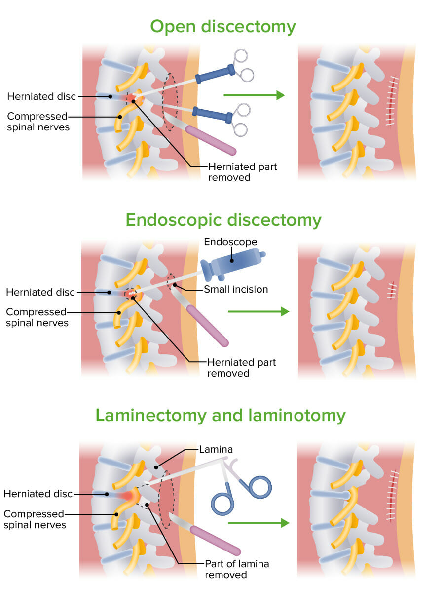
Spinal Cord Decompression Surgery (SCDS)
Definition
Spinal cordSpinal cordThe spinal cord is the major conduction pathway connecting the brain to the body; it is part of the CNS. In cross section, the spinal cord is divided into an H-shaped area of gray matter (consisting of synapsing neuronal cell bodies) and a surrounding area of white matter (consisting of ascending and descending tracts of myelinated axons). Spinal Cord: Anatomydecompression surgeryDecompression surgeryA surgical operation for the relief of pressure in a body compartment or on a body part.Cranial Nerve Palsies is a group of surgical interventions performed in the vertebral columnVertebral columnThe human spine, or vertebral column, is the most important anatomical and functional axis of the human body. It consists of 7 cervical vertebrae, 12 thoracic vertebrae, and 5 lumbar vertebrae and is limited cranially by the skull and caudally by the sacrum. Vertebral Column: Anatomy with the goal of alleviating direct compressionCompressionBlunt Chest Trauma on the spinal cordSpinal cordThe spinal cord is the major conduction pathway connecting the brain to the body; it is part of the CNS. In cross section, the spinal cord is divided into an H-shaped area of gray matter (consisting of synapsing neuronal cell bodies) and a surrounding area of white matter (consisting of ascending and descending tracts of myelinated axons). Spinal Cord: Anatomy.
Decompression techniques include:
Laminectomy
Diskectomy/microdiskectomy
Corpectomy
Foraminotomy
OsteophyteOsteophyteBony outgrowth usually found around joints and often seen in conditions such as arthritis.Osteoarthritis removal
Decompression techniques are classified as direct (permits visualization of the dural sac) and indirect (does not permit visualization of the dural sac).
Indications
Nerve root compressionCompressionBlunt Chest Trauma due to intervertebral disk herniationHerniationOmphalocele causing radiculopathyRadiculopathyDisease involving a spinal nerve root which may result from compression related to intervertebral disk displacement; spinal cord injuries; spinal diseases; and other conditions. Clinical manifestations include radicular pain, weakness, and sensory loss referable to structures innervated by the involved nerve root.Rheumatoid Arthritis
Cauda equina syndromeCauda Equina SyndromeCompressive lesion affecting the nerve roots of the cauda equina (e.g., compression, herniation, inflammation, rupture, or stenosis), which controls the function of the bladder and bowel. Symptoms may include neurological dysfunction of bladder or bowels, loss of sexual sensation and altered sensation or paralysis in the lower extremities.Ankylosing Spondylitis
Contraindication
Spinal instability is a contraindication for SCDS.
Operative care
Spinal cordSpinal cordThe spinal cord is the major conduction pathway connecting the brain to the body; it is part of the CNS. In cross section, the spinal cord is divided into an H-shaped area of gray matter (consisting of synapsing neuronal cell bodies) and a surrounding area of white matter (consisting of ascending and descending tracts of myelinated axons). Spinal Cord: Anatomydecompression surgeryDecompression surgeryA surgical operation for the relief of pressure in a body compartment or on a body part.Cranial Nerve Palsies is performed as a combination of different procedures according to the needs in each individual case. The following are among the most commonly performed decompression techniques.
Laminectomy (open approach):
A posterior midline incision is made. The extent of the incision depends on the number of laminae to be removed.
The subcutaneous soft tissues and paraspinal muscles are dissected and retracted.
Dissection continues until the ligamentum flavum is reached, which is resected using a Woodson elevator and spatula.
Once the spinous processes and laminae are fully exposed, they are sharply debulked and dissected with a rongeur to expose the spinal cordSpinal cordThe spinal cord is the major conduction pathway connecting the brain to the body; it is part of the CNS. In cross section, the spinal cord is divided into an H-shaped area of gray matter (consisting of synapsing neuronal cell bodies) and a surrounding area of white matter (consisting of ascending and descending tracts of myelinated axons). Spinal Cord: Anatomy dura and spinal nerve roots.
The compressive lesion (e.g., spinal cordSpinal cordThe spinal cord is the major conduction pathway connecting the brain to the body; it is part of the CNS. In cross section, the spinal cord is divided into an H-shaped area of gray matter (consisting of synapsing neuronal cell bodies) and a surrounding area of white matter (consisting of ascending and descending tracts of myelinated axons). Spinal Cord: AnatomytumorTumorInflammation, boneBoneBone is a compact type of hardened connective tissue composed of bone cells, membranes, an extracellular mineralized matrix, and central bone marrow. The 2 primary types of bone are compact and spongy. Bones: Structure and Types fragments, foreign bodies) is removed.
Interbody cages may be used to stabilize the spineSpineThe human spine, or vertebral column, is the most important anatomical and functional axis of the human body. It consists of 7 cervical vertebrae, 12 thoracic vertebrae, and 5 lumbar vertebrae and is limited cranially by the skull and caudally by the sacrum.Vertebral Column: Anatomy after the laminectomy.
Diskectomy (open approach):
A posterior incision is made parallel to the midline ipsilateral to the defect.
The subcutaneous soft tissues and paraspinal muscles are dissected and retracted down to the laminar junction.
A Cobb elevator is used to continue the dissection to the facet joints.
The ligamentum flavum is released from its attachment on the anterior aspect of the lamina of the superior vertebra using a curette.
The ligamentum is incised and retracted with a Penfield elevator to gain visualization of the nerve root.
The nerve root is retracted medially using a Penfield elevator to visualize the intervertebral space and the herniated disk.
Herniated tissue is removed.
If indicated, a total disk replacement is performed.
These procedures may also be done in a minimally invasive fashion; however, such procedures are beyond the scope of this review.
Illustration showing different procedures for spinal cord decompression surgery
MeningitisMeningitisMeningitis is inflammation of the meninges, the protective membranes of the brain, and spinal cord. The causes of meningitis are varied, with the most common being bacterial or viral infection. The classic presentation of meningitis is a triad of fever, altered mental status, and nuchal rigidity. Meningitis
Barrow, D. L., & Bendok, B. R. (2019). Introduction: what is neurosurgery? Operative Neurosurgery 17(Suppl 1), S1–S2. https://doi.org/10.1093/ons/opz071
Quick, C. R. G., Biers, S. M., & Arulampalam, T. H. A. (2020). Principles and techniques of operative surgery including neurosurgery. In Quick, C. R. G., Biers, S. M., & Arulampalam, T. H. A., Essential surgery: Problems, Diagnosis and Management, pp. 124–151. Elsevier.
Shayn Martin, R., & Wayne Meredith, J. (2018). Tratamiento de los traumatismos agudos. In Townsend, C. M., Beauchamp, R. D., Evers, B. M., & Mattox, K. L. (Eds.), Sabiston. Tratado de cirugía, pp. 408–448. Elsevier.
Vella, M. A., Crandall, M. L., & Patel, M. B. (2017). Acute Management of Traumatic Brain Injury. The Surgical clinics of North America, 97(5), 1015–1030. https://doi.org/10.1016/j.suc.2017.06.003
Fernández-de Thomas R. J., & De Jesus O. (2021). Craniotomy. StatPearls. Treasure Island (FL): StatPearls Publishing. Retrieved October 23, 2021, from https://www.ncbi.nlm.nih.gov/books/NBK560922/
Standring, S. (2021). Face and scalp. In Standring, S. (Ed.), Gray’s Anatomy, pp. 607–635. 41st ed. Elsevier.
Drake, R. L., Vogl, A. W., & Mitchell, A. W. M. (2020). Head and neck. In Drake, R. L., Vogl, A. W., & Mitchell, A. W. M. (Eds.), Gray’s Atlas of Anatomy, 3rd ed., pp. 477–610. Churchill Livingstone: Elsevier.
Xu, L. W., Grant, G. A., & Adelson, P. D. (2017). Management of head injury: special considerations in children. In Winn, H. R. (Ed.), Youmans and Winn Neurological Surgery, 3rd ed., pp. 1788–1795. Elsevier.
Turtz, A. R., & Barrese, J. C. (2019). Head injury. In Parrillo, J. E., & Dellinger, P. R. (Eds.), Critical Care Medicine: Principles of Diagnosis and Management in the Adult, 5th ed., pp. 1043–1072. Elsevier.
Berg, S. M., & Braehler, M. R. (2020). The postanesthesia care unit. In Gropper, M. A. (Ed.), Miller’s Anesthesia, 9th ed., pp. 2586–2613. Elsevier.
Munakomi S., & M Das, J. (2021). Ventriculostomy. StatPearls. Treasure Island (FL): StatPearls Publishing. Retrieved October 23, 2021, from http://www.ncbi.nlm.nih.gov/books/NBK545317/
Jandial, R. (2020). Core Techniques in Operative Neurosurgery. Philadelphia: Elsevier.
Baskin, J. J., & Spetzler, R. F. (2014). Lumbar puncture, ventriculostomy, and ventriculitis. In Aminoff, M. J., & Daroff, R. B. (Eds.), Encyclopedia of the Neurological Sciences, 2nd ed. Oxford: Academic Press, pp. 927–933.
Yadav, Y. R., Parihar, V., Pande, S., Namdev, H., & Agarwal, M. (2012). Endoscopic third ventriculostomy. Journal of Neurosciences in Rural Practice 3(2), pp. 163–173. https://doi.org/10.4103/0976-3147.98222
Harland, T. A., Winston, K. R., Jovanovich, A. J., & Johnson, R. J. (2018). Shunt Nephritis: An Increasingly Unfamiliar Diagnosis. World Neurosurgery 111, pp. 346–348. https://doi.org/10.1016/j.wneu.2018.01.017
Fowler, J. B., De Jesus, O., & Mesfin, F. B. (2021). Ventriculoperitoneal shunt. StatPearls. Treasure Island (FL): StatPearls Publishing. Retrieved October 23, 2021, from http://www.ncbi.nlm.nih.gov/books/NBK459351/
Estefan, M., & Camino Willhuber, G. O. (2021). Laminectomy. StatPearls. Treasure Island (FL): StatPearls Publishing. Retrieved October 23, 2021, from http://www.ncbi.nlm.nih.gov/books/NBK542274/
Wai, E. K., Roffey, D. M., Tricco, A. C., & Dagenais, S. (2012). Decompression surgery. Chapter 29 of S. Dagenais, & S. Haldeman (Eds.), Evidence-based management of low back pain. Saint Louis: Mosby, pp. 403–421. https://www.sciencedirect.com/science/article/pii/B9780323072939000295
Butler, A. J., & Donnally, C. J. III (2021). Discectomy. StatPearls. Treasure Island (FL): StatPearls Publishing. Retrieved October 23, 2021, from http://www.ncbi.nlm.nih.gov/books/NBK544281/
Phillips, N., & Hornacky, A. (2021). Berry & Kohn’s Operating Room Technique. St. Louis: Elsevier.
Create your free account or log in to continue reading!