An embolus is an intravascular solid, liquid, or gaseous material that is carried by the blood to a site distant from its point of origin. Emboli of all types warrant immediate medical attentionAttentionFocusing on certain aspects of current experience to the exclusion of others. It is the act of heeding or taking notice or concentrating.Psychiatric Assessment. The majority of emboli dislodge from a thrombus, forming a thromboembolus. Other less common nonthrombotic types of emboli are cholesterolCholesterolThe principal sterol of all higher animals, distributed in body tissues, especially the brain and spinal cord, and in animal fats and oils.Cholesterol Metabolism, fat, air, amniotic fluidAmniotic fluidA clear, yellowish liquid that envelopes the fetus inside the sac of amnion. In the first trimester, it is likely a transudate of maternal or fetal plasma. In the second trimester, amniotic fluid derives primarily from fetal lung and kidney. Cells or substances in this fluid can be removed for prenatal diagnostic tests (amniocentesis).Placenta, Umbilical Cord, and Amniotic Cavity, and tumorTumorInflammation emboli. The cause of the embolus depends on the type, as does the clinical presentation, diagnosis, and management of each embolic condition. Due to their effects on circulationCirculationThe movement of the blood as it is pumped through the cardiovascular system.ABCDE Assessment, all emboli have the potential to result in end-organ failure and death.
Also known as atheroembolism or cholesterolCholesterolThe principal sterol of all higher animals, distributed in body tissues, especially the brain and spinal cord, and in animal fats and oils.Cholesterol Metabolism crystal embolism
Occurs when cholesterolCholesterolThe principal sterol of all higher animals, distributed in body tissues, especially the brain and spinal cord, and in animal fats and oils.Cholesterol Metabolism crystals within an atherosclerotic plaquePlaquePrimary Skin Lesions dislodge and embolize
CholesterolCholesterolThe principal sterol of all higher animals, distributed in body tissues, especially the brain and spinal cord, and in animal fats and oils.Cholesterol Metabolism crystals tend to:
Dislodge from proximal, large-caliber arteriesArteriesArteries are tubular collections of cells that transport oxygenated blood and nutrients from the heart to the tissues of the body. The blood passes through the arteries in order of decreasing luminal diameter, starting in the largest artery (the aorta) and ending in the small arterioles. Arteries are classified into 3 types: large elastic arteries, medium muscular arteries, and small arteries and arterioles. Arteries: Histology
Move to many more distal, small-to-medium arteriesArteriesArteries are tubular collections of cells that transport oxygenated blood and nutrients from the heart to the tissues of the body. The blood passes through the arteries in order of decreasing luminal diameter, starting in the largest artery (the aorta) and ending in the small arterioles. Arteries are classified into 3 types: large elastic arteries, medium muscular arteries, and small arteries and arterioles. Arteries: Histology → simultaneous damage to multiple end-organs
Epidemiology and etiology
IncidenceIncidenceThe number of new cases of a given disease during a given period in a specified population. It also is used for the rate at which new events occur in a defined population. It is differentiated from prevalence, which refers to all cases in the population at a given time.Measures of Disease Frequency: unknown (many unrecognized cases)
Typical patient characteristics:
Age: > 50 years
GenderGenderGender Dysphoria: higher prevalencePrevalenceThe total number of cases of a given disease in a specified population at a designated time. It is differentiated from incidence, which refers to the number of new cases in the population at a given time.Measures of Disease Frequency in men than in women
Medical history: atherosclerotic disease
Etiology:
IatrogenicIatrogenicAny adverse condition in a patient occurring as the result of treatment by a physician, surgeon, or other health professional, especially infections acquired by a patient during the course of treatment.Anterior Cord Syndrome following an invasive vascular procedure (75%):
Cardiac catheterizationCardiac CatheterizationProcedures in which placement of cardiac catheters is performed for therapeutic or diagnostic procedures.Cardiac Surgery
Endovascular grafting
Spontaneous (25%)
Risk factors (similar to those associated with atherosclerosisAtherosclerosisAtherosclerosis is a common form of arterial disease in which lipid deposition forms a plaque in the blood vessel walls. Atherosclerosis is an incurable disease, for which there are clearly defined risk factors that often can be reduced through a change in lifestyle and behavior of the patient. Atherosclerosis):
HypercholesterolemiaHypercholesterolemiaA condition with abnormally high levels of cholesterol in the blood. It is defined as a cholesterol value exceeding the 95th percentile for the population.Lipid Disorders
HypertensionHypertensionHypertension, or high blood pressure, is a common disease that manifests as elevated systemic arterial pressures. Hypertension is most often asymptomatic and is found incidentally as part of a routine physical examination or during triage for an unrelated medical encounter. Hypertension
DiabetesDiabetesDiabetes mellitus (DM) is a metabolic disease characterized by hyperglycemia and dysfunction of the regulation of glucose metabolism by insulin. Type 1 DM is diagnosed mostly in children and young adults as the result of autoimmune destruction of β cells in the pancreas and the resulting lack of insulin. Type 2 DM has a significant association with obesity and is characterized by insulin resistance.Diabetes Mellitus mellitus
ObesityObesityObesity is a condition associated with excess body weight, specifically with the deposition of excessive adipose tissue. Obesity is considered a global epidemic. Major influences come from the western diet and sedentary lifestyles, but the exact mechanisms likely include a mixture of genetic and environmental factors. Obesity
SmokingSmokingWillful or deliberate act of inhaling and exhaling smoke from burning substances or agents held by hand.Interstitial Lung Diseases
Abdominal aortic aneurysms
Worse atherosclerotic disease → higher risk for cholesterolCholesterolThe principal sterol of all higher animals, distributed in body tissues, especially the brain and spinal cord, and in animal fats and oils.Cholesterol MetabolismembolizationEmbolizationA method of hemostasis utilizing various agents such as gelfoam, silastic, metal, glass, or plastic pellets, autologous clot, fat, and muscle as emboli. It has been used in the treatment of spinal cord and intracranial arteriovenous malformations, renal arteriovenous fistulas, gastrointestinal bleeding, epistaxis, hypersplenism, certain highly vascular tumors, traumatic rupture of blood vessels, and control of operative hemorrhage.Gastrointestinal Bleeding
Pathophysiology
CholesterolCholesterolThe principal sterol of all higher animals, distributed in body tissues, especially the brain and spinal cord, and in animal fats and oils.Cholesterol Metabolism crystals within a ruptured atherosclerotic plaquePlaquePrimary Skin Lesions embolize → “showering” of debris into the circulationCirculationThe movement of the blood as it is pumped through the cardiovascular system.ABCDE Assessment → multiple emboli → lodge in arteriolesArteriolesThe smallest divisions of the arteries located between the muscular arteries and the capillaries.Arteries: Histology
Results in multiple occlusions that affect multiple organs
Clinical presentation
The classic presentation includes skinSkinThe skin, also referred to as the integumentary system, is the largest organ of the body. The skin is primarily composed of the epidermis (outer layer) and dermis (deep layer). The epidermis is primarily composed of keratinocytes that undergo rapid turnover, while the dermis contains dense layers of connective tissue.Skin: Structure and Functions findings, abdominal painAbdominal PainAcute Abdomen, and progressive renal failureRenal failureConditions in which the kidneys perform below the normal level in the ability to remove wastes, concentrate urine, and maintain electrolyte balance; blood pressure; and calcium metabolism. Renal insufficiency can be classified by the degree of kidney damage (as measured by the level of proteinuria) and reduction in glomerular filtration rate.Crush Syndrome following a vascular procedure. Neurological and ocular symptoms are also common if the emboli travel superiorly.
Symptoms vary and depend on several factors:
Location of the embolic source:
The aortic archAortic archMediastinum and Great Vessels: Anatomy may embolize to the brainBrainThe part of central nervous system that is contained within the skull (cranium). Arising from the neural tube, the embryonic brain is comprised of three major parts including prosencephalon (the forebrain); mesencephalon (the midbrain); and rhombencephalon (the hindbrain). The developed brain consists of cerebrum; cerebellum; and other structures in the brain stem.Nervous System: Anatomy, Structure, and Classification, eye, or upper extremity.
Extent of embolizationEmbolizationA method of hemostasis utilizing various agents such as gelfoam, silastic, metal, glass, or plastic pellets, autologous clot, fat, and muscle as emboli. It has been used in the treatment of spinal cord and intracranial arteriovenous malformations, renal arteriovenous fistulas, gastrointestinal bleeding, epistaxis, hypersplenism, certain highly vascular tumors, traumatic rupture of blood vessels, and control of operative hemorrhage.Gastrointestinal Bleeding
Partial versus complete occlusion of the affected vessels
Presence or absence of pre-existing vascular disease in the affected area
Common dermatological presentations:
CyanosisCyanosisA bluish or purplish discoloration of the skin and mucous membranes due to an increase in the amount of deoxygenated hemoglobin in the blood or a structural defect in the hemoglobin molecule.Pulmonary Examination
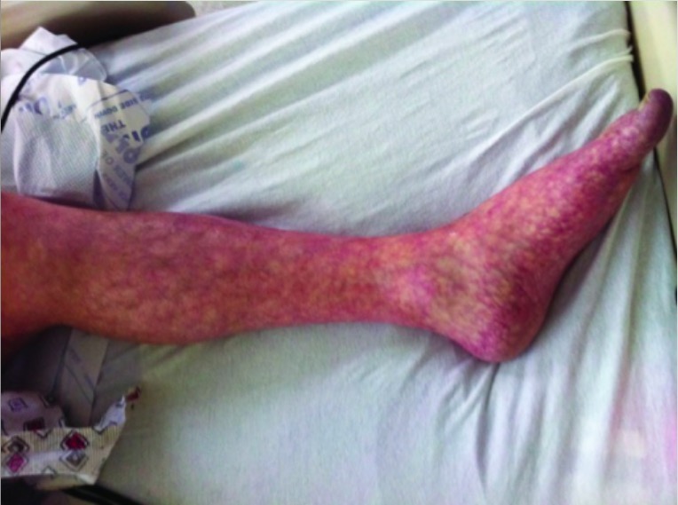
Livedo reticularisLivedo reticularisA condition characterized by a reticular or fishnet pattern on the skin of lower extremities and other parts of the body. This red and blue pattern is due to deoxygenated blood in unstable dermal blood vessels. The condition is intensified by cold exposure and relieved by rewarming.Chronic Kidney Disease: lace-like purplish discoloration of the skinSkinThe skin, also referred to as the integumentary system, is the largest organ of the body. The skin is primarily composed of the epidermis (outer layer) and dermis (deep layer). The epidermis is primarily composed of keratinocytes that undergo rapid turnover, while the dermis contains dense layers of connective tissue.Skin: Structure and Functions that blanches with pressure
AKIAKIAcute kidney injury refers to sudden and often reversible loss of renal function, which develops over days or weeks. Azotemia refers to elevated levels of nitrogen-containing substances in the blood that accompany AKI, which include BUN and creatinine. Acute Kidney Injury:
Most commonly causes distal parenchymal ischemiaIschemiaA hypoperfusion of the blood through an organ or tissue caused by a pathologic constriction or obstruction of its blood vessels, or an absence of blood circulation.Ischemic Cell Damage
Irregularly shaped emboli produce incomplete occlusion → secondary ischemic atrophyAtrophyDecrease in the size of a cell, tissue, organ, or multiple organs, associated with a variety of pathological conditions such as abnormal cellular changes, ischemia, malnutrition, or hormonal changes.Cellular Adaptation
RhabdomyolysisRhabdomyolysisRhabdomyolysis is characterized by muscle necrosis and the release of toxic intracellular contents, especially myoglobin, into the circulation.Rhabdomyolysis:
Red-brown urine (due to myoglobinuriaMyoglobinuriaThe presence of myoglobin in urine usually as a result of rhabdomyolysis.Rhabdomyolysis)
DiarrheaDiarrheaDiarrhea is defined as ≥ 3 watery or loose stools in a 24-hour period. There are a multitude of etiologies, which can be classified based on the underlying mechanism of disease. The duration of symptoms (acute or chronic) and characteristics of the stools (e.g., watery, bloody, steatorrheic, mucoid) can help guide further diagnostic evaluation. Diarrhea
GI bleeding
GI ischemiaIschemiaA hypoperfusion of the blood through an organ or tissue caused by a pathologic constriction or obstruction of its blood vessels, or an absence of blood circulation.Ischemic Cell Damage
Acute pancreatitisPancreatitisInflammation of the pancreas. Pancreatitis is classified as acute unless there are computed tomographic or endoscopic retrograde cholangiopancreatographic findings of chronic pancreatitis. The two most common forms of acute pancreatitis are alcoholic pancreatitis and gallstone pancreatitis.Acute Pancreatitis
Hepatic cell necrosisNecrosisThe death of cells in an organ or tissue due to disease, injury or failure of the blood supply.Ischemic Cell Damage
Necrotizing cholecystitisCholecystitisCholecystitis is the inflammation of the gallbladder (GB) usually caused by the obstruction of the cystic duct (acute cholecystitis). Mechanical irritation by gallstones can also produce chronic GB inflammation. Cholecystitis is one of the most common complications of cholelithiasis but inflammation without gallstones can occur in a minority of patients. Cholecystitis
Neurological presentations:
General symptoms:
HeadacheHeadacheThe symptom of pain in the cranial region. It may be an isolated benign occurrence or manifestation of a wide variety of headache disorders.Brain Abscess
Transient ischemic attackTransient ischemic attackTransient ischemic attack (TIA) is a temporary episode of neurologic dysfunction caused by ischemia without infarction that resolves completely when blood supply is restored. Transient ischemic attack is a neurologic emergency that warrants urgent medical attention. Transient Ischemic Attack (TIA)
Stroke
Ocular presentations:
Amaurosis fugaxAmaurosis fugaxTransient complete or partial monocular blindness due to retinal ischemia. This may be caused by emboli from the carotid artery (usually in association with carotid stenosis) and other locations that enter the central retinal artery.Carotid Artery Stenosis: temporary acute loss of visionVisionOphthalmic Exam due to a lack of blood flowBlood flowBlood flow refers to the movement of a certain volume of blood through the vasculature over a given unit of time (e.g., mL per minute).Vascular Resistance, Flow, and Mean Arterial Pressure to the eye
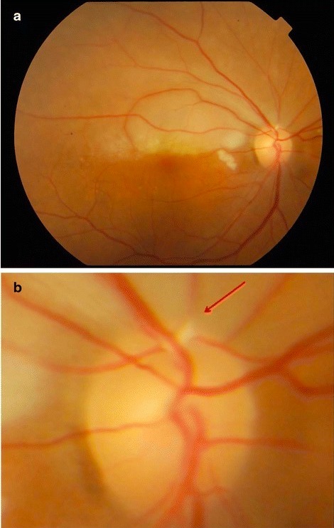
Hollenhorst plaquePlaquePrimary Skin Lesions on ophthalmoscopy: bright, refractile lesions in the retinaRetinaThe ten-layered nervous tissue membrane of the eye. It is continuous with the optic nerve and receives images of external objects and transmits visual impulses to the brain. Its outer surface is in contact with the choroid and the inner surface with the vitreous body. The outermost layer is pigmented, whereas the inner nine layers are transparent.Eye: Anatomy indicative of cholesterolCholesterolThe principal sterol of all higher animals, distributed in body tissues, especially the brain and spinal cord, and in animal fats and oils.Cholesterol Metabolism crystal embolizationEmbolizationA method of hemostasis utilizing various agents such as gelfoam, silastic, metal, glass, or plastic pellets, autologous clot, fat, and muscle as emboli. It has been used in the treatment of spinal cord and intracranial arteriovenous malformations, renal arteriovenous fistulas, gastrointestinal bleeding, epistaxis, hypersplenism, certain highly vascular tumors, traumatic rupture of blood vessels, and control of operative hemorrhage.Gastrointestinal Bleeding from a proximal source (e.g., carotid artery)
Eye painPainAn unpleasant sensation induced by noxious stimuli which are detected by nerve endings of nociceptive neurons.Pain: Types and Pathways
Image: “Livedo reticularis of left leg” by Texas Tech University Health Sciences Center, Lubbock, TX, USA. License: CC BY 3.0
a: photo of the fundus showing a pale retina superiorly b: close-up view of the optic disc from the above image showing a Hollenhorst plaque in the superotemporal vessel (arrow)
Image: “Right eye findings” by Department of Ophthalmology, Universiti Kebangsaan Malaysia Medical Centre, 56000 Cheras, Kuala Lumpur, Malaysia. License: CC BY 4.0
Diagnosis
The diagnosis is usually clinical. Definitive diagnosis requires a biopsyBiopsyRemoval and pathologic examination of specimens from the living body.Ewing Sarcoma.
Laboratory testing: generally nonspecific
CBC: ↑ eosinophilsEosinophilsGranular leukocytes with a nucleus that usually has two lobes connected by a slender thread of chromatin, and cytoplasm containing coarse, round granules that are uniform in size and stainable by eosin.Innate Immunity: Phagocytes and Antigen Presentation, ↓ RBCsRBCsErythrocytes, or red blood cells (RBCs), are the most abundant cells in the blood. While erythrocytes in the fetus are initially produced in the yolk sac then the liver, the bone marrow eventually becomes the main site of production.Erythrocytes: Histology, ↓ plateletsPlateletsPlatelets are small cell fragments involved in hemostasis. Thrombopoiesis takes place primarily in the bone marrow through a series of cell differentiation and is influenced by several cytokines. Platelets are formed after fragmentation of the megakaryocyte cytoplasm. Platelets: Histology
Organ-specific tests may be abnormal if the organ is affected:
↑ Creatinine/BUN (kidneysKidneysThe kidneys are a pair of bean-shaped organs located retroperitoneally against the posterior wall of the abdomen on either side of the spine. As part of the urinary tract, the kidneys are responsible for blood filtration and excretion of water-soluble waste in the urine.Kidneys: Anatomy)
↑ AmylaseAmylaseA group of amylolytic enzymes that cleave starch, glycogen, and related alpha-1, 4-glucans.Digestion and Absorption (pancreasPancreasThe pancreas lies mostly posterior to the stomach and extends across the posterior abdominal wall from the duodenum on the right to the spleen on the left. This organ has both exocrine and endocrine tissue. Pancreas: Anatomy)
↑ TransaminasesTransaminasesA subclass of enzymes of the transferase class that catalyze the transfer of an amino group from a donor (generally an amino acid) to an acceptor (generally a 2-keto acid). Most of these enzymes are pyridoxyl phosphate proteins.Autoimmune Hepatitis (liverLiverThe liver is the largest gland in the human body. The liver is found in the superior right quadrant of the abdomen and weighs approximately 1.5 kilograms. Its main functions are detoxification, metabolism, nutrient storage (e.g., iron and vitamins), synthesis of coagulation factors, formation of bile, filtration, and storage of blood. Liver: Anatomy)
Imaging:
Useful in assessing the extent of atherosclerotic disease
IdentificationIdentificationDefense Mechanisms of multiple complex plaques or multiple ischemic strokes assists in presumptive diagnosis.
Modalities:
Transesophageal echocardiographyEchocardiographyUltrasonic recording of the size, motion, and composition of the heart and surrounding tissues. The standard approach is transthoracic.Tricuspid Valve Atresia (TVA) (TEETEEUltrasonic recording of the size, motion, and composition of the heart and surrounding tissues using a transducer placed in the esophagus.Imaging of the Heart and Great Vessels): 1st-line approach for thoracic aortic sources
Chest CT
Chest MRI
BrainBrainThe part of central nervous system that is contained within the skull (cranium). Arising from the neural tube, the embryonic brain is comprised of three major parts including prosencephalon (the forebrain); mesencephalon (the midbrain); and rhombencephalon (the hindbrain). The developed brain consists of cerebrum; cerebellum; and other structures in the brain stem.Nervous System: Anatomy, Structure, and Classification imaging (may reveal multiple small ischemic lesions)
Histopathology:
BiopsyBiopsyRemoval and pathologic examination of specimens from the living body.Ewing Sarcoma sample can be obtained from any affected organs (e.g., skinSkinThe skin, also referred to as the integumentary system, is the largest organ of the body. The skin is primarily composed of the epidermis (outer layer) and dermis (deep layer). The epidermis is primarily composed of keratinocytes that undergo rapid turnover, while the dermis contains dense layers of connective tissue.Skin: Structure and Functions, kidney, muscle).
CholesterolCholesterolThe principal sterol of all higher animals, distributed in body tissues, especially the brain and spinal cord, and in animal fats and oils.Cholesterol Metabolism clefts (“ghosts”) within the arteriolesArteriolesThe smallest divisions of the arteries located between the muscular arteries and the capillaries.Arteries: Histology:
CholesterolCholesterolThe principal sterol of all higher animals, distributed in body tissues, especially the brain and spinal cord, and in animal fats and oils.Cholesterol Metabolism crystals are dissolved during tissue fixation leaving behind clefts.
Crescent-shaped clefts with pointed ends
Elongated ovoid spaces
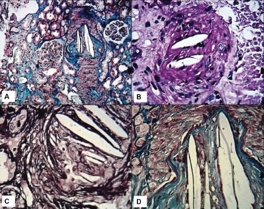
Kidney biopsies showing cholesterol clefts:
A: Low-power view showing an interlobular artery with luminal occlusion and needle-like crystals in the lumina. Two glomeruli are seen in the vicinity with minor changes on light microscopy. Mild patchy tubular atrophy is seen (Masson’s trichrome stain, ×100). B: Medium-power view showing a small artery with luminal cholesterol clefts causing luminal occlusion (Periodic acid-Schiff stain, ×200). C: High-power view showing typical empty-looking and needle-like cholesterol clefts in the lumina (Jones methenamine silver stain, ×400). D: High-power view showing luminal cholesterol clefts and intimal fibrosis. Media appears red on this stain (Masson’s trichrome stain, ×400).
Image: “” by Department of Nephrology, Division of Nephropathology, Isfahan University of Medical Sciences, Isfahan, Iran. License: CC BY 4.0
Management
Supportive management for presenting symptoms:
PainPainAn unpleasant sensation induced by noxious stimuli which are detected by nerve endings of nociceptive neurons.Pain: Types and Pathways management
Avoid further vascular instrumentation if possible.
Anti-thrombotic therapy is controversial and generally not recommended.
Prevention of kidney injury:
Aggressive saline hydration
BicarbonateBicarbonateInorganic salts that contain the -HCO3 radical. They are an important factor in determining the ph of the blood and the concentration of bicarbonate ions is regulated by the kidney. Levels in the blood are an index of the alkali reserve or buffering capacity.Electrolytes
MannitolMannitolA diuretic and renal diagnostic aid related to sorbitol. It has little significant energy value as it is largely eliminated from the body before any metabolism can take place. It can be used to treat oliguria associated with kidney failure or other manifestations of inadequate renal function and has been used for determination of glomerular filtration rate. Mannitol is also commonly used as a research tool in cell biological studies, usually to control osmolarity.Osmotic Diuretics
DialysisDialysisRenal replacement therapy refers to dialysis and/or kidney transplantation. Dialysis is a procedure by which toxins and excess water are removed from the circulation. Hemodialysis and peritoneal dialysis (PD) are the two types of dialysis, and their primary difference is the location of the filtration process (external to the body in hemodialysis versus inside the body for PD).Peritoneal Dialysis and Hemodialysis may be needed if the injury is significant.
Surgical plaquePlaquePrimary Skin Lesions removal or exclusion (if the embolic source is clearly identified)
Risk-factor reduction to prevent recurrent disease:
AspirinAspirinThe prototypical analgesic used in the treatment of mild to moderate pain. It has anti-inflammatory and antipyretic properties and acts as an inhibitor of cyclooxygenase which results in the inhibition of the biosynthesis of prostaglandins. Aspirin also inhibits platelet aggregation and is used in the prevention of arterial and venous thrombosis.Nonsteroidal Antiinflammatory Drugs (NSAIDs)
StatinsStatinsStatins are competitive inhibitors of HMG-CoA reductase in the liver. HMG-CoA reductase is the rate-limiting step in cholesterol synthesis. Inhibition results in lowered intrahepatocytic cholesterol formation, resulting in up-regulation of LDL receptors and, ultimately, lowering levels of serum LDL and triglycerides.Statins
BP control
SmokingSmokingWillful or deliberate act of inhaling and exhaling smoke from burning substances or agents held by hand.Interstitial Lung Diseases cessation
Glycemic control
Fat Embolism
Overview
Occurs when fat globules enter the pulmonary or systemic circulationCirculationThe movement of the blood as it is pumped through the cardiovascular system.ABCDE Assessment
Most common in patientsPatientsIndividuals participating in the health care system for the purpose of receiving therapeutic, diagnostic, or preventive procedures.Clinician–Patient Relationship with orthopedic trauma, especially:
Multiple fractures
Open (instead of closed) fractures
IncidenceIncidenceThe number of new cases of a given disease during a given period in a specified population. It also is used for the rate at which new events occur in a defined population. It is differentiated from prevalence, which refers to all cases in the population at a given time.Measures of Disease Frequency:
20%–30% in patientsPatientsIndividuals participating in the health care system for the purpose of receiving therapeutic, diagnostic, or preventive procedures.Clinician–Patient Relationship after trauma
1%–11% in long-bone fractures
Occurs in 90% of individuals with severe skeletal injuries, but < 10% show any clinical findings
GenderGenderGender DysphoriabiasBiasEpidemiological studies are designed to evaluate a hypothesized relationship between an exposure and an outcome; however, the existence and/or magnitude of these relationships may be erroneously affected by the design and execution of the study itself or by conscious or unconscious errors perpetrated by the investigators or the subjects. These systematic errors are called biases. Types of Biases: men > women
Age: most common in individuals between 10 and 40 years of age
Etiology
Traumatic causes:
Orthopedic causes (vast majority of cases):
Long-bone fractures
Pelvic fracturesPelvic FracturesPelvic fractures are a disruption in the cortex of a pelvic bone involving iliac wing fractures, acetabular fractures, or those causing loss of integrity of the pelvic ring (the sacrum and the 2 innominate bones). Patients often present with a history of trauma or a fall, limb length discrepancy, intense pain on palpation, and mechanical instability. Pelvic Fractures
Fractures of other marrow-containing bones (e.g., ribsRibsA set of twelve curved bones which connect to the vertebral column posteriorly, and terminate anteriorly as costal cartilage. Together, they form a protective cage around the internal thoracic organs.Chest Wall: Anatomy)
Severe burnsBurnsA burn is a type of injury to the skin and deeper tissues caused by exposure to heat, electricity, chemicals, friction, or radiation. Burns are classified according to their depth as superficial (1st-degree), partial-thickness (2nd-degree), full-thickness (3rd-degree), and 4th-degree burns. Burns
Crush injuries
Liposuction
Bone marrow transplantBone marrow transplantTransfer of hematopoietic stem cells from bone marrow or blood between individuals within the same species (homologous transplantation) or transfer within the same individual (autologous transplantation). Hematopoietic stem cell transplantation has been used as an alternative to bone marrow transplantation in the treatment of a variety of neoplasms.Organ Transplantation
Nontraumatic causes (very rare):
Acute pancreatitisPancreatitisInflammation of the pancreas. Pancreatitis is classified as acute unless there are computed tomographic or endoscopic retrograde cholangiopancreatographic findings of chronic pancreatitis. The two most common forms of acute pancreatitis are alcoholic pancreatitis and gallstone pancreatitis.Acute Pancreatitis
Sickle-cell hemoglobinopathiesHemoglobinopathiesA group of inherited disorders characterized by structural alterations within the hemoglobin molecule.Anemia: Overview and Types
Lipid infusion
Bone marrowBone marrowThe soft tissue filling the cavities of bones. Bone marrow exists in two types, yellow and red. Yellow marrow is found in the large cavities of large bones and consists mostly of fat cells and a few primitive blood cells. Red marrow is a hematopoietic tissue and is the site of production of erythrocytes and granular leukocytes. Bone marrow is made up of a framework of connective tissue containing branching fibers with the frame being filled with marrow cells.Bone Marrow: Composition and HematopoiesisnecrosisNecrosisThe death of cells in an organ or tissue due to disease, injury or failure of the blood supply.Ischemic Cell Damage
Pathogenesis
Pathogenesis may be by 1 or both of the following mechanisms:
Fat globules cause mechanical obstructionMechanical ObstructionAny impairment, arrest, or reversal of the normal flow of intestinal contents toward the anal canal.Imaging of the Intestines in the vasculature.
Embolized fat may degrade into toxic intermediaries → leads to inflammationInflammationInflammation is a complex set of responses to infection and injury involving leukocytes as the principal cellular mediators in the body’s defense against pathogenic organisms. Inflammation is also seen as a response to tissue injury in the process of wound healing. The 5 cardinal signs of inflammation are pain, heat, redness, swelling, and loss of function. Inflammation and endothelial injury
Clinical presentation
Signs and symptoms typically develop 24–72 hours after the inciting event.
DyspneaDyspneaDyspnea is the subjective sensation of breathing discomfort. Dyspnea is a normal manifestation of heavy physical or psychological exertion, but also may be caused by underlying conditions (both pulmonary and extrapulmonary). Dyspnea
TachycardiaTachycardiaAbnormally rapid heartbeat, usually with a heart rate above 100 beats per minute for adults. Tachycardia accompanied by disturbance in the cardiac depolarization (cardiac arrhythmia) is called tachyarrhythmia.Sepsis in Children
Neurological symptoms:
Changes in mental status
SeizuresSeizuresA seizure is abnormal electrical activity of the neurons in the cerebral cortex that can manifest in numerous ways depending on the region of the brain affected. Seizures consist of a sudden imbalance that occurs between the excitatory and inhibitory signals in cortical neurons, creating a net excitation. The 2 major classes of seizures are focal and generalized. Seizures
AnemiaAnemiaAnemia is a condition in which individuals have low Hb levels, which can arise from various causes. Anemia is accompanied by a reduced number of RBCs and may manifest with fatigue, shortness of breath, pallor, and weakness. Subtypes are classified by the size of RBCs, chronicity, and etiology. Anemia: Overview and Types
ThrombocytopeniaThrombocytopeniaThrombocytopenia occurs when the platelet count is < 150,000 per microliter. The normal range for platelets is usually 150,000-450,000/µL of whole blood. Thrombocytopenia can be a result of decreased production, increased destruction, or splenic sequestration of platelets. Patients are often asymptomatic until platelet counts are < 50,000/µL. Thrombocytopenia
Diagnosis and management
Fat embolism is usually a clinical diagnosis after excluding other possibilities.
Imaging:
Chest X-rayX-rayPenetrating electromagnetic radiation emitted when the inner orbital electrons of an atom are excited and release radiant energy. X-ray wavelengths range from 1 pm to 10 nm. Hard x-rays are the higher energy, shorter wavelength x-rays. Soft x-rays or grenz rays are less energetic and longer in wavelength. The short wavelength end of the x-ray spectrum overlaps the gamma rays wavelength range. The distinction between gamma rays and x-rays is based on their radiation source.Pulmonary Function Tests and/or CT in patientsPatientsIndividuals participating in the health care system for the purpose of receiving therapeutic, diagnostic, or preventive procedures.Clinician–Patient Relationship with respiratory symptoms
BrainBrainThe part of central nervous system that is contained within the skull (cranium). Arising from the neural tube, the embryonic brain is comprised of three major parts including prosencephalon (the forebrain); mesencephalon (the midbrain); and rhombencephalon (the hindbrain). The developed brain consists of cerebrum; cerebellum; and other structures in the brain stem.Nervous System: Anatomy, Structure, and Classification CT or MRI in patientsPatientsIndividuals participating in the health care system for the purpose of receiving therapeutic, diagnostic, or preventive procedures.Clinician–Patient Relationship with neurological symptoms
ProphylaxisProphylaxisCephalosporins for deep vein thrombosisThrombosisFormation and development of a thrombus or blood clot in the blood vessel.Epidemic Typhus (DVTDVTDeep vein thrombosis (DVT) usually occurs in the deep veins of the lower extremities. The affected veins include the femoral, popliteal, iliofemoral, and pelvic veins. Proximal DVT is more likely to cause a pulmonary embolism (PE) and is generally considered more serious. Deep Vein Thrombosis)
Early reduction and fixation of long-bone fractures
Air Embolism
Overview
An air embolism occurs when gas bubbles enter the circulationCirculationThe movement of the blood as it is pumped through the cardiovascular system.ABCDE Assessment and block blood flowBlood flowBlood flow refers to the movement of a certain volume of blood through the vasculature over a given unit of time (e.g., mL per minute).Vascular Resistance, Flow, and Mean Arterial Pressure.
2 types:
Venous air embolism
Arterial air embolism
IncidenceIncidenceThe number of new cases of a given disease during a given period in a specified population. It also is used for the rate at which new events occur in a defined population. It is differentiated from prevalence, which refers to all cases in the population at a given time.Measures of Disease Frequency: unknown
Obstruction occurs when the volume of air in a vessel exceeds the ability of the lungsLungsLungs are the main organs of the respiratory system. Lungs are paired viscera located in the thoracic cavity and are composed of spongy tissue. The primary function of the lungs is to oxygenate blood and eliminate CO2. Lungs: Anatomy to remove gas (approximately 50 mL).
Large bubbles obstruct the pulmonary arteriesArteriesArteries are tubular collections of cells that transport oxygenated blood and nutrients from the heart to the tissues of the body. The blood passes through the arteries in order of decreasing luminal diameter, starting in the largest artery (the aorta) and ending in the small arterioles. Arteries are classified into 3 types: large elastic arteries, medium muscular arteries, and small arteries and arterioles. Arteries: Histology → ↓ blood flowBlood flowBlood flow refers to the movement of a certain volume of blood through the vasculature over a given unit of time (e.g., mL per minute).Vascular Resistance, Flow, and Mean Arterial Pressure to lungsLungsLungs are the main organs of the respiratory system. Lungs are paired viscera located in the thoracic cavity and are composed of spongy tissue. The primary function of the lungs is to oxygenate blood and eliminate CO2. Lungs: Anatomy:
↑ Central venous pressureCentral venous pressureThe blood pressure in the central large veins of the body. It is distinguished from peripheral venous pressure which occurs in an extremity.Central Venous Catheter
Small bubbles obstruct the pulmonary arteriolesArteriolesThe smallest divisions of the arteries located between the muscular arteries and the capillaries.Arteries: Histology and microcirculation:
↑ Pulmonary vascular resistanceResistancePhysiologically, the opposition to flow of air caused by the forces of friction. As a part of pulmonary function testing, it is the ratio of driving pressure to the rate of air flow.Ventilation: Mechanics of Breathing
Very small bubbles may pass through the capillariesCapillariesCapillaries are the primary structures in the circulatory system that allow the exchange of gas, nutrients, and other materials between the blood and the extracellular fluid (ECF). Capillaries are the smallest of the blood vessels. Because a capillary diameter is so small, only 1 RBC may pass through at a time.Capillaries: Histology into arterial circulationCirculationThe movement of the blood as it is pumped through the cardiovascular system.ABCDE Assessment.
Fatal volume: estimated range between 50 and 500 mL in venous circulationCirculationThe movement of the blood as it is pumped through the cardiovascular system.ABCDE Assessment at a rate of 100 mL/sec
Arterial air embolism:
Air occludes microcirculation → end-organ ischemiaIschemiaA hypoperfusion of the blood through an organ or tissue caused by a pathologic constriction or obstruction of its blood vessels, or an absence of blood circulation.Ischemic Cell Damage
IschemiaIschemiaA hypoperfusion of the blood through an organ or tissue caused by a pathologic constriction or obstruction of its blood vessels, or an absence of blood circulation.Ischemic Cell Damage induces inflammationInflammationInflammation is a complex set of responses to infection and injury involving leukocytes as the principal cellular mediators in the body’s defense against pathogenic organisms. Inflammation is also seen as a response to tissue injury in the process of wound healing. The 5 cardinal signs of inflammation are pain, heat, redness, swelling, and loss of function. Inflammation → endothelial damage
Fatal volumes:
2 mL in cerebral arteriesArteriesArteries are tubular collections of cells that transport oxygenated blood and nutrients from the heart to the tissues of the body. The blood passes through the arteries in order of decreasing luminal diameter, starting in the largest artery (the aorta) and ending in the small arterioles. Arteries are classified into 3 types: large elastic arteries, medium muscular arteries, and small arteries and arterioles. Arteries: Histology
0.5–1 mL in coronary arteriesArteriesArteries are tubular collections of cells that transport oxygenated blood and nutrients from the heart to the tissues of the body. The blood passes through the arteries in order of decreasing luminal diameter, starting in the largest artery (the aorta) and ending in the small arterioles. Arteries are classified into 3 types: large elastic arteries, medium muscular arteries, and small arteries and arterioles. Arteries: Histology
Table: Etiologies of air embolisms
Etiology
Examples
Surgical procedures
Head and neckNeckThe part of a human or animal body connecting the head to the rest of the body.Peritonsillar Abscess surgery:
Most common
Affects up to 80% of patientsPatientsIndividuals participating in the health care system for the purpose of receiving therapeutic, diagnostic, or preventive procedures.Clinician–Patient Relationship who have undergone neurosurgeryNeurosurgeryNeurosurgery is a specialized field focused on the surgical management of pathologies of the brain, spine, spinal cord, and peripheral nerves. General neurosurgery includes cases of trauma and emergencies. There are a number of specialized neurosurgical practices, including oncologic neurosurgery, spinal neurosurgery, and pediatric neurosurgery. Neurosurgery, especially when surgery is performed on patientsPatientsIndividuals participating in the health care system for the purpose of receiving therapeutic, diagnostic, or preventive procedures.Clinician–Patient Relationship sitting upright
Cardiopulmonary bypass
Lung resection or biopsyBiopsyRemoval and pathologic examination of specimens from the living body.Ewing Sarcoma
Cardiac catheter ablations
PacemakerPacemakerA device designed to stimulate, by electric impulses, contraction of the heart muscles. It may be temporary (external) or permanent (internal or internal-external).Bradyarrhythmias placement
Endoscopies when gas is used as a distension media:
LaparoscopyLaparoscopyLaparoscopy is surgical exploration and interventions performed through small incisions with a camera and long instruments. Laparotomy and Laparoscopy
HemodialysisHemodialysisProcedures which temporarily or permanently remedy insufficient cleansing of body fluids by the kidneys.Crush Syndrome catheters
IV contrast injection
Can occur during catheter insertion, removal, or while in place
Trauma
Head and neckNeckThe part of a human or animal body connecting the head to the rest of the body.Peritonsillar Abscess injuries
Positive pressure ventilationPositive pressure ventilationApplication of positive pressure to the inspiratory phase when the patient has an artificial airway in place and is connected to a ventilator.Flail Chest
Decompression sickness in scuba divers
Clinical presentation
Presentation depends on the site and size of the embolism.
Venous air embolism → air travels to the right ventricle → pulmonary circulationCirculationThe movement of the blood as it is pumped through the cardiovascular system.ABCDE Assessment:
Mimics a thrombotic pulmonary embolismPulmonary EmbolismPulmonary embolism (PE) is a potentially fatal condition that occurs as a result of intraluminal obstruction of the main pulmonary artery or its branches. The causative factors include thrombi, air, amniotic fluid, and fat. In PE, gas exchange is impaired due to the decreased return of deoxygenated blood to the lungs. Pulmonary Embolism (PE)
Symptoms:
Sudden-onset dyspneaDyspneaDyspnea is the subjective sensation of breathing discomfort. Dyspnea is a normal manifestation of heavy physical or psychological exertion, but also may be caused by underlying conditions (both pulmonary and extrapulmonary). Dyspnea
TachycardiaTachycardiaAbnormally rapid heartbeat, usually with a heart rate above 100 beats per minute for adults. Tachycardia accompanied by disturbance in the cardiac depolarization (cardiac arrhythmia) is called tachyarrhythmia.Sepsis in Children
Arterial air embolism → air travels to end organs → ischemiaIschemiaA hypoperfusion of the blood through an organ or tissue caused by a pathologic constriction or obstruction of its blood vessels, or an absence of blood circulation.Ischemic Cell Damage:
Most commonly affected organs:
BrainBrainThe part of central nervous system that is contained within the skull (cranium). Arising from the neural tube, the embryonic brain is comprised of three major parts including prosencephalon (the forebrain); mesencephalon (the midbrain); and rhombencephalon (the hindbrain). The developed brain consists of cerebrum; cerebellum; and other structures in the brain stem.Nervous System: Anatomy, Structure, and Classification → stroke
Heart → MIMIMI is ischemia and death of an area of myocardial tissue due to insufficient blood flow and oxygenation, usually from thrombus formation on a ruptured atherosclerotic plaque in the epicardial arteries. Clinical presentation is most commonly with chest pain, but women and patients with diabetes may have atypical symptoms.Myocardial Infarction
SeizuresSeizuresA seizure is abnormal electrical activity of the neurons in the cerebral cortex that can manifest in numerous ways depending on the region of the brain affected. Seizures consist of a sudden imbalance that occurs between the excitatory and inhibitory signals in cortical neurons, creating a net excitation. The 2 major classes of seizures are focal and generalized. Seizures
Chest painPainAn unpleasant sensation induced by noxious stimuli which are detected by nerve endings of nociceptive neurons.Pain: Types and Pathways/dyspneaDyspneaDyspnea is the subjective sensation of breathing discomfort. Dyspnea is a normal manifestation of heavy physical or psychological exertion, but also may be caused by underlying conditions (both pulmonary and extrapulmonary). Dyspnea
Diagnosis
Diagnosis is made by determining air in the intravascular space or organs of a patient with known risk factors. However, air is often rapidly reabsorbed and no longer present by the time the patient undergoes diagnostic testing; thus, the diagnosis is often made clinically.
Imaging:
CT:
Head (most helpful): intraparenchymal gas, diffuse edemaEdemaEdema is a condition in which excess serous fluid accumulates in the body cavity or interstitial space of connective tissues. Edema is a symptom observed in several medical conditions. It can be categorized into 2 types, namely, peripheral (in the extremities) and internal (in an organ or body cavity). Edema, areas of infarction
Chest: may demonstrate air in the cardiac chambers or large vessels
ECGECGAn electrocardiogram (ECG) is a graphic representation of the electrical activity of the heart plotted against time. Adhesive electrodes are affixed to the skin surface allowing measurement of cardiac impulses from many angles. The ECG provides 3-dimensional information about the conduction system of the heart, the myocardium, and other cardiac structures. Electrocardiogram (ECG):
Metabolic acidosisAcidosisA pathologic condition of acid accumulation or depletion of base in the body. The two main types are respiratory acidosis and metabolic acidosis, due to metabolic acid build up.Respiratory Acidosis
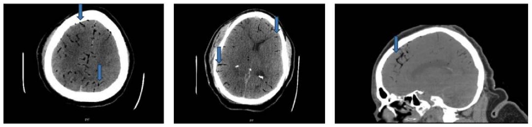
CT scan of head: axial and sagittal cuts showing foci of gas in bilateral cerebral arteries (blue arrows)
Image: “Case 2 CT scan of head” by Rashmi Mishra et al. License: CC BY 4.0
High flowFlowBlood flows through the heart, arteries, capillaries, and veins in a closed, continuous circuit. Flow is the movement of volume per unit of time. Flow is affected by the pressure gradient and the resistance fluid encounters between 2 points. Vascular resistance is the opposition to flow, which is caused primarily by blood friction against vessel walls.Vascular Resistance, Flow, and Mean Arterial Pressure of oxygen
Keeps air in the right ventricle and away from the pulmonary outflow tract
Arterial embolism:
Supine position
ArteriesArteriesArteries are tubular collections of cells that transport oxygenated blood and nutrients from the heart to the tissues of the body. The blood passes through the arteries in order of decreasing luminal diameter, starting in the largest artery (the aorta) and ending in the small arterioles. Arteries are classified into 3 types: large elastic arteries, medium muscular arteries, and small arteries and arterioles. Arteries: Histology are strong enough to overcome positioning; the supine position attempts to minimize cerebral edemaCerebral edemaIncreased intracellular or extracellular fluid in brain tissue. Cytotoxic brain edema (swelling due to increased intracellular fluid) is indicative of a disturbance in cell metabolism, and is commonly associated with hypoxic or ischemic injuries. An increase in extracellular fluid may be caused by increased brain capillary permeability (vasogenic edema), an osmotic gradient, local blockages in interstitial fluid pathways, or by obstruction of CSF flow (e.g., obstructive hydrocephalus).Increased Intracranial Pressure (ICP) caused by a cerebral air embolism.
Definitive therapies:
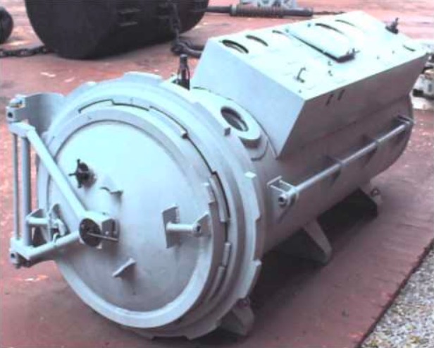
Hyperbaric oxygenHyperbaric oxygenThe therapeutic intermittent administration of oxygen in a chamber at greater than sea-level atmospheric pressures (three atmospheres). It is considered effective treatment for air and gas embolisms, smoke inhalation, acute carbon monoxide poisoning, caisson disease, clostridial gangrene, etc. The list of treatment modalities includes stroke.Decompression Sickness:
↑↑ Levels of blood oxygen create a large gradient for nitrogenNitrogenAn element with the atomic symbol n, atomic number 7, and atomic weight [14. 00643; 14. 00728]. Nitrogen exists as a diatomic gas and makes up about 78% of the earth’s atmosphere by volume. It is a constituent of proteins and nucleic acids and found in all living cells.Urea Cycle to move from the air bubbles into the blood → ↓ the size of the bubbles
↑ Oxygen helps treat tissue ischemiaIschemiaA hypoperfusion of the blood through an organ or tissue caused by a pathologic constriction or obstruction of its blood vessels, or an absence of blood circulation.Ischemic Cell Damage (secondary benefit)
Removal of embolized air (e.g., aspiration through a catheter)
Image: “Hyperbaric oxygen therapy chamber” by Mark Murphy. License: Public Domain
Amniotic Fluid Embolism
Overview
Amniotic fluid embolismAmniotic Fluid EmbolismBlocking of maternal circulation by amniotic fluid that is forced into uterine veins by strong uterine contraction near the end of pregnancy. It is characterized by the sudden onset of severe respiratory distress and hypotension that can lead to maternal death.Complications during Childbirth (AFEAFEBlocking of maternal circulation by amniotic fluid that is forced into uterine veins by strong uterine contraction near the end of pregnancy. It is characterized by the sudden onset of severe respiratory distress and hypotension that can lead to maternal death.Complications during Childbirth) is a complication during labor and the immediate postpartum periodPostpartum periodIn females, the period that is shortly after giving birth (parturition).Postpartum Complications.
Caused by the entry of the amniotic fluidAmniotic fluidA clear, yellowish liquid that envelopes the fetus inside the sac of amnion. In the first trimester, it is likely a transudate of maternal or fetal plasma. In the second trimester, amniotic fluid derives primarily from fetal lung and kidney. Cells or substances in this fluid can be removed for prenatal diagnostic tests (amniocentesis).Placenta, Umbilical Cord, and Amniotic Cavity into the maternal circulationCirculationThe movement of the blood as it is pumped through the cardiovascular system.ABCDE Assessment by:
Placental tears
Uterine vein rupture
IncidenceIncidenceThe number of new cases of a given disease during a given period in a specified population. It also is used for the rate at which new events occur in a defined population. It is differentiated from prevalence, which refers to all cases in the population at a given time.Measures of Disease Frequency: 1:40,000 deliveries
AFEAFEBlocking of maternal circulation by amniotic fluid that is forced into uterine veins by strong uterine contraction near the end of pregnancy. It is characterized by the sudden onset of severe respiratory distress and hypotension that can lead to maternal death.Complications during Childbirth causes 10% of maternal deaths in developed countries.
Risk factors
Cesarean deliveryCesarean DeliveryCesarean delivery (CD) is the operative delivery of ≥ 1 infants through a surgical incision in the maternal abdomen and uterus. Cesarean deliveries may be indicated for a number of either maternal or fetal reasons, most commonly including fetal intolerance to labor, arrest of labor, a history of prior uterine surgery, fetal malpresentation, and placental abnormalities. Cesarean Delivery
Placental abnormalitiesPlacental abnormalitiesNormal placental structure and function are essential for a healthy pregnancy. Some of the most common placental abnormalities include structural anomalies (such as a succenturiate lobe or velamentous cord insertion), implantation anomalies (such as placenta accreta and placenta previa), and functional anomalies (such as placental insufficiency). Placental Abnormalities (e.g., placenta previaPlacenta PreviaAbnormal placentation in which the placenta implants in the lower segment of the uterus (the zone of dilation) and may cover part or all of the opening of the cervix. It is often associated with serious antepartum bleeding and premature labor.Placental Abnormalities)
PreeclampsiaPreeclampsiaA complication of pregnancy, characterized by a complex of symptoms including maternal hypertension and proteinuria with or without pathological edema. Symptoms may range between mild and severe. Pre-eclampsia usually occurs after the 20th week of gestation, but may develop before this time in the presence of trophoblastic disease.Hypertensive Pregnancy Disorders/eclampsiaEclampsiaOnset of hyperreflexia; seizures; or coma in a previously diagnosed pre-eclamptic patient (pre-eclampsia).Hypertensive Pregnancy Disorders
Pathogenesis
Unclear
Amniotic fluidAmniotic fluidA clear, yellowish liquid that envelopes the fetus inside the sac of amnion. In the first trimester, it is likely a transudate of maternal or fetal plasma. In the second trimester, amniotic fluid derives primarily from fetal lung and kidney. Cells or substances in this fluid can be removed for prenatal diagnostic tests (amniocentesis).Placenta, Umbilical Cord, and Amniotic Cavity enters maternal circulationCirculationThe movement of the blood as it is pumped through the cardiovascular system.ABCDE Assessment and triggers:
↑ Pulmonary pressure → right ventricular failure → systemic hypotensionHypotensionHypotension is defined as low blood pressure, specifically < 90/60 mm Hg, and is most commonly a physiologic response. Hypotension may be mild, serious, or life threatening, depending on the cause. Hypotension
Pulmonary edemaPulmonary edemaPulmonary edema is a condition caused by excess fluid within the lung parenchyma and alveoli as a consequence of a disease process. Based on etiology, pulmonary edema is classified as cardiogenic or noncardiogenic. Patients may present with progressive dyspnea, orthopnea, cough, or respiratory failure.Pulmonary Edema
Abnormal immune responses are activated:
Intense inflammatory response (similar to SIRS)
Inflammatory mediators activate the coagulation cascadeCoagulation cascadeThe coagulation cascade is a series of reactions that ultimately generates a strong, cross-linked fibrin clot.Hemostasis systemically → disseminated intravascular coagulationDisseminated intravascular coagulationDisseminated intravascular coagulation (DIC) is a condition characterized by systemic bodywide activation of the coagulation cascade. This cascade results in both widespread microvascular thrombi contributing to multiple organ dysfunction and consumption of clotting factors and platelets, leading to hemorrhage. Disseminated Intravascular Coagulation (DICDICDisseminated intravascular coagulation (DIC) is a condition characterized by systemic bodywide activation of the coagulation cascade. This cascade results in both widespread microvascular thrombi contributing to multiple organ dysfunction and consumption of clotting factors and platelets, leading to hemorrhage. Disseminated Intravascular Coagulation)
DICDICDisseminated intravascular coagulation (DIC) is a condition characterized by systemic bodywide activation of the coagulation cascade. This cascade results in both widespread microvascular thrombi contributing to multiple organ dysfunction and consumption of clotting factors and platelets, leading to hemorrhage. Disseminated Intravascular Coagulation leads to:
Hemorrhage → further hemodynamic instability
Ischemic multi-organ failure
Mechanical obstructionMechanical ObstructionAny impairment, arrest, or reversal of the normal flow of intestinal contents toward the anal canal.Imaging of the Intestines from the amniotic-fluid debris likely does not play a significant role.
Clinical presentation
AFEAFEBlocking of maternal circulation by amniotic fluid that is forced into uterine veins by strong uterine contraction near the end of pregnancy. It is characterized by the sudden onset of severe respiratory distress and hypotension that can lead to maternal death.Complications during Childbirth typically presents dramatically, as sudden-onset cardiopulmonary collapse occurring during labor or within 30 minutes after delivery.
Signs:
Cardiopulmonary collapse: loss of breathing and pulse
DyspneaDyspneaDyspnea is the subjective sensation of breathing discomfort. Dyspnea is a normal manifestation of heavy physical or psychological exertion, but also may be caused by underlying conditions (both pulmonary and extrapulmonary). Dyspnea
HypotensionHypotensionHypotension is defined as low blood pressure, specifically < 90/60 mm Hg, and is most commonly a physiologic response. Hypotension may be mild, serious, or life threatening, depending on the cause. Hypotension
TachycardiaTachycardiaAbnormally rapid heartbeat, usually with a heart rate above 100 beats per minute for adults. Tachycardia accompanied by disturbance in the cardiac depolarization (cardiac arrhythmia) is called tachyarrhythmia.Sepsis in Children
Other symptoms:
NauseaNauseaAn unpleasant sensation in the stomach usually accompanied by the urge to vomit. Common causes are early pregnancy, sea and motion sickness, emotional stress, intense pain, food poisoning, and various enteroviruses.Antiemetics and vomitingVomitingThe forcible expulsion of the contents of the stomach through the mouth.Hypokalemia
Changes in mental status
Seizure
DICDICDisseminated intravascular coagulation (DIC) is a condition characterized by systemic bodywide activation of the coagulation cascade. This cascade results in both widespread microvascular thrombi contributing to multiple organ dysfunction and consumption of clotting factors and platelets, leading to hemorrhage. Disseminated Intravascular Coagulation:
Usually develops shortly after an AFEAFEBlocking of maternal circulation by amniotic fluid that is forced into uterine veins by strong uterine contraction near the end of pregnancy. It is characterized by the sudden onset of severe respiratory distress and hypotension that can lead to maternal death.Complications during Childbirth
Leads to obstetric hemorrhage
Fetal heart-rate abnormalities indicating distress (e.g., late decelerations, terminal bradycardiaBradycardiaBradyarrhythmia is a rhythm in which the heart rate is less than 60/min. Bradyarrhythmia can be physiologic, without symptoms or hemodynamic change. Pathologic bradyarrhythmia results in reduced cardiac output and hemodynamic instability causing syncope, dizziness, or dyspnea.Bradyarrhythmias) if still pregnant
Diagnosis
AFEAFEBlocking of maternal circulation by amniotic fluid that is forced into uterine veins by strong uterine contraction near the end of pregnancy. It is characterized by the sudden onset of severe respiratory distress and hypotension that can lead to maternal death.Complications during Childbirth is a clinical diagnosis based on presentation.
↑ Prothrombin timeProthrombin timeClotting time of plasma recalcified in the presence of excess tissue thromboplastin. Factors measured are fibrinogen; prothrombin; factor V; factor VII; and factor X.Hemostasis
↓ FibrinogenFibrinogenPlasma glycoprotein clotted by thrombin, composed of a dimer of three non-identical pairs of polypeptide chains (alpha, beta, gamma) held together by disulfide bonds. Fibrinogen clotting is a sol-gel change involving complex molecular arrangements: whereas fibrinogen is cleaved by thrombin to form polypeptides a and b, the proteolytic action of other enzymes yields different fibrinogen degradation products.Hemostasis
AnemiaAnemiaAnemia is a condition in which individuals have low Hb levels, which can arise from various causes. Anemia is accompanied by a reduced number of RBCs and may manifest with fatigue, shortness of breath, pallor, and weakness. Subtypes are classified by the size of RBCs, chronicity, and etiology. Anemia: Overview and Types
ThrombocytopeniaThrombocytopeniaThrombocytopenia occurs when the platelet count is < 150,000 per microliter. The normal range for platelets is usually 150,000-450,000/µL of whole blood. Thrombocytopenia can be a result of decreased production, increased destruction, or splenic sequestration of platelets. Patients are often asymptomatic until platelet counts are < 50,000/µL. Thrombocytopenia
AcidosisAcidosisA pathologic condition of acid accumulation or depletion of base in the body. The two main types are respiratory acidosis and metabolic acidosis, due to metabolic acid build up.Respiratory Acidosis (both respiratory and metabolic)
Imaging (once the patient is stable):
Chest radiography: bilateral diffuse infiltrates
EchocardiographyEchocardiographyUltrasonic recording of the size, motion, and composition of the heart and surrounding tissues. The standard approach is transthoracic.Tricuspid Valve Atresia (TVA) to assess cardiac function
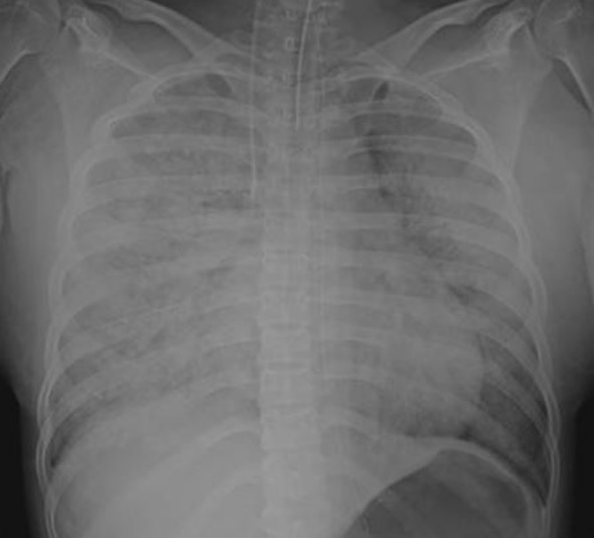
Chest X-ray of a patient with amniotic fluid embolism: Diffuse infiltration is observed throughout the lungs.
Image: “X-ray” by Department of Emergency and Critical Care, The University of Tokushima Graduate School, Kuramoto Tokushima, 770-8503, Japan. License: CC BY 2.0
CirculationCirculationThe movement of the blood as it is pumped through the cardiovascular system.ABCDE Assessment:
High-quality CPRCPRThe artificial substitution of heart and lung action as indicated for heart arrest resulting from electric shock, drowning, respiratory arrest, or other causes. The two major components of cardiopulmonary resuscitation are artificial ventilation and closed-chest cardiac massage.Cardiac Arrest
Transfuse to combat DICDICDisseminated intravascular coagulation (DIC) is a condition characterized by systemic bodywide activation of the coagulation cascade. This cascade results in both widespread microvascular thrombi contributing to multiple organ dysfunction and consumption of clotting factors and platelets, leading to hemorrhage. Disseminated Intravascular Coagulation; typically a 1:1:1 ratio of:
Immediate delivery if the patient is still pregnant
Complications
Hematological: DICDICDisseminated intravascular coagulation (DIC) is a condition characterized by systemic bodywide activation of the coagulation cascade. This cascade results in both widespread microvascular thrombi contributing to multiple organ dysfunction and consumption of clotting factors and platelets, leading to hemorrhage. Disseminated Intravascular Coagulation
Cardiovascular: hemorrhage and cardiac arrestCardiac arrestCardiac arrest is the sudden, complete cessation of cardiac output with hemodynamic collapse. Patients present as pulseless, unresponsive, and apneic. Rhythms associated with cardiac arrest are ventricular fibrillation/tachycardia, asystole, or pulseless electrical activity. Cardiac Arrest
Pulmonary complications: pulmonary edemaPulmonary edemaPulmonary edema is a condition caused by excess fluid within the lung parenchyma and alveoli as a consequence of a disease process. Based on etiology, pulmonary edema is classified as cardiogenic or noncardiogenic. Patients may present with progressive dyspnea, orthopnea, cough, or respiratory failure.Pulmonary Edema and ARDS
Permanent neurological deficits due to cerebral hypoxiaHypoxiaSub-optimal oxygen levels in the ambient air of living organisms.Ischemic Cell Damage (85% of survivors)
EmbolizationEmbolizationA method of hemostasis utilizing various agents such as gelfoam, silastic, metal, glass, or plastic pellets, autologous clot, fat, and muscle as emboli. It has been used in the treatment of spinal cord and intracranial arteriovenous malformations, renal arteriovenous fistulas, gastrointestinal bleeding, epistaxis, hypersplenism, certain highly vascular tumors, traumatic rupture of blood vessels, and control of operative hemorrhage.Gastrointestinal Bleeding of tumorTumorInflammation within the pulmonary vessels
Can occur with any malignancyMalignancyHemothorax, though most commonly associated with:
Renal-cell carcinoma
Hepatocellular carcinomaHepatocellular carcinomaHepatocellular carcinoma (HCC) typically arises in a chronically diseased or cirrhotic liver and is the most common primary liver cancer. Diagnosis may include ultrasound, CT, MRI, biopsy (if inconclusive imaging), and/or biomarkers. Hepatocellular Carcinoma (HCC) and Liver Metastases
Adenocarcinomas:
Breast
StomachStomachThe stomach is a muscular sac in the upper left portion of the abdomen that plays a critical role in digestion. The stomach develops from the foregut and connects the esophagus with the duodenum. Structurally, the stomach is C-shaped and forms a greater and lesser curvature and is divided grossly into regions: the cardia, fundus, body, and pylorus. Stomach: Anatomy
ColonColonThe large intestines constitute the last portion of the digestive system. The large intestine consists of the cecum, appendix, colon (with ascending, transverse, descending, and sigmoid segments), rectum, and anal canal. The primary function of the colon is to remove water and compact the stool prior to expulsion from the body via the rectum and anal canal. Colon, Cecum, and Appendix: Anatomy
Lung
Clinical presentation:
Subacute progressive dyspneaDyspneaDyspnea is the subjective sensation of breathing discomfort. Dyspnea is a normal manifestation of heavy physical or psychological exertion, but also may be caused by underlying conditions (both pulmonary and extrapulmonary). Dyspnea
Cough, with or without hemoptysisHemoptysisHemoptysis is defined as the expectoration of blood originating in the lower respiratory tract. Hemoptysis is a consequence of another disease process and can be classified as either life threatening or non-life threatening. Hemoptysis can result in significant morbidity and mortality due to both drowning (reduced gas exchange as the lungs fill with blood) and hemorrhagic shock. Hemoptysis
May present with signs of venous thromboembolismThromboembolismObstruction of a blood vessel (embolism) by a blood clot (thrombus) in the blood stream.Systemic Lupus Erythematosus (dyspneaDyspneaDyspnea is the subjective sensation of breathing discomfort. Dyspnea is a normal manifestation of heavy physical or psychological exertion, but also may be caused by underlying conditions (both pulmonary and extrapulmonary). Dyspnea, hypoxiaHypoxiaSub-optimal oxygen levels in the ambient air of living organisms.Ischemic Cell Damage, right heart strain)
Diagnosis is often made on autopsy:
Imaging studies are insensitive.
Lab studies are nonspecific.
Poor prognosisPrognosisA prediction of the probable outcome of a disease based on a individual’s condition and the usual course of the disease as seen in similar situations.Non-Hodgkin Lymphomas
Foreign-body embolism
Most commonly involves silicone and occurs after cosmetic or therapeutic injection
FeverFeverFever is defined as a measured body temperature of at least 38°C (100.4°F). Fever is caused by circulating endogenous and/or exogenous pyrogens that increase levels of prostaglandin E2 in the hypothalamus. Fever is commonly associated with chills, rigors, sweating, and flushing of the skin. Fever
CholesterolCholesterolThe principal sterol of all higher animals, distributed in body tissues, especially the brain and spinal cord, and in animal fats and oils.Cholesterol Metabolism embolism
From thoracic sources:
SkinSkinThe skin, also referred to as the integumentary system, is the largest organ of the body. The skin is primarily composed of the epidermis (outer layer) and dermis (deep layer). The epidermis is primarily composed of keratinocytes that undergo rapid turnover, while the dermis contains dense layers of connective tissue.Skin: Structure and Functions: livedo reticularisLivedo reticularisA condition characterized by a reticular or fishnet pattern on the skin of lower extremities and other parts of the body. This red and blue pattern is due to deoxygenated blood in unstable dermal blood vessels. The condition is intensified by cold exposure and relieved by rewarming.Chronic Kidney Disease, blue toe
Renal: AKIAKIAcute kidney injury refers to sudden and often reversible loss of renal function, which develops over days or weeks. Azotemia refers to elevated levels of nitrogen-containing substances in the blood that accompany AKI, which include BUN and creatinine. Acute Kidney Injury
GI symptoms
From aortic-arch sources:
BrainBrainThe part of central nervous system that is contained within the skull (cranium). Arising from the neural tube, the embryonic brain is comprised of three major parts including prosencephalon (the forebrain); mesencephalon (the midbrain); and rhombencephalon (the hindbrain). The developed brain consists of cerebrum; cerebellum; and other structures in the brain stem.Nervous System: Anatomy, Structure, and Classification: TIATIATransient ischemic attack (TIA) is a temporary episode of neurologic dysfunction caused by ischemia without infarction that resolves completely when blood supply is restored. Transient ischemic attack is a neurologic emergency that warrants urgent medical attention. Transient Ischemic Attack (TIA), stroke
Eyes symptoms
Focus on risk reduction in atherosclerotic disease:
AspirinAspirinThe prototypical analgesic used in the treatment of mild to moderate pain. It has anti-inflammatory and antipyretic properties and acts as an inhibitor of cyclooxygenase which results in the inhibition of the biosynthesis of prostaglandins. Aspirin also inhibits platelet aggregation and is used in the prevention of arterial and venous thrombosis.Nonsteroidal Antiinflammatory Drugs (NSAIDs)
StatinsStatinsStatins are competitive inhibitors of HMG-CoA reductase in the liver. HMG-CoA reductase is the rate-limiting step in cholesterol synthesis. Inhibition results in lowered intrahepatocytic cholesterol formation, resulting in up-regulation of LDL receptors and, ultimately, lowering levels of serum LDL and triglycerides.Statins
SmokingSmokingWillful or deliberate act of inhaling and exhaling smoke from burning substances or agents held by hand.Interstitial Lung Diseases cessation
Head and neckNeckThe part of a human or animal body connecting the head to the rest of the body.Peritonsillar Abscess surgeries
Endoscopies
IV catheterization
Symptoms:
Venous: mimics PE
Arterial: stroke or MIMIMI is ischemia and death of an area of myocardial tissue due to insufficient blood flow and oxygenation, usually from thrombus formation on a ruptured atherosclerotic plaque in the epicardial arteries. Clinical presentation is most commonly with chest pain, but women and patients with diabetes may have atypical symptoms.Myocardial Infarction
Amniotic fluid embolismAmniotic Fluid EmbolismBlocking of maternal circulation by amniotic fluid that is forced into uterine veins by strong uterine contraction near the end of pregnancy. It is characterized by the sudden onset of severe respiratory distress and hypotension that can lead to maternal death.Complications during Childbirth
During labor or within 30 minutes of delivery:
Acute cardiopulmonary collapse
DICDICDisseminated intravascular coagulation (DIC) is a condition characterized by systemic bodywide activation of the coagulation cascade. This cascade results in both widespread microvascular thrombi contributing to multiple organ dysfunction and consumption of clotting factors and platelets, leading to hemorrhage. Disseminated Intravascular Coagulation
Progressive dyspneaDyspneaDyspnea is the subjective sensation of breathing discomfort. Dyspnea is a normal manifestation of heavy physical or psychological exertion, but also may be caused by underlying conditions (both pulmonary and extrapulmonary). Dyspnea
May mimic PE
Poor prognosisPrognosisA prediction of the probable outcome of a disease based on a individual’s condition and the usual course of the disease as seen in similar situations.Non-Hodgkin Lymphomas
Agrawal, A., et al. (2017). Cholesterol embolization syndrome: An under‐recognized entity in cardiovascular interventions. https://doi.org/10.1111/joic.12483