A physician’s diagnostic and therapeutic tool kit must include a variety of basic procedures that can be performed in the outpatient setting. These procedures include emergency intervention of the airway; drainage of fluid from the abdomen, joints, and spinal canal; and incision and drainage of abscesses. Although these procedures may be of reduced complexity, there are still inherent risks associated with invasive procedures, and these risks must be reduced through consistent aseptic and procedural techniques.
Cricothyroidotomy: surgical creation of an artificial opening on the cricothyroidCricothyroidLarynx: Anatomy membrane
Tracheotomy: surgical creation of an artificial opening on the anterior wall of the tracheaTracheaThe trachea is a tubular structure that forms part of the lower respiratory tract. The trachea is continuous superiorly with the larynx and inferiorly becomes the bronchial tree within the lungs. The trachea consists of a support frame of semicircular, or C-shaped, rings made out of hyaline cartilage and reinforced by collagenous connective tissue. Trachea: Anatomy
Used synonymously with the term tracheostomyTracheostomySurgical formation of an opening into the trachea through the neck, or the opening so created.Laryngomalacia and Tracheomalacia
Tracheotomy technically refers to an incision into the tracheaTracheaThe trachea is a tubular structure that forms part of the lower respiratory tract. The trachea is continuous superiorly with the larynx and inferiorly becomes the bronchial tree within the lungs. The trachea consists of a support frame of semicircular, or C-shaped, rings made out of hyaline cartilage and reinforced by collagenous connective tissue. Trachea: Anatomy, whereas tracheostomyTracheostomySurgical formation of an opening into the trachea through the neck, or the opening so created.Laryngomalacia and Tracheomalacia refers to the placement of a cannula into that opening.
Usually, when the pharynxPharynxThe pharynx is a component of the digestive system that lies posterior to the nasal cavity, oral cavity, and larynx. The pharynx can be divided into the oropharynx, nasopharynx, and laryngopharynx. Pharyngeal muscles play an integral role in vital processes such as breathing, swallowing, and speaking. Pharynx: Anatomy and larynxLarynxThe larynx, also commonly called the voice box, is a cylindrical space located in the neck at the level of the C3-C6 vertebrae. The major structures forming the framework of the larynx are the thyroid cartilage, cricoid cartilage, and epiglottis. The larynx serves to produce sound (phonation), conducts air to the trachea, and prevents large molecules from reaching the lungs.Larynx: Anatomy need to be bypassed (e.g., obstruction)
To provide easy access to the lower airways for bronchial aspiration in the case of tracheostomyTracheostomySurgical formation of an opening into the trachea through the neck, or the opening so created.Laryngomalacia and Tracheomalacia
Functional obstruction of the upper airwayAirwayABCDE Assessment (e.g., carcinoma of larynxLarynxThe larynx, also commonly called the voice box, is a cylindrical space located in the neck at the level of the C3-C6 vertebrae. The major structures forming the framework of the larynx are the thyroid cartilage, cricoid cartilage, and epiglottis. The larynx serves to produce sound (phonation), conducts air to the trachea, and prevents large molecules from reaching the lungs.Larynx: Anatomy)
Long-term ventilatory support
Chronic aspiration
ContraindicationsContraindicationsA condition or factor associated with a recipient that makes the use of a drug, procedure, or physical agent improper or inadvisable. Contraindications may be absolute (life threatening) or relative (higher risk of complications in which benefits may outweigh risks).Noninvasive Ventilation
ThyroidThyroidThe thyroid gland is one of the largest endocrine glands in the human body. The thyroid gland is a highly vascular, brownish-red gland located in the visceral compartment of the anterior region of the neck.Thyroid Gland: Anatomy/cricoid cartilageCartilageCartilage is a type of connective tissue derived from embryonic mesenchyme that is responsible for structural support, resilience, and the smoothness of physical actions. Perichondrium (connective tissue membrane surrounding cartilage) compensates for the absence of vasculature in cartilage by providing nutrition and support. Cartilage: HistologyfractureFractureA fracture is a disruption of the cortex of any bone and periosteum and is commonly due to mechanical stress after an injury or accident. Open fractures due to trauma can be a medical emergency. Fractures are frequently associated with automobile accidents, workplace injuries, and trauma.Overview of Bone Fractures
Individual < 8 years old
Relative:
Massive neckNeckThe part of a human or animal body connecting the head to the rest of the body.Peritonsillar AbscessswellingSwellingInflammation or hematomaHematomaA collection of blood outside the blood vessels. Hematoma can be localized in an organ, space, or tissue.Intussusception
Cervical trauma
Procedure
Both procedures can be performed at the bedside; however, it is preferable for tracheotomies to be performed in the OR.
Preoperative preparation:
In an emergency cricothyroidotomy, some of the steps below may be skipped.
Explain the procedure to the individual and obtain informed consentInformed consentInformed consent is a medicolegal term describing the documented conversation between a patient and their physician wherein the physician discloses all relevant and necessary information to a patient who is competent to make an informed and voluntary decision regarding their care. Competency, disclosure, and voluntariness are the key elements upon which IC is based.Informed Consent, if possible.
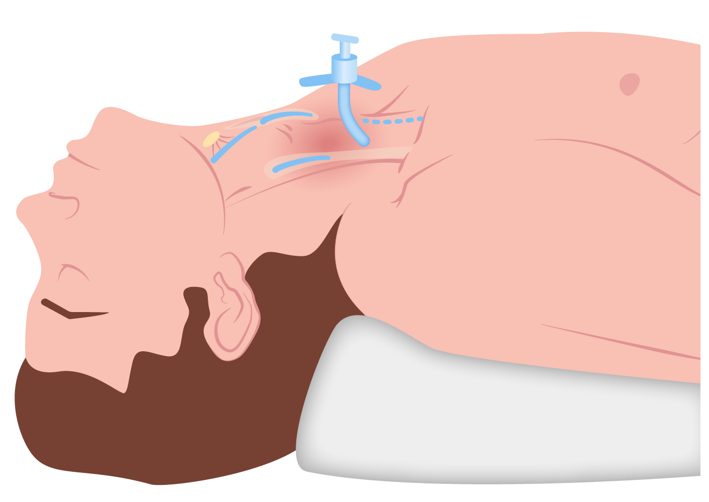
Place individual in the supine position, with the neckNeckThe part of a human or animal body connecting the head to the rest of the body.Peritonsillar Abscess extended, unless contraindicated, with the chinChinThe anatomical frontal portion of the mandible, also known as the mentum, that contains the line of fusion of the two separate halves of the mandible (symphysis menti). This line of fusion divides inferiorly to enclose a triangular area called the mental protuberance. On each side, inferior to the second premolar tooth, is the mental foramen for the passage of blood vessels and a nerve.Melasma aligned with the midline.
Administer preoperative antibiotics at least 30 minutes before incision, if possible.
If possible, sedate and intubate the individual.
Identify the thyroidThyroidThe thyroid gland is one of the largest endocrine glands in the human body. The thyroid gland is a highly vascular, brownish-red gland located in the visceral compartment of the anterior region of the neck.Thyroid Gland: Anatomy and cricoid cartilages.
Wash the site of the incision with an antiseptic solution and cover with sterile drapes.
Continuous monitoring:
HR
Blood pressure
Oxygen saturation
ECGECGAn electrocardiogram (ECG) is a graphic representation of the electrical activity of the heart plotted against time. Adhesive electrodes are affixed to the skin surface allowing measurement of cardiac impulses from many angles. The ECG provides 3-dimensional information about the conduction system of the heart, the myocardium, and other cardiac structures. Electrocardiogram (ECG) rhythm monitor
In either the ED or OR, the operator should don adequate PPE.
Cricothyroidotomy:
Local anesthetic (1% lidocaineLidocaineA local anesthetic and cardiac depressant used as an antiarrhythmic agent. Its actions are more intense and its effects more prolonged than those of procaine but its duration of action is shorter than that of bupivacaine or prilocaine.Local Anesthetics) is infiltrated into the proposed incision site.
The thyroidThyroidThe thyroid gland is one of the largest endocrine glands in the human body. The thyroid gland is a highly vascular, brownish-red gland located in the visceral compartment of the anterior region of the neck.Thyroid Gland: AnatomycartilageCartilageCartilage is a type of connective tissue derived from embryonic mesenchyme that is responsible for structural support, resilience, and the smoothness of physical actions. Perichondrium (connective tissue membrane surrounding cartilage) compensates for the absence of vasculature in cartilage by providing nutrition and support. Cartilage: Histology is immobilized with the nondominant handHandThe hand constitutes the distal part of the upper limb and provides the fine, precise movements needed in activities of daily living. It consists of 5 metacarpal bones and 14 phalanges, as well as numerous muscles innervated by the median and ulnar nerves. Hand: Anatomy, and the skinSkinThe skin, also referred to as the integumentary system, is the largest organ of the body. The skin is primarily composed of the epidermis (outer layer) and dermis (deep layer). The epidermis is primarily composed of keratinocytes that undergo rapid turnover, while the dermis contains dense layers of connective tissue.Skin: Structure and Functions is held under tension over the cricothyroidCricothyroidLarynx: Anatomy membrane.
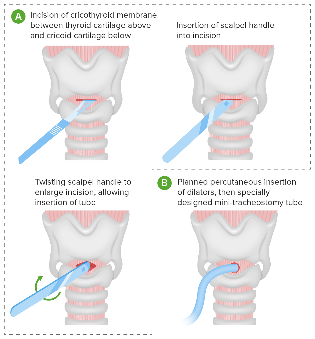
A 3-cm vertical incision is made on the midline, over the cricothyroidCricothyroidLarynx: Anatomy membrane. Horizontal incision is avoided, since in emergency situations, the incision could be extended too far laterally and the jugular veinsVeinsVeins are tubular collections of cells, which transport deoxygenated blood and waste from the capillary beds back to the heart. Veins are classified into 3 types: small veins/venules, medium veins, and large veins. Each type contains 3 primary layers: tunica intima, tunica media, and tunica adventitia. Veins: Histology could be injured.
Manual retraction of the edges of the incision is performed with the nondominant handHandThe hand constitutes the distal part of the upper limb and provides the fine, precise movements needed in activities of daily living. It consists of 5 metacarpal bones and 14 phalanges, as well as numerous muscles innervated by the median and ulnar nerves. Hand: Anatomy.
Subcutaneous tissueSubcutaneous tissueLoose connective tissue lying under the dermis, which binds skin loosely to subjacent tissues. It may contain a pad of adipocytes, which vary in number according to the area of the body and vary in size according to the nutritional state.Soft Tissue Abscess and strap muscles are dissected at the midline to expose the thyroidThyroidThe thyroid gland is one of the largest endocrine glands in the human body. The thyroid gland is a highly vascular, brownish-red gland located in the visceral compartment of the anterior region of the neck.Thyroid Gland: Anatomy and cricoid cartilages and the cricothyroidCricothyroidLarynx: Anatomy membrane.
The incision is made as low as possible to avoid vocal cord damage.
Note: Once the airwayAirwayABCDE Assessment is accessed, be on guard for sputum and secretions when the individual tries to exhale.
Once the incision is made, the handle of the scalpel is introduced into the incision and twisted 90 degrees to expand the incision.
The index finger of the nondominant handHandThe hand constitutes the distal part of the upper limb and provides the fine, precise movements needed in activities of daily living. It consists of 5 metacarpal bones and 14 phalanges, as well as numerous muscles innervated by the median and ulnar nerves. Hand: Anatomy is used to keep the incision open while the scalpel is flipped.
The scalpel is maintained in the opening to maintain patency and facilitate passage of a cannula.
An endotracheal or tracheostomyTracheostomySurgical formation of an opening into the trachea through the neck, or the opening so created.Laryngomalacia and Tracheomalacia tube is inserted into the airwayAirwayABCDE Assessment through the incision, using the scalpel handle as a retractor and guide.
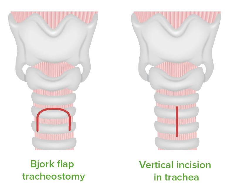
Horizontal: 1 cm below the cricoid cartilageCartilageCartilage is a type of connective tissue derived from embryonic mesenchyme that is responsible for structural support, resilience, and the smoothness of physical actions. Perichondrium (connective tissue membrane surrounding cartilage) compensates for the absence of vasculature in cartilage by providing nutrition and support. Cartilage: Histology
Vertical: from the inferior aspect of the cricoid cartilageCartilageCartilage is a type of connective tissue derived from embryonic mesenchyme that is responsible for structural support, resilience, and the smoothness of physical actions. Perichondrium (connective tissue membrane surrounding cartilage) compensates for the absence of vasculature in cartilage by providing nutrition and support. Cartilage: Histology, extending 2–3 cm caudally
Subcutaneous tissueSubcutaneous tissueLoose connective tissue lying under the dermis, which binds skin loosely to subjacent tissues. It may contain a pad of adipocytes, which vary in number according to the area of the body and vary in size according to the nutritional state.Soft Tissue Abscess and strap muscles along the midline are dissected to expose the tracheaTracheaThe trachea is a tubular structure that forms part of the lower respiratory tract. The trachea is continuous superiorly with the larynx and inferiorly becomes the bronchial tree within the lungs. The trachea consists of a support frame of semicircular, or C-shaped, rings made out of hyaline cartilage and reinforced by collagenous connective tissue. Trachea: Anatomy and thyroidThyroidThe thyroid gland is one of the largest endocrine glands in the human body. The thyroid gland is a highly vascular, brownish-red gland located in the visceral compartment of the anterior region of the neck.Thyroid Gland: Anatomy gland.
The thyroidThyroidThe thyroid gland is one of the largest endocrine glands in the human body. The thyroid gland is a highly vascular, brownish-red gland located in the visceral compartment of the anterior region of the neck.Thyroid Gland: AnatomyisthmusIsthmusUterus, Cervix, and Fallopian Tubes: Anatomy is retracted away or divided.
An incision is made between the 2nd and 3rd tracheal rings and is dilated.
The surgeon must communicate with the anesthesiologist before accessing the airwayAirwayABCDE Assessment.
The existing endotracheal tube will be manipulated to allow passage of the tracheostomyTracheostomySurgical formation of an opening into the trachea through the neck, or the opening so created.Laryngomalacia and Tracheomalacia tube.
The endotracheal tube is elevated to just above the tracheotomy.
The tracheostomyTracheostomySurgical formation of an opening into the trachea through the neck, or the opening so created.Laryngomalacia and Tracheomalacia tube is placed through the opening in the tracheaTracheaThe trachea is a tubular structure that forms part of the lower respiratory tract. The trachea is continuous superiorly with the larynx and inferiorly becomes the bronchial tree within the lungs. The trachea consists of a support frame of semicircular, or C-shaped, rings made out of hyaline cartilage and reinforced by collagenous connective tissue. Trachea: Anatomy and the cuff is inflated.
The tube is connected to the mechanical ventilator.
The endotracheal tube is extracted, if one was present, and the tracheostomyTracheostomySurgical formation of an opening into the trachea through the neck, or the opening so created.Laryngomalacia and Tracheomalacia tube is secured using sutures.
The airwayAirwayABCDE Assessment mask bag unit is connected to the tube and the individual is ventilated with 100% O2.
Both sides are auscultated to ensure placement of the tube above the carina.
Arterial blood gases and a chest x-rayX-rayPenetrating electromagnetic radiation emitted when the inner orbital electrons of an atom are excited and release radiant energy. X-ray wavelengths range from 1 pm to 10 nm. Hard x-rays are the higher energy, shorter wavelength x-rays. Soft x-rays or grenz rays are less energetic and longer in wavelength. The short wavelength end of the x-ray spectrum overlaps the gamma rays wavelength range. The distinction between gamma rays and x-rays is based on their radiation source.Pulmonary Function Tests should be requested.
Complications
Intraoperative and postoperative hemorrhagePostoperative HemorrhageHemorrhage following any surgical procedure. It may be immediate or delayed and is not restricted to the surgical wound.Postoperative Care
Improper placement with asphyxiaAsphyxiaA pathological condition caused by lack of oxygen, manifested in impending or actual cessation of life.Drowning
Accidental decannulation:
Individuals with altered mental statusAltered Mental StatusSepsis in Children (e.g., deliriumDeliriumDelirium is a medical condition characterized by acute disturbances in attention and awareness. Symptoms may fluctuate during the course of a day and involve memory deficits and disorientation. Delirium) are at risk.
Temporary restraints may be used during the procedure.
Tube obstruction
Tracheoesophageal fistulaFistulaAbnormal communication most commonly seen between two internal organs, or between an internal organ and the surface of the body.Anal Fistula
DisplacementDisplacementThe process by which an emotional or behavioral response that is appropriate for one situation appears in another situation for which it is inappropriate.Defense Mechanisms of the tracheostomyTracheostomySurgical formation of an opening into the trachea through the neck, or the opening so created.Laryngomalacia and Tracheomalacia tube
PneumothoraxPneumothoraxA pneumothorax is a life-threatening condition in which air collects in the pleural space, causing partial or full collapse of the lung. A pneumothorax can be traumatic or spontaneous. Patients present with a sudden onset of sharp chest pain, dyspnea, and diminished breath sounds on exam.Pneumothorax/pneumomediastinumPneumomediastinumMediastinitis (rare)
Steps for a cricothyroidotomy
Image by Lecturio.
Insertion of tracheostomy tube into a cricothyroidotomy incision
Image by Lecturio.
Types of tracheotomy incisions:
Left: Tracheal flap Right: Vertical incision on the 2nd and 3rd tracheal rings Image by Lecturio.
Paracentesis
Definition
The evacuation of fluid within the peritoneal cavityPeritoneal CavityThe space enclosed by the peritoneum. It is divided into two portions, the greater sac and the lesser sac or omental bursa, which lies behind the stomach. The two sacs are connected by the foramen of winslow, or epiploic foramen.Peritoneum: Anatomy via a needle and/or catheter for diagnostic and/or therapeutic purposes
Also known as abdominal tap
Therapeutic goals
Diagnosis of the causative disease process for peritoneal fluid accumulation
Temporary relief of symptoms caused by tense or chronic ascitesAscitesAscites is the pathologic accumulation of fluid within the peritoneal cavity that occurs due to an osmotic and/or hydrostatic pressure imbalance secondary to portal hypertension (cirrhosis, heart failure) or non-portal hypertension (hypoalbuminemia, malignancy, infection).Ascites:
Respiratory (e.g., dyspneaDyspneaDyspnea is the subjective sensation of breathing discomfort. Dyspnea is a normal manifestation of heavy physical or psychological exertion, but also may be caused by underlying conditions (both pulmonary and extrapulmonary). Dyspnea)
GI (e.g., diarrheaDiarrheaDiarrhea is defined as ≥ 3 watery or loose stools in a 24-hour period. There are a multitude of etiologies, which can be classified based on the underlying mechanism of disease. The duration of symptoms (acute or chronic) and characteristics of the stools (e.g., watery, bloody, steatorrheic, mucoid) can help guide further diagnostic evaluation. Diarrhea, early satietyEarly SatietyBariatric Surgery)
Indications
New-onset ascitesAscitesAscites is the pathologic accumulation of fluid within the peritoneal cavity that occurs due to an osmotic and/or hydrostatic pressure imbalance secondary to portal hypertension (cirrhosis, heart failure) or non-portal hypertension (hypoalbuminemia, malignancy, infection).Ascites
Suspected malignant ascitesAscitesAscites is the pathologic accumulation of fluid within the peritoneal cavity that occurs due to an osmotic and/or hydrostatic pressure imbalance secondary to portal hypertension (cirrhosis, heart failure) or non-portal hypertension (hypoalbuminemia, malignancy, infection).Ascites
Suspected intraabdominal infection
ContraindicationsContraindicationsA condition or factor associated with a recipient that makes the use of a drug, procedure, or physical agent improper or inadvisable. Contraindications may be absolute (life threatening) or relative (higher risk of complications in which benefits may outweigh risks).Noninvasive Ventilation
Absolute:
Acute abdomenAcute AbdomenAcute abdomen, which is in many cases a surgical emergency, is the sudden onset of abdominal pain that may be caused by inflammation, infection, perforation, ischemia, or obstruction. The location of the pain, its characteristics, and associated symptoms (e.g., jaundice) are important tools that help narrow the differential diagnosis.Acute Abdomen
DICDICDisseminated intravascular coagulation (DIC) is a condition characterized by systemic bodywide activation of the coagulation cascade. This cascade results in both widespread microvascular thrombi contributing to multiple organ dysfunction and consumption of clotting factors and platelets, leading to hemorrhage. Disseminated Intravascular Coagulation
Relative:
Current bowel obstructionBowel obstructionAny impairment, arrest, or reversal of the normal flow of intestinal contents toward the anal canal.Ascaris/Ascariasis or severe bowel distention
Previous abdominal surgery (because of the likelihood of adhesions)
Anticoagulated individuals with no evidence of DICDICDisseminated intravascular coagulation (DIC) is a condition characterized by systemic bodywide activation of the coagulation cascade. This cascade results in both widespread microvascular thrombi contributing to multiple organ dysfunction and consumption of clotting factors and platelets, leading to hemorrhage. Disseminated Intravascular Coagulation
Explain the procedure to the individual and obtain informed consentInformed consentInformed consent is a medicolegal term describing the documented conversation between a patient and their physician wherein the physician discloses all relevant and necessary information to a patient who is competent to make an informed and voluntary decision regarding their care. Competency, disclosure, and voluntariness are the key elements upon which IC is based.Informed Consent.
Individual is placed in the supine or lateral decubitus position.
Lateral to the rectus abdominisRectus AbdominisA long flat muscle that extends along the whole length of both sides of the abdomen. It flexes the vertebral column, particularly the lumbar portion; it also tenses the anterior abdominal wall and assists in compressing the abdominal contents. It is frequently the site of hematomas. In reconstructive surgery it is often used for the creation of myocutaneous flaps.Anterior Abdominal Wall: Anatomy muscle
Avoid surgical scars because there may be adhesions underneath within the peritoneal cavityPeritoneal CavityThe space enclosed by the peritoneum. It is divided into two portions, the greater sac and the lesser sac or omental bursa, which lies behind the stomach. The two sacs are connected by the foramen of winslow, or epiploic foramen.Peritoneum: Anatomy, increasing the risk of bowel injury
The site of the incision is washed with an antiseptic solution (povidone–iodineIodineA nonmetallic element of the halogen group that is represented by the atomic symbol I, atomic number 53, and atomic weight of 126. 90. It is a nutritionally essential element, especially important in thyroid hormone synthesis. In solution, it has anti-infective properties and is used topically.Thyroid Hormones or chlorhexidine) and covered with sterile drapes.
Continuous monitoring:
HR
Blood pressure
StomachStomachThe stomach is a muscular sac in the upper left portion of the abdomen that plays a critical role in digestion. The stomach develops from the foregut and connects the esophagus with the duodenum. Structurally, the stomach is C-shaped and forms a greater and lesser curvature and is divided grossly into regions: the cardia, fundus, body, and pylorus. Stomach: Anatomy decompression with nasogastric tubeNasogastric tubeMalnutrition in children in resource-limited countries, if necessary.
BladderBladderA musculomembranous sac along the urinary tract. Urine flows from the kidneys into the bladder via the ureters, and is held there until urination.Pyelonephritis and Perinephric Abscess decompression with Foley catheter. If necessary.
Technique:
Local anesthetic (1% lidocaineLidocaineA local anesthetic and cardiac depressant used as an antiarrhythmic agent. Its actions are more intense and its effects more prolonged than those of procaine but its duration of action is shorter than that of bupivacaine or prilocaine.Local Anesthetics) is infiltrated into the proposed puncture site.
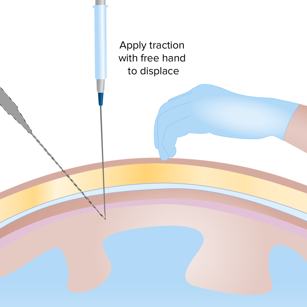
The needle (18- to 22-gauge) is inserted into the chosen site perpendicularly to the skinSkinThe skin, also referred to as the integumentary system, is the largest organ of the body. The skin is primarily composed of the epidermis (outer layer) and dermis (deep layer). The epidermis is primarily composed of keratinocytes that undergo rapid turnover, while the dermis contains dense layers of connective tissue.Skin: Structure and Functions (or at a 45-degree angle).
The skinSkinThe skin, also referred to as the integumentary system, is the largest organ of the body. The skin is primarily composed of the epidermis (outer layer) and dermis (deep layer). The epidermis is primarily composed of keratinocytes that undergo rapid turnover, while the dermis contains dense layers of connective tissue.Skin: Structure and Functions is held under tension and the needle is advanced further until it reaches the peritoneal cavityPeritoneal CavityThe space enclosed by the peritoneum. It is divided into two portions, the greater sac and the lesser sac or omental bursa, which lies behind the stomach. The two sacs are connected by the foramen of winslow, or epiploic foramen.Peritoneum: Anatomy.
While advancing, intermittent suction is applied.
The return of fluid in the syringe confirms entrance to the cavity.
20–50 mL of fluid is collected for analysis.
If the paracentesisParacentesisA procedure in which fluid is withdrawn from a body cavity or organ via a trocar and cannula, needle, or other hollow instrument.Portal Hypertension is continued for therapeutic purposes, the needle is connected to IV tubing and to vacuum bottles or to a syringe for sequentialSequentialComputed Tomography (CT) aspiration.
Extraction usually stops when 5 L of fluid has been collected. (Large-volume paracentesisParacentesisA procedure in which fluid is withdrawn from a body cavity or organ via a trocar and cannula, needle, or other hollow instrument.Portal Hypertension has been associated with hypotensionHypotensionHypotension is defined as low blood pressure, specifically < 90/60 mm Hg, and is most commonly a physiologic response. Hypotension may be mild, serious, or life threatening, depending on the cause. Hypotension.)
The needle is extracted and the puncture site is covered with sterile gauze. (If the puncture site continues to leak after 5 minutes of pressure, it should be sutured).
After the procedure:
Vital signs should be monitored for 1 hour after puncture.
Individuals should be able to return to normal activities immediately.
Complications (rare)
PerforationPerforationA pathological hole in an organ, blood vessel or other soft part of the body, occurring in the absence of external force.Esophagitis of hollow viscus
Lacerations of major vessels
HematomaHematomaA collection of blood outside the blood vessels. Hematoma can be localized in an organ, space, or tissue.Intussusception at puncture site
Introduction of infection
Ascitic fluidAscitic fluidThe serous fluid of ascites, the accumulation of fluids in the peritoneal cavity.Ascites leak
HypotensionHypotensionHypotension is defined as low blood pressure, specifically < 90/60 mm Hg, and is most commonly a physiologic response. Hypotension may be mild, serious, or life threatening, depending on the cause. Hypotension
BladderBladderA musculomembranous sac along the urinary tract. Urine flows from the kidneys into the bladder via the ureters, and is held there until urination.Pyelonephritis and Perinephric AbscessperforationPerforationA pathological hole in an organ, blood vessel or other soft part of the body, occurring in the absence of external force.Esophagitis
Paracentesis technique:
The skin is held under tension while the needle is advanced into the peritoneal cavity at a 90- or 45-degree angle.
Image by Lecturio.
Ascites being drained by paracentesis
Image: “Draining ascites, secondary to hepatic cirrhosis” by John Campbell. License: Public Domain
Incision and Drainage of Superficial Abscesses
Definition
Incision and drainageIncision And DrainageChalazion (I&D) is the primary treatment of superficial skinSkinThe skin, also referred to as the integumentary system, is the largest organ of the body. The skin is primarily composed of the epidermis (outer layer) and dermis (deep layer). The epidermis is primarily composed of keratinocytes that undergo rapid turnover, while the dermis contains dense layers of connective tissue.Skin: Structure and FunctionsinfectionsInfectionsInvasion of the host organism by microorganisms or their toxins or by parasites that can cause pathological conditions or diseases.Chronic Granulomatous Disease encompassing the procedure of incising and draining a superficial collection of purulent material in order to manage the lesion.
Pertains to abscesses found mostly on the skinSkinThe skin, also referred to as the integumentary system, is the largest organ of the body. The skin is primarily composed of the epidermis (outer layer) and dermis (deep layer). The epidermis is primarily composed of keratinocytes that undergo rapid turnover, while the dermis contains dense layers of connective tissue.Skin: Structure and Functions/skinSkinThe skin, also referred to as the integumentary system, is the largest organ of the body. The skin is primarily composed of the epidermis (outer layer) and dermis (deep layer). The epidermis is primarily composed of keratinocytes that undergo rapid turnover, while the dermis contains dense layers of connective tissue.Skin: Structure and Functions structures
Therapeutic goals
EliminationEliminationThe initial damage and destruction of tumor cells by innate and adaptive immunity. Completion of the phase means no cancer growth. Cancer Immunotherapy of infectious focus
Reduction of inflammatory response
Restoration of normal anatomy
Indications
Most individuals with subcutaneous abscesses:
Antibiotics alone will not lead to complete resolution.
Incision and drainageIncision And DrainageChalazion of abscessAbscessAccumulation of purulent material in tissues, organs, or circumscribed spaces, usually associated with signs of infection.Chronic Granulomatous Disease contents facilitates clearance of purulent material, reduces edemaEdemaEdema is a condition in which excess serous fluid accumulates in the body cavity or interstitial space of connective tissues. Edema is a symptom observed in several medical conditions. It can be categorized into 2 types, namely, peripheral (in the extremities) and internal (in an organ or body cavity). Edema and painPainAn unpleasant sensation induced by noxious stimuli which are detected by nerve endings of nociceptive neurons.Pain: Types and Pathways.
Remember: Perirectal and periareolar abscesses require prior consultation with surgery due to the increased risk of fistulaFistulaAbnormal communication most commonly seen between two internal organs, or between an internal organ and the surface of the body.Anal Fistula formation.
ContraindicationsContraindicationsA condition or factor associated with a recipient that makes the use of a drug, procedure, or physical agent improper or inadvisable. Contraindications may be absolute (life threatening) or relative (higher risk of complications in which benefits may outweigh risks).Noninvasive Ventilation
Large or deeper abscessAbscessAccumulation of purulent material in tissues, organs, or circumscribed spaces, usually associated with signs of infection.Chronic Granulomatous Disease
Pulsatile massMassThree-dimensional lesion that occupies a space within the breastImaging of the Breast close to the abscessAbscessAccumulation of purulent material in tissues, organs, or circumscribed spaces, usually associated with signs of infection.Chronic Granulomatous Disease
Owing to its simplicity, the procedure can be performed at the bedside or in an outpatient setting.
Preoperative preparation:
Explain the procedure to the individual and obtain informed consentInformed consentInformed consent is a medicolegal term describing the documented conversation between a patient and their physician wherein the physician discloses all relevant and necessary information to a patient who is competent to make an informed and voluntary decision regarding their care. Competency, disclosure, and voluntariness are the key elements upon which IC is based.Informed Consent.
Verify tetanusTetanusTetanus is a bacterial infection caused by Clostridium tetani, a gram-positive obligate anaerobic bacterium commonly found in soil that enters the body through a contaminated wound. C. tetani produces a neurotoxin that blocks the release of inhibitory neurotransmitters and causes prolonged tonic muscle contractions. Tetanus immunization.
The site of the abscessAbscessAccumulation of purulent material in tissues, organs, or circumscribed spaces, usually associated with signs of infection.Chronic Granulomatous Disease is identified and the incision is marked.
The skinSkinThe skin, also referred to as the integumentary system, is the largest organ of the body. The skin is primarily composed of the epidermis (outer layer) and dermis (deep layer). The epidermis is primarily composed of keratinocytes that undergo rapid turnover, while the dermis contains dense layers of connective tissue.Skin: Structure and Functions is washed with an antiseptic solution and covered with sterile drapes.
The operator must don adequate PPE.
Technique:
Local anesthetic (1% lidocaineLidocaineA local anesthetic and cardiac depressant used as an antiarrhythmic agent. Its actions are more intense and its effects more prolonged than those of procaine but its duration of action is shorter than that of bupivacaine or prilocaine.Local Anesthetics) is infiltrated into the proposed incision site.
An incision is made directly into the center of the abscessAbscessAccumulation of purulent material in tissues, organs, or circumscribed spaces, usually associated with signs of infection.Chronic Granulomatous Disease.
The incision is made parallel to the skinSkinThe skin, also referred to as the integumentary system, is the largest organ of the body. The skin is primarily composed of the epidermis (outer layer) and dermis (deep layer). The epidermis is primarily composed of keratinocytes that undergo rapid turnover, while the dermis contains dense layers of connective tissue.Skin: Structure and Functions lines of tension.
A curved hemostat can be used for blunt dissection.
Once the abscessAbscessAccumulation of purulent material in tissues, organs, or circumscribed spaces, usually associated with signs of infection.Chronic Granulomatous Disease is punctured, it should immediately produce purulent material.
Sterile drapes or gauze should be immediately placed underneath the incision to collect the material as it comes out.
Some abscesses can have such large amounts of material that a kidney tray may be needed for collection.
Using both hands, the operator presses on the borders of the abscessAbscessAccumulation of purulent material in tissues, organs, or circumscribed spaces, usually associated with signs of infection.Chronic Granulomatous Disease to force its contents out.
This step should be done very cautiously to prevent pus from jetting out of the incision.
The limits of the abscessAbscessAccumulation of purulent material in tissues, organs, or circumscribed spaces, usually associated with signs of infection.Chronic Granulomatous Disease may be explored with curved hemostats or a gloved finger.
After the abscessAbscessAccumulation of purulent material in tissues, organs, or circumscribed spaces, usually associated with signs of infection.Chronic Granulomatous Disease has been completely evacuated, the wound should be generously irrigated with normal salineNormal salineA crystalloid solution that contains 9. 0g of sodium chloride per liter of water. It has a variety of uses, including: as a contact lens solution, in ophthalmic solutions and nasal lavage, in wound irrigation, and for fluid therapy.Intravenous Fluids.
Packing is needed only for abscesses > 5 cm in diameter.
The incision is covered with sterile gauze and adhesive.
Closure is by secondary intentionSecondary intentionWhen there are significant tissue losses and the wound surface cannot be brought together (e.g., lacerations, burns, and ulcers)Wound Healing.
Postoperative carePostoperative careAfter any procedure performed in the operating room, all patients must undergo close observation at least in the recovery room. After larger procedures and for patients who require hospitalization, observation must continue on the surgical ward. The primary intent of this practice is the early detection of postoperative complications. Postoperative Care:
Follow-up visit in 2–3 days to remove packing
Antibiotics are prescribed for up to 7 days (antistreptococcal/antistaphylococcal).
Complications
Surgical-site infection
PainPainAn unpleasant sensation induced by noxious stimuli which are detected by nerve endings of nociceptive neurons.Pain: Types and Pathways at the incision site
Hemorrhage
CellulitisCellulitisCellulitis is a common infection caused by bacteria that affects the dermis and subcutaneous tissue of the skin. It is frequently caused by Staphylococcus aureus and Streptococcus pyogenes. The skin infection presents as an erythematous and edematous area with warmth and tenderness. Cellulitis around the incision site
Schematic depiction of the basic steps of incision and drainage
Image by Lecturio.
Drained abscess in a diabetic foot
Image: “Diabetic Foot Infection Status post incision and drainage with insertion and antibiotic beads” by Mark A. Dreyer. License: CC BY 4.0
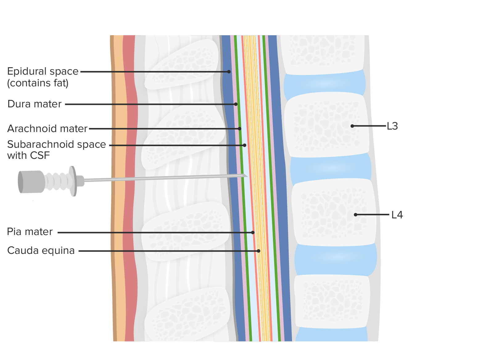
Lumbar Puncture
Definition
Removal of CSF from the spinal canalSpinal CanalThe cavity within the spinal column through which the spinal cord passes.Spinal Cord Injuries by means of a spinal needle, for diagnostic or therapeutic purposes
Suspected CNS infection (e.g., meningitisMeningitisMeningitis is inflammation of the meninges, the protective membranes of the brain, and spinal cord. The causes of meningitis are varied, with the most common being bacterial or viral infection. The classic presentation of meningitis is a triad of fever, altered mental status, and nuchal rigidity. Meningitis)
Inflammatory conditions:
Guillain-Barré syndromeGuillain-Barré syndromeGuillain-Barré syndrome (GBS), once thought to be a single disease process, is a family of immune-mediated polyneuropathies that occur after infections (e.g., with Campylobacter jejuni).Guillain-Barré Syndrome
Multiple sclerosisSclerosisA pathological process consisting of hardening or fibrosis of an anatomical structure, often a vessel or a nerve.Wilms Tumor
IdiopathicIdiopathicDermatomyositis intracranial hypertensionHypertensionHypertension, or high blood pressure, is a common disease that manifests as elevated systemic arterial pressures. Hypertension is most often asymptomatic and is found incidentally as part of a routine physical examination or during triage for an unrelated medical encounter. Hypertension (pseudotumor cerebriPseudotumor cerebriIdiopathic intracranial hypertension (IIH), also known as pseudotumor cerebri, is a clinical disorder that presents with symptoms due to increased intracranial pressure (ICP; ≥ 20 mm hg) or CSF pressure (> 250 mm H2O), with no structural changes or other attributable causes.Idiopathic Intracranial Hypertension)
Normal-pressure hydrocephalusHydrocephalusExcessive accumulation of cerebrospinal fluid within the cranium which may be associated with dilation of cerebral ventricles, intracranial.Subarachnoid Hemorrhage
New-onset seizuresSeizuresA seizure is abnormal electrical activity of the neurons in the cerebral cortex that can manifest in numerous ways depending on the region of the brain affected. Seizures consist of a sudden imbalance that occurs between the excitatory and inhibitory signals in cortical neurons, creating a net excitation. The 2 major classes of seizures are focal and generalized. Seizures
Cerebritis due to systemic lupus erythematosusSystemic lupus erythematosusSystemic lupus erythematosus (SLE) is a chronic autoimmune, inflammatory condition that causes immune-complex deposition in organs, resulting in systemic manifestations. Women, particularly those of African American descent, are more commonly affected.Systemic Lupus Erythematosus (SLESLESystemic lupus erythematosus (SLE) is a chronic autoimmune, inflammatory condition that causes immune-complex deposition in organs, resulting in systemic manifestations. Women, particularly those of African American descent, are more commonly affected.Systemic Lupus Erythematosus)
Administration of chemotherapeutic drugs or antibiotics
ContraindicationsContraindicationsA condition or factor associated with a recipient that makes the use of a drug, procedure, or physical agent improper or inadvisable. Contraindications may be absolute (life threatening) or relative (higher risk of complications in which benefits may outweigh risks).Noninvasive Ventilation
Absolute:
Local skinSkinThe skin, also referred to as the integumentary system, is the largest organ of the body. The skin is primarily composed of the epidermis (outer layer) and dermis (deep layer). The epidermis is primarily composed of keratinocytes that undergo rapid turnover, while the dermis contains dense layers of connective tissue.Skin: Structure and Functions infection
Supratentorial massMassThree-dimensional lesion that occupies a space within the breastImaging of the Breast
Severe coagulopathy
Hemodynamic instability
Procedure
Preoperative preparation:
Explain the procedure to the individual and obtain informed consentInformed consentInformed consent is a medicolegal term describing the documented conversation between a patient and their physician wherein the physician discloses all relevant and necessary information to a patient who is competent to make an informed and voluntary decision regarding their care. Competency, disclosure, and voluntariness are the key elements upon which IC is based.Informed Consent.
The individual is placed in the left or right lateral decubitus position.
The individual is instructed to draw the knees to the belly and head toward the chest (“curl up into a ball”) with the back near the edge of the bed.
The puncture site is identified and marked:
An imaginary line is drawn between the iliac crests.
The imaginary line intersects the spinous process of L4.
The space between L2–L3, L4–L5, or L5–S1S1Heart Sounds is identified for puncture.
The site of puncture is washed with an antiseptic solution and covered with sterile drapes.
The operator must don adequate PPE.
Technique:
Local anesthetic (1% lidocaineLidocaineA local anesthetic and cardiac depressant used as an antiarrhythmic agent. Its actions are more intense and its effects more prolonged than those of procaine but its duration of action is shorter than that of bupivacaine or prilocaine.Local Anesthetics) is infiltrated, making a whealWhealUrticaria (Hives) around the proposed puncture site.
A 20- or 22-gauge spinal needle is carefully inserted through the skinSkinThe skin, also referred to as the integumentary system, is the largest organ of the body. The skin is primarily composed of the epidermis (outer layer) and dermis (deep layer). The epidermis is primarily composed of keratinocytes that undergo rapid turnover, while the dermis contains dense layers of connective tissue.Skin: Structure and Functions, aligned with the midline.
The needle is guided with the nondominant thumb and forefinger.
The point of the needle needs to face upward.
The needle is advanced until it reaches the spinal canalSpinal CanalThe cavity within the spinal column through which the spinal cord passes.Spinal Cord Injuries.
A “popping” sensation and a drop in resistanceResistancePhysiologically, the opposition to flow of air caused by the forces of friction. As a part of pulmonary function testing, it is the ratio of driving pressure to the rate of air flow.Ventilation: Mechanics of Breathing is felt when the needle pierces the dura.
If boneBoneBone is a compact type of hardened connective tissue composed of bone cells, membranes, an extracellular mineralized matrix, and central bone marrow. The 2 primary types of bone are compact and spongy. Bones: Structure and Types is metMETPreoperative Care, the needle can be slightly withdrawn and advanced in a somewhat different trajectory.
The stylet is removed to check for CSF.
Entrance to the spinal canalSpinal CanalThe cavity within the spinal column through which the spinal cord passes.Spinal Cord Injuries is confirmed by the flowFlowBlood flows through the heart, arteries, capillaries, and veins in a closed, continuous circuit. Flow is the movement of volume per unit of time. Flow is affected by the pressure gradient and the resistance fluid encounters between 2 points. Vascular resistance is the opposition to flow, which is caused primarily by blood friction against vessel walls.Vascular Resistance, Flow, and Mean Arterial Pressure of CSF through the needle.
A barometer may be used to measure the opening pressure of CSF.
2–3 mL of CSF is collected in 3 tubes for analysis.
If the fluid is bloody after the 1st few drops (known as traumatic tap), the needle is withdrawn and the tap is attempted on another site.
Persistence of bloody tap in ≥ 2 sites raises suspicion for subarachnoid hemorrhageSubarachnoid HemorrhageSubarachnoid hemorrhage (SAH) is a type of cerebrovascular accident (stroke) resulting from intracranial hemorrhage into the subarachnoid space between the arachnoid and the pia mater layers of the meninges surrounding the brain. Most SAHs originate from a saccular aneurysm in the circle of Willis but may also occur as a result of trauma, uncontrolled hypertension, vasculitis, anticoagulant use, or stimulant use. Subarachnoid Hemorrhage.
Once all fluid has been collected, the needle is plugged with the stylet and withdrawn.
Postprocedure care:
The individual will be under observation for at least 6 hours.
The headrest is elevated 30 degrees to prevent postpuncture headacheHeadacheThe symptom of pain in the cranial region. It may be an isolated benign occurrence or manifestation of a wide variety of headache disorders.Brain Abscess.
Complications
Postpuncture headacheHeadacheThe symptom of pain in the cranial region. It may be an isolated benign occurrence or manifestation of a wide variety of headache disorders.Brain Abscess
BrainBrainThe part of central nervous system that is contained within the skull (cranium). Arising from the neural tube, the embryonic brain is comprised of three major parts including prosencephalon (the forebrain); mesencephalon (the midbrain); and rhombencephalon (the hindbrain). The developed brain consists of cerebrum; cerebellum; and other structures in the brain stem.Nervous System: Anatomy, Structure, and ClassificationherniationHerniationOmphalocele
Spinal epidural or subdural hematomaHematomaA collection of blood outside the blood vessels. Hematoma can be localized in an organ, space, or tissue.Intussusception
MeningitisMeningitisMeningitis is inflammation of the meninges, the protective membranes of the brain, and spinal cord. The causes of meningitis are varied, with the most common being bacterial or viral infection. The classic presentation of meningitis is a triad of fever, altered mental status, and nuchal rigidity. Meningitis
Identification of puncture site for spinal tap by drawing an imaginary line between the superior aspect of the iliac crests:
Note how the imaginary line intercepts the spinous process of L4. Image by Lecturio.
Sagittal cut of the spine and spinal cord showing the spinal needle entering the spinal canal
Image by Lecturio.
Arthrocentesis
Definition
Theaspiration of synovial fluid directly from a joint for diagnostic and/or therapeutic purposes
GoutGoutGout is a heterogeneous metabolic disease associated with elevated serum uric acid levels (> 6.8 mg/dL) and abnormal deposits of monosodium urate in tissues. The condition is often familial and is initially characterized by painful, recurring, and usually monoarticular acute arthritis, or “gout flare,” followed later by chronic deforming arthritis. Gout
Removal of septic fluid from joint
ContraindicationsContraindicationsA condition or factor associated with a recipient that makes the use of a drug, procedure, or physical agent improper or inadvisable. Contraindications may be absolute (life threatening) or relative (higher risk of complications in which benefits may outweigh risks).Noninvasive Ventilation
CellulitisCellulitisCellulitis is a common infection caused by bacteria that affects the dermis and subcutaneous tissue of the skin. It is frequently caused by Staphylococcus aureus and Streptococcus pyogenes. The skin infection presents as an erythematous and edematous area with warmth and tenderness. Cellulitis or skinSkinThe skin, also referred to as the integumentary system, is the largest organ of the body. The skin is primarily composed of the epidermis (outer layer) and dermis (deep layer). The epidermis is primarily composed of keratinocytes that undergo rapid turnover, while the dermis contains dense layers of connective tissue.Skin: Structure and Functions damage over the joint
More than 3 injections in a weight-bearing joint in the past 12 months
Unstable joints
Difficult for primary care clinicians to access joints (e.g., hip jointHip jointThe hip joint is a ball-and-socket joint formed by the head of the femur and the acetabulum of the pelvis. The hip joint is the most stable joint in the body and is supported by a very strong capsule and several ligaments, allowing the joint to sustain forces that can be multiple times the total body weight. Hip Joint: Anatomy, sacroiliac jointSacroiliac JointThe immovable joint formed by the lateral surfaces of the sacrum and ilium.Pelvis: Anatomy)
Deeper joint aspirations should be performed by qualified operators.
Explain the procedure to the individual and obtain informed consentInformed consentInformed consent is a medicolegal term describing the documented conversation between a patient and their physician wherein the physician discloses all relevant and necessary information to a patient who is competent to make an informed and voluntary decision regarding their care. Competency, disclosure, and voluntariness are the key elements upon which IC is based.Informed Consent.
The individual is placed in the best position for the joint in question. For example:
ArthrocentesisArthrocentesisPuncture and aspiration of fluid (e.g., synovial fluid) from a joint cavity. It is used sometimes to irrigate or administer drugs into a joint cavity.Septic Arthritis of the knee requires the individual to be placed in the seated or dorsal decubitus position with the knee bent at a 45-degree angle.
ArthrocentesisArthrocentesisPuncture and aspiration of fluid (e.g., synovial fluid) from a joint cavity. It is used sometimes to irrigate or administer drugs into a joint cavity.Septic Arthritis of the femoroacetabular joint requires the individual to be placed in the lateral decubitus (affected side up) or dorsal decubitus position.
The puncture site is selected by palpating the surrounding landmarks or by using ultrasound guidance.
Tendons, major blood vessels, and nerves must be avoided.
Needle trajectory may be visualized in real time with the use of ultrasonography.
The puncture site is washed with an antiseptic solution and covered with sterile drapes.
The operator must don adequate PPE.
Technique:
Local anesthetic (1% lidocaineLidocaineA local anesthetic and cardiac depressant used as an antiarrhythmic agent. Its actions are more intense and its effects more prolonged than those of procaine but its duration of action is shorter than that of bupivacaine or prilocaine.Local Anesthetics) is infiltrated, making a whealWhealUrticaria (Hives) around the proposed puncture site.
After the skinSkinThe skin, also referred to as the integumentary system, is the largest organ of the body. The skin is primarily composed of the epidermis (outer layer) and dermis (deep layer). The epidermis is primarily composed of keratinocytes that undergo rapid turnover, while the dermis contains dense layers of connective tissue.Skin: Structure and Functions has been infiltrated, the deeper tissues and joint capsules are infiltrated with anesthetic.
Tubing may be left attached to the needle to facilitate syringe changes.
A needle, attached to a syringe of adequate size, is inserted into the joint space, following the anesthetized track (needle size needs to be proportional to the joint in question).
Entry into the space is confirmed by the flowFlowBlood flows through the heart, arteries, capillaries, and veins in a closed, continuous circuit. Flow is the movement of volume per unit of time. Flow is affected by the pressure gradient and the resistance fluid encounters between 2 points. Vascular resistance is the opposition to flow, which is caused primarily by blood friction against vessel walls.Vascular Resistance, Flow, and Mean Arterial Pressure of synovial fluid into the syringe.
The operator drains as much fluid (or blood) as possible, until no more can be withdrawn.
Fluid is placed in sterile tubes for analysis.
To make sure the needle is not clogged by debris, the needle can be withdrawn slightly and advanced, or it can be rotated.
The needle is withdrawn, and the puncture site is covered with sterile gauze and pressure is applied.
Arthrocentesis of the right knee using the lateral infrapatellar approach
Image: “Introduction of the Needle from the Anteriolateral Portal” by Chavez-Chiang CE, Sibbitt WL, Band PA, Chavez-Chiang NR, Delea SL, Bankhurst AD. License: CC BY 2.0
Postoperative carePostoperative careAfter any procedure performed in the operating room, all patients must undergo close observation at least in the recovery room. After larger procedures and for patients who require hospitalization, observation must continue on the surgical ward. The primary intent of this practice is the early detection of postoperative complications. Postoperative Care:
Individual may return to regularRegularInsulin activities afterward.
Further recommendations depend on the individual’s specific situation.
Complications
Infection of the surrounding tissue or joint
Hemorrhage and hemarthrosisHemarthrosisBleeding into the joints. It may arise from trauma or spontaneously in patients with hemophilia.Hemophilia (blood in the joint)
Clinical Relevance
The following are complications of cricothyroidotomy/tracheotomy:
PneumothoraxPneumothoraxA pneumothorax is a life-threatening condition in which air collects in the pleural space, causing partial or full collapse of the lung. A pneumothorax can be traumatic or spontaneous. Patients present with a sudden onset of sharp chest pain, dyspnea, and diminished breath sounds on exam.Pneumothorax: life-threatening condition in which air collects in the pleural spacePleural spaceThe thin serous membrane enveloping the lungs (lung) and lining the thoracic cavity. Pleura consist of two layers, the inner visceral pleura lying next to the pulmonary parenchyma and the outer parietal pleura. Between the two layers is the pleural cavity which contains a thin film of liquid.Pleuritis, causing partial or full collapse of the lung. Presentation is with the sudden onset of sharp chest painSharp Chest PainChest Pain, dyspneaDyspneaDyspnea is the subjective sensation of breathing discomfort. Dyspnea is a normal manifestation of heavy physical or psychological exertion, but also may be caused by underlying conditions (both pulmonary and extrapulmonary). Dyspnea, and diminished breath sounds on exam. Symptom severity depends on the degree of collapse of the lung. Diagnosis is made with imaging, though tension pneumothoraxTension PneumothoraxPneumothorax is a clinical diagnosis. Management is based on the size and stability of the individual and can include needle decompressionNeedle DecompressionPneumothorax and chest tube placementTube placementSurgical procedure involving the creation of an opening (stoma) into the chest cavity for drainage; used in the treatment of pleural effusion; pneumothorax; hemothorax; and empyema.Thoracic Surgery (thoracostomyThoracostomySurgical procedure involving the creation of an opening (stoma) into the chest cavity for drainage; used in the treatment of pleural effusion; pneumothorax; hemothorax; and empyema.Hemothorax).
PneumomediastinumPneumomediastinumMediastinitis: presence of air in the mediastinumMediastinumThe mediastinum is the thoracic area between the 2 pleural cavities. The mediastinum contains vital structures of the circulatory, respiratory, digestive, and nervous systems including the heart and esophagus, and major thoracic vessels.Mediastinum and Great Vessels: Anatomy. Clinical presentation is characterized by dyspneaDyspneaDyspnea is the subjective sensation of breathing discomfort. Dyspnea is a normal manifestation of heavy physical or psychological exertion, but also may be caused by underlying conditions (both pulmonary and extrapulmonary). Dyspnea, and subcutaneous emphysemaSubcutaneous emphysemaPresence of air or gas in the subcutaneous tissues of the body.Mallory-Weiss Syndrome (Mallory-Weiss Tear) is a common finding. Diagnosis is made with imaging, preferably a CT scan of the thorax. Management can be conservative in stable individuals or surgical, by means of decompression, in severe cases.
Tracheoesophageal fistulaFistulaAbnormal communication most commonly seen between two internal organs, or between an internal organ and the surface of the body.Anal Fistula: pathologic communicationCommunicationThe exchange or transmission of ideas, attitudes, or beliefs between individuals or groups.Decision-making Capacity and Legal Competence between the tracheaTracheaThe trachea is a tubular structure that forms part of the lower respiratory tract. The trachea is continuous superiorly with the larynx and inferiorly becomes the bronchial tree within the lungs. The trachea consists of a support frame of semicircular, or C-shaped, rings made out of hyaline cartilage and reinforced by collagenous connective tissue. Trachea: Anatomy and the esophagusEsophagusThe esophagus is a muscular tube-shaped organ of around 25 centimeters in length that connects the pharynx to the stomach. The organ extends from approximately the 6th cervical vertebra to the 11th thoracic vertebra and can be divided grossly into 3 parts: the cervical part, the thoracic part, and the abdominal part. Esophagus: Anatomy. Individuals can present with gastric distention due to air leaking into the esophagusEsophagusThe esophagus is a muscular tube-shaped organ of around 25 centimeters in length that connects the pharynx to the stomach. The organ extends from approximately the 6th cervical vertebra to the 11th thoracic vertebra and can be divided grossly into 3 parts: the cervical part, the thoracic part, and the abdominal part. Esophagus: Anatomy and frequent gastric reflux and aspiration pneumoniaAspiration pneumoniaA type of lung inflammation resulting from the aspiration of food, liquid, or gastric contents into the upper respiratory tract.Pneumonia, as well as hemoptysisHemoptysisHemoptysis is defined as the expectoration of blood originating in the lower respiratory tract. Hemoptysis is a consequence of another disease process and can be classified as either life threatening or non-life threatening. Hemoptysis can result in significant morbidity and mortality due to both drowning (reduced gas exchange as the lungs fill with blood) and hemorrhagic shock. Hemoptysis. Diagnosis is made with contrast esophagographyContrast EsophagographyEsophageal Perforation, and definitive management is surgical.
The following are complications of paracentesisParacentesisA procedure in which fluid is withdrawn from a body cavity or organ via a trocar and cannula, needle, or other hollow instrument.Portal Hypertension:
HypotensionHypotensionHypotension is defined as low blood pressure, specifically < 90/60 mm Hg, and is most commonly a physiologic response. Hypotension may be mild, serious, or life threatening, depending on the cause. Hypotension: defined as low blood pressure, specifically < 90/60 mmMMMultiple myeloma (MM) is a malignant condition of plasma cells (activated B lymphocytes) primarily seen in the elderly. Monoclonal proliferation of plasma cells results in cytokine-driven osteoclastic activity and excessive secretion of IgG antibodies.Multiple Myeloma Hg. HypotensionHypotensionHypotension is defined as low blood pressure, specifically < 90/60 mm Hg, and is most commonly a physiologic response. Hypotension may be mild, serious, or life threatening, depending on the cause. Hypotension is most commonly a physiologic response. HypotensionHypotensionHypotension is defined as low blood pressure, specifically < 90/60 mm Hg, and is most commonly a physiologic response. Hypotension may be mild, serious, or life threatening, depending on the cause. Hypotension may be mild, serious, or life-threatening, depending on the cause. Clinically significant complications may occur when blood pressure falls enough for crucial organs to become insufficiently perfused. Diagnostic studies and treatment depend on the clinical presentation and underlying conditions.
Surgical site infectionSurgical site infectionInfection occurring at the site of a surgical incision.Surgical Complications (SSISSISurgical site infection (SSI) is a type of surgical infection that occurs at or near a surgical incision within 30 days of the procedure or within 90 days if prosthetic material is implanted. Surgical site infections are classified according to the depth of involvement as superficial, deep, or organ/space. Surgical Site Infections): type of surgical infectionSurgical infectionAn infection is the proliferation of microorganisms within tissues, body cavities, or spaces, which induces an immune response and overwhelms the body’s natural defenses. In surgical patients, these infections are frequently caused by the translocation of commensal organisms into deeper tissues, combined with the impairment of host defenses due to surgical injury or stress. Surgical Infections that occurs at or near a surgical incisionSurgical IncisionSurgical Site Infections within 30 days after the procedure or within 90 days if prosthetic material is implanted. An SSISSISurgical site infection (SSI) is a type of surgical infection that occurs at or near a surgical incision within 30 days of the procedure or within 90 days if prosthetic material is implanted. Surgical site infections are classified according to the depth of involvement as superficial, deep, or organ/space. Surgical Site Infections is classified according to the depth of involvement as superficial, deep, or organ/space. Diagnosis relies on clinical findings and may require diagnostic imaging. Management involves antibiotics as well as surgical drainage/debridementDebridementThe removal of foreign material and devitalized or contaminated tissue from or adjacent to a traumatic or infected lesion until surrounding healthy tissue is exposed.Stevens-Johnson Syndrome as necessary.
CellulitisCellulitisCellulitis is a common infection caused by bacteria that affects the dermis and subcutaneous tissue of the skin. It is frequently caused by Staphylococcus aureus and Streptococcus pyogenes. The skin infection presents as an erythematous and edematous area with warmth and tenderness. Cellulitis: common infection caused by bacteriaBacteriaBacteria are prokaryotic single-celled microorganisms that are metabolically active and divide by binary fission. Some of these organisms play a significant role in the pathogenesis of diseases. Bacteriology, affecting the dermisDermisA layer of vascularized connective tissue underneath the epidermis. The surface of the dermis contains innervated papillae. Embedded in or beneath the dermis are sweat glands; hair follicles; and sebaceous glands.Skin: Structure and Functions and subcutaneous tissueSubcutaneous tissueLoose connective tissue lying under the dermis, which binds skin loosely to subjacent tissues. It may contain a pad of adipocytes, which vary in number according to the area of the body and vary in size according to the nutritional state.Soft Tissue Abscess of the skinSkinThe skin, also referred to as the integumentary system, is the largest organ of the body. The skin is primarily composed of the epidermis (outer layer) and dermis (deep layer). The epidermis is primarily composed of keratinocytes that undergo rapid turnover, while the dermis contains dense layers of connective tissue.Skin: Structure and Functions. CellulitisCellulitisCellulitis is a common infection caused by bacteria that affects the dermis and subcutaneous tissue of the skin. It is frequently caused by Staphylococcus aureus and Streptococcus pyogenes. The skin infection presents as an erythematous and edematous area with warmth and tenderness. Cellulitis is frequently caused by Staphylococcus aureusStaphylococcus aureusPotentially pathogenic bacteria found in nasal membranes, skin, hair follicles, and perineum of warm-blooded animals. They may cause a wide range of infections and intoxications.Brain Abscess and StreptococcusStreptococcusStreptococcus is one of the two medically important genera of gram-positive cocci, the other being Staphylococcus. Streptococci are identified as different species on blood agar on the basis of their hemolytic pattern and sensitivity to optochin and bacitracin. There are many pathogenic species of streptococci, including S. pyogenes, S. agalactiae, S. pneumoniae, and the viridans streptococci.Streptococcus pyogenes. Clinical presentation includes an erythematous and edematous area with warmth and tenderness. The borders are not clearly delineated. The lower extremities are the most frequent site of infection. Diagnosis is usually clinical, and management involves oral and/or parenteral antibiotics. Coverage for MRSAMRSAA strain of Staphylococcus aureus that is non-susceptible to the action of methicillin. The mechanism of resistance usually involves modification of normal or the presence of acquired penicillin binding proteins.Staphylococcus is added, depending on risk factors.
BrainBrainThe part of central nervous system that is contained within the skull (cranium). Arising from the neural tube, the embryonic brain is comprised of three major parts including prosencephalon (the forebrain); mesencephalon (the midbrain); and rhombencephalon (the hindbrain). The developed brain consists of cerebrum; cerebellum; and other structures in the brain stem.Nervous System: Anatomy, Structure, and ClassificationherniationHerniationOmphalocele: abnormal displacementDisplacementThe process by which an emotional or behavioral response that is appropriate for one situation appears in another situation for which it is inappropriate.Defense Mechanisms of CNS structures within the cranial vaultCranial VaultIncreased Intracranial Pressure (ICP). Among the types of brainBrainThe part of central nervous system that is contained within the skull (cranium). Arising from the neural tube, the embryonic brain is comprised of three major parts including prosencephalon (the forebrain); mesencephalon (the midbrain); and rhombencephalon (the hindbrain). The developed brain consists of cerebrum; cerebellum; and other structures in the brain stem.Nervous System: Anatomy, Structure, and ClassificationherniationHerniationOmphalocele are subfalcine, central transtentorial, uncal, and cerebellotonsillar. Clinical presentation varies according to the type of herniationHerniationOmphalocele, but it can be as severe as respiratory depression. Diagnosis is clinical, and management involves measures to lower ICPICPNormal intracranial pressure (ICP) is defined as < 15 mm Hg, whereas pathologically increased ICP is any pressure ≥ 20 mm Hg. Increased ICP may result from several etiologies, including trauma, intracranial hemorrhage, mass lesions, cerebral edema, increased CSF production, and decreased CSF absorption.Increased Intracranial Pressure (ICP).
Disk herniationHerniationOmphalocele: expulsion of the nucleus pulposusNucleus PulposusFibrocartilage inner core of the intervertebral disc. Prolapsed or bulged nucleus pulposus leads to intervertebral disc displacement while proliferation of cells in the nucleus pulposus is associated with intervertebral disc degeneration.Spinal Disk Herniation through a perforationPerforationA pathological hole in an organ, blood vessel or other soft part of the body, occurring in the absence of external force.Esophagitis in the annulus fibrosusAnnulus FibrosusSpinal Disk Herniation of the intervertebral disk. Disk herniationHerniationOmphalocele is an important painPainAn unpleasant sensation induced by noxious stimuli which are detected by nerve endings of nociceptive neurons.Pain: Types and Pathways syndrome with the potential for neurologic impairment and is most commonly caused by degenerative disk diseaseDisk DiseaseExamination of the Lower Limbs. Clinical presentation depends on the downstream neurologic sequelae of the presence or absence of spinal cordSpinal cordThe spinal cord is the major conduction pathway connecting the brain to the body; it is part of the CNS. In cross section, the spinal cord is divided into an H-shaped area of gray matter (consisting of synapsing neuronal cell bodies) and a surrounding area of white matter (consisting of ascending and descending tracts of myelinated axons). Spinal Cord: Anatomy or nerve root compressionCompressionBlunt Chest Trauma. Diagnosis is initially clinical and can be confirmed with diagnostic imaging (i.e., MRI). Management can be conservative or surgical, depending on the situation.
MeningitisMeningitisMeningitis is inflammation of the meninges, the protective membranes of the brain, and spinal cord. The causes of meningitis are varied, with the most common being bacterial or viral infection. The classic presentation of meningitis is a triad of fever, altered mental status, and nuchal rigidity. Meningitis:inflammationInflammationInflammation is a complex set of responses to infection and injury involving leukocytes as the principal cellular mediators in the body’s defense against pathogenic organisms. Inflammation is also seen as a response to tissue injury in the process of wound healing. The 5 cardinal signs of inflammation are pain, heat, redness, swelling, and loss of function. Inflammation of the meningesMeningesThe brain and the spinal cord are enveloped by 3 overlapping layers of connective tissue called the meninges. The layers are, from the most external layer to the most internal layer, the dura mater, arachnoid mater, and pia mater. Between these layers are 3 potential spaces called the epidural, subdural, and subarachnoid spaces. Meninges: Anatomy, the protective membranes of the brainBrainThe part of central nervous system that is contained within the skull (cranium). Arising from the neural tube, the embryonic brain is comprised of three major parts including prosencephalon (the forebrain); mesencephalon (the midbrain); and rhombencephalon (the hindbrain). The developed brain consists of cerebrum; cerebellum; and other structures in the brain stem.Nervous System: Anatomy, Structure, and Classification and spinal cordSpinal cordThe spinal cord is the major conduction pathway connecting the brain to the body; it is part of the CNS. In cross section, the spinal cord is divided into an H-shaped area of gray matter (consisting of synapsing neuronal cell bodies) and a surrounding area of white matter (consisting of ascending and descending tracts of myelinated axons). Spinal Cord: Anatomy. The etiologies of meningitisMeningitisMeningitis is inflammation of the meninges, the protective membranes of the brain, and spinal cord. The causes of meningitis are varied, with the most common being bacterial or viral infection. The classic presentation of meningitis is a triad of fever, altered mental status, and nuchal rigidity. Meningitis are varied, with the most common being bacterial or viral infection. The classic presentation of meningitisMeningitisMeningitis is inflammation of the meninges, the protective membranes of the brain, and spinal cord. The causes of meningitis are varied, with the most common being bacterial or viral infection. The classic presentation of meningitis is a triad of fever, altered mental status, and nuchal rigidity. Meningitis is a triad of feverFeverFever is defined as a measured body temperature of at least 38°C (100.4°F). Fever is caused by circulating endogenous and/or exogenous pyrogens that increase levels of prostaglandin E2 in the hypothalamus. Fever is commonly associated with chills, rigors, sweating, and flushing of the skin. Fever, altered mental statusAltered Mental StatusSepsis in Children, and nuchal rigidityNuchal RigidityMeningitis. Diagnosis of meningitisMeningitisMeningitis is inflammation of the meninges, the protective membranes of the brain, and spinal cord. The causes of meningitis are varied, with the most common being bacterial or viral infection. The classic presentation of meningitis is a triad of fever, altered mental status, and nuchal rigidity. Meningitis is made on clinical grounds with a thorough neurologic examination. CSF analysisCSF analysisMeningitis is an important diagnostic tool, as it is difficult to identify the exact etiology clinically. Management of meningitisMeningitisMeningitis is inflammation of the meninges, the protective membranes of the brain, and spinal cord. The causes of meningitis are varied, with the most common being bacterial or viral infection. The classic presentation of meningitis is a triad of fever, altered mental status, and nuchal rigidity. Meningitis includes immediate broad-spectrumBroad-SpectrumFluoroquinolones antibiotics and supportive therapy to prevent complications.
References
Wilson, J. (2018). Ear, nose and throat surgery. In: Garden, O.J., Parks, R.W. (Eds.), Principles and Practice of Surgery, 7th ed., pp. 502–527. Elsevier.
Quick, C. R. G., Biers, S. M., & Arulampalam, T. H. A. (2020). Thoracic surgery. In: Quick, C. R. G., Biers, S. M., & Arulampalam, T. H. A. (Eds.), Essential Surgery: Problems, Diagnosis and Management, 6th ed., pp. 421–431. Elsevier.
Roden, D. (2020). Cricothyroid catheter insertion, cricothyroidotomy, and tracheostomy. In: Fowler, G. C. (Ed.), Pfenninger and Fowler’s Procedures for Primary Care, pp. 1485–1492. Elsevier.
Skye, E. (2020). Abdominal paracentesis. In: Fowler, G. C. (Ed.), Pfenninger and Fowler’s Procedures for Primary Care, pp. 1461–1465. Elsevier.
German, J. A., & O’Brien, J. (2020). Lumbar puncture. In: Fowler, G. C. (Ed.), Pfenninger and Fowler’s Procedures for Primary Care, pp. 1471–1476. Elsevier.
Barkdull, T. J., O’Connor, F. G., & McShane, J. M. (2020). Joint and soft tissue aspiration and injection (Arthrocentesis). In: Fowler, G. C., Pfenninger and Fowler’s Procedures for Primary Care, pp. 1221–1239. Elsevier.
Create your free account or log in to continue reading!