The hip joint is a ball-and-socket joint formed by the head of the femur and the acetabulum of the pelvis Pelvis The pelvis consists of the bony pelvic girdle, the muscular and ligamentous pelvic floor, and the pelvic cavity, which contains viscera, vessels, and multiple nerves and muscles. The pelvic girdle, composed of 2 "hip" bones and the sacrum, is a ring-like bony structure of the axial skeleton that links the vertebral column with the lower extremities. Pelvis: Anatomy. The hip joint is the most stable joint in the body and is supported by a very strong capsule Capsule An envelope of loose gel surrounding a bacterial cell which is associated with the virulence of pathogenic bacteria. Some capsules have a well-defined border, whereas others form a slime layer that trails off into the medium. Most capsules consist of relatively simple polysaccharides but there are some bacteria whose capsules are made of polypeptides. Bacteroides and several ligaments, allowing the joint to sustain forces that can be multiple times the total body weight. Tolerating these forces is possible thanks to the bony alignment and substantial support from the static and dynamic stabilizers of the hip. Several muscle groups attach to the components of the hip joint, allowing for the joint's range of motion Range of motion The distance and direction to which a bone joint can be extended. Range of motion is a function of the condition of the joints, muscles, and connective tissues involved. Joint flexibility can be improved through appropriate muscle strength exercises. Examination of the Upper Limbs. The muscles that attach to the hip joint include those of the gluteal region Gluteal region The gluteal region is located posterior to the pelvic girdle and extends distally into the upper leg as the posterior thigh. The gluteal region consists of the gluteal muscles and several clinically important arteries, veins, and nerves. The muscles of the gluteal region help to move the hip joint during walking, running, standing, and sitting. Gluteal Region: Anatomy and thigh Thigh The thigh is the region of the lower limb found between the hip and the knee joint. There is a single bone in the thigh called the femur, which is surrounded by large muscles grouped into 3 fascial compartments. Thigh: Anatomy.
Last updated: Dec 15, 2025
Contents
The hip joint is a multi-axial joint that connects the pelvis Pelvis The pelvis consists of the bony pelvic girdle, the muscular and ligamentous pelvic floor, and the pelvic cavity, which contains viscera, vessels, and multiple nerves and muscles. The pelvic girdle, composed of 2 “hip” bones and the sacrum, is a ring-like bony structure of the axial skeleton that links the vertebral column with the lower extremities. Pelvis: Anatomy to the lower extremities. As compared with the glenohumeral joint (shoulder), the hip has less range of motion Range of motion The distance and direction to which a bone joint can be extended. Range of motion is a function of the condition of the joints, muscles, and connective tissues involved. Joint flexibility can be improved through appropriate muscle strength exercises. Examination of the Upper Limbs and is designed primarily for weight bearing and stability.
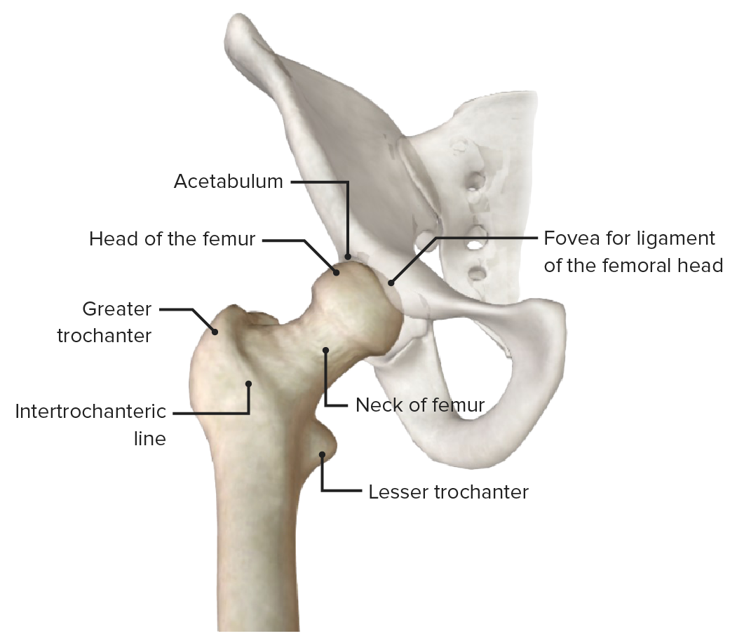
Anterior view of the hip joint (pelvis faded), featuring the bony landmarks of the proximal end of the femur
Image by BioDigital, edited by Lecturio.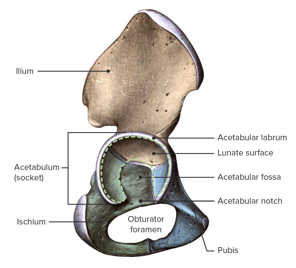
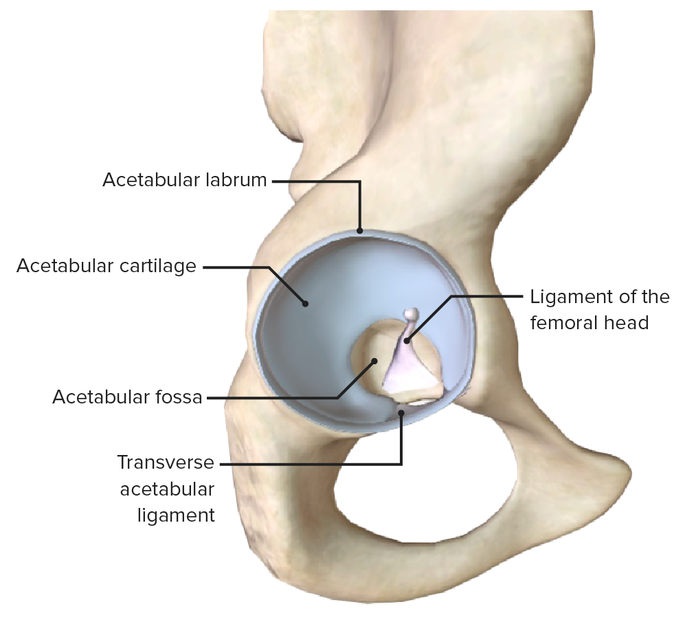
The hip bone featuring the acetabulum and acetabular labrum, which are the pelvic components of the hip joint
Image by BioDigital, edited by Lecturio.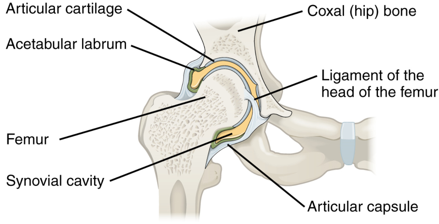
Frontal section through the right hip joint featuring the ball-and-socket joint
Image: “916 Hip Joint” by OpenStax College. License: CC BY 3.0, edited by Lecturio.Consists of 2 layers:
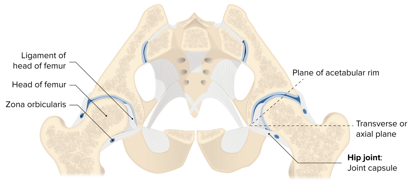
Cross-section of the hip joint, featuring the insertions of the articular capsule and supporting ligaments
Image by Lecturio.Can be divided into 2 groups:
| Insertions | Functions | |
|---|---|---|
| Ligament of the femoral head (or round ligament Round ligament A fibromuscular band that attaches to the uterus and then passes along the broad ligament, out through the inguinal ring, and into the labium majus. Uterus, Cervix, and Fallopian Tubes: Anatomy of the femur) | Apex of the femoral fovea Fovea An area approximately 1. 5 millimeters in diameter within the macula lutea where the retina thins out greatly because of the oblique shifting of all layers except the pigment epithelium layer. It includes the sloping walls of the fovea (clivus) and contains a few rods in its periphery. In its center (foveola) are the cones most adapted to yield high visual acuity, each cone being connected to only one ganglion cell. Eye: Anatomy to either side of the acetabular notch | Limits abduction Abduction Examination of the Upper Limbs and lateral rotation Rotation Motion of an object in which either one or more points on a line are fixed. It is also the motion of a particle about a fixed point. X-rays when the thigh Thigh The thigh is the region of the lower limb found between the hip and the knee joint. There is a single bone in the thigh called the femur, which is surrounded by large muscles grouped into 3 fascial compartments. Thigh: Anatomy is semiflexed; carries the artery to the head of the femur |
| Transverse ligament of the acetabulum | Fibrous Fibrous Fibrocystic Change structure that converts the acetabular notch into a foramen | Allows passage of the neurovasculature into the joint |
| Insertions | Functions | |
|---|---|---|
| Iliofemoral ligament | Anterior inferior iliac spine Spine The human spine, or vertebral column, is the most important anatomical and functional axis of the human body. It consists of 7 cervical vertebrae, 12 thoracic vertebrae, and 5 lumbar vertebrae and is limited cranially by the skull and caudally by the sacrum. Vertebral Column: Anatomy and the acetabular rim to the intertrochanteric line and the greater trochanter |
|
| Pubofemoral ligament | Pubic part of the acetabular rim and the superior pubic ramus to the lower part of the femoral neck Neck The part of a human or animal body connecting the head to the rest of the body. Peritonsillar Abscess |
|
| Ischiofemoral ligament | Ischial region of the acetabulum to the neck Neck The part of a human or animal body connecting the head to the rest of the body. Peritonsillar Abscess of the femur medial to the greater trochanter |
|
| Zona orbicularis | Annular ligament Annular Ligament Radial Head Subluxation (Nursemaid’s Elbow) made of the deep circular fibers of the fibrous Fibrous Fibrocystic Change capsule Capsule An envelope of loose gel surrounding a bacterial cell which is associated with the virulence of pathogenic bacteria. Some capsules have a well-defined border, whereas others form a slime layer that trails off into the medium. Most capsules consist of relatively simple polysaccharides but there are some bacteria whose capsules are made of polypeptides. Bacteroides, which contains fibers from all extra-articular ligaments | Stabilizes the hip |
Intra-articular ligaments of the hip joint
Image by BioDigital, edited by Lecturio.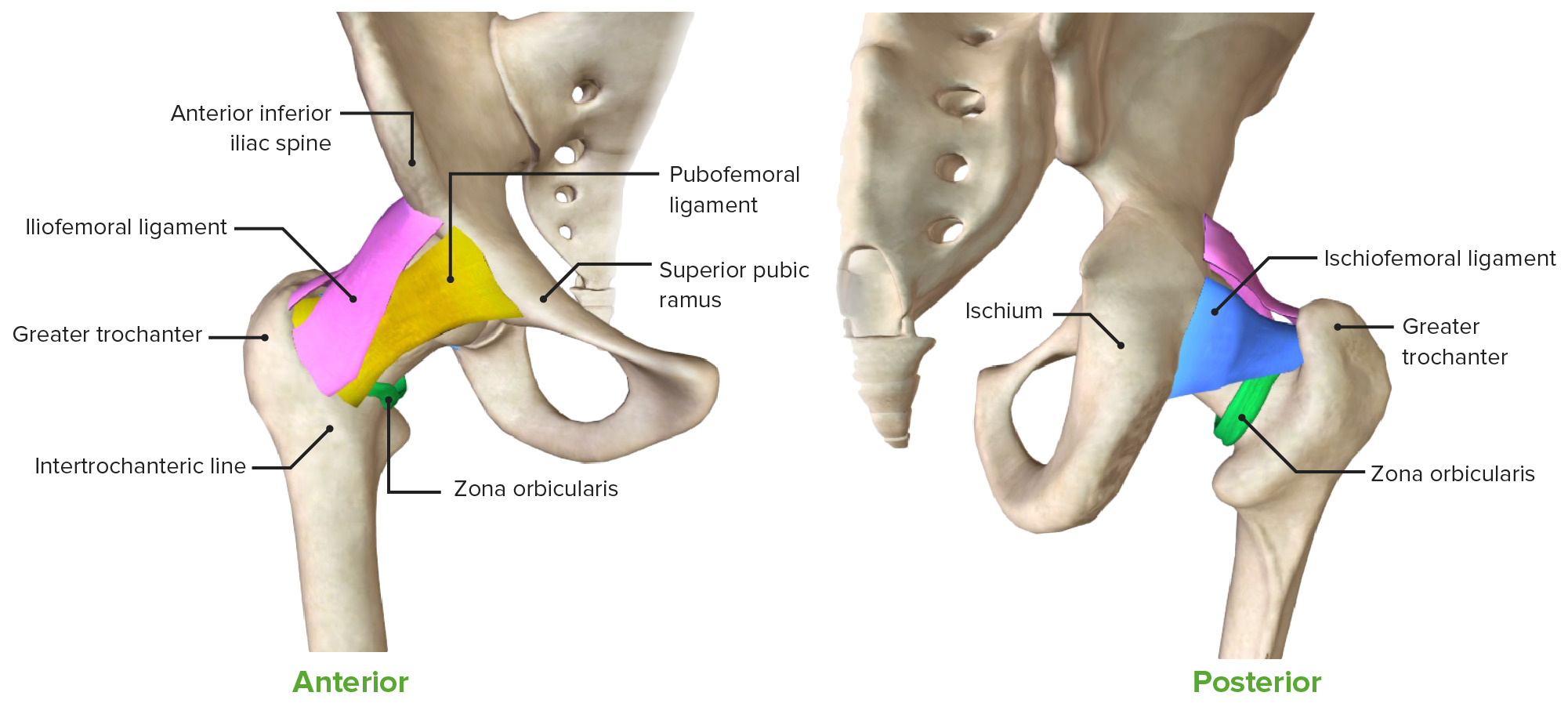
Anterior and posterior views of the hip joint, featuring the extra-articular ligaments
Image by BioDigital, edited by Lecturio.Bursae are small, synovial fluid-filled sacs that reduce friction between the bony components of the joint and the surrounding muscles.
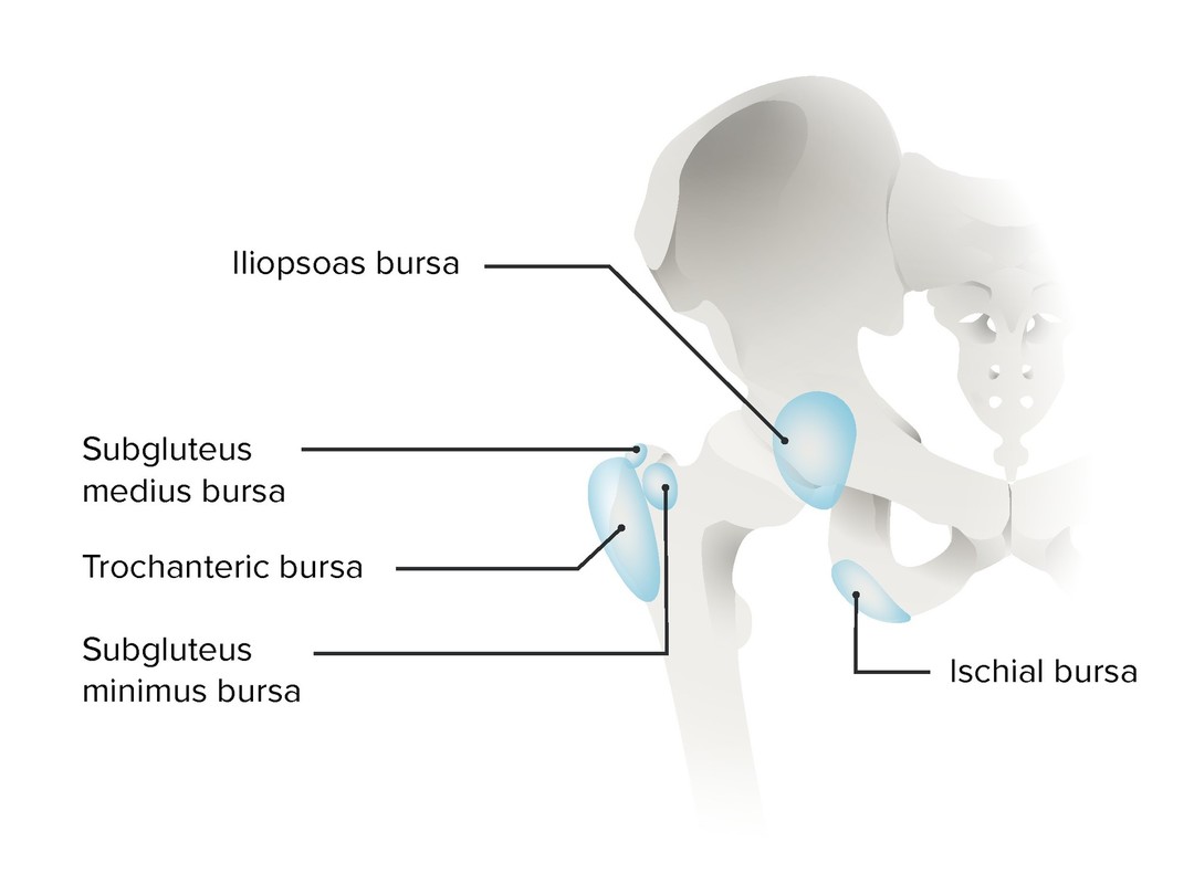
Bursae of the hip joint
Image by Lecturio.The primary flexor muscles of the hip are the iliopsoas and rectus femoris Rectus femoris Thigh: Anatomy.
| Muscle | Origin | Insertion | Innervation |
|---|---|---|---|
| Iliopsoas | Iliacus: lateral edge of the sacrum Sacrum Five fused vertebrae forming a triangle-shaped structure at the back of the pelvis. It articulates superiorly with the lumbar vertebrae, inferiorly with the coccyx, and anteriorly with the ilium of the pelvis. The sacrum strengthens and stabilizes the pelvis. Vertebral Column: Anatomy and iliac fossa | Iliopsoas tendon: lesser trochanter of the femur | Femoral nerve Femoral Nerve A nerve originating in the lumbar spinal cord (usually L2 to L4) and traveling through the lumbar plexus to provide motor innervation to extensors of the thigh and sensory innervation to parts of the thigh, lower leg, and foot, and to the hip and knee joints. Femoral Region and Hernias: Anatomy (L2–L4) |
| Psoas major Psoas major Posterior Abdominal Wall: Anatomy: transverse processes of vertebrae T12–L5 | Lumbar plexus (L1–L3) | ||
| Psoas minor Psoas minor Posterior Abdominal Wall: Anatomy: vertebral bodies of T12–L1 | Iliopubic ramus | Anterior ramus of nerve L1 | |
| Rectus femoris Rectus femoris Thigh: Anatomy (quadriceps) | Anterior-inferior iliac spine Spine The human spine, or vertebral column, is the most important anatomical and functional axis of the human body. It consists of 7 cervical vertebrae, 12 thoracic vertebrae, and 5 lumbar vertebrae and is limited cranially by the skull and caudally by the sacrum. Vertebral Column: Anatomy, superior rim of the femoral acetabulum | Base of the patella Patella The flat, triangular bone situated at the anterior part of the knee. Knee Joint: Anatomy via the quadriceps tendon Quadriceps tendon Knee Joint: Anatomy | Femoral nerve Femoral Nerve A nerve originating in the lumbar spinal cord (usually L2 to L4) and traveling through the lumbar plexus to provide motor innervation to extensors of the thigh and sensory innervation to parts of the thigh, lower leg, and foot, and to the hip and knee joints. Femoral Region and Hernias: Anatomy |
| Tensor fasciae latae Tensor fasciae latae Gluteal Region: Anatomy | Anterior superior iliac spine Anterior Superior Iliac Spine Chronic Apophyseal Injury | Iliotibial tract Iliotibial tract Thigh: Anatomy | Superior gluteal nerve Superior gluteal nerve Gluteal Region: Anatomy (L4–L5) |
| Sartorius Sartorius Thigh: Anatomy | Anterior superior iliac spine Anterior Superior Iliac Spine Chronic Apophyseal Injury | Upper medial side of the tibia Tibia The second longest bone of the skeleton. It is located on the medial side of the lower leg, articulating with the fibula laterally, the talus distally, and the femur proximally. Knee Joint: Anatomy | Femoral nerve Femoral Nerve A nerve originating in the lumbar spinal cord (usually L2 to L4) and traveling through the lumbar plexus to provide motor innervation to extensors of the thigh and sensory innervation to parts of the thigh, lower leg, and foot, and to the hip and knee joints. Femoral Region and Hernias: Anatomy (L2–L3) |
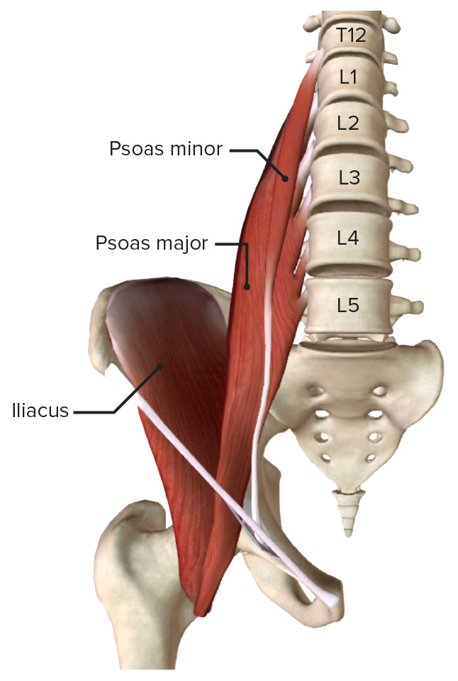
Iliopsoas muscle
Image by BioDigital, edited by Lecturio.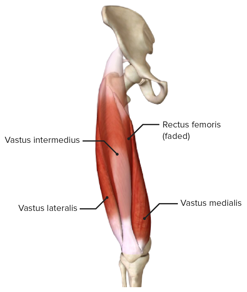
Rectus femoris muscle (quadriceps)
Image by BioDigital, edited by Lecturio.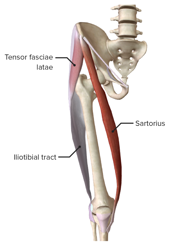
Tensor fasciae latae and sartorius muscles
Image by BioDigital, edited by Lecturio.
Superficial muscles of the anterior compartment of the thigh, featuring the main flexors of the hip:
iliopsoas, tensor fasciae latae, sartorius, and rectus femoris muscles
The primary extensor muscle of the hip is the gluteus maximus Gluteus maximus Gluteal Region: Anatomy, assisted by the biceps femoris Biceps femoris Thigh: Anatomy, semitendinosus Semitendinosus Thigh: Anatomy, and semimembranosus Semimembranosus Thigh: Anatomy muscles.
| Muscle | Origin | Insertion | Innervation |
|---|---|---|---|
| Gluteus maximus Gluteus maximus Gluteal Region: Anatomy | Ilium, sacrum Sacrum Five fused vertebrae forming a triangle-shaped structure at the back of the pelvis. It articulates superiorly with the lumbar vertebrae, inferiorly with the coccyx, and anteriorly with the ilium of the pelvis. The sacrum strengthens and stabilizes the pelvis. Vertebral Column: Anatomy, coccyx Coccyx The last bone in the vertebral column in tailless primates considered to be a vestigial tail-bone consisting of three to five fused vertebrae. Vertebral Column: Anatomy, and the sacrotuberous ligament | Gluteal tuberosity of the femur and iliotibial band Iliotibial band Thigh: Anatomy | Inferior gluteal nerve Inferior gluteal nerve Gluteal Region: Anatomy (L4– S1 S1 Heart Sounds) |
| Biceps femoris Biceps femoris Thigh: Anatomy |
|
|
|
| Semitendinosus Semitendinosus Thigh: Anatomy | Ischial tuberosity Ischial Tuberosity Chronic Apophyseal Injury | Superomedial surface of the tibia Tibia The second longest bone of the skeleton. It is located on the medial side of the lower leg, articulating with the fibula laterally, the talus distally, and the femur proximally. Knee Joint: Anatomy | Tibial nerve Tibial Nerve The medial terminal branch of the sciatic nerve. The tibial nerve fibers originate in lumbar and sacral spinal segments (L4 to S2). They supply motor and sensory innervation to parts of the calf and foot. Popliteal Fossa: Anatomy (L5– S2 S2 Heart Sounds) |
| Semimembranosus Semimembranosus Thigh: Anatomy | Medial condyle of the tibia Tibia The second longest bone of the skeleton. It is located on the medial side of the lower leg, articulating with the fibula laterally, the talus distally, and the femur proximally. Knee Joint: Anatomy |
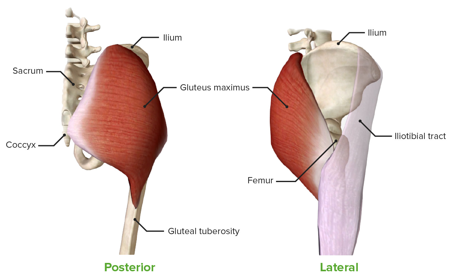
Gluteus maximus muscle: featuring its origin and insertion in posterior and lateral views
Image by BioDigital, edited by Lecturio.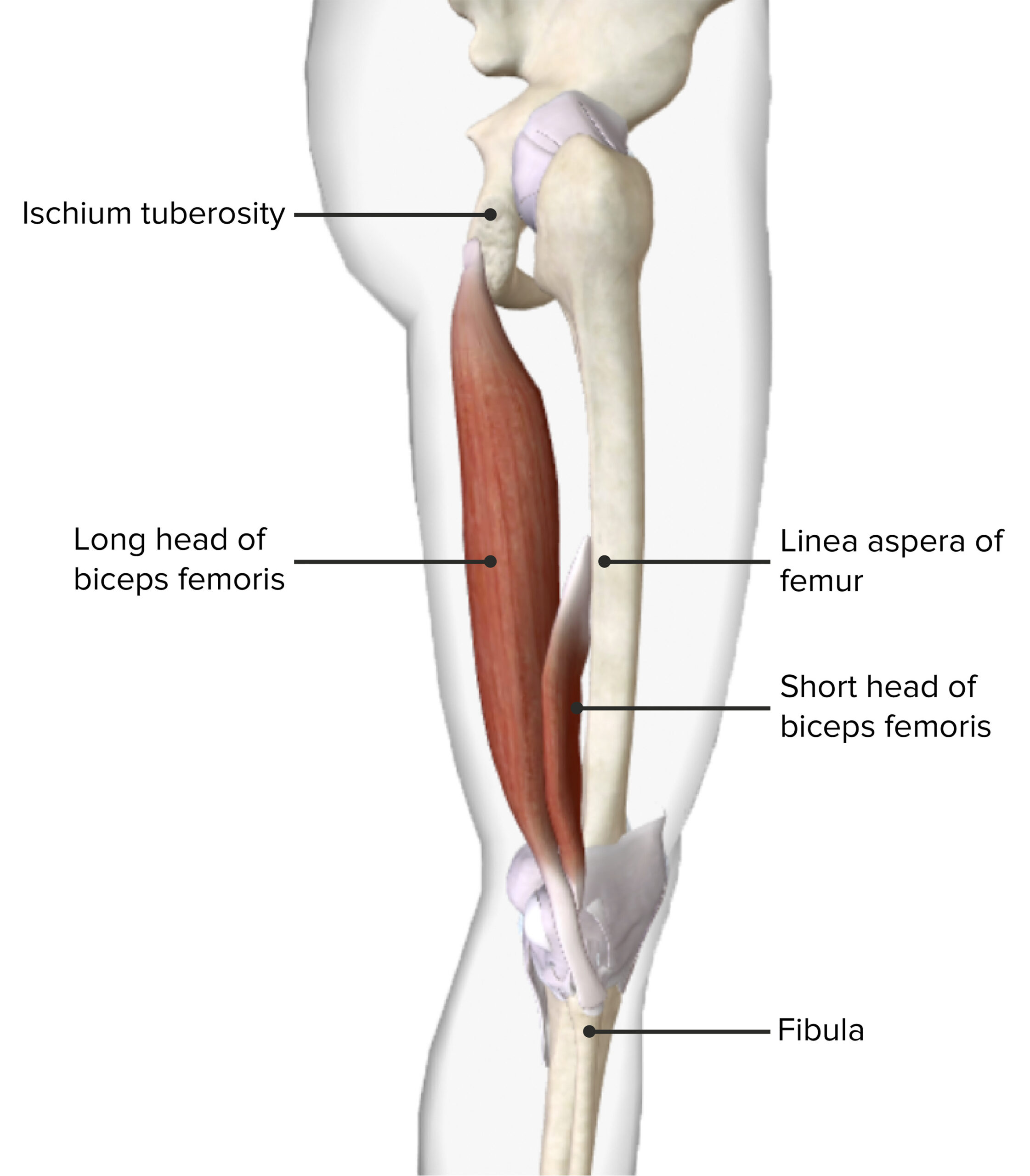
Lateral view of the thigh featuring the origin and insertion of the biceps femoris muscle
Image by BioDigital, edited by Lecturio.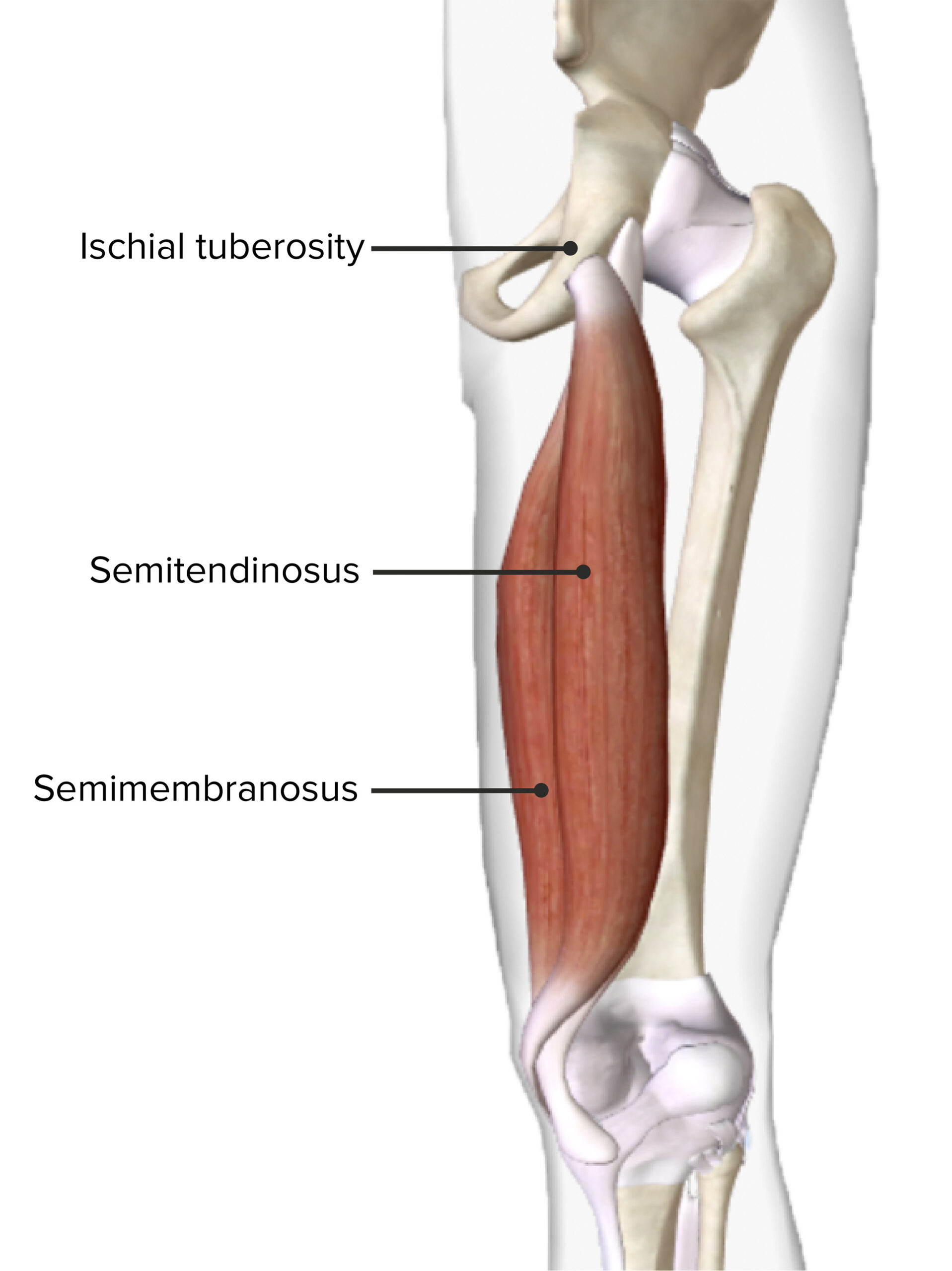
Posterior view of the right thigh featuring the origin and insertion of the semitendinosus and semimembranosus muscles
Image by BioDigital, edited by Lecturio.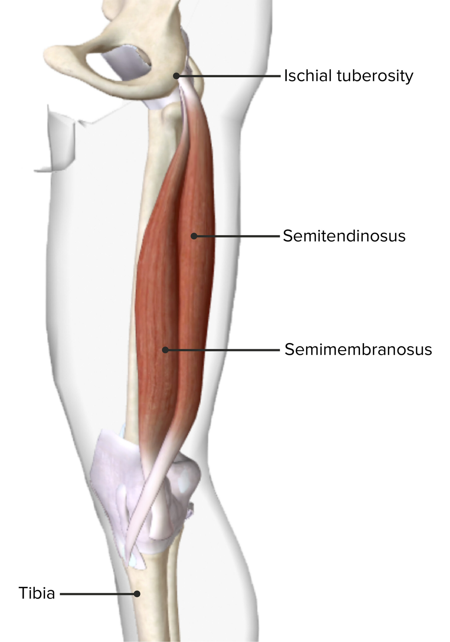
Medial view of the right thigh featuring the origin and insertion of the semitendinosus and semimembranosus muscles
Image by BioDigital, edited by Lecturio.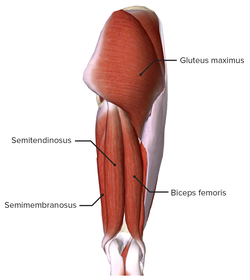
Posterior view of the thigh, featuring the main extensors of the hip: the semitendinosus, semimembranosus, and biceps femoris muscles
Image by BioDigital, edited by Lecturio.| Muscle | Origin | Insertion | Innervation |
|---|---|---|---|
| Gluteus medius Gluteus medius Gluteal Region: Anatomy | Outer surface of the ilium, between the iliac crest, and the anterior and posterior gluteal lines | Greater trochanter | Superior gluteal nerve Superior gluteal nerve Gluteal Region: Anatomy (L4– S1 S1 Heart Sounds) |
| Gluteus minimus Gluteus minimus Gluteal Region: Anatomy | Outer surface of the ilium, between the anterior and posterior gluteal lines | ||
| Piriformis Piriformis Vagina, Vulva, and Pelvic Floor: Anatomy | Anterior surface of the sacrum Sacrum Five fused vertebrae forming a triangle-shaped structure at the back of the pelvis. It articulates superiorly with the lumbar vertebrae, inferiorly with the coccyx, and anteriorly with the ilium of the pelvis. The sacrum strengthens and stabilizes the pelvis. Vertebral Column: Anatomy and sacrotuberous ligament | Nerve to the piriformis Piriformis Vagina, Vulva, and Pelvic Floor: Anatomy (L5– S2 S2 Heart Sounds) | |
| Tensor fascia Fascia Layers of connective tissue of variable thickness. The superficial fascia is found immediately below the skin; the deep fascia invests muscles, nerves, and other organs. Cellulitis latae | Anterior superior iliac spine Anterior Superior Iliac Spine Chronic Apophyseal Injury, lip of the iliac crest | Iliotibial tract Iliotibial tract Thigh: Anatomy | Superior gluteal nerve Superior gluteal nerve Gluteal Region: Anatomy (L4– S1 S1 Heart Sounds) |

Gluteus medius muscle, featuring its origin and insertion in posterior and lateral views
Image by BioDigital, edited by Lecturio.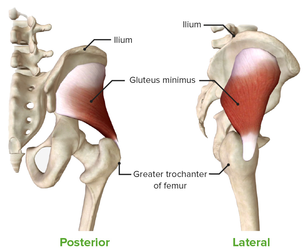
Gluteus minimus muscle, featuring its origin and insertion in posterior and lateral views
Image by BioDigital, edited by Lecturio.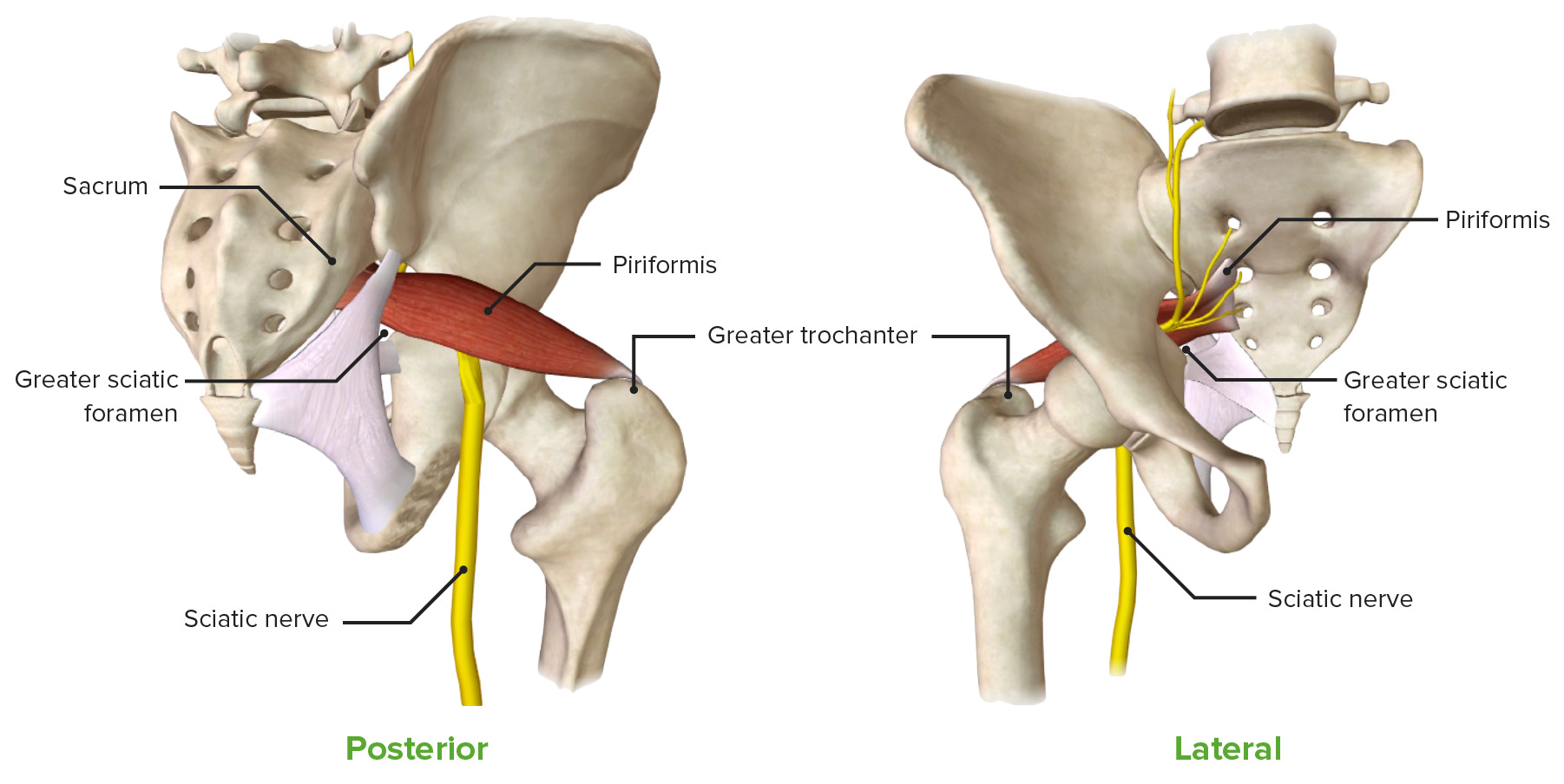
Piriformis muscle, featuring its origin and insertion in posterior and anterior views, along with its close spatial relationship to the sciatic nerve
Image by BioDigital, edited by Lecturio.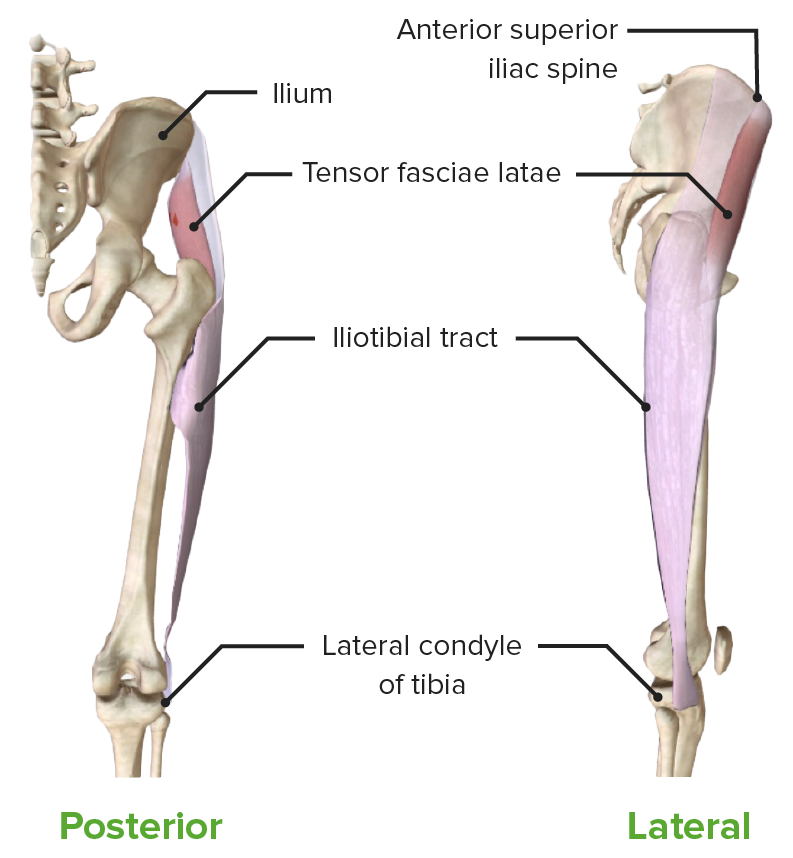
Tensor fasciae latae muscle, featuring its origin and insertion in posterior and lateral views
Image by BioDigital, edited by Lecturio.| Muscle | Origin | Insertion | Innervation |
|---|---|---|---|
| Pectineus Pectineus Thigh: Anatomy | Pectineal line of the pubis and pubic tubercle | Pectineal line of the femur | Obturator and femoral nerves (L2–L4) |
| Gracilis Gracilis Thigh: Anatomy | Inferior pubic ramus | Medial side of the tibial tuberosity Tibial tuberosity Leg: Anatomy | Obturator nerve Obturator Nerve A nerve originating in the lumbar spinal cord (L2 to L4) and traveling through the lumbar plexus to the lower extremity. The obturator nerve provides motor innervation to the adductor muscles of the thigh and cutaneous sensory innervation of the inner thigh. Thigh: Anatomy (L2–L4) |
| Adductor longus Adductor longus Thigh: Anatomy | Pubic bone Bone Bone is a compact type of hardened connective tissue composed of bone cells, membranes, an extracellular mineralized matrix, and central bone marrow. The 2 primary types of bone are compact and spongy. Bones: Structure and Types, between the crest and symphysis | Linea aspera of the femur | |
| Adductor brevis Adductor brevis Thigh: Anatomy | Body and inferior ramus of the pubis | ||
| Adductor magnus Adductor magnus Thigh: Anatomy | Ischial tuberosity Ischial Tuberosity Chronic Apophyseal Injury and inferior ramus of the pubis | Linea aspera and the adductor tubercle Adductor tubercle Knee Joint: Anatomy | Obturator and tibial nerves (L3– S2 S2 Heart Sounds) |
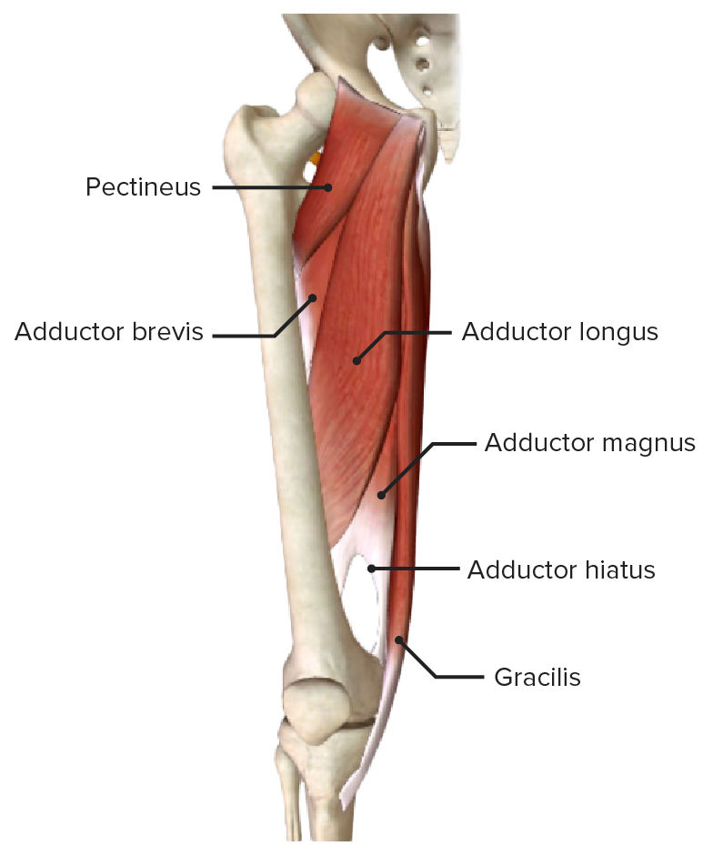
Adductor muscles of the hip joint, located in the medial thigh
Image by BioDigital, edited by Lecturio.
The conjoined tendon of the gracilis, sartorius, and semitendinosus muscles forming the pes anserinus (“goose foot”)
Image by BioDigital, edited by Lecturio.| Muscle | Origin | Insertion | Innervation |
|---|---|---|---|
| Obturator internus Obturator internus Vagina, Vulva, and Pelvic Floor: Anatomy | Obturator membrane and ischiopubic rami | Greater trochanter | Nerve to the obturator internus Obturator internus Vagina, Vulva, and Pelvic Floor: Anatomy (L5– S2 S2 Heart Sounds) |
| Obturator externus | Lateral area of the obturator foramen, outer obturator membrane, and ischiopubic ramus | Intertrochanteric fossa of the femur | Nerve to the obturator muscles (L3–L4) |
| Gemelli muscles |
|
|
|
| Quadratus femoris Quadratus femoris Gluteal Region: Anatomy | Ischial tuberosity Ischial Tuberosity Chronic Apophyseal Injury | Intertrochanteric crest of the femur | Nerve to the quadratus femoris Quadratus femoris Gluteal Region: Anatomy (L4– S1 S1 Heart Sounds) |
| Piriformis Piriformis Vagina, Vulva, and Pelvic Floor: Anatomy | Anterior surface of the sacrum Sacrum Five fused vertebrae forming a triangle-shaped structure at the back of the pelvis. It articulates superiorly with the lumbar vertebrae, inferiorly with the coccyx, and anteriorly with the ilium of the pelvis. The sacrum strengthens and stabilizes the pelvis. Vertebral Column: Anatomy and sacrotuberous ligament | Greater trochanter | Nerve to the piriformis Piriformis Vagina, Vulva, and Pelvic Floor: Anatomy (L5– S2 S2 Heart Sounds) |
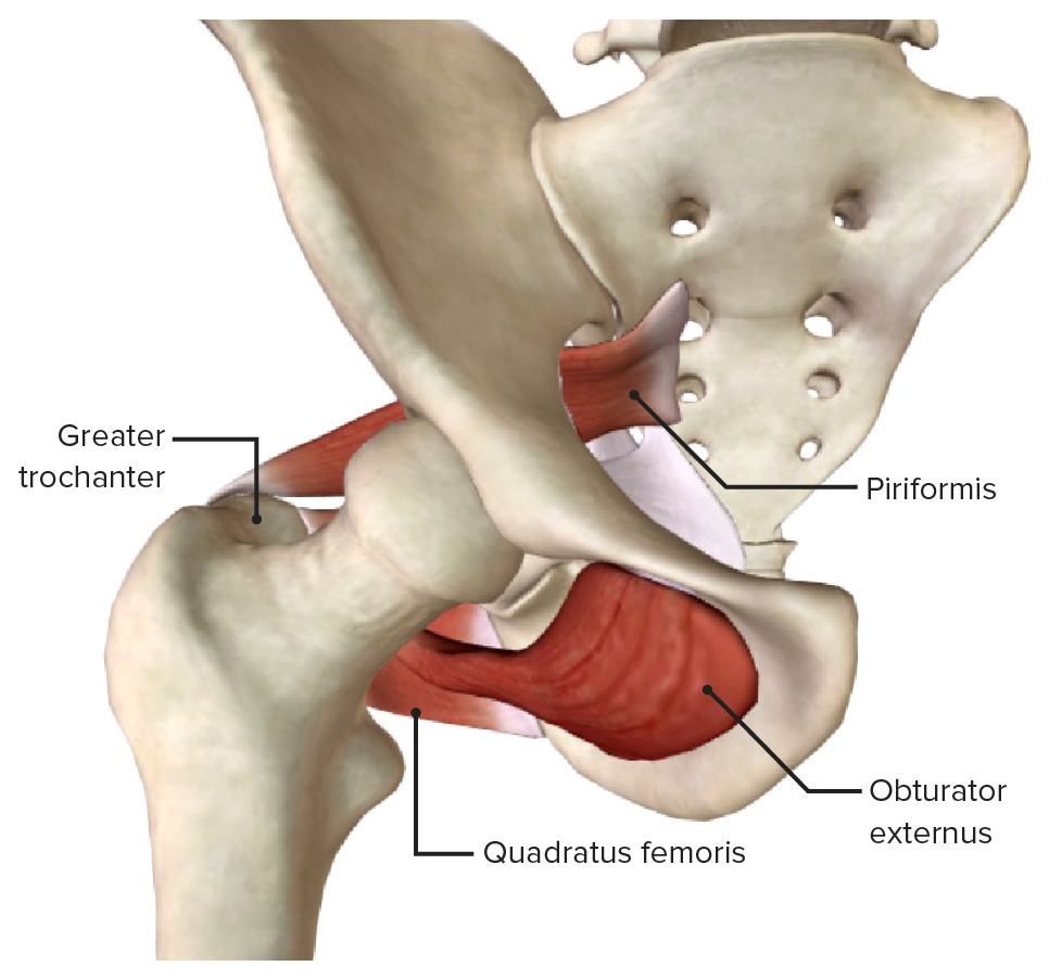
Anterior view
The external rotators of the hip, including the obturator internus, obturator externus, gemellus superior, gemellus inferior, quadratus femoris, and piriformis muscles
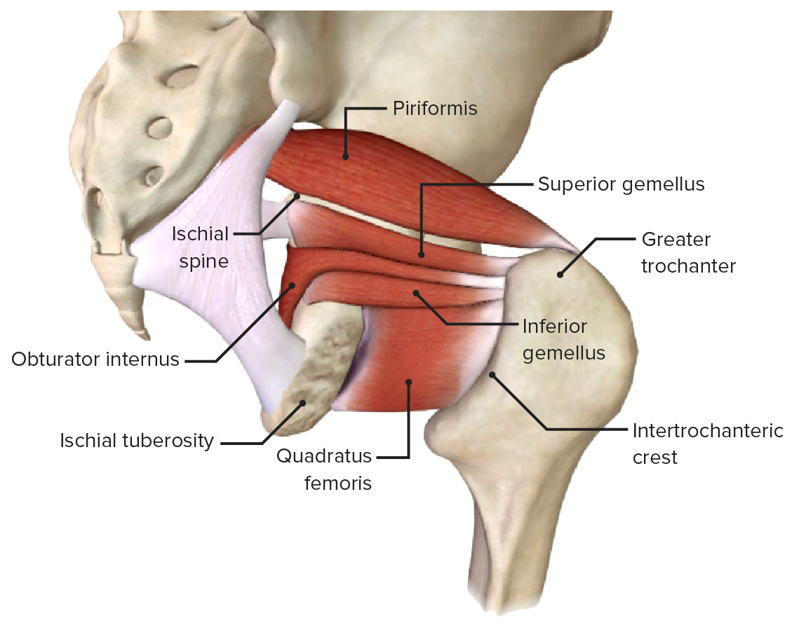
Posterolateral view
The external rotators of the hip, including the obturator internus, obturator externus, gemellus superior, gemellus inferior, quadratus femoris, and piriformis muscles
The arteries Arteries Arteries are tubular collections of cells that transport oxygenated blood and nutrients from the heart to the tissues of the body. The blood passes through the arteries in order of decreasing luminal diameter, starting in the largest artery (the aorta) and ending in the small arterioles. Arteries are classified into 3 types: large elastic arteries, medium muscular arteries, and small arteries and arterioles. Arteries: Histology that supply the hip joint originate from the common iliac artery, which bifurcates into the internal and external iliac arteries Arteries Arteries are tubular collections of cells that transport oxygenated blood and nutrients from the heart to the tissues of the body. The blood passes through the arteries in order of decreasing luminal diameter, starting in the largest artery (the aorta) and ending in the small arterioles. Arteries are classified into 3 types: large elastic arteries, medium muscular arteries, and small arteries and arterioles. Arteries: Histology.
The veins Veins Veins are tubular collections of cells, which transport deoxygenated blood and waste from the capillary beds back to the heart. Veins are classified into 3 types: small veins/venules, medium veins, and large veins. Each type contains 3 primary layers: tunica intima, tunica media, and tunica adventitia. Veins: Histology of the hip joint accompany the arteries Arteries Arteries are tubular collections of cells that transport oxygenated blood and nutrients from the heart to the tissues of the body. The blood passes through the arteries in order of decreasing luminal diameter, starting in the largest artery (the aorta) and ending in the small arterioles. Arteries are classified into 3 types: large elastic arteries, medium muscular arteries, and small arteries and arterioles. Arteries: Histology in trajectory and name.
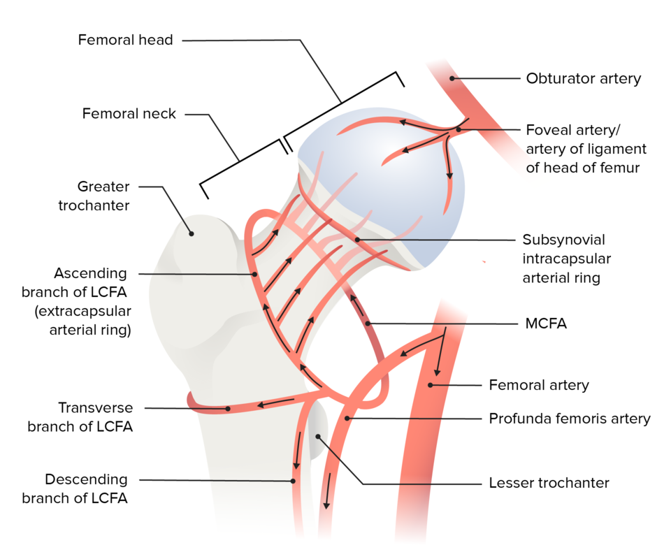
Schematic diagram of the cruciate anastomosis around the hip joint
Image by Lecturio.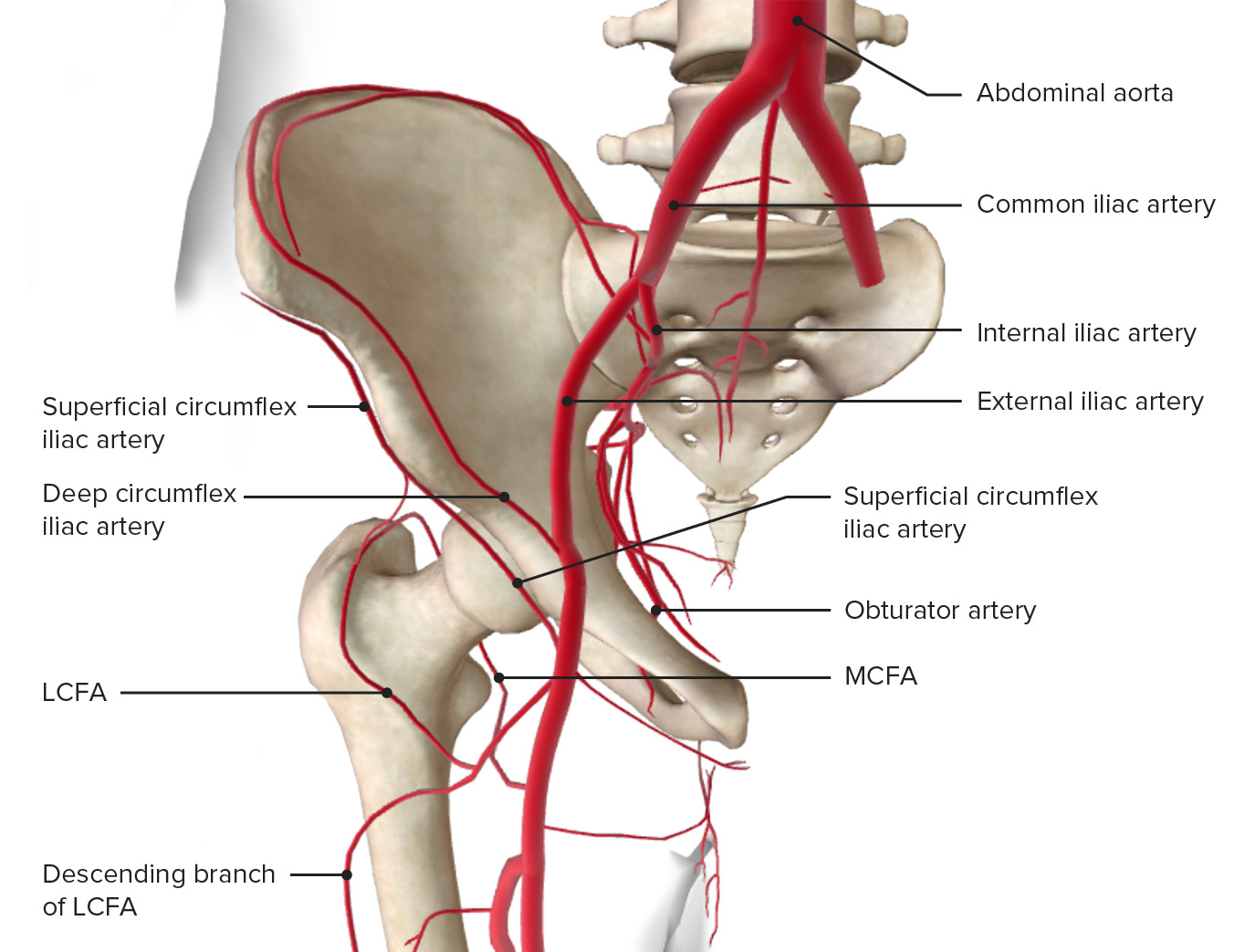
Blood supply of the hip joint
Image by BioDigital, edited by Lecturio.The femoral and obturator nerves, arising from the lumbar plexus (T12–L4), and multiple smaller nerves arising from the sacral plexus Sacral plexus Pelvis: Anatomy (L4– S4 S4 Heart Sounds), innervate the hip joint.
Clinical tip: The knee joint Knee joint The knee joint is made up of the articulations between the femur, tibia, and patella bones, and is one of the largest and most complex joints of the human body. The knee is classified as a synovial hinge joint, which primarily allows for flexion and extension with a more limited degree of translation and rotation. Knee Joint: Anatomy is also innervated by the femoral, obturator, and sciatic nerves, explaining the pain Pain An unpleasant sensation induced by noxious stimuli which are detected by nerve endings of nociceptive neurons. Pain: Types and Pathways referral patterns from the knee to the hip.
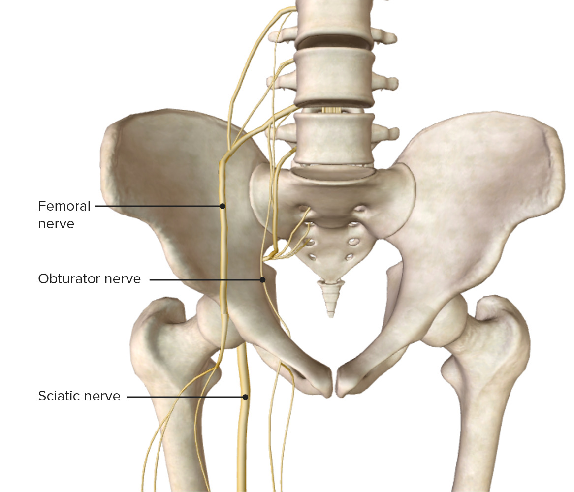
Anterior view of the pelvis and hip joint, featuring the innervation of the hip
Image by BioDigital, edited by Lecturio.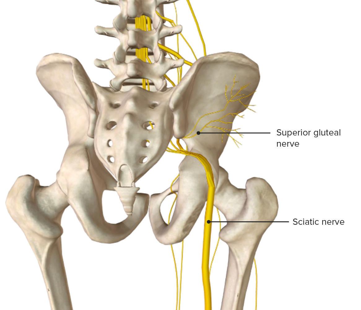
Posterior view of the pelvis and hip joint, featuring the innervation of the hip
Image by BioDigital, edited by Lecturio.The following are clinically relevant to the hip joint and region: