Achieve Mastery of Medical Concepts

Study for medical school and boards with Lecturio

Disseminated Intravascular Coagulation

Disseminated intravascular coagulation (DIC) is a condition characterized by systemic bodywide activation of the coagulation cascade Coagulation cascade The coagulation cascade is a series of reactions that ultimately generates a strong, cross-linked fibrin clot. Hemostasis. This cascade results in both widespread microvascular thrombi contributing to multiple organ dysfunction and consumption of clotting factors and platelets Platelets Platelets are small cell fragments involved in hemostasis. Thrombopoiesis takes place primarily in the bone marrow through a series of cell differentiation and is influenced by several cytokines. Platelets are formed after fragmentation of the megakaryocyte cytoplasm. Platelets: Histology, leading to hemorrhage. DIC is always triggered by another (often serious) condition, including severe sepsis Severe Sepsis Sepsis in Children, trauma, malignancy Malignancy Hemothorax, or obstetric complications. Acute DIC often presents dramatically, with rapid onset of spontaneous bleeding from multiple locations throughout the body simultaneously. A chronic form of DIC also exists, typically in patients Patients Individuals participating in the health care system for the purpose of receiving therapeutic, diagnostic, or preventive procedures. Clinician–Patient Relationship with late-stage malignancies. Management involves stabilizing the patient, replacing consumed platelets Platelets Platelets are small cell fragments involved in hemostasis. Thrombopoiesis takes place primarily in the bone marrow through a series of cell differentiation and is influenced by several cytokines. Platelets are formed after fragmentation of the megakaryocyte cytoplasm. Platelets: Histology and coagulation factors Coagulation factors Endogenous substances, usually proteins, that are involved in the blood coagulation process. Hemostasis, and treating the triggering condition.

Last updated: Nov 2, 2022

Editorial responsibility: Stanley Oiseth, Lindsay Jones, Evelin Maza

Overview

Definition

Disseminated intravascular coagulation (DIC) is a condition characterized by systemic bodywide activation of the coagulation cascade Coagulation cascade The coagulation cascade is a series of reactions that ultimately generates a strong, cross-linked fibrin clot. Hemostasis that results in both:

  • Widespread microvascular thrombi contributing to multiple organ dysfunction
  • Consumption of clotting factors and platelets Platelets Platelets are small cell fragments involved in hemostasis. Thrombopoiesis takes place primarily in the bone marrow through a series of cell differentiation and is influenced by several cytokines. Platelets are formed after fragmentation of the megakaryocyte cytoplasm. Platelets: Histology leading to hemorrhage

Epidemiology

  • Incidence Incidence The number of new cases of a given disease during a given period in a specified population. It also is used for the rate at which new events occur in a defined population. It is differentiated from prevalence, which refers to all cases in the population at a given time. Measures of Disease Frequency rates:
    • 1% of all hospitalized patients Patients Individuals participating in the health care system for the purpose of receiving therapeutic, diagnostic, or preventive procedures. Clinician–Patient Relationship in tertiary care centers
    • 30%–50% of septic patients Patients Individuals participating in the health care system for the purpose of receiving therapeutic, diagnostic, or preventive procedures. Clinician–Patient Relationship
    • Approximately 40% of severe trauma patients Patients Individuals participating in the health care system for the purpose of receiving therapeutic, diagnostic, or preventive procedures. Clinician–Patient Relationship
    • 20% of pregnant patients Patients Individuals participating in the health care system for the purpose of receiving therapeutic, diagnostic, or preventive procedures. Clinician–Patient Relationship with HELLP syndrome HELLP syndrome A severe form of preeclampsia with Hemolysis (with: LDH > 600 IU/L, ↑ Bilirubin, Schistocytes on blood smear, Anemia), ↑ Liver enzymes (with AST and/or AST > 2 times upper limit of normal), ↓ Platelet count, and thrombocytopenia (< 100,000). Hypertensive Pregnancy Disorders
    • 66% of pregnant patients Patients Individuals participating in the health care system for the purpose of receiving therapeutic, diagnostic, or preventive procedures. Clinician–Patient Relationship with amniotic fluid embolism Amniotic Fluid Embolism Blocking of maternal circulation by amniotic fluid that is forced into uterine veins by strong uterine contraction near the end of pregnancy. It is characterized by the sudden onset of severe respiratory distress and hypotension that can lead to maternal death. Complications during Childbirth
  • Gender Gender Gender Dysphoria bias Bias Epidemiological studies are designed to evaluate a hypothesized relationship between an exposure and an outcome; however, the existence and/or magnitude of these relationships may be erroneously affected by the design and execution of the study itself or by conscious or unconscious errors perpetrated by the investigators or the subjects. These systematic errors are called biases. Types of Biases: none, equal incidence Incidence The number of new cases of a given disease during a given period in a specified population. It also is used for the rate at which new events occur in a defined population. It is differentiated from prevalence, which refers to all cases in the population at a given time. Measures of Disease Frequency between sexes
  • Age bias Bias Epidemiological studies are designed to evaluate a hypothesized relationship between an exposure and an outcome; however, the existence and/or magnitude of these relationships may be erroneously affected by the design and execution of the study itself or by conscious or unconscious errors perpetrated by the investigators or the subjects. These systematic errors are called biases. Types of Biases: none, may develop at any age
  • Ethnic bias Bias Epidemiological studies are designed to evaluate a hypothesized relationship between an exposure and an outcome; however, the existence and/or magnitude of these relationships may be erroneously affected by the design and execution of the study itself or by conscious or unconscious errors perpetrated by the investigators or the subjects. These systematic errors are called biases. Types of Biases: none, occurs in all races
  • Prognosis Prognosis A prediction of the probable outcome of a disease based on a individual’s condition and the usual course of the disease as seen in similar situations. Non-Hodgkin Lymphomas
    • Depends on the severity of the coagulopathy and the underlying condition that led to the disease
    • Generally poor if untreated

Classification

Acute DIC:

  • Sudden overwhelming activation of systemic coagulation and subsequent fibrinolysis
  • Occurs in the setting of an acute process:
    • Trauma
    • Sepsis Sepsis Systemic inflammatory response syndrome with a proven or suspected infectious etiology. When sepsis is associated with organ dysfunction distant from the site of infection, it is called severe sepsis. When sepsis is accompanied by hypotension despite adequate fluid infusion, it is called septic shock. Sepsis and Septic Shock
    • Pregnancy Pregnancy The status during which female mammals carry their developing young (embryos or fetuses) in utero before birth, beginning from fertilization to birth. Pregnancy: Diagnosis, Physiology, and Care complications
  • Frequently results in significant hemorrhage and end-organ damage

Chronic DIC:

  • Insidious presentation due to continuous exposure to systemic procoagulants
  • Typically occurs in late-stage malignancies
  • More likely to result in thrombotic (rather than hemorrhagic) complications

Etiology

Disseminated intravascular coagulation is always a complication of other conditions:

Table: Etiologies of disseminated intravascular coagulation
Infection
  • Bacterial:
    • Sepsis Sepsis Systemic inflammatory response syndrome with a proven or suspected infectious etiology. When sepsis is associated with organ dysfunction distant from the site of infection, it is called severe sepsis. When sepsis is accompanied by hypotension despite adequate fluid infusion, it is called septic shock. Sepsis and Septic Shock (both gram-positive Gram-Positive Penicillins and gram-negative species)
    • Escherichia coli Escherichia coli The gram-negative bacterium Escherichia coli is a key component of the human gut microbiota. Most strains of E. coli are avirulent, but occasionally they escape the GI tract, infecting the urinary tract and other sites. Less common strains of E. coli are able to cause disease within the GI tract, most commonly presenting as abdominal pain and diarrhea. Escherichia coli
    • Neisseria meningitidis Neisseria meningitidis A species of gram-negative, aerobic bacteria. It is a commensal and pathogen only of humans, and can be carried asymptomatically in the nasopharynx. When found in cerebrospinal fluid it is the causative agent of cerebrospinal meningitis. It is also found in venereal discharges and blood. There are at least 13 serogroups based on antigenic differences in the capsular polysaccharides; the ones causing most meningitis infections being a, b, c, y, and w-135. Each serogroup can be further classified by serotype, serosubtype, and immunotype. Neisseria
  • Viral:
    • HIV HIV Anti-HIV Drugs
    • Cytomegalovirus Cytomegalovirus CMV is a ubiquitous double-stranded DNA virus belonging to the Herpesviridae family. CMV infections can be transmitted in bodily fluids, such as blood, saliva, urine, semen, and breast milk. The initial infection is usually asymptomatic in the immunocompetent host, or it can present with symptoms of mononucleosis. Cytomegalovirus
    • Varicella
    • Hepatitis
  • Parasitic: malaria Malaria Malaria is an infectious parasitic disease affecting humans and other animals. Most commonly transmitted via the bite of a female Anopheles mosquito infected with microorganisms of the Plasmodium genus. Patients present with fever, chills, myalgia, headache, and diaphoresis. Plasmodium/Malaria
Inflammation Inflammation Inflammation is a complex set of responses to infection and injury involving leukocytes as the principal cellular mediators in the body’s defense against pathogenic organisms. Inflammation is also seen as a response to tissue injury in the process of wound healing. The 5 cardinal signs of inflammation are pain, heat, redness, swelling, and loss of function. Inflammation
  • Pancreatitis Pancreatitis Inflammation of the pancreas. Pancreatitis is classified as acute unless there are computed tomographic or endoscopic retrograde cholangiopancreatographic findings of chronic pancreatitis. The two most common forms of acute pancreatitis are alcoholic pancreatitis and gallstone pancreatitis. Acute Pancreatitis
  • Fulminant liver failure Liver failure Severe inability of the liver to perform its normal metabolic functions, as evidenced by severe jaundice and abnormal serum levels of ammonia; bilirubin; alkaline phosphatase; aspartate aminotransferase; lactate dehydrogenases; and albumin/globulin ratio. Autoimmune Hepatitis
Malignancy Malignancy Hemothorax
  • Acute promyelocytic leukemia Acute promyelocytic leukemia An acute myeloid leukemia in which abnormal promyelocytes predominate. It is frequently associated with disseminated intravascular coagulation. Acute Myeloid Leukemia
  • Metastatic adenocarcinomas, especially:
    • Pancreas Pancreas The pancreas lies mostly posterior to the stomach and extends across the posterior abdominal wall from the duodenum on the right to the spleen on the left. This organ has both exocrine and endocrine tissue. Pancreas: Anatomy
    • Stomach Stomach The stomach is a muscular sac in the upper left portion of the abdomen that plays a critical role in digestion. The stomach develops from the foregut and connects the esophagus with the duodenum. Structurally, the stomach is C-shaped and forms a greater and lesser curvature and is divided grossly into regions: the cardia, fundus, body, and pylorus. Stomach: Anatomy
    • Ovary
    • Prostate Prostate The prostate is a gland in the male reproductive system. The gland surrounds the bladder neck and a portion of the urethra. The prostate is an exocrine gland that produces a weakly acidic secretion, which accounts for roughly 20% of the seminal fluid.
Obstetric causes
  • Amniotic fluid embolism Amniotic Fluid Embolism Blocking of maternal circulation by amniotic fluid that is forced into uterine veins by strong uterine contraction near the end of pregnancy. It is characterized by the sudden onset of severe respiratory distress and hypotension that can lead to maternal death. Complications during Childbirth
  • Placental abruption Placental Abruption Premature separation of the normally implanted placenta from the uterus. Signs of varying degree of severity include uterine bleeding, uterine muscle hypertonia, and fetal distress or fetal death. Antepartum Hemorrhage
  • Postpartum hemorrhage Postpartum hemorrhage Postpartum hemorrhage is one of the most common and deadly obstetric complications. Since 2017, postpartum hemorrhage has been defined as blood loss greater than 1,000 mL for both cesarean and vaginal deliveries, or excessive blood loss with signs of hemodynamic instability. Postpartum Hemorrhage
  • Preeclampsia Preeclampsia A complication of pregnancy, characterized by a complex of symptoms including maternal hypertension and proteinuria with or without pathological edema. Symptoms may range between mild and severe. Pre-eclampsia usually occurs after the 20th week of gestation, but may develop before this time in the presence of trophoblastic disease. Hypertensive Pregnancy Disorders/ eclampsia Eclampsia Onset of hyperreflexia; seizures; or coma in a previously diagnosed pre-eclamptic patient (pre-eclampsia). Hypertensive Pregnancy Disorders/ HELLP syndrome HELLP syndrome A severe form of preeclampsia with Hemolysis (with: LDH > 600 IU/L, ↑ Bilirubin, Schistocytes on blood smear, Anemia), ↑ Liver enzymes (with AST and/or AST > 2 times upper limit of normal), ↓ Platelet count, and thrombocytopenia (< 100,000). Hypertensive Pregnancy Disorders
  • Retained stillbirth
  • Septic abortion Abortion Expulsion of the product of fertilization before completing the term of gestation and without deliberate interference. Spontaneous Abortion
  • Acute fatty liver Liver The liver is the largest gland in the human body. The liver is found in the superior right quadrant of the abdomen and weighs approximately 1.5 kilograms. Its main functions are detoxification, metabolism, nutrient storage (e.g., iron and vitamins), synthesis of coagulation factors, formation of bile, filtration, and storage of blood. Liver: Anatomy of pregnancy Pregnancy The status during which female mammals carry their developing young (embryos or fetuses) in utero before birth, beginning from fertilization to birth. Pregnancy: Diagnosis, Physiology, and Care
Trauma
  • Burns Burns A burn is a type of injury to the skin and deeper tissues caused by exposure to heat, electricity, chemicals, friction, or radiation. Burns are classified according to their depth as superficial (1st-degree), partial-thickness (2nd-degree), full-thickness (3rd-degree), and 4th-degree burns. Burns
  • Frostbite Frostbite Injuries due to cold weather are common among children and athletes who are involved in sports played in cold conditions. Frostbite is a direct freezing injury to the peripheral tissues and occurs when the skin temperature drops below 0°C (32°F). Common sites of frostbite include the nose, ears, fingers, and toes. Frostbite
  • Head injuries
  • Snake Snake Limbless reptiles of the suborder serpentes. Snakebites bites
Hematologic and vascular causes

Pathophysiology

In DIC, coagulation and subsequent fibrinolysis are activated systemically, which is abnormal and leads to microthrombi throughout the vasculature. These microthrombi consume large amounts of platelets Platelets Platelets are small cell fragments involved in hemostasis. Thrombopoiesis takes place primarily in the bone marrow through a series of cell differentiation and is influenced by several cytokines. Platelets are formed after fragmentation of the megakaryocyte cytoplasm. Platelets: Histology and coagulation factors Coagulation factors Endogenous substances, usually proteins, that are involved in the blood coagulation process. Hemostasis, leading to a severe coagulopathy and clinical bleeding.

Normal activation of coagulation

  • Platelets Platelets Platelets are small cell fragments involved in hemostasis. Thrombopoiesis takes place primarily in the bone marrow through a series of cell differentiation and is influenced by several cytokines. Platelets are formed after fragmentation of the megakaryocyte cytoplasm. Platelets: Histology bind BIND Hyperbilirubinemia of the Newborn exposed von Willebrand factor von Willebrand factor A high-molecular-weight plasma protein, produced by endothelial cells and megakaryocytes, that is part of the factor VIII/von Willebrand factor complex. The von Willebrand factor has receptors for collagen, platelets, and ristocetin activity as well as the immunologically distinct antigenic determinants. It functions in adhesion of platelets to collagen and hemostatic plug formation. The prolonged bleeding time in von Willebrand diseases is due to the deficiency of this factor. Hemostasis (VWF) on injured endothelial cells:
    • Adherent platelets Platelets Platelets are small cell fragments involved in hemostasis. Thrombopoiesis takes place primarily in the bone marrow through a series of cell differentiation and is influenced by several cytokines. Platelets are formed after fragmentation of the megakaryocyte cytoplasm. Platelets: Histology activate and express glycoprotein (Gp) IIb/IIIa receptors Receptors Receptors are proteins located either on the surface of or within a cell that can bind to signaling molecules known as ligands (e.g., hormones) and cause some type of response within the cell. Receptors.
    • GpIIb/IIIa receptors Receptors Receptors are proteins located either on the surface of or within a cell that can bind to signaling molecules known as ligands (e.g., hormones) and cause some type of response within the cell. Receptors bind BIND Hyperbilirubinemia of the Newborn fibrinogen Fibrinogen Plasma glycoprotein clotted by thrombin, composed of a dimer of three non-identical pairs of polypeptide chains (alpha, beta, gamma) held together by disulfide bonds. Fibrinogen clotting is a sol-gel change involving complex molecular arrangements: whereas fibrinogen is cleaved by thrombin to form polypeptides a and b, the proteolytic action of other enzymes yields different fibrinogen degradation products. Hemostasis.
    • Fibrinogen Fibrinogen Plasma glycoprotein clotted by thrombin, composed of a dimer of three non-identical pairs of polypeptide chains (alpha, beta, gamma) held together by disulfide bonds. Fibrinogen clotting is a sol-gel change involving complex molecular arrangements: whereas fibrinogen is cleaved by thrombin to form polypeptides a and b, the proteolytic action of other enzymes yields different fibrinogen degradation products. Hemostasis can bind BIND Hyperbilirubinemia of the Newborn one platelet on each side → platelet aggregation Platelet aggregation The attachment of platelets to one another. This clumping together can be induced by a number of agents (e.g., thrombin; collagen) and is part of the mechanism leading to the formation of a thrombus. Hemostasis/formation of the platelet plug Platelet plug Hemostasis
    • Activated platelets Platelets Platelets are small cell fragments involved in hemostasis. Thrombopoiesis takes place primarily in the bone marrow through a series of cell differentiation and is influenced by several cytokines. Platelets are formed after fragmentation of the megakaryocyte cytoplasm. Platelets: Histology secrete substances that augment further platelet aggregation Platelet aggregation The attachment of platelets to one another. This clumping together can be induced by a number of agents (e.g., thrombin; collagen) and is part of the mechanism leading to the formation of a thrombus. Hemostasis and the coagulation cascade Coagulation cascade The coagulation cascade is a series of reactions that ultimately generates a strong, cross-linked fibrin clot. Hemostasis.
  • Tissue factor (TF) binds circulating factor VII Factor VII Heat- and storage-stable plasma protein that is activated by tissue thromboplastin to form factor viia in the extrinsic pathway of blood coagulation. The activated form then catalyzes the activation of factor X to factor Xa. Hemostasis and activates the extrinsic pathway Extrinsic pathway The extrinsic pathway is the primary physiological mechanism by which clotting is initiated Hemostasis (initial activation of the coagulation cascade Coagulation cascade The coagulation cascade is a series of reactions that ultimately generates a strong, cross-linked fibrin clot. Hemostasis):
    • Membrane-bound glycoprotein expressed on injured endothelial cells
    • Restricts activation of the cascade primarily to local sites of injury
  • The initial thrombin Thrombin An enzyme formed from prothrombin that converts fibrinogen to fibrin. Hemostasis produced activates factor XI in the intrinsic pathway Intrinsic pathway The intrinsic pathway is mainly responsible for the amplification of factor X activation Hemostasis.
    • The intrinsic pathway Intrinsic pathway The intrinsic pathway is mainly responsible for the amplification of factor X activation Hemostasis significantly amplifies the local response.
    • Coagulation factors Coagulation factors Endogenous substances, usually proteins, that are involved in the blood coagulation process. Hemostasis form multicomponent enzyme complexes, which are also bound to membrane surfaces.
    • Keeps coagulation localized
  • Thrombin Thrombin An enzyme formed from prothrombin that converts fibrinogen to fibrin. Hemostasis converts fibrinogen Fibrinogen Plasma glycoprotein clotted by thrombin, composed of a dimer of three non-identical pairs of polypeptide chains (alpha, beta, gamma) held together by disulfide bonds. Fibrinogen clotting is a sol-gel change involving complex molecular arrangements: whereas fibrinogen is cleaved by thrombin to form polypeptides a and b, the proteolytic action of other enzymes yields different fibrinogen degradation products. Hemostasis to fibrin Fibrin A protein derived from fibrinogen in the presence of thrombin, which forms part of the blood clot. Rapidly Progressive Glomerulonephritis, forming a stable clot.
  • Plasmin Plasmin A product of the lysis of plasminogen (profibrinolysin) by plasminogen activators. It is composed of two polypeptide chains, light (b) and heavy (a), with a molecular weight of 75, 000. It is the major proteolytic enzyme involved in blood clot retraction or the lysis of fibrin and quickly inactivated by antiplasmins. Hemostasis is produced to break down fibrin Fibrin A protein derived from fibrinogen in the presence of thrombin, which forms part of the blood clot. Rapidly Progressive Glomerulonephritis.
  • Fibrin Fibrin A protein derived from fibrinogen in the presence of thrombin, which forms part of the blood clot. Rapidly Progressive Glomerulonephritis degradation products (FDPs):
    • Have anticoagulant properties
    • Are cleared by the liver Liver The liver is the largest gland in the human body. The liver is found in the superior right quadrant of the abdomen and weighs approximately 1.5 kilograms. Its main functions are detoxification, metabolism, nutrient storage (e.g., iron and vitamins), synthesis of coagulation factors, formation of bile, filtration, and storage of blood. Liver: Anatomy

Activation of systemic coagulation (abnormal)

  • Abnormal systemic activation of the coagulation cascade Coagulation cascade The coagulation cascade is a series of reactions that ultimately generates a strong, cross-linked fibrin clot. Hemostasis may occur owing to:
    • An overwhelming systemic inflammatory response (e.g., severe sepsis Severe Sepsis Sepsis in Children)
    • Sudden exposure of the blood to procoagulants, including:
      • TF
      • Thromboplastin
      • VWF
    • Examples of procoagulant exposure:
      • Sepsis Sepsis Systemic inflammatory response syndrome with a proven or suspected infectious etiology. When sepsis is associated with organ dysfunction distant from the site of infection, it is called severe sepsis. When sepsis is accompanied by hypotension despite adequate fluid infusion, it is called septic shock. Sepsis and Septic Shock: Bacterial endotoxins Endotoxins Toxins closely associated with the living cytoplasm or cell wall of certain microorganisms, which do not readily diffuse into the culture medium, but are released upon lysis of the cells. Bacteriology can trigger Trigger The type of signal that initiates the inspiratory phase by the ventilator Invasive Mechanical Ventilation release Release Release of a virus from the host cell following virus assembly and maturation. Egress can occur by host cell lysis, exocytosis, or budding through the plasma membrane. Virology of circulating TF.
      • Malignancy Malignancy Hemothorax: Some cancers can produce circulating TF.
      • Trauma: Significant vascular endothelial damage results in ↑ amounts of TF.
      • Obstetric: The placenta Placenta A highly vascularized mammalian fetal-maternal organ and major site of transport of oxygen, nutrients, and fetal waste products. It includes a fetal portion (chorionic villi) derived from trophoblasts and a maternal portion (decidua) derived from the uterine endometrium. The placenta produces an array of steroid, protein and peptide hormones (placental hormones). Placenta, Umbilical Cord, and Amniotic Cavity can release Release Release of a virus from the host cell following virus assembly and maturation. Egress can occur by host cell lysis, exocytosis, or budding through the plasma membrane. Virology a proteolytic enzyme capable of activating factor X Factor X Storage-stable glycoprotein blood coagulation factor that can be activated to factor Xa by both the intrinsic and extrinsic pathways. A deficiency of factor X, sometimes called stuart-prower factor deficiency, may lead to a systemic coagulation disorder. Hemostasis.
  • Effects of systemic coagulation:
    • With systemic activation of the coagulation cascade Coagulation cascade The coagulation cascade is a series of reactions that ultimately generates a strong, cross-linked fibrin clot. Hemostasis, thrombi are formed throughout the vasculature.
    • Thrombi can affect both small and large vessels.
    • Extensive, systemwide coagulation consumes large amounts of platelets Platelets Platelets are small cell fragments involved in hemostasis. Thrombopoiesis takes place primarily in the bone marrow through a series of cell differentiation and is influenced by several cytokines. Platelets are formed after fragmentation of the megakaryocyte cytoplasm. Platelets: Histology and coagulation factors Coagulation factors Endogenous substances, usually proteins, that are involved in the blood coagulation process. Hemostasis.
  • Fibrinolysis:
    • Activated in conjunction with coagulation cascade Coagulation cascade The coagulation cascade is a series of reactions that ultimately generates a strong, cross-linked fibrin clot. Hemostasis
    • Plasmin Plasmin A product of the lysis of plasminogen (profibrinolysin) by plasminogen activators. It is composed of two polypeptide chains, light (b) and heavy (a), with a molecular weight of 75, 000. It is the major proteolytic enzyme involved in blood clot retraction or the lysis of fibrin and quickly inactivated by antiplasmins. Hemostasis cleaves fibrin Fibrin A protein derived from fibrinogen in the presence of thrombin, which forms part of the blood clot. Rapidly Progressive Glomerulonephritis polymers and produces FDPs.
    • ↑ Levels of FDPs interfere with new fibrin Fibrin A protein derived from fibrinogen in the presence of thrombin, which forms part of the blood clot. Rapidly Progressive Glomerulonephritis clot formation and platelet aggregation Platelet aggregation The attachment of platelets to one another. This clumping together can be induced by a number of agents (e.g., thrombin; collagen) and is part of the mechanism leading to the formation of a thrombus. Hemostasis → impaired hemostasis Hemostasis Hemostasis refers to the innate, stepwise body processes that occur following vessel injury, resulting in clot formation and cessation of bleeding. Hemostasis occurs in 2 phases, namely, primary and secondary. Primary hemostasis involves forming a plug that stops the bleeding temporarily. Secondary hemostasis involves the activation of the coagulation cascade. Hemostasis
  • End-organ damage occurs by:
  • Organ dysfunction can include:
    • Change in mental status
    • Acute kidney injury Acute Kidney Injury Acute kidney injury refers to sudden and often reversible loss of renal function, which develops over days or weeks. Azotemia refers to elevated levels of nitrogen-containing substances in the blood that accompany AKI, which include BUN and creatinine. Acute Kidney Injury
    • Respiratory dysfunction
    • Hepatic dysfunction
    • Cardiac tamponade Tamponade Pericardial effusion, usually of rapid onset, exceeding ventricular filling pressures and causing collapse of the heart with a markedly reduced cardiac output. Pericarditis
    • Hemothorax Hemothorax A hemothorax is a collection of blood in the pleural cavity. Hemothorax most commonly occurs due to damage to the intercostal arteries or from a lung laceration following chest trauma. Hemothorax can also occur as a complication of disease, or hemothorax may be spontaneous or iatrogenic. Hemothorax
    • Intracerebral hemorrhage Intracerebral Hemorrhage Intracerebral hemorrhage (ICH) refers to a spontaneous or traumatic bleed into the brain parenchyma and is the 2nd-most common cause of cerebrovascular accidents (CVAs), commonly known as stroke, after ischemic CVAs. Intracerebral Hemorrhage
    • Gangrene Gangrene Death and putrefaction of tissue usually due to a loss of blood supply. Small Bowel Obstruction and loss of digits

Acute versus chronic disseminated intravascular coagulation

Acute DIC is decompensated DIC:

  • Rapid systemic activation of coagulation 
  • Leads to rapid consumption of platelets Platelets Platelets are small cell fragments involved in hemostasis. Thrombopoiesis takes place primarily in the bone marrow through a series of cell differentiation and is influenced by several cytokines. Platelets are formed after fragmentation of the megakaryocyte cytoplasm. Platelets: Histology and coagulation factors Coagulation factors Endogenous substances, usually proteins, that are involved in the blood coagulation process. Hemostasis 
  • Consumption significantly outpaces production.
  • ↑ Number of thrombi → ↑ fibrinolysis → ↑ FDPs → pathologically prevents new clots and platelet aggregation Platelet aggregation The attachment of platelets to one another. This clumping together can be induced by a number of agents (e.g., thrombin; collagen) and is part of the mechanism leading to the formation of a thrombus. Hemostasis
  • End result: severe clinical bleeding/hemorrhage plus thrombotic end-organ damage

Chronic DIC is compensated DIC:

  • Typically occurs in patients Patients Individuals participating in the health care system for the purpose of receiving therapeutic, diagnostic, or preventive procedures. Clinician–Patient Relationship with:
  • Develops when blood is continuously (or intermittently) exposed to smaller amounts of procoagulants
  • Compensation Compensation Respiratory Acidosis mechanisms:
    • New production of platelets Platelets Platelets are small cell fragments involved in hemostasis. Thrombopoiesis takes place primarily in the bone marrow through a series of cell differentiation and is influenced by several cytokines. Platelets are formed after fragmentation of the megakaryocyte cytoplasm. Platelets: Histology and coagulation factors Coagulation factors Endogenous substances, usually proteins, that are involved in the blood coagulation process. Hemostasis
    • Liver Liver The liver is the largest gland in the human body. The liver is found in the superior right quadrant of the abdomen and weighs approximately 1.5 kilograms. Its main functions are detoxification, metabolism, nutrient storage (e.g., iron and vitamins), synthesis of coagulation factors, formation of bile, filtration, and storage of blood. Liver: Anatomy is able to clear FDPs.
  • Compensation Compensation Respiratory Acidosis mechanisms are able to balance the ongoing consumption of products and production of FDPs.
  • Thrombotic complications are more common than bleeding.

Clinical Presentation

Acute disseminated intravascular coagulation

Acute DIC typically presents rapidly and dramatically, with acute and severe bleeding from multiple sites simultaneously throughout the body. Other manifestations are related to thrombotic end-organ dysfunction.

Table: Clinical manifestations of acute disseminated intravascular coagulation
Vital signs
  • Hypovolemic shock Hypovolemic Shock Types of Shock
  • Hypotension Hypotension Hypotension is defined as low blood pressure, specifically < 90/60 mm Hg, and is most commonly a physiologic response. Hypotension may be mild, serious, or life threatening, depending on the cause. Hypotension
  • Tachycardia Tachycardia Abnormally rapid heartbeat, usually with a heart rate above 100 beats per minute for adults. Tachycardia accompanied by disturbance in the cardiac depolarization (cardiac arrhythmia) is called tachyarrhythmia. Sepsis in Children
Skin Skin The skin, also referred to as the integumentary system, is the largest organ of the body. The skin is primarily composed of the epidermis (outer layer) and dermis (deep layer). The epidermis is primarily composed of keratinocytes that undergo rapid turnover, while the dermis contains dense layers of connective tissue. Skin: Structure and Functions
Mucosal
Renal
  • Hematuria Hematuria Presence of blood in the urine. Renal Cell Carcinoma
  • Oliguria Oliguria Decreased urine output that is below the normal range. Oliguria can be defined as urine output of less than or equal to 0. 5 or 1 ml/kg/hr depending on the age. Renal Potassium Regulation
  • Azotemia Azotemia A biochemical abnormality referring to an elevation of blood urea nitrogen and creatinine. Azotemia can be produced by kidney diseases or other extrarenal disorders. When azotemia becomes associated with a constellation of clinical signs, it is termed uremia. Acute Kidney Injury
Pulmonary
  • Hemoptysis Hemoptysis Hemoptysis is defined as the expectoration of blood originating in the lower respiratory tract. Hemoptysis is a consequence of another disease process and can be classified as either life threatening or non-life threatening. Hemoptysis can result in significant morbidity and mortality due to both drowning (reduced gas exchange as the lungs fill with blood) and hemorrhagic shock. Hemoptysis
  • Hemothorax Hemothorax A hemothorax is a collection of blood in the pleural cavity. Hemothorax most commonly occurs due to damage to the intercostal arteries or from a lung laceration following chest trauma. Hemothorax can also occur as a complication of disease, or hemothorax may be spontaneous or iatrogenic. Hemothorax
  • ARDS
GI
Neurologic
Skin manifestation of dic - purpura

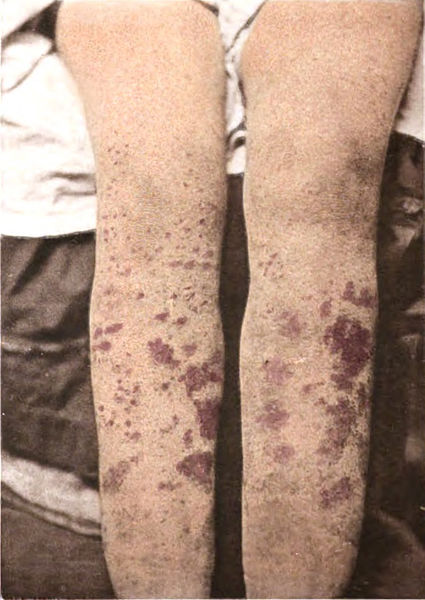
Skin manifestation of DIC:
Note the petechiae and purpura.

Image: “ Purpura” by George Henry Fox. License: Public Domain

Chronic disseminated intravascular coagulation

  • Frequently asymptomatic
  • Thrombotic sequelae are more common than bleeding complications.
  • Manifestations can include:

Diagnosis

The diagnosis is made on the basis of clinical history and laboratory findings, though no single test is diagnostic for DIC. Laboratory tests also aid in management.

Laboratory tests

In DIC, coagulation factors Coagulation factors Endogenous substances, usually proteins, that are involved in the blood coagulation process. Hemostasis and platelets Platelets Platelets are small cell fragments involved in hemostasis. Thrombopoiesis takes place primarily in the bone marrow through a series of cell differentiation and is influenced by several cytokines. Platelets are formed after fragmentation of the megakaryocyte cytoplasm. Platelets: Histology are rapidly consumed in the formation of systemwide microthrombi. Lab findings in DIC include:

  • CBC:
    • Mild-to-moderate thrombocytopenia Thrombocytopenia Thrombocytopenia occurs when the platelet count is < 150,000 per microliter. The normal range for platelets is usually 150,000-450,000/µL of whole blood. Thrombocytopenia can be a result of decreased production, increased destruction, or splenic sequestration of platelets. Patients are often asymptomatic until platelet counts are < 50,000/µL. Thrombocytopenia ( platelets Platelets Platelets are small cell fragments involved in hemostasis. Thrombopoiesis takes place primarily in the bone marrow through a series of cell differentiation and is influenced by several cytokines. Platelets are formed after fragmentation of the megakaryocyte cytoplasm. Platelets: Histology are consumed)
    • Anemia Anemia Anemia is a condition in which individuals have low Hb levels, which can arise from various causes. Anemia is accompanied by a reduced number of RBCs and may manifest with fatigue, shortness of breath, pallor, and weakness. Subtypes are classified by the size of RBCs, chronicity, and etiology. Anemia: Overview and Types (due to hemorrhage and hemolysis)
  • Peripheral blood smear Peripheral Blood Smear Anemia: Overview and Types:
  • PT: 
    • ↑ (prolonged) in DIC
    • Interpretation: Factors in the extrinsic and/or common pathways have been consumed.
  • aPTT: 
    • ↑ (prolonged) in DIC
    • Interpretation: Factors in the intrinsic and/or common pathways have been consumed.
  • Fibrinogen Fibrinogen Plasma glycoprotein clotted by thrombin, composed of a dimer of three non-identical pairs of polypeptide chains (alpha, beta, gamma) held together by disulfide bonds. Fibrinogen clotting is a sol-gel change involving complex molecular arrangements: whereas fibrinogen is cleaved by thrombin to form polypeptides a and b, the proteolytic action of other enzymes yields different fibrinogen degradation products. Hemostasis:
    • Usually ↓ owing to consumption
    • May be normal despite significant consumption if fibrinogen Fibrinogen Plasma glycoprotein clotted by thrombin, composed of a dimer of three non-identical pairs of polypeptide chains (alpha, beta, gamma) held together by disulfide bonds. Fibrinogen clotting is a sol-gel change involving complex molecular arrangements: whereas fibrinogen is cleaved by thrombin to form polypeptides a and b, the proteolytic action of other enzymes yields different fibrinogen degradation products. Hemostasis was originally ↑, which occurs in patients Patients Individuals participating in the health care system for the purpose of receiving therapeutic, diagnostic, or preventive procedures. Clinician–Patient Relationship with:
      • Sepsis Sepsis Systemic inflammatory response syndrome with a proven or suspected infectious etiology. When sepsis is associated with organ dysfunction distant from the site of infection, it is called severe sepsis. When sepsis is accompanied by hypotension despite adequate fluid infusion, it is called septic shock. Sepsis and Septic Shock
      • Malignancy Malignancy Hemothorax
      • Other inflammatory conditions
  • D-dimer D-dimer Deep Vein Thrombosis
    • ↑↑ in DIC
    • An FDP, which indicates recent or ongoing thrombosis Thrombosis Formation and development of a thrombus or blood clot in the blood vessel. Epidemic Typhus
    • Interpretation: A negative test for D-dimer D-dimer Deep Vein Thrombosis essentially rules out DIC.
  • Findings in chronic DIC:
    • Thrombocytopenia Thrombocytopenia Thrombocytopenia occurs when the platelet count is < 150,000 per microliter. The normal range for platelets is usually 150,000-450,000/µL of whole blood. Thrombocytopenia can be a result of decreased production, increased destruction, or splenic sequestration of platelets. Patients are often asymptomatic until platelet counts are < 50,000/µL. Thrombocytopenia (mild)
    • ↓ or normal PT and aPTT
    • D-dimer D-dimer Deep Vein Thrombosis
Schistocytes as diagnostic of dic

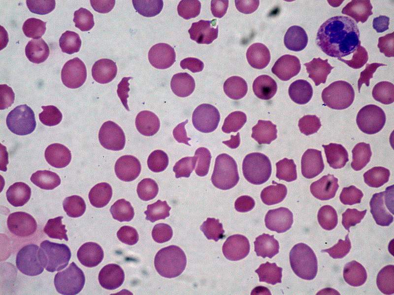
Peripheral blood smear showing schistocytes, which are indicative of intravascular hemolysis

Image: “Disseminated carcinomatosis with DIC” by Ed Uthman, MD. License: Public Domain

Imaging

No specific imaging study confirms the diagnosis of DIC. Specific imaging methods may be indicated to assist in:

  • The diagnosis of the underlying etiology (e.g., abdominal imaging for suspected malignancy Malignancy Hemothorax)
  • Evaluation of end-organ damage (e.g., head CT in patients Patients Individuals participating in the health care system for the purpose of receiving therapeutic, diagnostic, or preventive procedures. Clinician–Patient Relationship with evidence of stroke)

Management

The most important principles in management are to treat the underlying cause and stabilize the patient.

Acute disseminated intravascular coagulation

  • Emergency care: airway Airway ABCDE Assessment, breathing, circulation Circulation The movement of the blood as it is pumped through the cardiovascular system. ABCDE Assessment:
  • Product Product A molecule created by the enzymatic reaction. Basics of Enzymes replacement: coagulation factors Coagulation factors Endogenous substances, usually proteins, that are involved in the blood coagulation process. Hemostasis and platelets Platelets Platelets are small cell fragments involved in hemostasis. Thrombopoiesis takes place primarily in the bone marrow through a series of cell differentiation and is influenced by several cytokines. Platelets are formed after fragmentation of the megakaryocyte cytoplasm. Platelets: Histology (see Blood component replacement below)
  • Treat the underlying cause: treatment may include:
    • Antimicrobial agents
    • Delivery of fetus in obstetric cases

Blood component replacement

  • Platelets Platelets Platelets are small cell fragments involved in hemostasis. Thrombopoiesis takes place primarily in the bone marrow through a series of cell differentiation and is influenced by several cytokines. Platelets are formed after fragmentation of the megakaryocyte cytoplasm. Platelets: Histology:
    • 1 pool of 5–6 units should ↑ platelet counts Platelet counts The number of platelets per unit volume in a sample of venous blood. Coagulation Studies by approximately 30,000/µL 
    • Indications:
      • Active bleeding
      • Platelets Platelets Platelets are small cell fragments involved in hemostasis. Thrombopoiesis takes place primarily in the bone marrow through a series of cell differentiation and is influenced by several cytokines. Platelets are formed after fragmentation of the megakaryocyte cytoplasm. Platelets: Histology < 50,000/µL with need for urgent surgery
      • Platelets Platelets Platelets are small cell fragments involved in hemostasis. Thrombopoiesis takes place primarily in the bone marrow through a series of cell differentiation and is influenced by several cytokines. Platelets are formed after fragmentation of the megakaryocyte cytoplasm. Platelets: Histology < 10,000/µL
  • Fresh frozen plasma Fresh Frozen Plasma Transfusion Products ( FFP FFP Transfusion Products)
    • 1 unit = 200–300 mL
    • Contains all soluble plasma Plasma The residual portion of blood that is left after removal of blood cells by centrifugation without prior blood coagulation. Transfusion Products proteins Proteins Linear polypeptides that are synthesized on ribosomes and may be further modified, crosslinked, cleaved, or assembled into complex proteins with several subunits. The specific sequence of amino acids determines the shape the polypeptide will take, during protein folding, and the function of the protein. Energy Homeostasis and clotting factors 
    • Indications: 
      • Serious bleeding and ↑ PT or aPTT 
      • Serious bleeding and ↓ fibrinogen Fibrinogen Plasma glycoprotein clotted by thrombin, composed of a dimer of three non-identical pairs of polypeptide chains (alpha, beta, gamma) held together by disulfide bonds. Fibrinogen clotting is a sol-gel change involving complex molecular arrangements: whereas fibrinogen is cleaved by thrombin to form polypeptides a and b, the proteolytic action of other enzymes yields different fibrinogen degradation products. Hemostasis
  • Cryoprecipitate Cryoprecipitate Transfusion Products
    • 1 unit = 10–20 mL 
    • Contains:
      • Fibrinogen Fibrinogen Plasma glycoprotein clotted by thrombin, composed of a dimer of three non-identical pairs of polypeptide chains (alpha, beta, gamma) held together by disulfide bonds. Fibrinogen clotting is a sol-gel change involving complex molecular arrangements: whereas fibrinogen is cleaved by thrombin to form polypeptides a and b, the proteolytic action of other enzymes yields different fibrinogen degradation products. Hemostasis
      • Factors VIII and XIII
      • VWF
    • Indication: fibrinogen Fibrinogen Plasma glycoprotein clotted by thrombin, composed of a dimer of three non-identical pairs of polypeptide chains (alpha, beta, gamma) held together by disulfide bonds. Fibrinogen clotting is a sol-gel change involving complex molecular arrangements: whereas fibrinogen is cleaved by thrombin to form polypeptides a and b, the proteolytic action of other enzymes yields different fibrinogen degradation products. Hemostasis < 100 mg/dL
    • Used to help boost fibrinogen Fibrinogen Plasma glycoprotein clotted by thrombin, composed of a dimer of three non-identical pairs of polypeptide chains (alpha, beta, gamma) held together by disulfide bonds. Fibrinogen clotting is a sol-gel change involving complex molecular arrangements: whereas fibrinogen is cleaved by thrombin to form polypeptides a and b, the proteolytic action of other enzymes yields different fibrinogen degradation products. Hemostasis without significant extra volume

Chronic disseminated intravascular coagulation

Differential Diagnosis

  • Cirrhosis Cirrhosis Cirrhosis is a late stage of hepatic parenchymal necrosis and scarring (fibrosis) most commonly due to hepatitis C infection and alcoholic liver disease. Patients may present with jaundice, ascites, and hepatosplenomegaly. Cirrhosis can also cause complications such as hepatic encephalopathy, portal hypertension, portal vein thrombosis, and hepatorenal syndrome. Cirrhosis: The liver Liver The liver is the largest gland in the human body. The liver is found in the superior right quadrant of the abdomen and weighs approximately 1.5 kilograms. Its main functions are detoxification, metabolism, nutrient storage (e.g., iron and vitamins), synthesis of coagulation factors, formation of bile, filtration, and storage of blood. Liver: Anatomy is the primary site of synthesis Synthesis Polymerase Chain Reaction (PCR) for a majority of the clotting factors. In addition to impaired synthesis Synthesis Polymerase Chain Reaction (PCR) of clotting factors, cirrhosis Cirrhosis Cirrhosis is a late stage of hepatic parenchymal necrosis and scarring (fibrosis) most commonly due to hepatitis C infection and alcoholic liver disease. Patients may present with jaundice, ascites, and hepatosplenomegaly. Cirrhosis can also cause complications such as hepatic encephalopathy, portal hypertension, portal vein thrombosis, and hepatorenal syndrome. Cirrhosis also may independently result in thrombocytopenia Thrombocytopenia Thrombocytopenia occurs when the platelet count is < 150,000 per microliter. The normal range for platelets is usually 150,000-450,000/µL of whole blood. Thrombocytopenia can be a result of decreased production, increased destruction, or splenic sequestration of platelets. Patients are often asymptomatic until platelet counts are < 50,000/µL. Thrombocytopenia owing to splenic sequestration Splenic sequestration Severe Congenital Neutropenia of platelets Platelets Platelets are small cell fragments involved in hemostasis. Thrombopoiesis takes place primarily in the bone marrow through a series of cell differentiation and is influenced by several cytokines. Platelets are formed after fragmentation of the megakaryocyte cytoplasm. Platelets: Histology and decreased thrombopoietin Thrombopoietin A humoral factor that stimulates the production of thrombocytes (blood platelets). Thrombopoietin stimulates the proliferation of bone marrow megakaryocytes and their release of blood platelets. The process is called thrombopoiesis. Platelets: Histology production. Platelets Platelets Platelets are small cell fragments involved in hemostasis. Thrombopoiesis takes place primarily in the bone marrow through a series of cell differentiation and is influenced by several cytokines. Platelets are formed after fragmentation of the megakaryocyte cytoplasm. Platelets: Histology themselves may also be dysfunctional. Like DIC, cirrhosis Cirrhosis Cirrhosis is a late stage of hepatic parenchymal necrosis and scarring (fibrosis) most commonly due to hepatitis C infection and alcoholic liver disease. Patients may present with jaundice, ascites, and hepatosplenomegaly. Cirrhosis can also cause complications such as hepatic encephalopathy, portal hypertension, portal vein thrombosis, and hepatorenal syndrome. Cirrhosis can lead to both bleeding and thrombosis Thrombosis Formation and development of a thrombus or blood clot in the blood vessel. Epidemic Typhus. A history of liver Liver The liver is the largest gland in the human body. The liver is found in the superior right quadrant of the abdomen and weighs approximately 1.5 kilograms. Its main functions are detoxification, metabolism, nutrient storage (e.g., iron and vitamins), synthesis of coagulation factors, formation of bile, filtration, and storage of blood. Liver: Anatomy injury and abnormal liver function tests Liver function tests Liver function tests, also known as hepatic function panels, are one of the most commonly performed screening blood tests. Such tests are also used to detect, evaluate, and monitor acute and chronic liver diseases. Liver Function Tests can help distinguish cirrhosis Cirrhosis Cirrhosis is a late stage of hepatic parenchymal necrosis and scarring (fibrosis) most commonly due to hepatitis C infection and alcoholic liver disease. Patients may present with jaundice, ascites, and hepatosplenomegaly. Cirrhosis can also cause complications such as hepatic encephalopathy, portal hypertension, portal vein thrombosis, and hepatorenal syndrome. Cirrhosis from DIC. Of note, fulminant liver failure Liver failure Severe inability of the liver to perform its normal metabolic functions, as evidenced by severe jaundice and abnormal serum levels of ammonia; bilirubin; alkaline phosphatase; aspartate aminotransferase; lactate dehydrogenases; and albumin/globulin ratio. Autoimmune Hepatitis may actually trigger Trigger The type of signal that initiates the inspiratory phase by the ventilator Invasive Mechanical Ventilation DIC, as well.
  • Thrombotic thrombocytopenic purpura Thrombotic thrombocytopenic purpura Thrombotic thrombocytopenic purpura (TTP) is a life-threatening condition due to either a congenital or an acquired deficiency of ADAMTS-13, a metalloproteinase that cleaves multimers of von Willebrand factor (VWF). The large multimers then aggregate excessive platelets resulting in microvascular thrombosis and an increase in consumption of platelets. Thrombotic Thrombocytopenic Purpura ( TTP TTP Thrombotic thrombocytopenic purpura (TTP) is a life-threatening condition due to either a congenital or an acquired deficiency of adamts-13, a metalloproteinase that cleaves multimers of von Willebrand factor (vWF). The large multimers then aggregate excessive platelets resulting in microvascular thrombosis and an increase in consumption of platelets. Thrombotic Thrombocytopenic Purpura): a bleeding disorder marked by a pentad of microangiopathic hemolytic anemia Microangiopathic Hemolytic Anemia Hemolytic Uremic Syndrome, thrombocytopenia Thrombocytopenia Thrombocytopenia occurs when the platelet count is < 150,000 per microliter. The normal range for platelets is usually 150,000-450,000/µL of whole blood. Thrombocytopenia can be a result of decreased production, increased destruction, or splenic sequestration of platelets. Patients are often asymptomatic until platelet counts are < 50,000/µL. Thrombocytopenia, renal failure Renal failure Conditions in which the kidneys perform below the normal level in the ability to remove wastes, concentrate urine, and maintain electrolyte balance; blood pressure; and calcium metabolism. Renal insufficiency can be classified by the degree of kidney damage (as measured by the level of proteinuria) and reduction in glomerular filtration rate. Crush Syndrome, neurologic symptoms, and fever Fever Fever is defined as a measured body temperature of at least 38°C (100.4°F). Fever is caused by circulating endogenous and/or exogenous pyrogens that increase levels of prostaglandin E2 in the hypothalamus. Fever is commonly associated with chills, rigors, sweating, and flushing of the skin. Fever: Thrombotic thrombocytopenic purpura Thrombotic thrombocytopenic purpura Thrombotic thrombocytopenic purpura (TTP) is a life-threatening condition due to either a congenital or an acquired deficiency of ADAMTS-13, a metalloproteinase that cleaves multimers of von Willebrand factor (VWF). The large multimers then aggregate excessive platelets resulting in microvascular thrombosis and an increase in consumption of platelets. Thrombotic Thrombocytopenic Purpura occurs because of a congenital Congenital Chorioretinitis or acquired deficiency of ADAMTS13, which is a metalloproteinase that cleaves VWF. Deficiencies of ADAMTS13 result in large VWF multimers that increase platelet aggregation Platelet aggregation The attachment of platelets to one another. This clumping together can be induced by a number of agents (e.g., thrombin; collagen) and is part of the mechanism leading to the formation of a thrombus. Hemostasis, leading to microvascular thrombosis Thrombosis Formation and development of a thrombus or blood clot in the blood vessel. Epidemic Typhus and consumption of platelets Platelets Platelets are small cell fragments involved in hemostasis. Thrombopoiesis takes place primarily in the bone marrow through a series of cell differentiation and is influenced by several cytokines. Platelets are formed after fragmentation of the megakaryocyte cytoplasm. Platelets: Histology. Unlike DIC, in TTP TTP Thrombotic thrombocytopenic purpura (TTP) is a life-threatening condition due to either a congenital or an acquired deficiency of adamts-13, a metalloproteinase that cleaves multimers of von Willebrand factor (vWF). The large multimers then aggregate excessive platelets resulting in microvascular thrombosis and an increase in consumption of platelets. Thrombotic Thrombocytopenic Purpura, the PT and PTT are usually normal (or only mildly elevated) and fibrinogen Fibrinogen Plasma glycoprotein clotted by thrombin, composed of a dimer of three non-identical pairs of polypeptide chains (alpha, beta, gamma) held together by disulfide bonds. Fibrinogen clotting is a sol-gel change involving complex molecular arrangements: whereas fibrinogen is cleaved by thrombin to form polypeptides a and b, the proteolytic action of other enzymes yields different fibrinogen degradation products. Hemostasis is normal or even increased.
  • Immune thrombocytopenia Thrombocytopenia Thrombocytopenia occurs when the platelet count is < 150,000 per microliter. The normal range for platelets is usually 150,000-450,000/µL of whole blood. Thrombocytopenia can be a result of decreased production, increased destruction, or splenic sequestration of platelets. Patients are often asymptomatic until platelet counts are < 50,000/µL. Thrombocytopenia ( ITP ITP Immune thrombocytopenic purpura (ITP), formerly known as idiopathic thrombocytopenic purpura, is a condition that develops secondary to immune-mediated destruction of platelets, resulting in thrombocytopenia (platelet count < 100,000/mm³). Immune thrombocytopenic purpura can be either primary or secondary due to drugs or underlying disease. Immune Thrombocytopenic Purpura): an autoimmune disorder Autoimmune Disorder Septic Arthritis characterized by anti-GpIIb/IIIa autoantibodies Autoantibodies Antibodies that react with self-antigens (autoantigens) of the organism that produced them. Blotting Techniques that lead to the destruction of platelets Platelets Platelets are small cell fragments involved in hemostasis. Thrombopoiesis takes place primarily in the bone marrow through a series of cell differentiation and is influenced by several cytokines. Platelets are formed after fragmentation of the megakaryocyte cytoplasm. Platelets: Histology: Immune thrombocytopenia Thrombocytopenia Thrombocytopenia occurs when the platelet count is < 150,000 per microliter. The normal range for platelets is usually 150,000-450,000/µL of whole blood. Thrombocytopenia can be a result of decreased production, increased destruction, or splenic sequestration of platelets. Patients are often asymptomatic until platelet counts are < 50,000/µL. Thrombocytopenia often occurs after GI or respiratory viral infections Infections Invasion of the host organism by microorganisms or their toxins or by parasites that can cause pathological conditions or diseases. Chronic Granulomatous Disease, though it can also be drug-induced. Clinically, it may present with prolonged bleeding, petechiae Petechiae Primary Skin Lesions, easy bruising Easy bruising Chédiak-Higashi Syndrome, and/or purpura. Unlike DIC, in ITP ITP Immune thrombocytopenic purpura (ITP), formerly known as idiopathic thrombocytopenic purpura, is a condition that develops secondary to immune-mediated destruction of platelets, resulting in thrombocytopenia (platelet count < 100,000/mm³). Immune thrombocytopenic purpura can be either primary or secondary due to drugs or underlying disease. Immune Thrombocytopenic Purpura, there is no coagulation defect, so the PT and PTT are normal. 

References

  1. Marcel, M. (2020). Disseminated intravascular coagulation (DIC). Retrieved March 21, 2021, from https://emedicine.medscape.com/article/199627-overview
  2. Leng, L. (2021). Disseminated intravascular coagulation (DIC) in adults: Evaluation and management. Retrieved March 21, 2021, from https://www.uptodate.com/contents/disseminated-intravascular-coagulation-dic-in-adults-evaluation-and-management
  3. Longo, D., Fauci, A., Kasper, D., Hauser, S., Jameson, J., Loscalzo, J. (2012). Harrison’s Manual of Medicine, 18th ed. McGraw-Hill Professional, pages 2188–2191.

Create your free account or log in to continue reading!

Sign up now and get free access to Lecturio with concept pages, medical videos, and questions for your medical education.

User Reviews

Details