Gastrointestinal bleeding (GIB) is a symptom of multiple diseases within the gastrointestinal (GI) tract. Gastrointestinal bleeding is designated as upper or lower based on the etiology’s location to the ligament of Treitz. It is more common to have bleeding in the upper GI tract, with peptic ulcer Peptic ulcer Peptic ulcer disease (PUD) refers to the full-thickness ulcerations of duodenal or gastric mucosa. The ulcerations form when exposure to acid and digestive enzymes overcomes mucosal defense mechanisms. The most common etiologies include Helicobacter pylori (H. pylori) infection and prolonged use of non-steroidal anti-inflammatory drugs (NSAIDs). Peptic Ulcer Disease disease being the most frequent cause. Depending on the location of the bleeding, the patient may present with hematemesis Hematemesis Vomiting of blood that is either fresh bright red, or older 'coffee-ground' in character. It generally indicates bleeding of the upper gastrointestinal tract. Mallory-Weiss Syndrome (Mallory-Weiss Tear) ( vomiting Vomiting The forcible expulsion of the contents of the stomach through the mouth. Hypokalemia blood), melena (black, tarry stool), or hematochezia (fresh blood in stools). Some patients Patients Individuals participating in the health care system for the purpose of receiving therapeutic, diagnostic, or preventive procedures. Clinician–Patient Relationship presenting with GIB can be hemodynamically unstable and require emergent stabilization and evaluation. The source of the bleed can often be located and treated with endoscopy Endoscopy Procedures of applying endoscopes for disease diagnosis and treatment. Endoscopy involves passing an optical instrument through a small incision in the skin i.e., percutaneous; or through a natural orifice and along natural body pathways such as the digestive tract; and/or through an incision in the wall of a tubular structure or organ, i.e. Transluminal, to examine or perform surgery on the interior parts of the body. Gastroesophageal Reflux Disease (GERD).
Last updated: 8 Jan, 2021
Upper gastrointestinal bleed (proximal to the ligament of Treitz):
Lower GIB (distal to the ligament of Treitz):
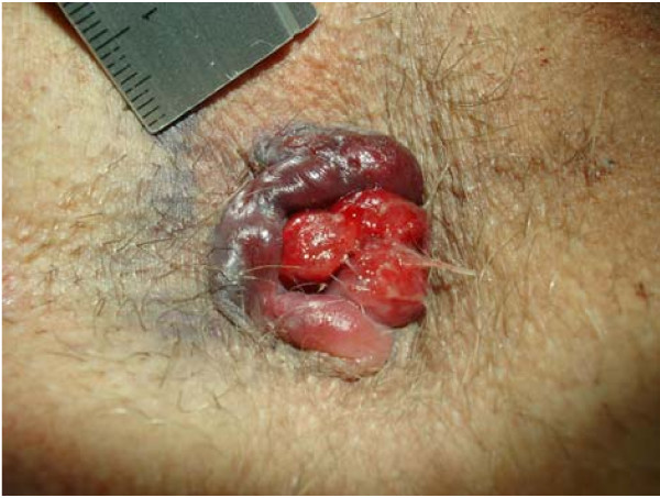
Thrombosed external hemorrhoid External hemorrhoid Hemorrhoids with prolapsing internal hemorrhoid Internal hemorrhoid Hemorrhoids
Image: “Circular perianal thrombosis Thrombosis Formation and development of a thrombus or blood clot in the blood vessel. Epidemic Typhus” by Rohde et al AL Amyloidosis; licensee BioMed Central Ltd.. License: CC BY 2.0Diagnosis and management of GIB tend to go hand Hand The hand constitutes the distal part of the upper limb and provides the fine, precise movements needed in activities of daily living. It consists of 5 metacarpal bones and 14 phalanges, as well as numerous muscles innervated by the median and ulnar nerves. Hand: Anatomy-in- hand Hand The hand constitutes the distal part of the upper limb and provides the fine, precise movements needed in activities of daily living. It consists of 5 metacarpal bones and 14 phalanges, as well as numerous muscles innervated by the median and ulnar nerves. Hand: Anatomy and will vary depending on the hemodynamic stability of the patient.
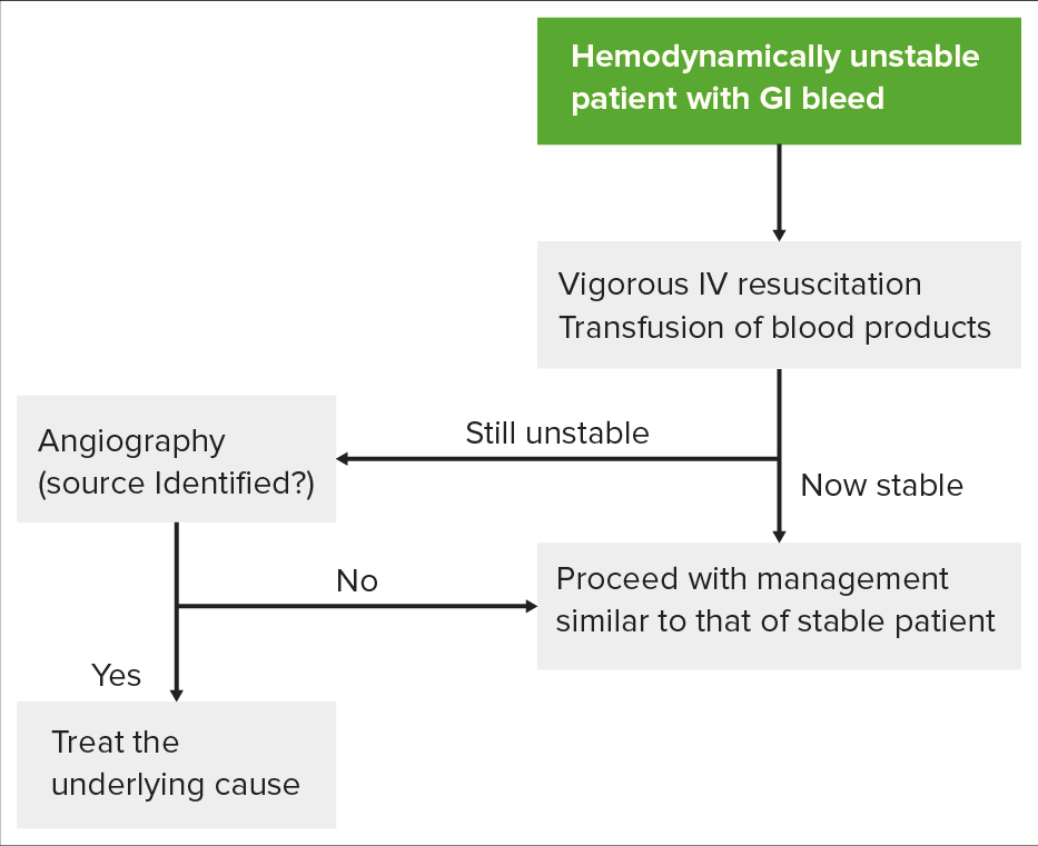
Algorithm for evaluating a hemodynamically unstable patient: The first step requires attempting to stabilize the patient before pursuing workup. The patient’s response will then determine whether the patient should go straight to angiography Angiography Radiography of blood vessels after injection of a contrast medium. Cardiac Surgery for diagnosis and treatment, or endoscopy Endoscopy Procedures of applying endoscopes for disease diagnosis and treatment. Endoscopy involves passing an optical instrument through a small incision in the skin i.e., percutaneous; or through a natural orifice and along natural body pathways such as the digestive tract; and/or through an incision in the wall of a tubular structure or organ, i.e. Transluminal, to examine or perform surgery on the interior parts of the body. Gastroesophageal Reflux Disease (GERD).
Image by Lecturio.
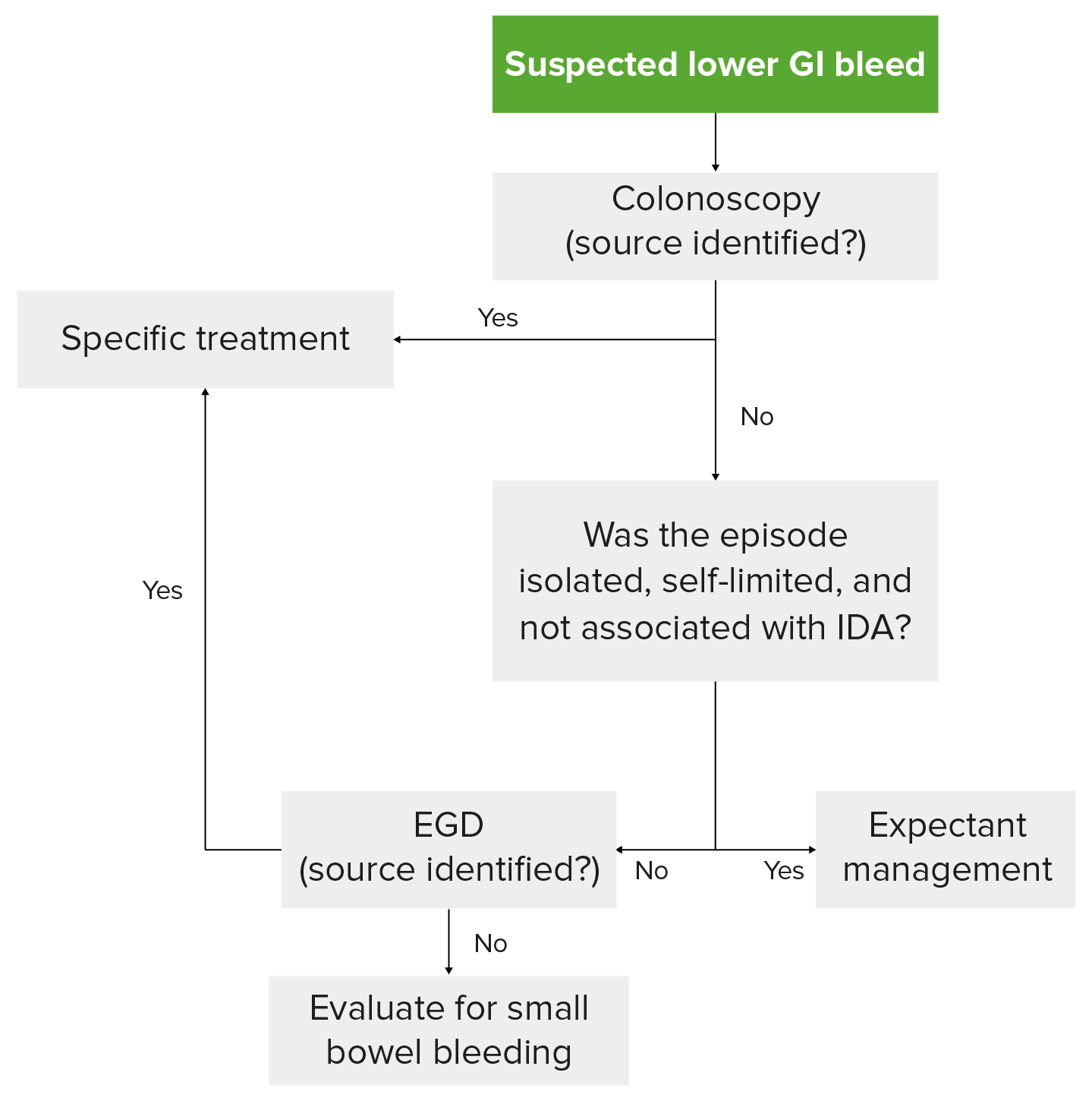
Diagnostic algorithm for LGIB in a hemodynamically stable patient: Colonoscopy Colonoscopy Endoscopic examination, therapy or surgery of the luminal surface of the colon. Colorectal Cancer Screening is the preferred 1st step. If colonoscopy Colonoscopy Endoscopic examination, therapy or surgery of the luminal surface of the colon. Colorectal Cancer Screening is unremarkable, the bleed has ceased and not recurred, and the patient does not have iron Iron A metallic element with atomic symbol fe, atomic number 26, and atomic weight 55. 85. It is an essential constituent of hemoglobins; cytochromes; and iron-binding proteins. It plays a role in cellular redox reactions and in the transport of oxygen. Trace Elements deficiency anemia Anemia Anemia is a condition in which individuals have low Hb levels, which can arise from various causes. Anemia is accompanied by a reduced number of RBCs and may manifest with fatigue, shortness of breath, pallor, and weakness. Subtypes are classified by the size of RBCs, chronicity, and etiology. Anemia: Overview and Types ( IDA IDA Iron deficiency anemia is the most common type of anemia worldwide. This form of anemia is caused by insufficient iron due to a decreased supply, an increased loss, or an increased demand. Iron deficiency anemia is seen across all ages, sexes, and socioeconomic strata; however, children, women of childbearing age, and patients from lower socioeconomic strata are at higher risk. Iron Deficiency Anemia), then expectant management is pursued.
Image by Lecturio.
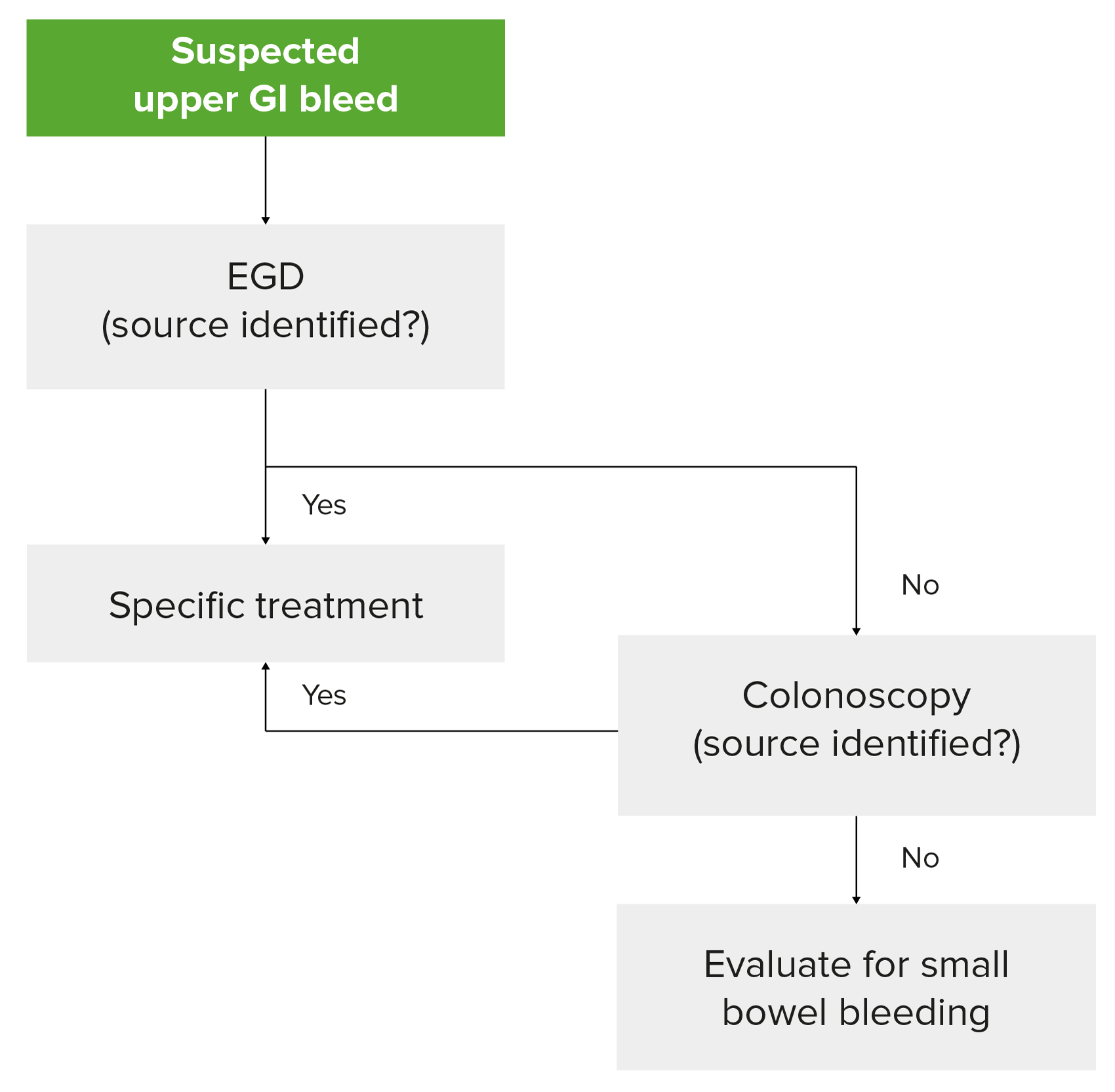
Diagnostic algorithm for UGIB in a hemodynamically stable patient: Notice that capsule Capsule An envelope of loose gel surrounding a bacterial cell which is associated with the virulence of pathogenic bacteria. Some capsules have a well-defined border, whereas others form a slime layer that trails off into the medium. Most capsules consist of relatively simple polysaccharides but there are some bacteria whose capsules are made of polypeptides. Bacteroides endoscopy Endoscopy Procedures of applying endoscopes for disease diagnosis and treatment. Endoscopy involves passing an optical instrument through a small incision in the skin i.e., percutaneous; or through a natural orifice and along natural body pathways such as the digestive tract; and/or through an incision in the wall of a tubular structure or organ, i.e. Transluminal, to examine or perform surgery on the interior parts of the body. Gastroesophageal Reflux Disease (GERD) to evaluate the small bowel Small bowel The small intestine is the longest part of the GI tract, extending from the pyloric orifice of the stomach to the ileocecal junction. The small intestine is the major organ responsible for chemical digestion and absorption of nutrients. It is divided into 3 segments: the duodenum, the jejunum, and the ileum. Small Intestine: Anatomy should be considered if EGD and colonoscopy Colonoscopy Endoscopic examination, therapy or surgery of the luminal surface of the colon. Colorectal Cancer Screening do not reveal a source.
Image by Lecturio.
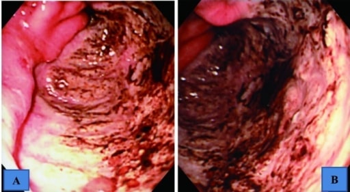
Upper GI endoscopic findings showing a giant ulcer in the
greater curvature
Greater curvature
Stomach: Anatomy of the antrum, which occupied halfway around the lumen
A: intermediate view
B: near view
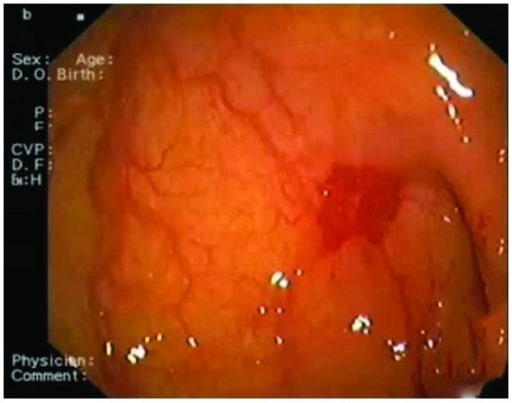
Endoscopic picture obtained from an 83-year-old woman showing the site of angiodysplasia between the 1st and the 2nd portion of the duodenum Duodenum The shortest and widest portion of the small intestine adjacent to the pylorus of the stomach. It is named for having the length equal to about the width of 12 fingers. Small Intestine: Anatomy
Image: “Angiodysplasia” by Department of Cardiology, San Raffaele Hospital, Via Olgettina 60, 20131 Milan, Italy. License: CC BY 3.0
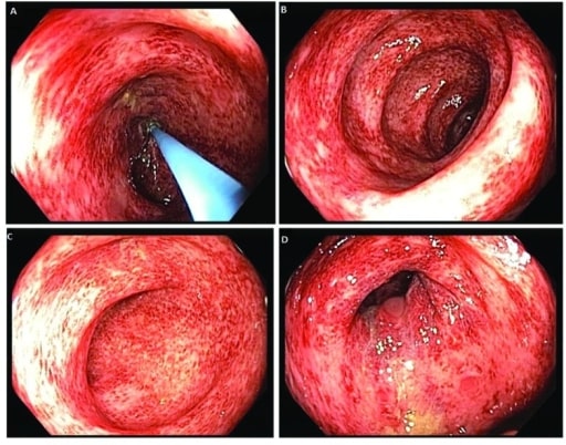
Colonoscopy Colonoscopy Endoscopic examination, therapy or surgery of the luminal surface of the colon. Colorectal Cancer Screening images revealing patchy and diffuse colitis Colitis Inflammation of the colon section of the large intestine, usually with symptoms such as diarrhea (often with blood and mucus), abdominal pain, and fever. Pseudomembranous Colitis throughout the colon Colon The large intestines constitute the last portion of the digestive system. The large intestine consists of the cecum, appendix, colon (with ascending, transverse, descending, and sigmoid segments), rectum, and anal canal. The primary function of the colon is to remove water and compact the stool prior to expulsion from the body via the rectum and anal canal. Colon, Cecum, and Appendix: Anatomy
Image: “ Colonoscopy Colonoscopy Endoscopic examination, therapy or surgery of the luminal surface of the colon. Colorectal Cancer Screening” by US National Library of Medicine. License: CC BY 2.0Assess the patient’s hemodynamics Hemodynamics The movement and the forces involved in the movement of the blood through the cardiovascular system. Vascular Resistance, Flow, and Mean Arterial Pressure and stabilize: