Syncope is a short-term loss of consciousness and loss of postural stability followed by spontaneous return of consciousness to the previous neurologic baseline without the need for resuscitationResuscitationThe restoration to life or consciousness of one apparently dead. .Neonatal Respiratory Distress Syndrome. The condition is caused by transient interruption of cerebral blood flowBlood flowBlood flow refers to the movement of a certain volume of blood through the vasculature over a given unit of time (e.g., mL per minute).Vascular Resistance, Flow, and Mean Arterial Pressure that may be benignBenignFibroadenoma or related to an underlying life-threatening condition. Syncope is not a distinct disease entity; rather, it is a symptom of another pathologic process, whether it be transient or a more established disease process. Syncope may be accompanied by other symptoms, such as light-headedness, sweating, palpitationsPalpitationsEbstein’s Anomaly, nauseaNauseaAn unpleasant sensation in the stomach usually accompanied by the urge to vomit. Common causes are early pregnancy, sea and motion sickness, emotional stress, intense pain, food poisoning, and various enteroviruses.Antiemetics, feeling warm or cold, and visual blurring. Workup includes a detailed history and physical examination, electrocardiographyElectrocardiographyRecording of the moment-to-moment electromotive forces of the heart as projected onto various sites on the body's surface, delineated as a scalar function of time. The recording is monitored by a tracing on slow moving chart paper or by observing it on a cardioscope, which is a cathode ray tube display.Electrocardiogram (ECG), echocardiographyEchocardiographyUltrasonic recording of the size, motion, and composition of the heart and surrounding tissues. The standard approach is transthoracic.Tricuspid Valve Atresia (TVA), provocative testing (tilt-table test), or imaging of the suspected culprit vasculature. In many cases, a definite etiology is not found. Management is based on the underlying cause and can include physical countermaneuvers, stopping offending drugs, volume resuscitationResuscitationThe restoration to life or consciousness of one apparently dead. .Neonatal Respiratory Distress Syndrome, blood transfusion, and/or cardiac or vascular interventions.
A spontaneous return to baseline levels of neurologic function without the need for resuscitationResuscitationThe restoration to life or consciousness of one apparently dead. .Neonatal Respiratory Distress Syndrome or intervention is typical.
Presyncope (also known as near-syncope) is part of the syncope spectrum.
Like syncope, it may present with prodromal symptoms.
However, there is no loss of consciousness.
Epidemiology
Accounts for:
> 2% of all ED encounters
> 5% of all hospital admissions
Lifetime prevalencePrevalenceThe total number of cases of a given disease in a specified population at a designated time. It is differentiated from incidence, which refers to the number of new cases in the population at a given time.Measures of Disease Frequency in general population: approximately 10%–20%
Occurrence has a bimodal age distribution:
A peak in late adolescence to early adulthood (mostly vasovagal origin)
Second peak in older age, with a sharp rise thereafter
Etiology
Regardless of the underlying cause, syncope is a manifestation of hypoperfusion to either the cerebral cortexCerebral cortexThe cerebral cortex is the largest and most developed part of the human brain and CNS. Occupying the upper part of the cranial cavity, the cerebral cortex has 4 lobes and is divided into 2 hemispheres that are joined centrally by the corpus callosum. Cerebral Cortex: Anatomy (bilateral) or the reticular activating system (RASRASRenal artery stenosis (RAS) is the narrowing of one or both renal arteries, usually caused by atherosclerotic disease or by fibromuscular dysplasia. If the stenosis is severe enough, the stenosis causes decreased renal blood flow, which activates the renin-angiotensin-aldosterone system (RAAS) and leads to renovascular hypertension (RVH).Renal Artery Stenosis).
Neurocardiogenic syncope
Also known as vasovagal syncope
Neurocardiogenic symptoms most common cause of syncope
Usually a benignBenignFibroadenoma, self-limited episode of systemic hypotensionHypotensionHypotension is defined as low blood pressure, specifically < 90/60 mm Hg, and is most commonly a physiologic response. Hypotension may be mild, serious, or life threatening, depending on the cause. Hypotension caused by a reflex that increases vagal tone and/or decreases sympathetic tone (i.e., excessive autonomic reflex activity)
BradycardiaBradycardiaBradyarrhythmia is a rhythm in which the heart rate is less than 60/min. Bradyarrhythmia can be physiologic, without symptoms or hemodynamic change. Pathologic bradyarrhythmia results in reduced cardiac output and hemodynamic instability causing syncope, dizziness, or dyspnea.Bradyarrhythmias
These dynamics result in:
Cerebral hypoperfusion
Loss of consciousness
Loss of cortical stimulation of postural tone
Situational causes (as a group, referred to as “situational syncope”) include:
Emotional stress:
Witnessing trauma
Sight of needles or blood
Extreme anxietyAnxietyFeelings or emotions of dread, apprehension, and impending disaster but not disabling as with anxiety disorders.Generalized Anxiety Disorder or panic attackPanic attackA panic attack is not a mental disorder. Rather, this disorder is a sudden, spontaneous, time-limited period (minutes to an hour) of heightened anxiety or intense fear, often with physical symptoms.Panic Disorder
Extreme painPainAn unpleasant sensation induced by noxious stimuli which are detected by nerve endings of nociceptive neurons.Pain: Types and Pathways
SwallowingSwallowingThe act of taking solids and liquids into the gastrointestinal tract through the mouth and throat.Gastrointestinal Motility
Coughing/sneezingSneezingThe sudden, forceful, involuntary expulsion of air from the nose and mouth caused by irritation to the mucous membranes of the upper respiratory tract.Rhinovirus
Carotid sinusCarotid sinusThe dilated portion of the common carotid artery at its bifurcation into external and internal carotids. It contains baroreceptors which, when stimulated, cause slowing of the heart, vasodilatation, and a fall in blood pressure.Carotid Arterial System: Anatomy hypersensitivity:
Syncope (or presyncope) resulting from excessive reflex response to carotid sinusCarotid sinusThe dilated portion of the common carotid artery at its bifurcation into external and internal carotids. It contains baroreceptors which, when stimulated, cause slowing of the heart, vasodilatation, and a fall in blood pressure.Carotid Arterial System: Anatomy stimulation
Stimuli include head turning, tight neckwear, shaving.
Orthostatic hypotensionOrthostatic hypotensionA significant drop in blood pressure after assuming a standing position. Orthostatic hypotension is a finding, and defined as a 20-mm hg decrease in systolic pressure or a 10-mm hg decrease in diastolic pressure 3 minutes after the person has risen from supine to standing. Symptoms generally include dizziness, blurred vision, and syncope.Hypotension
Defined as a decrease in systolic BP ≥ 20 mm Hg or diastolic BP ≥ 10 mm Hg within 3 minutes of standing or head-up tilt.
Volume depletionVolume depletionVolume status is a balance between water and solutes, the majority of which is Na. Volume depletion refers to a loss of both water and Na, whereas dehydration refers only to a loss of water. Volume depletion can be caused by GI losses, renal losses, bleeding, poor oral Na intake, or third spacing of fluids.Volume Depletion and Dehydration:
Splenic ruptureSplenic ruptureSplenic rupture is a medical emergency that carries a significant risk of hypovolemic shock and death. Injury to the spleen accounts for nearly half of all injuries to intra-abdominal organs. The most common reason for a rupture of the spleen is blunt abdominal trauma, specifically, motor vehicle accidents. Rupture of the Spleen
Obstetric/gynecologic blood loss
GI losses:
VomitingVomitingThe forcible expulsion of the contents of the stomach through the mouth.Hypokalemia
DiarrheaDiarrheaDiarrhea is defined as ≥ 3 watery or loose stools in a 24-hour period. There are a multitude of etiologies, which can be classified based on the underlying mechanism of disease. The duration of symptoms (acute or chronic) and characteristics of the stools (e.g., watery, bloody, steatorrheic, mucoid) can help guide further diagnostic evaluation. Diarrhea
Diminished thirst drive (primarily in older individuals)
Pure autonomic failurePure autonomic failureA degenerative disease of the autonomic nervous system that is characterized by idiopathic orthostatic hypotension and a greatly reduced level of catecholamines. No other neurological deficits are present.Hypotension
Parkinson diseaseParkinson diseaseParkinson’s disease (PD) is a chronic, progressive neurodegenerative disorder. Although the cause is unknown, several genetic and environmental risk factors are currently being studied. Individuals present clinically with resting tremor, bradykinesia, rigidity, and postural instability.Parkinson’s Disease
Multiple system atrophyAtrophyDecrease in the size of a cell, tissue, organ, or multiple organs, associated with a variety of pathological conditions such as abnormal cellular changes, ischemia, malnutrition, or hormonal changes.Cellular Adaptation
Lewy body dementiaDementiaMajor neurocognitive disorders (NCD), also known as dementia, are a group of diseases characterized by decline in a person’s memory and executive function. These disorders are progressive and persistent diseases that are the leading cause of disability among elderly people worldwide.Major Neurocognitive Disorders
Secondary:
DiabetesDiabetesDiabetes mellitus (DM) is a metabolic disease characterized by hyperglycemia and dysfunction of the regulation of glucose metabolism by insulin. Type 1 DM is diagnosed mostly in children and young adults as the result of autoimmune destruction of β cells in the pancreas and the resulting lack of insulin. Type 2 DM has a significant association with obesity and is characterized by insulin resistance.Diabetes Mellitus mellitus
AmyloidosisAmyloidosisAmyloidosis is a disease caused by abnormal extracellular tissue deposition of fibrils composed of various misfolded low-molecular-weight protein subunits. These proteins are frequently byproducts of other pathological processes (e.g., multiple myeloma). Amyloidosis
Spinal cordSpinal cordThe spinal cord is the major conduction pathway connecting the brain to the body; it is part of the CNS. In cross section, the spinal cord is divided into an H-shaped area of gray matter (consisting of synapsing neuronal cell bodies) and a surrounding area of white matter (consisting of ascending and descending tracts of myelinated axons). Spinal Cord: Anatomy injury
BradyarrhythmiasBradyarrhythmiasBradyarrhythmia is a rhythm in which the heart rate is less than 60/min. Bradyarrhythmia can be physiologic, without symptoms or hemodynamic change. Pathologic bradyarrhythmia results in reduced cardiac output and hemodynamic instability causing syncope, dizziness, or dyspnea. Bradyarrhythmias (with inadequate ventricular compensationCompensationRespiratory Acidosis):
Sinus node dysfunctionSinus node dysfunctionA condition caused by dysfunctions related to the sinoatrial node including impulse generation (cardiac sinus arrest) and impulse conduction (sinoatrial exit block). It is characterized by persistent bradycardia, chronic atrial fibrillation, and failure to resume sinus rhythm following cardioversion. This syndrome can be congenital or acquired, particularly after surgical correction for heart defects.Bradyarrhythmias
Atrioventricular blockAtrioventricular blockAtrioventricular (AV) block is a bradyarrhythmia caused by delay, or interruption, in the electrical conduction between the atria and the ventricles. Atrioventricular block occurs due to either anatomic or functional impairment, and is classified into 3 types. Atrioventricular block (AV block)
Other:
Long QT syndromeLong QT syndromeLong QT syndrome (LQTS) is a disorder of ventricular myocardial repolarization that produces QT prolongation on electrocardiogram (ECG). Long QT syndrome is associated with an increased risk of developing life-threatening cardiac arrhythmias, specifically torsades de pointes.Long QT Syndrome
Brugada syndromeBrugada syndromeAn autosomal dominant defect of cardiac conduction that is characterized by an abnormal st-segment in leads v1-v3 on the electrocardiogram resembling a right bundle-branch block; high risk of ventricular tachycardia; or ventricular fibrillation; syncopal episode; and possible sudden death. This syndrome is linked to mutations of gene encoding the cardiac sodium channel alpha subunit.Ventricular Tachycardia
PacemakerPacemakerA device designed to stimulate, by electric impulses, contraction of the heart muscles. It may be temporary (external) or permanent (internal or internal-external).Bradyarrhythmias failure
Myocardial ischemiaMyocardial ischemiaA disorder of cardiac function caused by insufficient blood flow to the muscle tissue of the heart. The decreased blood flow may be due to narrowing of the coronary arteries (coronary artery disease), to obstruction by a thrombus (coronary thrombosis), or less commonly, to diffuse narrowing of arterioles and other small vessels within the heart.Coronary Heart Disease:
MIMIMI is ischemia and death of an area of myocardial tissue due to insufficient blood flow and oxygenation, usually from thrombus formation on a ruptured atherosclerotic plaque in the epicardial arteries. Clinical presentation is most commonly with chest pain, but women and patients with diabetes may have atypical symptoms.Myocardial Infarction
Hypertrophic cardiomyopathyHypertrophic CardiomyopathyHypertrophic cardiomyopathy (HCM) is the most commonly inherited cardiomyopathy, which is characterized by an asymmetric increase in thickness (hypertrophy) of the left ventricular wall, diastolic dysfunction, and often left ventricular outflow tract obstruction. Hypertrophic Cardiomyopathy
Cardiac tamponadeTamponadePericardial effusion, usually of rapid onset, exceeding ventricular filling pressures and causing collapse of the heart with a markedly reduced cardiac output.Pericarditis
Cardiac masses and tumors (e.g., atrial myxomaMyxomaA benign neoplasm derived from connective tissue, consisting chiefly of polyhedral and stellate cells that are loosely embedded in a soft mucoid matrix, thereby resembling primitive mesenchymal tissue. It occurs frequently intramuscularly where it may be mistaken for a sarcoma. It appears also in the jaws and the skin.Cardiac Myxoma)
Syncope related to pathology of the great vessels
Pulmonary embolismPulmonary EmbolismPulmonary embolism (PE) is a potentially fatal condition that occurs as a result of intraluminal obstruction of the main pulmonary artery or its branches. The causative factors include thrombi, air, amniotic fluid, and fat. In PE, gas exchange is impaired due to the decreased return of deoxygenated blood to the lungs. Pulmonary Embolism (PE; saddle embolusSaddle embolusEmboli at the bifurcation of the main pulmonary artery.Pulmonary Embolism)
Severe pulmonary hypertensionPulmonary HypertensionPulmonary hypertension (PH) or pulmonary arterial hypertension (PAH) is characterized by elevated pulmonary arterial pressure, which can lead to chronic progressive right heart failure. Pulmonary hypertension is grouped into 5 categories based on etiology, which include primary PAH, and PH due to cardiac disease, lung or hypoxic disease, chronic thromboembolic disease, and multifactorial or unclear etiologies. Pulmonary Hypertension
Aortic dissectionAortic dissectionAortic dissection occurs due to shearing stress from pulsatile pressure causing a tear in the tunica intima of the aortic wall. This tear allows blood to flow into the media, creating a “false lumen.” Aortic dissection is most commonly caused by uncontrolled hypertension.Aortic Dissection
Cerebrovascular causes of syncope
Bilateral carotid artery disease
Subclavian stealSubclavian stealA clinically significant reduction in blood supply to the brain stem and cerebellum (i.e., vertebrobasilar insufficiency) resulting from reversal of blood flow through the vertebral artery from occlusion or stenosis of the proximal subclavian or brachiocephalic artery. Common symptoms include vertigo, syncope, and intermittent claudication of the involved upper extremity. Subclavian steal may also occur in asymptomatic individuals.Subclavian Steal Syndrome syndrome
Subarachnoid hemorrhageSubarachnoid HemorrhageSubarachnoid hemorrhage (SAH) is a type of cerebrovascular accident (stroke) resulting from intracranial hemorrhage into the subarachnoid space between the arachnoid and the pia mater layers of the meninges surrounding the brain. Most SAHs originate from a saccular aneurysm in the circle of Willis but may also occur as a result of trauma, uncontrolled hypertension, vasculitis, anticoagulant use, or stimulant use. Subarachnoid Hemorrhage
Transient ischemic attackTransient ischemic attackTransient ischemic attack (TIA) is a temporary episode of neurologic dysfunction caused by ischemia without infarction that resolves completely when blood supply is restored. Transient ischemic attack is a neurologic emergency that warrants urgent medical attention. Transient Ischemic Attack (TIA)
Medication-related syncope
Medication-related syncope is generally related to orthostasis or an effect on cardiovascular function.
May induce volume depletionVolume depletionVolume status is a balance between water and solutes, the majority of which is Na. Volume depletion refers to a loss of both water and Na, whereas dehydration refers only to a loss of water. Volume depletion can be caused by GI losses, renal losses, bleeding, poor oral Na intake, or third spacing of fluids.Volume Depletion and Dehydration
May induce electrolyte disturbances
Vasoactive medications (e.g., calciumCalciumA basic element found in nearly all tissues. It is a member of the alkaline earth family of metals with the atomic symbol ca, atomic number 20, and atomic weight 40. Calcium is the most abundant mineral in the body and combines with phosphorus to form calcium phosphate in the bones and teeth. It is essential for the normal functioning of nerves and muscles and plays a role in blood coagulation (as factor IV) and in many enzymatic processes.Electrolytes channel blockers, beta blockers, alpha blockers, nitratesNitratesNitrates are a class of medications that cause systemic vasodilation (veins > arteries) by smooth muscle relaxation. Nitrates are primarily indicated for the treatment of angina, where preferential venodilation causes pooling of blood, decreased preload, and ultimately decreased myocardial O2 demand.Nitrates, etcETCThe electron transport chain (ETC) sends electrons through a series of proteins, which generate an electrochemical proton gradient that produces energy in the form of adenosine triphosphate (ATP).Electron Transport Chain (ETC).):
May induce bradycardiaBradycardiaBradyarrhythmia is a rhythm in which the heart rate is less than 60/min. Bradyarrhythmia can be physiologic, without symptoms or hemodynamic change. Pathologic bradyarrhythmia results in reduced cardiac output and hemodynamic instability causing syncope, dizziness, or dyspnea.Bradyarrhythmias or suppress vascular autoregulationAutoregulationSystemic and Special Circulations
HypoglycemiaHypoglycemiaHypoglycemia is an emergency condition defined as a serum glucose level ≤ 70 mg/dL (≤ 3.9 mmol/L) in diabetic patients. In nondiabetic patients, there is no specific or defined limit for normal serum glucose levels, and hypoglycemia is defined mainly by its clinical features. Hypoglycemia
Intoxication:
Alcohol
Illicit drugsIllicit DrugsDrugs that are manufactured, obtained, or sold illegally. They include prescription drugs obtained or sold without prescription and non-prescription drugs. Illicit drugs are widely distributed, tend to be grossly impure and may cause unexpected toxicity.Delirium
Prescription medication use disorder
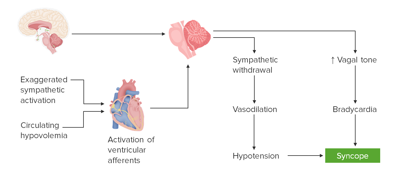
Dynamics of neurocardiogenic (also known as vasovagal, reflex, or neurally mediated) syncope: Normally, the heart and the CNS provide hemodynamic inputs to the brain stem, which then balances sympathetic and parasympathetic tone to maintain perfusion. A failure of this mechanism, in the face of physiologic stress, results in a paradoxical withdrawal of sympathetic tone simultaneous to increased parasympathetic discharge. Vasodilation and relative hypotension combined with bradycardia lead to poor cerebral perfusion and syncope.
Image by Lecturio.
Mnemonic
Causes of syncope “SVNCOPE”
Situational
Vasovagal
Neurogenic
Cardiac
Orthostatic hypotensionHypotensionHypotension is defined as low blood pressure, specifically < 90/60 mm Hg, and is most commonly a physiologic response. Hypotension may be mild, serious, or life threatening, depending on the cause. Hypotension
May or may not be associated with an identifiable triggerTriggerThe type of signal that initiates the inspiratory phase by the ventilatorInvasive Mechanical Ventilation associated with situational syncope
May or may not be attributed to an underlying pathologic disturbance
Loss of postural tone (e.g., a fall if standing, a slump if seated)
May or may not be accompanied by brief convulsive activity
May be mistaken for seizure
Distinguished from seizure by brevity of convulsionsConvulsionsSeizures and absence of postictalPostictalPeriod after the seizure episode during which the individual is disoriented.Seizures state
Postural tone returns to normal after individual regains consciousness
Major or minor trauma associated with loss of postural tone
Spontaneous return of consciousness:
Individual may report fatigueFatigueThe state of weariness following a period of exertion, mental or physical, characterized by a decreased capacity for work and reduced efficiency to respond to stimuli.Fibromyalgia or tiredness.
Generally, return to neurologic baseline level of function, unless:
Cause of syncope is cerebrovascular in nature.
Neurologic trauma is sustained during postural loss.
Prodromal symptoms
The following symptoms are associated with imminent syncope or presyncope:
NauseaNauseaAn unpleasant sensation in the stomach usually accompanied by the urge to vomit. Common causes are early pregnancy, sea and motion sickness, emotional stress, intense pain, food poisoning, and various enteroviruses.Antiemetics or nonspecific abdominal discomfort
Visual blurring; can proceed to temporary darkening
Diminution of hearing and occurrence of unusual sounds
Pallor reported by onlookers
Red flags
Certain presentations suggest more serious causes of syncope:
Syncope during exertion
Syncope while supine
Multiple recurrences within a short period of time
Heart murmur or other findings suggesting structural abnormalities
SeizuresSeizuresA seizure is abnormal electrical activity of the neurons in the cerebral cortex that can manifest in numerous ways depending on the region of the brain affected. Seizures consist of a sudden imbalance that occurs between the excitatory and inhibitory signals in cortical neurons, creating a net excitation. The 2 major classes of seizures are focal and generalized. Seizures
The etiology of approximately ½ of syncope cases remains undetermined despite an exhaustive workup. It is imperative to rule out life-threatening etiologies, such as cardiac syncope, PE, subarachnoid hemorrhageSubarachnoid HemorrhageSubarachnoid hemorrhage (SAH) is a type of cerebrovascular accident (stroke) resulting from intracranial hemorrhage into the subarachnoid space between the arachnoid and the pia mater layers of the meninges surrounding the brain. Most SAHs originate from a saccular aneurysm in the circle of Willis but may also occur as a result of trauma, uncontrolled hypertension, vasculitis, anticoagulant use, or stimulant use. Subarachnoid Hemorrhage, and blood loss.
History
Number, frequency, and duration of episodes
Onset
Position
Trauma sustained during loss of postural tone
Provocative factors:
During or immediately after exertion/exercise (red flag)
SwallowingSwallowingThe act of taking solids and liquids into the gastrointestinal tract through the mouth and throat.Gastrointestinal Motility
While in a warm and/or crowded place
During prolonged standing
During the postprandial period
In association with:
Emotional stress
Fear
Intense painPainAn unpleasant sensation induced by noxious stimuli which are detected by nerve endings of nociceptive neurons.Pain: Types and Pathways
Immediately following carotid sinusCarotid sinusThe dilated portion of the common carotid artery at its bifurcation into external and internal carotids. It contains baroreceptors which, when stimulated, cause slowing of the heart, vasodilatation, and a fall in blood pressure.Carotid Arterial System: Anatomy stimulation
While supine (suggestive of a serious problem)
Associated symptoms preceding and/or following the event:
NauseaNauseaAn unpleasant sensation in the stomach usually accompanied by the urge to vomit. Common causes are early pregnancy, sea and motion sickness, emotional stress, intense pain, food poisoning, and various enteroviruses.Antiemetics
VomitingVomitingThe forcible expulsion of the contents of the stomach through the mouth.Hypokalemia
Shortness of breathShortness of breathDyspnea is the subjective sensation of breathing discomfort. Dyspnea is a normal manifestation of heavy physical or psychological exertion, but also may be caused by underlying conditions (both pulmonary and extrapulmonary).Dyspnea
Chest painPainAn unpleasant sensation induced by noxious stimuli which are detected by nerve endings of nociceptive neurons.Pain: Types and Pathways
Additional symptoms following the syncopal event:
Confusion
FatigueFatigueThe state of weariness following a period of exertion, mental or physical, characterized by a decreased capacity for work and reduced efficiency to respond to stimuli.Fibromyalgia
Injury
BladderBladderA musculomembranous sac along the urinary tract. Urine flows from the kidneys into the bladder via the ureters, and is held there until urination.Pyelonephritis and Perinephric Abscess or bowel incontinence
Recurrent syncope
Witnessed signs:
Manner in which collapse happened
External appearance of individual
Estimated duration of loss of consciousness
Physical movements noted
Any breathing changes seen
Associated trauma
Preexisting medical conditions:
Structural heart disease:
Ischemic heart diseaseIschemic heart diseaseCoronary heart disease (CHD), or ischemic heart disease, describes a situation in which an inadequate supply of blood to the myocardium exists due to a stenosis of the coronary arteries, typically from atherosclerosis. Coronary Heart Disease
Valvular heart disease
Congenital heart disease
CardiomyopathiesCardiomyopathiesA group of diseases in which the dominant feature is the involvement of the cardiac muscle itself. Cardiomyopathies are classified according to their predominant pathophysiological features (dilated cardiomyopathy; hypertrophic cardiomyopathy; restrictive cardiomyopathy) or their etiological/pathological factors (cardiomyopathy, alcoholic; endocardial fibroelastosis).Cardiomyopathy: Overview and Types
Prior cardiac surgeryCardiac surgeryCardiac surgery is the surgical management of cardiac abnormalities and of the great vessels of the thorax. In general terms, surgical intervention of the heart is performed to directly restore adequate pump function, correct inherent structural issues, and reestablish proper blood supply via the coronary circulation.Cardiac Surgery
Neurologic conditions:
Seizure disorders
MigraineMigraineMigraine headache is a primary headache disorder and is among the most prevalent disorders in the world. Migraine is characterized by episodic, moderate to severe headaches that may be associated with increased sensitivity to light and sound, as well as nausea and/or vomiting. Migraine Headache headaches
Parkinson diseaseParkinson diseaseParkinson’s disease (PD) is a chronic, progressive neurodegenerative disorder. Although the cause is unknown, several genetic and environmental risk factors are currently being studied. Individuals present clinically with resting tremor, bradykinesia, rigidity, and postural instability.Parkinson’s Disease
Stroke
DiabetesDiabetesDiabetes mellitus (DM) is a metabolic disease characterized by hyperglycemia and dysfunction of the regulation of glucose metabolism by insulin. Type 1 DM is diagnosed mostly in children and young adults as the result of autoimmune destruction of β cells in the pancreas and the resulting lack of insulin. Type 2 DM has a significant association with obesity and is characterized by insulin resistance.Diabetes Mellitus mellitus:
Predisposition to cardiovascular/cerebrovascular disease
Illicit drugsIllicit DrugsDrugs that are manufactured, obtained, or sold illegally. They include prescription drugs obtained or sold without prescription and non-prescription drugs. Illicit drugs are widely distributed, tend to be grossly impure and may cause unexpected toxicity.Delirium
Prescription narcotics (e.g., opioidsOpioidsOpiates are drugs that are derived from the sap of the opium poppy. Opiates have been used since antiquity for the relief of acute severe pain. Opioids are synthetic opiates with properties that are substantially similar to those of opiates. Opioid Analgesics, benzodiazepinesBenzodiazepinesBenzodiazepines work on the gamma-aminobutyric acid type A (GABAA) receptor to produce inhibitory effects on the CNS. Benzodiazepines do not mimic GABA, the main inhibitory neurotransmitter in humans, but instead potentiate GABA activity. Benzodiazepines, amphetaminesAmphetaminesAnalogs or derivatives of amphetamine. Many are sympathomimetics and central nervous system stimulators causing excitation, vasopressin, bronchodilation, and to varying degrees, anorexia, analepsis, nasal decongestion, and some smooth muscle relaxation.Stimulants)
Familial cardiomyopathyCardiomyopathyCardiomyopathy refers to a group of myocardial diseases associated with structural changes of the heart muscles (myocardium) and impaired systolic and/or diastolic function in the absence of other heart disorders (coronary artery disease, hypertension, valvular disease, and congenital heart disease). Cardiomyopathy: Overview and Types
Seizure disorders or migraineMigraineMigraine headache is a primary headache disorder and is among the most prevalent disorders in the world. Migraine is characterized by episodic, moderate to severe headaches that may be associated with increased sensitivity to light and sound, as well as nausea and/or vomiting. Migraine Headache headaches
Familial predisposition to syncope
Physical examination
Vital signs:
Pulse and blood pressure taken with individual supine, seated, and standing (orthostatic vital signs)
Drop of systolic BP ≥ 20 mm Hg or diastolic BP ≥ 10 mm Hg within 3 minutes of standing is diagnostic of orthostatic hypotensionOrthostatic hypotensionA significant drop in blood pressure after assuming a standing position. Orthostatic hypotension is a finding, and defined as a 20-mm hg decrease in systolic pressure or a 10-mm hg decrease in diastolic pressure 3 minutes after the person has risen from supine to standing. Symptoms generally include dizziness, blurred vision, and syncope.Hypotension
≥ 30 mm Hg systolic thresholdThresholdMinimum voltage necessary to generate an action potential (an all-or-none response)Skeletal Muscle Contraction in those with supine hypertensionHypertensionHypertension, or high blood pressure, is a common disease that manifests as elevated systemic arterial pressures. Hypertension is most often asymptomatic and is found incidentally as part of a routine physical examination or during triage for an unrelated medical encounter. Hypertension
Note speed and regularity of pulse.
Note rate, regularity, and intensity of breathing effort.
Cardiac examination:
Note presence of heart murmur, especially if new or worsened.
Comparative pulse timing and blood pressure:
Incongruence between upper limbs indicative of proximal aortic dissectionAortic dissectionAortic dissection occurs due to shearing stress from pulsatile pressure causing a tear in the tunica intima of the aortic wall. This tear allows blood to flow into the media, creating a “false lumen.” Aortic dissection is most commonly caused by uncontrolled hypertension.Aortic Dissection
Incongruence between upper and lower limbs indicative of distal aortic dissectionAortic dissectionAortic dissection occurs due to shearing stress from pulsatile pressure causing a tear in the tunica intima of the aortic wall. This tear allows blood to flow into the media, creating a “false lumen.” Aortic dissection is most commonly caused by uncontrolled hypertension.Aortic Dissection
ECGECGAn electrocardiogram (ECG) is a graphic representation of the electrical activity of the heart plotted against time. Adhesive electrodes are affixed to the skin surface allowing measurement of cardiac impulses from many angles. The ECG provides 3-dimensional information about the conduction system of the heart, the myocardium, and other cardiac structures. Electrocardiogram (ECG)
ECGECGAn electrocardiogram (ECG) is a graphic representation of the electrical activity of the heart plotted against time. Adhesive electrodes are affixed to the skin surface allowing measurement of cardiac impulses from many angles. The ECG provides 3-dimensional information about the conduction system of the heart, the myocardium, and other cardiac structures. Electrocardiogram (ECG) is indicated for all individuals presenting with syncope, regardless of suspected etiology. ECGECGAn electrocardiogram (ECG) is a graphic representation of the electrical activity of the heart plotted against time. Adhesive electrodes are affixed to the skin surface allowing measurement of cardiac impulses from many angles. The ECG provides 3-dimensional information about the conduction system of the heart, the myocardium, and other cardiac structures. Electrocardiogram (ECG) monitoring should be continued throughout the ED or hospital stay. Notable findings may include:
Arrhythmias
ECGECGAn electrocardiogram (ECG) is a graphic representation of the electrical activity of the heart plotted against time. Adhesive electrodes are affixed to the skin surface allowing measurement of cardiac impulses from many angles. The ECG provides 3-dimensional information about the conduction system of the heart, the myocardium, and other cardiac structures. Electrocardiogram (ECG) changes suggestive of cardiac ischemiaIschemiaA hypoperfusion of the blood through an organ or tissue caused by a pathologic constriction or obstruction of its blood vessels, or an absence of blood circulation.Ischemic Cell Damage
Right heart strain pattern (S1S1Heart Sounds, Q3, T3T3A T3 thyroid hormone normally synthesized and secreted by the thyroid gland in much smaller quantities than thyroxine (T4). Most T3 is derived from peripheral monodeiodination of T4 at the 5′ position of the outer ring of the iodothyronine nucleus. The hormone finally delivered and used by the tissues is mainly t3.Thyroid Hormones) suggestive of PE
Conduction blocks
Specific signs of congenital or acquired structural heart disease
EchocardiographyEchocardiographyUltrasonic recording of the size, motion, and composition of the heart and surrounding tissues. The standard approach is transthoracic.Tricuspid Valve Atresia (TVA)
Used to screen for structural heart disease in known or suspected cases
May detect:
Valvular abnormalities
Wall-motion abnormalities
Left ventricular dysfunction
Elevated pulmonary pressures (suggestive of PE)
Pericardial effusionPericardial effusionFluid accumulation within the pericardium. Serous effusions are associated with pericardial diseases. Hemopericardium is associated with trauma. Lipid-containing effusion (chylopericardium) results from leakage of thoracic duct. Severe cases can lead to cardiac tamponade.Pericardial Effusion and Cardiac Tamponade
Masses
Vegetations
Laboratory evaluation
CBC:
RBC indices for:
AnemiaAnemiaAnemia is a condition in which individuals have low Hb levels, which can arise from various causes. Anemia is accompanied by a reduced number of RBCs and may manifest with fatigue, shortness of breath, pallor, and weakness. Subtypes are classified by the size of RBCs, chronicity, and etiology. Anemia: Overview and Types
Blood loss
Erythrocytosis
WBC indices for:
Evidence of infection
Lymphoproliferation
Platelet count for:
Bleeding
Thrombotic tendencies
CMP to evaluate for:
Renal or hepatic dysfunction
Electrolyte disturbance
Acid–base imbalance
HypoglycemiaHypoglycemiaHypoglycemia is an emergency condition defined as a serum glucose level ≤ 70 mg/dL (≤ 3.9 mmol/L) in diabetic patients. In nondiabetic patients, there is no specific or defined limit for normal serum glucose levels, and hypoglycemia is defined mainly by its clinical features. Hypoglycemia
Coagulation studiesCoagulation studiesCoagulation studies are a group of hematologic laboratory studies that reflect the function of blood vessels, platelets, and coagulation factors, which all interact with one another to achieve hemostasis. Coagulation studies are usually ordered to evaluate patients with bleeding or hypercoagulation disorders.Coagulation Studies:
PT/PTT to evaluate for coagulopathy
Especially in suspected intracerebral/cerebrovascular or GI hemorrhage
Cardiac biomarkers:
Includes:
MB isoenzyme of creatineCreatineAn amino acid that occurs in vertebrate tissues and in urine. In muscle tissue, creatine generally occurs as phosphocreatine. Creatine is excreted as creatinine in the urine.Acute Kidney Injury kinase (CKMB)
Cardiac troponins
Beta-natriuretic peptide
Evaluate for the presence of ischemic heart diseaseIschemic heart diseaseCoronary heart disease (CHD), or ischemic heart disease, describes a situation in which an inadequate supply of blood to the myocardium exists due to a stenosis of the coronary arteries, typically from atherosclerosis. Coronary Heart Disease and/or heart failureHeart FailureA heterogeneous condition in which the heart is unable to pump out sufficient blood to meet the metabolic need of the body. Heart failure can be caused by structural defects, functional abnormalities (ventricular dysfunction), or a sudden overload beyond its capacity. Chronic heart failure is more common than acute heart failure which results from sudden insult to cardiac function, such as myocardial infarction.Total Anomalous Pulmonary Venous Return (TAPVR)
Urine toxicology screen
Urine hCG for women of childbearing age
Imaging
NeuroimagingNeuroimagingNon-invasive methods of visualizing the central nervous system, especially the brain, by various imaging modalities.Febrile Infant (CT, MRI of head/brainBrainThe part of central nervous system that is contained within the skull (cranium). Arising from the neural tube, the embryonic brain is comprised of three major parts including prosencephalon (the forebrain); mesencephalon (the midbrain); and rhombencephalon (the hindbrain). The developed brain consists of cerebrum; cerebellum; and other structures in the brain stem.Nervous System: Anatomy, Structure, and Classification) for:
Suspected intracranial massMassThree-dimensional lesion that occupies a space within the breastImaging of the Breast
Intracranial hemorrhageIntracranial hemorrhageSubarachnoid hemorrhage (SAH) is a type of cerebrovascular accident (stroke) resulting from intracranial hemorrhage into the subarachnoid space between the arachnoid and the pia mater layers of the meninges surrounding the brain. Most sahs originate from a saccular aneurysm in the circle of willis but may also occur as a result of trauma, uncontrolled hypertension, vasculitis, anticoagulant use, or stimulant use.Subarachnoid Hemorrhage
Cerebrovascular accidentCerebrovascular accidentAn ischemic stroke (also known as cerebrovascular accident) is an acute neurologic injury that occurs as a result of brain ischemia; this condition may be due to cerebral blood vessel occlusion by thrombosis or embolism, or rarely due to systemic hypoperfusion. Ischemic Stroke
CTACTAA non-invasive method that uses a ct scanner for capturing images of blood vessels and tissues. A contrast material is injected, which helps produce detailed images that aid in diagnosing vascular diseases.Pulmonary Function Tests of the chest or ventilationVentilationThe total volume of gas inspired or expired per unit of time, usually measured in liters per minute.Ventilation: Mechanics of Breathing/perfusion (VQ) scan for suspected PE
Carotid DopplerDopplerUltrasonography applying the doppler effect, with frequency-shifted ultrasound reflections produced by moving targets (usually red blood cells) in the bloodstream along the ultrasound axis in direct proportion to the velocity of movement of the targets, to determine both direction and velocity of blood flow.Ultrasound (Sonography) scan for suspected carotid vascular disease
Abdominal CT or ultrasonography to evaluate for:
Splenic ruptureSplenic ruptureSplenic rupture is a medical emergency that carries a significant risk of hypovolemic shock and death. Injury to the spleen accounts for nearly half of all injuries to intra-abdominal organs. The most common reason for a rupture of the spleen is blunt abdominal trauma, specifically, motor vehicle accidents. Rupture of the Spleen
Abdominopelvic ultrasonography to evaluate for ectopic pregnancyEctopic pregnancyEctopic pregnancy refers to the implantation of a fertilized egg (embryo) outside the uterine cavity. The main cause is disruption of the normal anatomy of the fallopian tube. Ectopic Pregnancy or gynecologic sources of hemorrhage
Lower-extremity ultrasonography to evaluate for deep vein thrombosisThrombosisFormation and development of a thrombus or blood clot in the blood vessel.Epidemic Typhus (DVTDVTDeep vein thrombosis (DVT) usually occurs in the deep veins of the lower extremities. The affected veins include the femoral, popliteal, iliofemoral, and pelvic veins. Proximal DVT is more likely to cause a pulmonary embolism (PE) and is generally considered more serious. Deep Vein Thrombosis)
Specific imaging indicated for evaluation of other suspected etiologies
Other tests
Tilt-table test: changes in posture from lying to standing to evaluate cause of syncope
Holter monitoring or loop recording for cardiac rhythm disturbances that manifest during the initial ED visit or hospital stay
Other specific testing indicated for evaluation of other suspected etiologies
Management
Much of the management of syncope will be specific to the confirmed or suspected etiology. Because the specific etiology of syncope often goes undiagnosed, general measures are discussed here.
Treatment of prodromal symptoms
This includes physical countermaneuvers, such as:
LegLegThe lower leg, or just “leg” in anatomical terms, is the part of the lower limb between the knee and the ankle joint. The bony structure is composed of the tibia and fibula bones, and the muscles of the leg are grouped into the anterior, lateral, and posterior compartments by extensions of fascia.Leg: Anatomy crossing: simultaneous tensing of legLegThe lower leg, or just “leg” in anatomical terms, is the part of the lower limb between the knee and the ankle joint. The bony structure is composed of the tibia and fibula bones, and the muscles of the leg are grouped into the anterior, lateral, and posterior compartments by extensions of fascia.Leg: Anatomy, abdominal, and buttock muscles
Handgrip: consists of maximum grip on a rubber ball or similar object
ArmArmThe arm, or “upper arm” in common usage, is the region of the upper limb that extends from the shoulder to the elbow joint and connects inferiorly to the forearm through the cubital fossa. It is divided into 2 fascial compartments (anterior and posterior).Arm: Anatomy tensing: involves gripping one handHandThe hand constitutes the distal part of the upper limb and provides the fine, precise movements needed in activities of daily living. It consists of 5 metacarpal bones and 14 phalanges, as well as numerous muscles innervated by the median and ulnar nerves. Hand: Anatomy with the other while simultaneously abducting both hands
Immediate treatment
Assist the individual to the ground, chair, or stretcher to avoid traumatic injury.
Lay individual supine with legs elevated to help with venous return to the heart and to eventually restore cerebral perfusion.
Assess vital signs (blood pressure, pulse, respiratory rateRespiratory rateThe number of times an organism breathes with the lungs (respiration) per unit time, usually per minute.Pulmonary Examination).
Observe other signs (pallor, diaphoresis, seizure activity).
Get additional assistance:
Call 911.
“Is there a doctor in the house?”
Attempt to arouse the individual.
If high-risk factors are present, admit to the most appropriate unit in the hospital (e.g., telemetryTelemetryTransmission of the readings of instruments to a remote location by means of wires, radio waves, or other means.Crush Syndrome, ICUICUHospital units providing continuous surveillance and care to acutely ill patients.West Nile Virus).
Risk assessmentRisk assessmentThe qualitative or quantitative estimation of the likelihood of adverse effects that may result from exposure to specified health hazards or from the absence of beneficial influences.Preoperative Care
Low risk for poor outcomes if no evidence of heart disease is identified
High-risk features associated with poor outcomes:
Evidence of structural or ischemic heart diseaseIschemic heart diseaseCoronary heart disease (CHD), or ischemic heart disease, describes a situation in which an inadequate supply of blood to the myocardium exists due to a stenosis of the coronary arteries, typically from atherosclerosis. Coronary Heart Disease
History of structural or ischemic heart diseaseIschemic heart diseaseCoronary heart disease (CHD), or ischemic heart disease, describes a situation in which an inadequate supply of blood to the myocardium exists due to a stenosis of the coronary arteries, typically from atherosclerosis. Coronary Heart Disease
Chest painPainAn unpleasant sensation induced by noxious stimuli which are detected by nerve endings of nociceptive neurons.Pain: Types and Pathways at time of syncope
DyspneaDyspneaDyspnea is the subjective sensation of breathing discomfort. Dyspnea is a normal manifestation of heavy physical or psychological exertion, but also may be caused by underlying conditions (both pulmonary and extrapulmonary). Dyspnea at time of syncope
Syncope without prodromeProdromeSymptoms that appear 24–48 hours prior to migraine onset.Migraine Headache
Family historyFamily HistoryAdult Health Maintenance of sudden cardiac deathSudden cardiac deathCardiac arrest is the sudden, complete cessation of cardiac output with hemodynamic collapse. Patients present as pulseless, unresponsive, and apneic. Rhythms associated with cardiac arrest are ventricular fibrillation/tachycardia, asystole, or pulseless electrical activity.Cardiac Arrest
Abnormal ECGECGAn electrocardiogram (ECG) is a graphic representation of the electrical activity of the heart plotted against time. Adhesive electrodes are affixed to the skin surface allowing measurement of cardiac impulses from many angles. The ECG provides 3-dimensional information about the conduction system of the heart, the myocardium, and other cardiac structures. Electrocardiogram (ECG)
Persistently low blood pressure
Low hematocritHematocritThe volume of packed red blood cells in a blood specimen. The volume is measured by centrifugation in a tube with graduated markings, or with automated blood cell counters. It is an indicator of erythrocyte status in disease. For example, anemia shows a low value; polycythemia, a high value.Neonatal Polycythemia
Promptly rule out life-threatening causes of syncope or syncope mimics (seizure and cerebrovascular accidentCerebrovascular accidentAn ischemic stroke (also known as cerebrovascular accident) is an acute neurologic injury that occurs as a result of brain ischemia; this condition may be due to cerebral blood vessel occlusion by thrombosis or embolism, or rarely due to systemic hypoperfusion. Ischemic Stroke are not true causes of syncope):
MIMIMI is ischemia and death of an area of myocardial tissue due to insufficient blood flow and oxygenation, usually from thrombus formation on a ruptured atherosclerotic plaque in the epicardial arteries. Clinical presentation is most commonly with chest pain, but women and patients with diabetes may have atypical symptoms.Myocardial Infarction
Nonperfusing cardiac arrhythmia
PE
Cerebrovascular accidentCerebrovascular accidentAn ischemic stroke (also known as cerebrovascular accident) is an acute neurologic injury that occurs as a result of brain ischemia; this condition may be due to cerebral blood vessel occlusion by thrombosis or embolism, or rarely due to systemic hypoperfusion. Ischemic Stroke
Intracranial hemorrhageIntracranial hemorrhageSubarachnoid hemorrhage (SAH) is a type of cerebrovascular accident (stroke) resulting from intracranial hemorrhage into the subarachnoid space between the arachnoid and the pia mater layers of the meninges surrounding the brain. Most sahs originate from a saccular aneurysm in the circle of willis but may also occur as a result of trauma, uncontrolled hypertension, vasculitis, anticoagulant use, or stimulant use.Subarachnoid Hemorrhage
Aortic rupture
Massive hemorrhage
Seizure
Therapies to prevent syncope recurrence
Reflex syncope:carotid sinusCarotid sinusThe dilated portion of the common carotid artery at its bifurcation into external and internal carotids. It contains baroreceptors which, when stimulated, cause slowing of the heart, vasodilatation, and a fall in blood pressure.Carotid Arterial System: Anatomy syncope:
Avoid mechanical manipulation of the carotid sinuses (e.g., abrupt turning of the neckNeckThe part of a human or animal body connecting the head to the rest of the body.Peritonsillar Abscess, wearing tight collars).
MidodrineMidodrineAn ethanolamine derivative that is an adrenergic alpha-1 agonist. It is used as a vasoconstrictor agent in the treatment of hypotension.Sympathomimetic Drugs
Consider fludrocortisoneFludrocortisoneA synthetic mineralocorticoid with anti-inflammatory activity.Mineralocorticoids in selected patientsPatientsIndividuals participating in the health care system for the purpose of receiving therapeutic, diagnostic, or preventive procedures.Clinician–Patient Relationship
Beta blockers may be considered in older adults
Pacemakers (those with cardioinhibitory responses)
Orthostatic hypotensionOrthostatic hypotensionA significant drop in blood pressure after assuming a standing position. Orthostatic hypotension is a finding, and defined as a 20-mm hg decrease in systolic pressure or a 10-mm hg decrease in diastolic pressure 3 minutes after the person has risen from supine to standing. Symptoms generally include dizziness, blurred vision, and syncope.Hypotension:
Medication-induced:
Discontinue offending medication.
Substitute with an alternative agent.
Adjust dose.
Change timing of drug administration.
Volume depletionVolume depletionVolume status is a balance between water and solutes, the majority of which is Na. Volume depletion refers to a loss of both water and Na, whereas dehydration refers only to a loss of water. Volume depletion can be caused by GI losses, renal losses, bleeding, poor oral Na intake, or third spacing of fluids.Volume Depletion and Dehydration:
Treat underlying cause (e.g., gastroenteritisGastroenteritisGastroenteritis is inflammation of the stomach and intestines, commonly caused by infections from bacteria, viruses, or parasites. Transmission may be foodborne, fecal-oral, or through animal contact. Common clinical features include abdominal pain, diarrhea, vomiting, fever, and dehydration.Gastroenteritis, hemorrhage).
Causes of syncope that warrant immediate admission/intervention
Cardiac emergencies:
Arrhythmias:
Documented, suspected, or induced ventricular tachycardiaTachycardiaAbnormally rapid heartbeat, usually with a heart rate above 100 beats per minute for adults. Tachycardia accompanied by disturbance in the cardiac depolarization (cardiac arrhythmia) is called tachyarrhythmia.Sepsis in Children:
Advanced cardiac life support (ACLS) protocol if indicated
Antiarrhythmics
Catheter ablation
Implantable cardioverter–defibrillatorDefibrillatorCardiac electrical stimulators that apply brief high-voltage electroshocks to the heart. These stimulators are used to restore normal rhythm and contractile function in hearts of patients who are experiencing ventricular fibrillation or ventricular tachycardia that is not accompanied by a palpable pulse. Some defibrillators may also be used to correct certain noncritical dysrhythmias (called synchronized defibrillation or cardioversion), using relatively low-level discharges synchronized to the patient’s ECG waveform.Cardiac Arrest
Supraventricular arrhythmias:
ACLS protocol if indicated
Antiarrhythmics
Catheter ablation
BradyarrhythmiasBradyarrhythmiasBradyarrhythmia is a rhythm in which the heart rate is less than 60/min. Bradyarrhythmia can be physiologic, without symptoms or hemodynamic change. Pathologic bradyarrhythmia results in reduced cardiac output and hemodynamic instability causing syncope, dizziness, or dyspnea. Bradyarrhythmias: permanent pacemakers
Ischemic heart diseaseIschemic heart diseaseCoronary heart disease (CHD), or ischemic heart disease, describes a situation in which an inadequate supply of blood to the myocardium exists due to a stenosis of the coronary arteries, typically from atherosclerosis. Coronary Heart Disease (IHDIHDCoronary heart disease (CHD), or ischemic heart disease, describes a situation in which an inadequate supply of blood to the myocardium exists due to a stenosis of the coronary arteries, typically from atherosclerosis.Coronary Heart Disease) or acute coronary syndrome (ACS):
Activate ACS protocol if indicated.
Coronary intervention or cardiac surgeryCardiac surgeryCardiac surgery is the surgical management of cardiac abnormalities and of the great vessels of the thorax. In general terms, surgical intervention of the heart is performed to directly restore adequate pump function, correct inherent structural issues, and reestablish proper blood supply via the coronary circulation.Cardiac Surgery if indicated
Immediate noncontrast CT of head if cerebrovascular accidentCerebrovascular accidentAn ischemic stroke (also known as cerebrovascular accident) is an acute neurologic injury that occurs as a result of brain ischemia; this condition may be due to cerebral blood vessel occlusion by thrombosis or embolism, or rarely due to systemic hypoperfusion. Ischemic Stroke (hemorrhagic or ischemic) or intracranial hemorrhageIntracranial hemorrhageSubarachnoid hemorrhage (SAH) is a type of cerebrovascular accident (stroke) resulting from intracranial hemorrhage into the subarachnoid space between the arachnoid and the pia mater layers of the meninges surrounding the brain. Most sahs originate from a saccular aneurysm in the circle of willis but may also occur as a result of trauma, uncontrolled hypertension, vasculitis, anticoagulant use, or stimulant use.Subarachnoid Hemorrhage is suspected
Appropriate admission/transfer/consultation depending on findings:
Neurology
NeurosurgeryNeurosurgeryNeurosurgery is a specialized field focused on the surgical management of pathologies of the brain, spine, spinal cord, and peripheral nerves. General neurosurgery includes cases of trauma and emergencies. There are a number of specialized neurosurgical practices, including oncologic neurosurgery, spinal neurosurgery, and pediatric neurosurgery. Neurosurgery
Interventional vascular surgeryVascular surgeryVascular surgery is the specialized field of medicine that focuses on the surgical management of the pathologies of the peripheral circulation. The main goal of most vascular procedures is to restore circulatory function to the affected vessels by relieving occlusions or by redirecting blood flow (e.g., bypass).Vascular Surgery/neurosurgeryNeurosurgeryNeurosurgery is a specialized field focused on the surgical management of pathologies of the brain, spine, spinal cord, and peripheral nerves. General neurosurgery includes cases of trauma and emergencies. There are a number of specialized neurosurgical practices, including oncologic neurosurgery, spinal neurosurgery, and pediatric neurosurgery. Neurosurgery
Neurologic ICUICUHospital units providing continuous surveillance and care to acutely ill patients.West Nile Virus monitoring
Seizure: abnormal electrical activity of the neuronsNeuronsThe basic cellular units of nervous tissue. Each neuron consists of a body, an axon, and dendrites. Their purpose is to receive, conduct, and transmit impulses in the nervous system.Nervous System: Histology in the cerebral cortexCerebral cortexThe cerebral cortex is the largest and most developed part of the human brain and CNS. Occupying the upper part of the cranial cavity, the cerebral cortex has 4 lobes and is divided into 2 hemispheres that are joined centrally by the corpus callosum. Cerebral Cortex: Anatomy that can manifest in numerous ways depending on the region of the brainBrainThe part of central nervous system that is contained within the skull (cranium). Arising from the neural tube, the embryonic brain is comprised of three major parts including prosencephalon (the forebrain); mesencephalon (the midbrain); and rhombencephalon (the hindbrain). The developed brain consists of cerebrum; cerebellum; and other structures in the brain stem.Nervous System: Anatomy, Structure, and Classification affected. There are numerous etiologies, and investigation of the root cause should be part of the initial evaluation. Diagnosis is made by a clinical evaluation, lab testing, neuroimagingNeuroimagingNon-invasive methods of visualizing the central nervous system, especially the brain, by various imaging modalities.Febrile Infant, electroencephalographyElectroencephalographySeizures, and antiseizure drug levels. Treatment is by eliminationEliminationThe initial damage and destruction of tumor cells by innate and adaptive immunity. Completion of the phase means no cancer growth. Cancer Immunotherapy of the cause, if possible, antiseizure drugs, and surgery when drugs are ineffective.
Traumatic brain injuryTraumatic brain injuryA form of acquired brain injury which occurs when a sudden trauma causes damage to the brain.Le Fort Fractures: physical injury to brainBrainThe part of central nervous system that is contained within the skull (cranium). Arising from the neural tube, the embryonic brain is comprised of three major parts including prosencephalon (the forebrain); mesencephalon (the midbrain); and rhombencephalon (the hindbrain). The developed brain consists of cerebrum; cerebellum; and other structures in the brain stem.Nervous System: Anatomy, Structure, and Classification tissue that temporarily or permanently impairs brainBrainThe part of central nervous system that is contained within the skull (cranium). Arising from the neural tube, the embryonic brain is comprised of three major parts including prosencephalon (the forebrain); mesencephalon (the midbrain); and rhombencephalon (the hindbrain). The developed brain consists of cerebrum; cerebellum; and other structures in the brain stem.Nervous System: Anatomy, Structure, and Classification function. Traumatic brain injuryTraumatic brain injuryA form of acquired brain injury which occurs when a sudden trauma causes damage to the brain.Le Fort Fractures can be caused by falls, motor vehicle accidentsMotor Vehicle AccidentsSpinal Cord Injuries, assaults, and sports activities. Individuals may present with loss of consciousness, confusion, amnesia, seizuresSeizuresA seizure is abnormal electrical activity of the neurons in the cerebral cortex that can manifest in numerous ways depending on the region of the brain affected. Seizures consist of a sudden imbalance that occurs between the excitatory and inhibitory signals in cortical neurons, creating a net excitation. The 2 major classes of seizures are focal and generalized. Seizures, and focal neurologic deficitsNeurologic DeficitsHigh-Risk Headaches. Diagnosis is by initial rapid trauma assessment, neurologic examination, and CT scan. Initial treatment is optimizing brainBrainThe part of central nervous system that is contained within the skull (cranium). Arising from the neural tube, the embryonic brain is comprised of three major parts including prosencephalon (the forebrain); mesencephalon (the midbrain); and rhombencephalon (the hindbrain). The developed brain consists of cerebrum; cerebellum; and other structures in the brain stem.Nervous System: Anatomy, Structure, and Classification perfusion and supportive care. Severe injuries may require timely surgical intervention.
Intoxication: reversible syndrome associated with substance use, which may cause physical and mental changes (varies depending on the substance that was ingested). Intoxication may lead to accidental death via overdose, and various substances may carry significant complications that increase morbidityMorbidityThe proportion of patients with a particular disease during a given year per given unit of population.Measures of Health Status and mortalityMortalityAll deaths reported in a given population.Measures of Health Status. Management is usually supportive, although some substances have reversible pharmacologic agents.
Conversion disorders: also called functional neurologic symptom disorder. Conversion disorders are psychiatric disorders with prominent motorMotorNeurons which send impulses peripherally to activate muscles or secretory cells.Nervous System: Histology or sensorySensoryNeurons which conduct nerve impulses to the central nervous system.Nervous System: Histology impairment that is not compatible with any known neurologic medical condition. The deficits are not consciously produced. Individuals are typically impaired in their social and professional life, but can also be inappropriately unconcerned with their symptoms. Treatment centers around education and psychotherapyPsychotherapyPsychotherapy is interpersonal treatment based on the understanding of psychological principles and mechanisms of mental disease. The treatment approach is often individualized, depending on the psychiatric condition(s) or circumstance. Psychotherapy.
Kharsa, A., & Wadhwa, R. (2022). Carotid sinus hypersensitivity. In StatPearls [Internet]. StatPearls Publishing. Retrieved September 22, 2025, from https://www.ncbi.nlm.nih.gov/books/NBK559059/