Achieve Mastery of Medical Concepts

Study for medical school and boards with Lecturio

Gastrointestinal Motility

The primary functions of the GI tract are digestion Digestion Digestion refers to the process of the mechanical and chemical breakdown of food into smaller particles, which can then be absorbed and utilized by the body. Digestion and Absorption and absorption Absorption Absorption involves the uptake of nutrient molecules and their transfer from the lumen of the GI tract across the enterocytes and into the interstitial space, where they can be taken up in the venous or lymphatic circulation. Digestion and Absorption, which require coordinated contractions of the smooth muscles Smooth muscles Unstriated and unstriped muscle, one of the muscles of the internal organs, blood vessels, hair follicles, etc. Contractile elements are elongated, usually spindle-shaped cells with centrally located nuclei. Smooth muscle fibers are bound together into sheets or bundles by reticular fibers and frequently elastic nets are also abundant. Muscle Tissue: Histology present in the GI tract. Peristaltic waves, segmentation contractions, and the migrating motor Motor Neurons which send impulses peripherally to activate muscles or secretory cells. Nervous System: Histology complex are all important contraction patterns that help to mix contents, get them in contact with the intestinal walls (where they are further digested by brush-border enzymes Enzymes Enzymes are complex protein biocatalysts that accelerate chemical reactions without being consumed by them. Due to the body's constant metabolic needs, the absence of enzymes would make life unsustainable, as reactions would occur too slowly without these molecules. Basics of Enzymes and absorbed into the enterocytes), and propel material down the tract at appropriate times and in appropriate amounts.

Last updated: Apr 18, 2023

Editorial responsibility: Stanley Oiseth, Lindsay Jones, Evelin Maza

Overview of GI Motility

Definition

GI motility refers to the contraction of smooth muscle within the walls of the GI tract, which:

  • Move foodstuff along the tract
  • Mix and grind
  • Store foodstuff (via contracted sphincters, keeping material in its current location)

Important types of movements/contractions

  1. Peristalsis
  2. Segmentation
  3. Sphincter contraction and relaxation

Peristalsis

  • Coordinated, wave-like contractions that move material forward through the tract, ultimately toward the rectum Rectum The rectum and anal canal are the most terminal parts of the lower GI tract/large intestine that form a functional unit and control defecation. Fecal continence is maintained by several important anatomic structures including rectal folds, anal valves, the sling-like puborectalis muscle, and internal and external anal sphincters. Rectum and Anal Canal: Anatomy
  • Acetylcholine Acetylcholine A neurotransmitter found at neuromuscular junctions, autonomic ganglia, parasympathetic effector junctions, a subset of sympathetic effector junctions, and at many sites in the central nervous system. Receptors and Neurotransmitters of the CNS ( ACh ACh A neurotransmitter found at neuromuscular junctions, autonomic ganglia, parasympathetic effector junctions, a subset of sympathetic effector junctions, and at many sites in the central nervous system. Receptors and Neurotransmitters of the CNS) contracts one small area of the tract.
  • NO and vasoactive intestinal peptide Vasoactive intestinal peptide A highly basic, 28 amino acid neuropeptide released from intestinal mucosa. It has a wide range of biological actions affecting the cardiovascular, gastrointestinal, and respiratory systems and is neuroprotective. It binds special receptors. Gastrointestinal Neural and Hormonal Signaling ( VIP VIP A highly basic, 28 amino acid neuropeptide released from intestinal mucosa. It has a wide range of biological actions affecting the cardiovascular, gastrointestinal, and respiratory systems and is neuroprotective. It binds special receptors. Gastrointestinal Neural and Hormonal Signaling) cause relaxation of the area immediately ahead of the contracting area.
  • This relaxation pushes foodstuff from areas of high pressure (area that is contracting) to areas of low pressure (area that is relaxing).
Peristalsis

Coordination of smooth muscle in peristalsis:
Acetylcholine (ACh) causes constriction of the muscle just behind the foodstuff, while NO and vasoactive intestinal peptide (VIP) cause relaxation ahead of it. This creates a pressure gradient, forcing the food downward. Nerves from the enteric nervous system (with input from the autonomic nervous system), coordinate the wave of constriction and relaxation.

Image by Lecturio.

Segmentation

  • Circumferential muscle contractions (i.e., squeezing)
  • Forces some of the contents forward and some backward
  • Segmentation contractions occur in multiple nearby locations all at once.
  • Allows for mixing, churning, and kneading of food material 
  • Occurs in the stomach Stomach The stomach is a muscular sac in the upper left portion of the abdomen that plays a critical role in digestion. The stomach develops from the foregut and connects the esophagus with the duodenum. Structurally, the stomach is C-shaped and forms a greater and lesser curvature and is divided grossly into regions: the cardia, fundus, body, and pylorus. Stomach: Anatomy and intestines
Segmentation contractions in the stomach

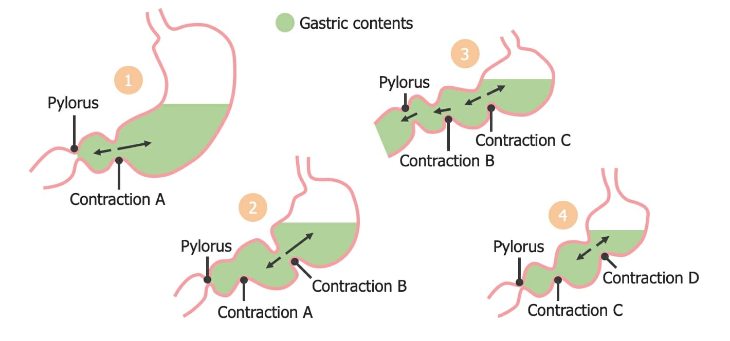
Segmentation contractions in the stomach:
Note how the subsequent contraction achieves mixing of contents and enough increase in pressure to overcome the relaxed pyloric sphincter.

Image by Lecturio.

Sphincter contraction and relaxation

  • Sphincters are circular bands of smooth muscle that are normally contracted at baseline → keep foodstuff from passing into the next segment of the tract 
  • Regulating sphincter relaxation allows for control of movement through the tract.
  • Baseline contraction is maintained by:
    • Enkephalins Enkephalins One of the three major families of endogenous opioid peptides. The enkephalins are pentapeptides that are widespread in the central and peripheral nervous systems and in the adrenal medulla. Receptors and Neurotransmitters of the CNS (neurotransmitters in the enteric nervous system Enteric nervous system Two ganglionated neural plexuses in the gut wall which form one of the three major divisions of the autonomic nervous system. The enteric nervous system innervates the gastrointestinal tract, the pancreas, and the gallbladder. It contains sensory neurons, interneurons, and motor neurons. Thus the circuitry can autonomously sense the tension and the chemical environment in the gut and regulate blood vessel tone, motility, secretions, and fluid transport. The system is itself governed by the central nervous system and receives both parasympathetic and sympathetic innervation. Autonomic Nervous System: Anatomy (ENS))
    • The ability of smooth muscle to maintain a latch-bridge state Latch-bridge state Smooth Muscle Contraction:
      • A state in which myosin Myosin A diverse superfamily of proteins that function as translocating proteins. They share the common characteristics of being able to bind actins and hydrolyze mgATP. Myosins generally consist of heavy chains which are involved in locomotion, and light chains which are involved in regulation. Within the structure of myosin heavy chain are three domains: the head, the neck and the tail. The head region of the heavy chain contains the actin binding domain and mgATPase domain which provides energy for locomotion. The neck region is involved in binding the light-chains. The tail region provides the anchoring point that maintains the position of the heavy chain. The superfamily of myosins is organized into structural classes based upon the type and arrangement of the subunits they contain. Skeletal Muscle Contraction is dephosphorylated (no longer cross-bridge cycle), however, remains attached to actin Actin Filamentous proteins that are the main constituent of the thin filaments of muscle fibers. The filaments (known also as filamentous or f-actin) can be dissociated into their globular subunits; each subunit is composed of a single polypeptide 375 amino acids long. This is known as globular or g-actin. In conjunction with myosins, actin is responsible for the contraction and relaxation of muscle. Skeletal Muscle Contraction = maintains some tension 
      • Allows the muscle to maintain tone without expending much energy
  • Relaxation is triggered by VIP VIP A highly basic, 28 amino acid neuropeptide released from intestinal mucosa. It has a wide range of biological actions affecting the cardiovascular, gastrointestinal, and respiratory systems and is neuroprotective. It binds special receptors. Gastrointestinal Neural and Hormonal Signaling.

Review of Nervous System Control over GI Smooth Muscle

GI motility is controlled primarily via the parasympathetic nervous system Nervous system The nervous system is a small and complex system that consists of an intricate network of neural cells (or neurons) and even more glial cells (for support and insulation). It is divided according to its anatomical components as well as its functional characteristics. The brain and spinal cord are referred to as the central nervous system, and the branches of nerves from these structures are referred to as the peripheral nervous system. Nervous System: Anatomy, Structure, and Classification (PNS) and the ENS.

Parasympathetic nervous system Nervous system The nervous system is a small and complex system that consists of an intricate network of neural cells (or neurons) and even more glial cells (for support and insulation). It is divided according to its anatomical components as well as its functional characteristics. The brain and spinal cord are referred to as the central nervous system, and the branches of nerves from these structures are referred to as the peripheral nervous system. Nervous System: Anatomy, Structure, and Classification

  • Stimulation of the PNS:
    • ↑ GI motility 
    • ↑ GI secretions
    • Blood flow Blood flow Blood flow refers to the movement of a certain volume of blood through the vasculature over a given unit of time (e.g., mL per minute). Vascular Resistance, Flow, and Mean Arterial Pressure
    • ↓ Constriction of GI sphincters (allows food to move through the tract)
  • Primary neural signaling molecules Signaling molecules Second Messengers:
    • ACh ACh A neurotransmitter found at neuromuscular junctions, autonomic ganglia, parasympathetic effector junctions, a subset of sympathetic effector junctions, and at many sites in the central nervous system. Receptors and Neurotransmitters of the CNS
    • VIP VIP A highly basic, 28 amino acid neuropeptide released from intestinal mucosa. It has a wide range of biological actions affecting the cardiovascular, gastrointestinal, and respiratory systems and is neuroprotective. It binds special receptors. Gastrointestinal Neural and Hormonal Signaling

Enteric nervous system Enteric nervous system Two ganglionated neural plexuses in the gut wall which form one of the three major divisions of the autonomic nervous system. The enteric nervous system innervates the gastrointestinal tract, the pancreas, and the gallbladder. It contains sensory neurons, interneurons, and motor neurons. Thus the circuitry can autonomously sense the tension and the chemical environment in the gut and regulate blood vessel tone, motility, secretions, and fluid transport. The system is itself governed by the central nervous system and receives both parasympathetic and sympathetic innervation. Autonomic Nervous System: Anatomy

A specialized portion of the ANS ANS The ans is a component of the peripheral nervous system that uses both afferent (sensory) and efferent (effector) neurons, which control the functioning of the internal organs and involuntary processes via connections with the CNS. The ans consists of the sympathetic and parasympathetic nervous systems. Autonomic Nervous System: Anatomy located within the walls of the GI tract

Consists of:

  • Meissner plexus Meissner plexus One of two ganglionated neural networks which together form the enteric nervous system. The submucous (Meissner’s) plexus is in the connective tissue of the submucosa. Its neurons innervate the epithelium, blood vessels, endocrine cells, other submucosal ganglia, and myenteric ganglia, and play an important role in regulating ion and water transport. Gastrointestinal Neural and Hormonal Signaling (also called the submucosal plexus):
    • Located in the submucosa
    • Controls the muscularis mucosa (independently of the muscular layer of the intestines)
  • Auerbach plexus Auerbach plexus One of two ganglionated neural networks which together form the enteric nervous system. The myenteric (Auerbach’s) plexus is located between the longitudinal and circular muscle layers of the gut. Its neurons project to the circular muscle, to other myenteric ganglia, to submucosal ganglia, or directly to the epithelium, and play an important role in regulating and patterning gut motility. Gastrointestinal Neural and Hormonal Signaling (also called the myenteric plexus Myenteric plexus One of two ganglionated neural networks which together form the enteric nervous system. The myenteric (Auerbach’s) plexus is located between the longitudinal and circular muscle layers of the gut. Its neurons project to the circular muscle, to other myenteric ganglia, to submucosal ganglia, or directly to the epithelium, and play an important role in regulating and patterning gut motility. Gastrointestinal Neural and Hormonal Signaling)
    • Located in the muscularis, between the circular and longitudinal layers 
    • Includes both motor Motor Neurons which send impulses peripherally to activate muscles or secretory cells. Nervous System: Histology and sensory Sensory Neurons which conduct nerve impulses to the central nervous system. Nervous System: Histology neurons Neurons The basic cellular units of nervous tissue. Each neuron consists of a body, an axon, and dendrites. Their purpose is to receive, conduct, and transmit impulses in the nervous system. Nervous System: Histology
    • Provides motor Motor Neurons which send impulses peripherally to activate muscles or secretory cells. Nervous System: Histology input to the smooth muscle → generates contractions
    • Receives sensory Sensory Neurons which conduct nerve impulses to the central nervous system. Nervous System: Histology input from chemoreceptors and mechanoreceptors and coordinates appropriate responses (often as a reflex) 
  • Interstitial cells of Cajal Interstitial cells of Cajal C-kit positive cells related to smooth muscle cells that are intercalated between the autonomic nerves and the effector smooth muscle cells of the gastrointestinal tract. Different phenotypic classes play roles as pacemakers, mediators of neural inputs, and mechanosensors. Gastrointestinal Neural and Hormonal Signaling (ICCs):
    • Specialized pacemaker Pacemaker A device designed to stimulate, by electric impulses, contraction of the heart muscles. It may be temporary (external) or permanent (internal or internal-external). Bradyarrhythmias cells located in the muscularis
    • Generate the slow-wave electrical activity responsible for triggering action potentials that result in baseline GI motility
Layers and folds in intestinal walls

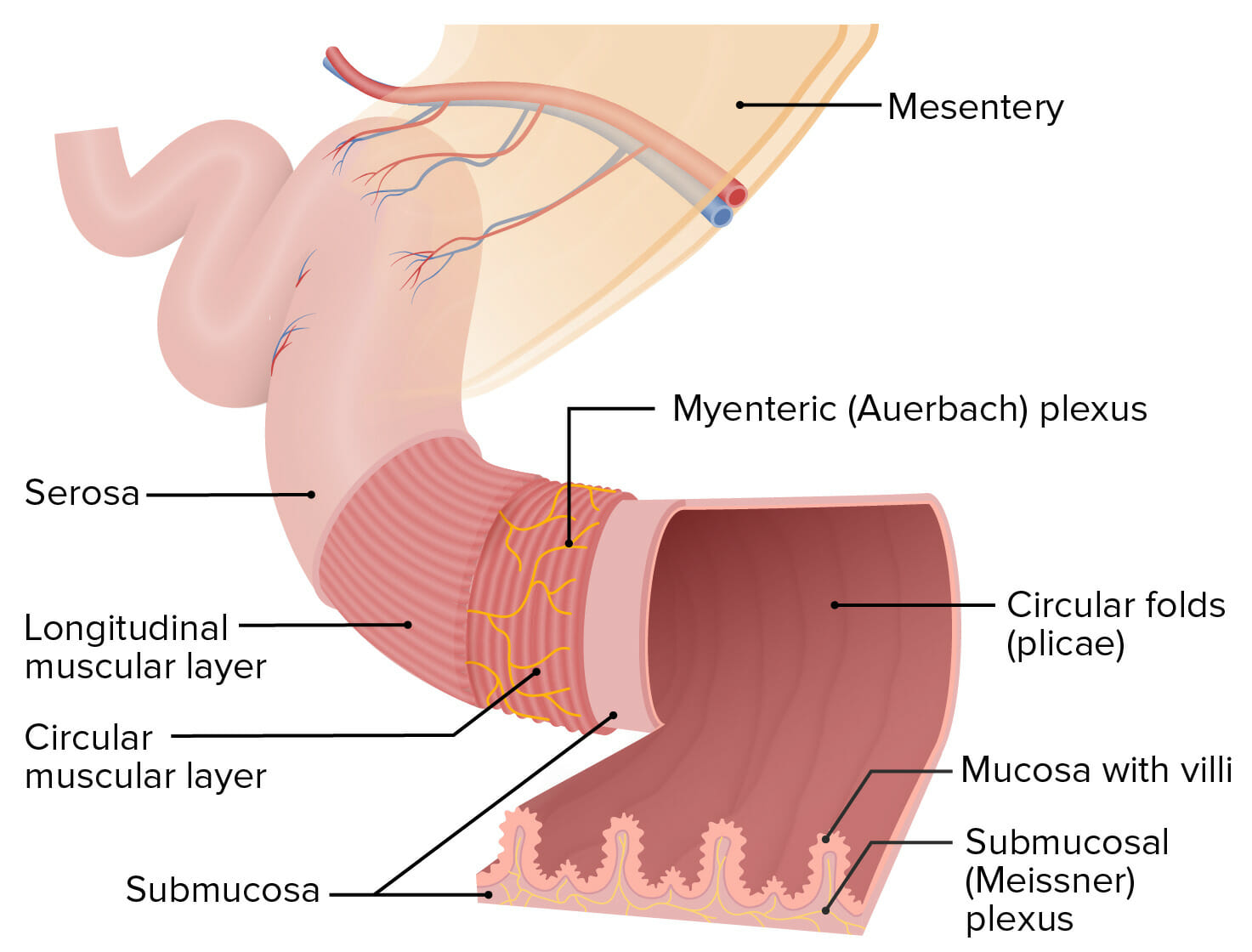
Structure of the intestinal walls:
The Meissner plexus is located within the submucosa, and the Auerbach plexus (also known as the myenteric plexus) is located between the circular and longitudinal muscular layers.

Image by Lecturio.

Function: local control of GI function

  • Controls peristalsis and segmentation
  • Stimulates the secretion Secretion Coagulation Studies of regulatory hormones Hormones Hormones are messenger molecules that are synthesized in one part of the body and move through the bloodstream to exert specific regulatory effects on another part of the body. Hormones play critical roles in coordinating cellular activities throughout the body in response to the constant changes in both the internal and external environments. Hormones: Overview and Types and neurotransmitters (see sections below)

Control and regulation of motility

  • The ICCs control the rate of smooth muscle contraction Smooth muscle contraction Smooth muscle is primarily found in the walls of hollow structures and some visceral organs, including the walls of the vasculature, GI, respiratory, and genitourinary tracts. Smooth muscle contracts more slowly and is regulated differently than skeletal muscle. Smooth muscle can be stimulated by nerve impulses, hormones, metabolic factors (like pH, CO2 or O2 levels), its own intrinsic pacemaker ability, or even mechanical stretch. Smooth Muscle Contraction via slow (electrical) waves:
    • Refers to the slowly changing membrane potential Membrane potential The membrane potential is the difference in electric charge between the interior and the exterior of a cell. All living cells maintain a potential difference across the membrane thanks to the insulating properties of their plasma membranes (PMs) and the selective transport of ions across this membrane by transporters. Membrane Potential of pacemaker Pacemaker A device designed to stimulate, by electric impulses, contraction of the heart muscles. It may be temporary (external) or permanent (internal or internal-external). Bradyarrhythmias cells (e.g., ICCs)
    • Pacemaker Pacemaker A device designed to stimulate, by electric impulses, contraction of the heart muscles. It may be temporary (external) or permanent (internal or internal-external). Bradyarrhythmias cells do not have a stable resting membrane potential Resting membrane potential Membrane Potential.
    • Instead, specific ion channels Channels The Cell: Cell Membrane allow for a slow flow Flow Blood flows through the heart, arteries, capillaries, and veins in a closed, continuous circuit. Flow is the movement of volume per unit of time. Flow is affected by the pressure gradient and the resistance fluid encounters between 2 points. Vascular resistance is the opposition to flow, which is caused primarily by blood friction against vessel walls. Vascular Resistance, Flow, and Mean Arterial Pressure of ions (i.e., current) across the membrane → slowly brings the membrane potential Membrane potential The membrane potential is the difference in electric charge between the interior and the exterior of a cell. All living cells maintain a potential difference across the membrane thanks to the insulating properties of their plasma membranes (PMs) and the selective transport of ions across this membrane by transporters. Membrane Potential closer to threshold Threshold Minimum voltage necessary to generate an action potential (an all-or-none response) Skeletal Muscle Contraction for depolarization Depolarization Membrane Potential
    • If threshold Threshold Minimum voltage necessary to generate an action potential (an all-or-none response) Skeletal Muscle Contraction is reached, an action potential Action Potential Abrupt changes in the membrane potential that sweep along the cell membrane of excitable cells in response to excitation stimuli. Membrane Potential (AP) occurs → muscle cells contract
    • These contractions are known as slow-wave contractions.
  • Frequencies of contractions:
    • Stomach Stomach The stomach is a muscular sac in the upper left portion of the abdomen that plays a critical role in digestion. The stomach develops from the foregut and connects the esophagus with the duodenum. Structurally, the stomach is C-shaped and forms a greater and lesser curvature and is divided grossly into regions: the cardia, fundus, body, and pylorus. Stomach: Anatomy: 3–5 contractions per minute
    • Small intestine Small intestine The small intestine is the longest part of the GI tract, extending from the pyloric orifice of the stomach to the ileocecal junction. The small intestine is the major organ responsible for chemical digestion and absorption of nutrients. It is divided into 3 segments: the duodenum, the jejunum, and the ileum. Small Intestine: Anatomy: 12–20 contractions per minute
    • Large intestine Large intestine The large intestines constitute the last portion of the digestive system. The large intestine consists of the cecum, appendix, colon (with ascending, transverse, descending, and sigmoid segments), rectum, and anal canal. The primary function of the colon is to remove water and compact the stool prior to expulsion from the body via the rectum and anal canal. Colon, Cecum, and Appendix: Anatomy: 6–8 contractions per minute
  • Frequency of contractions does not change significantly with regulation, but strength of contraction does.
  • Smooth muscle contractions are graded:
    • ↑ Release of Ca CA Condylomata acuminata are a clinical manifestation of genital HPV infection. Condylomata acuminata are described as raised, pearly, flesh-colored, papular, cauliflower-like lesions seen in the anogenital region that may cause itching, pain, or bleeding. Condylomata Acuminata (Genital Warts)2+ from the sarcoplasmic reticulum Sarcoplasmic Reticulum A network of tubules and sacs in the cytoplasm of skeletal muscle fibers that assist with muscle contraction and relaxation by releasing and storing calcium ions. Muscle Tissue: Histology (SR) in response to an AP → stronger contraction
    • Amount of Ca CA Condylomata acuminata are a clinical manifestation of genital HPV infection. Condylomata acuminata are described as raised, pearly, flesh-colored, papular, cauliflower-like lesions seen in the anogenital region that may cause itching, pain, or bleeding. Condylomata Acuminata (Genital Warts)2+ released from the SR can be regulated by the ANS ANS The ans is a component of the peripheral nervous system that uses both afferent (sensory) and efferent (effector) neurons, which control the functioning of the internal organs and involuntary processes via connections with the CNS. The ans consists of the sympathetic and parasympathetic nervous systems. Autonomic Nervous System: Anatomy and hormones Hormones Hormones are messenger molecules that are synthesized in one part of the body and move through the bloodstream to exert specific regulatory effects on another part of the body. Hormones play critical roles in coordinating cellular activities throughout the body in response to the constant changes in both the internal and external environments. Hormones: Overview and Types
  • The ENS receives input from:
    • Vagus nerve Vagus nerve The 10th cranial nerve. The vagus is a mixed nerve which contains somatic afferents (from skin in back of the ear and the external auditory meatus), visceral afferents (from the pharynx, larynx, thorax, and abdomen), parasympathetic efferents (to the thorax and abdomen), and efferents to striated muscle (of the larynx and pharynx). Pharynx: Anatomy (parasympathetic, stimulatory → ↑ motility)
    • Prevertebral ganglia (sympathetic, inhibitory → ↓ motility)
Slow electrical waves

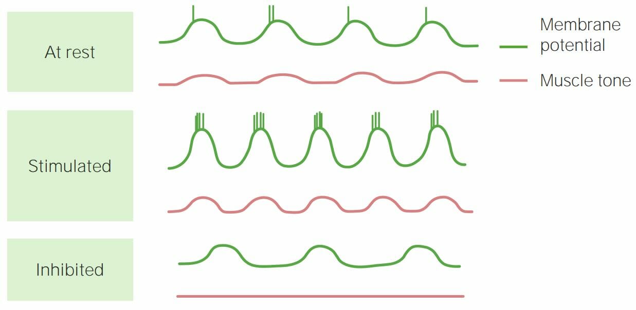
Schematic depiction of slow electrical waves and how they affect smooth muscle tone:
If the peak of the wave reaches the threshold required for an action potential (seen as a vertical spike in the membrane potential), muscle contraction occurs. When action potentials occur back to back in smooth muscle (Stimulated panel), contractions are stronger. If threshold is not reached (Inhibited panel), contractions do not occur.

Image by Lecturio.

Swallowing and Esophageal Motility

Swallowing

Swallowing (also called deglutition) is a complex process involving > 22 muscles in the mouth, pharynx Pharynx The pharynx is a component of the digestive system that lies posterior to the nasal cavity, oral cavity, and larynx. The pharynx can be divided into the oropharynx, nasopharynx, and laryngopharynx. Pharyngeal muscles play an integral role in vital processes such as breathing, swallowing, and speaking. Pharynx: Anatomy, and esophagus Esophagus The esophagus is a muscular tube-shaped organ of around 25 centimeters in length that connects the pharynx to the stomach. The organ extends from approximately the 6th cervical vertebra to the 11th thoracic vertebra and can be divided grossly into 3 parts: the cervical part, the thoracic part, and the abdominal part. Esophagus: Anatomy, which moves food in the mouth through the upper esophageal sphincter Upper esophageal sphincter The structure at the pharyngoesophageal junction consisting chiefly of the cricopharyngeus muscle. It normally occludes the lumen of the esophagus, except during swallowing. Esophagus: Anatomy and into the esophagus Esophagus The esophagus is a muscular tube-shaped organ of around 25 centimeters in length that connects the pharynx to the stomach. The organ extends from approximately the 6th cervical vertebra to the 11th thoracic vertebra and can be divided grossly into 3 parts: the cervical part, the thoracic part, and the abdominal part. Esophagus: Anatomy.
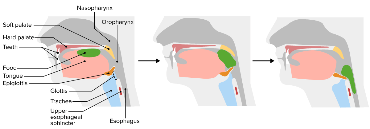
  • The tongue Tongue The tongue, on the other hand, is a complex muscular structure that permits tasting and facilitates the process of mastication and communication. The blood supply of the tongue originates from the external carotid artery, and the innervation is through cranial nerves. Lips and Tongue: Anatomy shapes and lubricates the bolus.
  • Initiation of swallowing is a voluntary process → tongue Tongue The tongue, on the other hand, is a complex muscular structure that permits tasting and facilitates the process of mastication and communication. The blood supply of the tongue originates from the external carotid artery, and the innervation is through cranial nerves. Lips and Tongue: Anatomy voluntarily moves the bolus toward the back of the mouth → initiates involuntary reflexive process
  • Mechanoreceptors in the oropharynx Oropharynx The middle portion of the pharynx that lies posterior to the mouth, inferior to the soft palate, and superior to the base of the tongue and epiglottis. It has a digestive function as food passes from the mouth into the oropharynx before entering esophagus. Pharynx: Anatomy are activated.
  • Afferents travel through the glossopharyngeal nerve Glossopharyngeal nerve The 9th cranial nerve. The glossopharyngeal nerve is a mixed motor and sensory nerve; it conveys somatic and autonomic efferents as well as general, special, and visceral afferents. Among the connections are motor fibers to the stylopharyngeus muscle, parasympathetic fibers to the parotid glands, general and taste afferents from the posterior third of the tongue, the nasopharynx, and the palate, and afferents from baroreceptors and chemoreceptor cells of the carotid sinus. Pharynx: Anatomy (IX) to the swallowing centers in the medulla oblongata Medulla Oblongata The lower portion of the brain stem. It is inferior to the pons and anterior to the cerebellum. Medulla oblongata serves as a relay station between the brain and the spinal cord, and contains centers for regulating respiratory, vasomotor, cardiac, and reflex activities. Brain Stem: Anatomy and pons Pons The front part of the hindbrain (rhombencephalon) that lies between the medulla and the midbrain (mesencephalon) ventral to the cerebellum. It is composed of two parts, the dorsal and the ventral. The pons serves as a relay station for neural pathways between the cerebellum to the cerebrum. Brain Stem: Anatomy.
  • Motor Motor Neurons which send impulses peripherally to activate muscles or secretory cells. Nervous System: Histology efferents travel through the vagus nerves (X) → stimulate the pharyngeal muscles Pharyngeal Muscles The muscles of the pharynx are voluntary muscles arranged in two layers. The external circular layer consists of three constrictors (superior, middle, and inferior). The internal longitudinal layer consists of the palatopharyngeus, the salpingopharyngeus, and the stylopharyngeus. During swallowing, the outer layer constricts the pharyngeal wall and the inner layer elevates pharynx and larynx. Pharynx: Anatomy:
    • Base of the tongue Tongue The tongue, on the other hand, is a complex muscular structure that permits tasting and facilitates the process of mastication and communication. The blood supply of the tongue originates from the external carotid artery, and the innervation is through cranial nerves. Lips and Tongue: Anatomy compresses against the hard palate Hard palate The anteriorly located rigid section of the palate. Palate: Anatomy → closes off the oropharynx Oropharynx The middle portion of the pharynx that lies posterior to the mouth, inferior to the soft palate, and superior to the base of the tongue and epiglottis. It has a digestive function as food passes from the mouth into the oropharynx before entering esophagus. Pharynx: Anatomy from the rest of the mouth
    • Soft palate Soft palate A movable fold suspended from the posterior border of the hard palate. The uvula hangs from the middle of the lower border. Palate: Anatomy moves upward → closes off the nasopharynx Nasopharynx The top portion of the pharynx situated posterior to the nose and superior to the soft palate. The nasopharynx is the posterior extension of the nasal cavities and has a respiratory function. Pharynx: Anatomy
    • Laryngeal muscles Laryngeal muscles The striated muscle groups which move the larynx as a whole or its parts, such as altering tension of the vocal cords, or size of the slit (rima glottidis). Larynx: Anatomy move forward and upward → close the airways
  • Upper esophageal sphincter Upper esophageal sphincter The structure at the pharyngoesophageal junction consisting chiefly of the cricopharyngeus muscle. It normally occludes the lumen of the esophagus, except during swallowing. Esophagus: Anatomy muscles first relax and then contract → force foods down into the esophagus Esophagus The esophagus is a muscular tube-shaped organ of around 25 centimeters in length that connects the pharynx to the stomach. The organ extends from approximately the 6th cervical vertebra to the 11th thoracic vertebra and can be divided grossly into 3 parts: the cervical part, the thoracic part, and the abdominal part. Esophagus: Anatomy and then close to prevent immediate reflux
  • Stretch in the esophagus Esophagus The esophagus is a muscular tube-shaped organ of around 25 centimeters in length that connects the pharynx to the stomach. The organ extends from approximately the 6th cervical vertebra to the 11th thoracic vertebra and can be divided grossly into 3 parts: the cervical part, the thoracic part, and the abdominal part. Esophagus: Anatomy triggers esophageal peristaltic waves
Swallowing

Swallowing:
First, the tongue compresses the food against the hard palate (note the glottis is open). Next, the tongue forces the bolus to the back of the mouth; the soft palate closes off the nasopharynx. Finally, as the foodstuff is forced downwards, the epiglottis is pulled forwards, closing the glottis (airway), and the upper esophageal sphincter relaxes, letting food into the esophagus. Peristaltic waves move the food down towards the stomach.

Image by Lecturio.

Normal esophageal motility

  • The 1st ⅓ of the esophagus Esophagus The esophagus is a muscular tube-shaped organ of around 25 centimeters in length that connects the pharynx to the stomach. The organ extends from approximately the 6th cervical vertebra to the 11th thoracic vertebra and can be divided grossly into 3 parts: the cervical part, the thoracic part, and the abdominal part. Esophagus: Anatomy has a striated muscle Striated muscle One of two types of muscle in the body, characterized by the array of bands observed under microscope. Striated muscles can be divided into two subtypes: the cardiac muscle and the skeletal muscle. Muscle Tissue: Histology layer.
  • The remaining ⅔ has a smooth muscle layer.
  • Primary peristalsis:
    • 1st wave of esophageal peristalsis that begins as food enters the esophagus Esophagus The esophagus is a muscular tube-shaped organ of around 25 centimeters in length that connects the pharynx to the stomach. The organ extends from approximately the 6th cervical vertebra to the 11th thoracic vertebra and can be divided grossly into 3 parts: the cervical part, the thoracic part, and the abdominal part. Esophagus: Anatomy
    • Coordinated by the PNS and ENS
  • Secondary peristalsis: a 2nd wave of peristalsis responsible for:
    • Clearing the esophagus Esophagus The esophagus is a muscular tube-shaped organ of around 25 centimeters in length that connects the pharynx to the stomach. The organ extends from approximately the 6th cervical vertebra to the 11th thoracic vertebra and can be divided grossly into 3 parts: the cervical part, the thoracic part, and the abdominal part. Esophagus: Anatomy of remnants of the foodstuff
    • Removing acidic content that may have refluxed 
  • Relaxation of the lower esophageal sphincter Lower Esophageal Sphincter Esophagus: Anatomy and upper stomach Stomach The stomach is a muscular sac in the upper left portion of the abdomen that plays a critical role in digestion. The stomach develops from the foregut and connects the esophagus with the duodenum. Structurally, the stomach is C-shaped and forms a greater and lesser curvature and is divided grossly into regions: the cardia, fundus, body, and pylorus. Stomach: Anatomy allowing the stomach Stomach The stomach is a muscular sac in the upper left portion of the abdomen that plays a critical role in digestion. The stomach develops from the foregut and connects the esophagus with the duodenum. Structurally, the stomach is C-shaped and forms a greater and lesser curvature and is divided grossly into regions: the cardia, fundus, body, and pylorus. Stomach: Anatomy to accept food (mediated by VIP VIP A highly basic, 28 amino acid neuropeptide released from intestinal mucosa. It has a wide range of biological actions affecting the cardiovascular, gastrointestinal, and respiratory systems and is neuroprotective. It binds special receptors. Gastrointestinal Neural and Hormonal Signaling)
Esophagus innervation

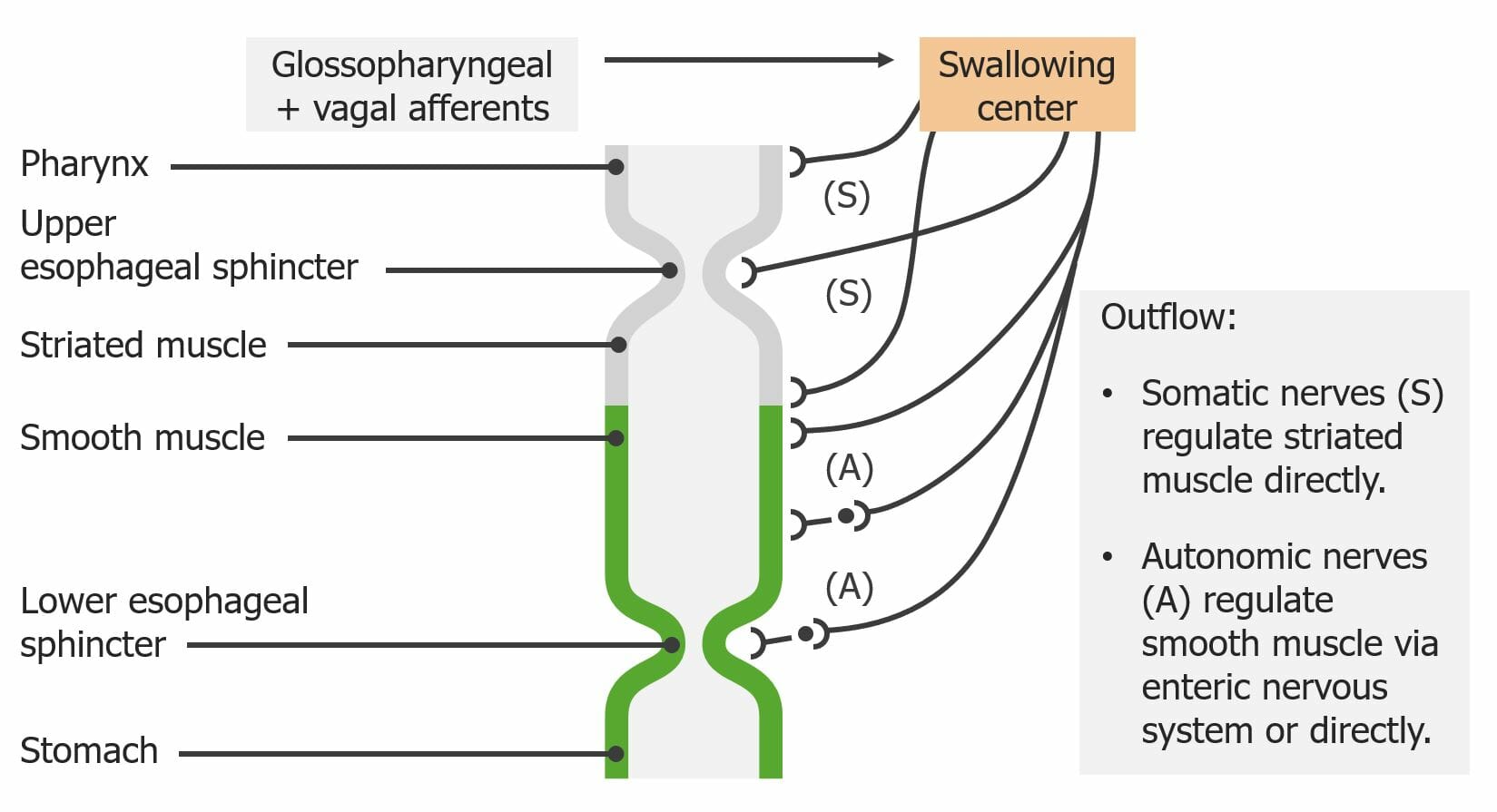
Innervation of the esophagus:
Note how the swallowing center integrates striated and smooth muscle activity.

Image by Lecturio.

Vomiting Vomiting The forcible expulsion of the contents of the stomach through the mouth. Hypokalemia

  • Induced by:
    • Excessive stretching of the stomach Stomach The stomach is a muscular sac in the upper left portion of the abdomen that plays a critical role in digestion. The stomach develops from the foregut and connects the esophagus with the duodenum. Structurally, the stomach is C-shaped and forms a greater and lesser curvature and is divided grossly into regions: the cardia, fundus, body, and pylorus. Stomach: Anatomy
    • Psychological stimuli
    • Chemical irritants (e.g., bacterial toxins, alcohol)
  • Controlled by the emetic center in the medulla, which stimulates:
    • Relaxation of the lower esophageal sphincter Lower Esophageal Sphincter Esophagus: Anatomy
    • Contraction of the diaphragm Diaphragm The diaphragm is a large, dome-shaped muscle that separates the thoracic cavity from the abdominal cavity. The diaphragm consists of muscle fibers and a large central tendon, which is divided into right and left parts. As the primary muscle of inspiration, the diaphragm contributes 75% of the total inspiratory muscle force. Diaphragm: Anatomy and abdominal muscles
  • These muscles compress the stomach Stomach The stomach is a muscular sac in the upper left portion of the abdomen that plays a critical role in digestion. The stomach develops from the foregut and connects the esophagus with the duodenum. Structurally, the stomach is C-shaped and forms a greater and lesser curvature and is divided grossly into regions: the cardia, fundus, body, and pylorus. Stomach: Anatomy → force food back up the esophagus Esophagus The esophagus is a muscular tube-shaped organ of around 25 centimeters in length that connects the pharynx to the stomach. The organ extends from approximately the 6th cervical vertebra to the 11th thoracic vertebra and can be divided grossly into 3 parts: the cervical part, the thoracic part, and the abdominal part. Esophagus: Anatomy

Gastric Motility

  • Regions of the stomach Stomach The stomach is a muscular sac in the upper left portion of the abdomen that plays a critical role in digestion. The stomach develops from the foregut and connects the esophagus with the duodenum. Structurally, the stomach is C-shaped and forms a greater and lesser curvature and is divided grossly into regions: the cardia, fundus, body, and pylorus. Stomach: Anatomy and their functions:
    • Lower esophageal sphincter Lower Esophageal Sphincter Esophagus: Anatomy: controls movement between the stomach Stomach The stomach is a muscular sac in the upper left portion of the abdomen that plays a critical role in digestion. The stomach develops from the foregut and connects the esophagus with the duodenum. Structurally, the stomach is C-shaped and forms a greater and lesser curvature and is divided grossly into regions: the cardia, fundus, body, and pylorus. Stomach: Anatomy and esophagus Esophagus The esophagus is a muscular tube-shaped organ of around 25 centimeters in length that connects the pharynx to the stomach. The organ extends from approximately the 6th cervical vertebra to the 11th thoracic vertebra and can be divided grossly into 3 parts: the cervical part, the thoracic part, and the abdominal part. Esophagus: Anatomy 
    • Cardia: relaxes to allow food entry into the stomach Stomach The stomach is a muscular sac in the upper left portion of the abdomen that plays a critical role in digestion. The stomach develops from the foregut and connects the esophagus with the duodenum. Structurally, the stomach is C-shaped and forms a greater and lesser curvature and is divided grossly into regions: the cardia, fundus, body, and pylorus. Stomach: Anatomy 
    • Fundus Fundus The superior portion of the body of the stomach above the level of the cardiac notch. Stomach: Anatomy and body:
      • Act as a reservoir Reservoir Animate or inanimate sources which normally harbor disease-causing organisms and thus serve as potential sources of disease outbreaks. Reservoirs are distinguished from vectors (disease vectors) and carriers, which are agents of disease transmission rather than continuing sources of potential disease outbreaks. Humans may serve both as disease reservoirs and carriers. Escherichia coli
      • Contain ICCs: pacemaker Pacemaker A device designed to stimulate, by electric impulses, contraction of the heart muscles. It may be temporary (external) or permanent (internal or internal-external). Bradyarrhythmias cells triggering regular Regular Insulin contractions in the stomach Stomach The stomach is a muscular sac in the upper left portion of the abdomen that plays a critical role in digestion. The stomach develops from the foregut and connects the esophagus with the duodenum. Structurally, the stomach is C-shaped and forms a greater and lesser curvature and is divided grossly into regions: the cardia, fundus, body, and pylorus. Stomach: Anatomy
    • Antrum: mixing and grinding 
    • Pylorus Pylorus The region between the sharp indentation at the lower third of the stomach (incisura angularis) and the junction of the pylorus with the duodenum. Pyloric antral glands contain mucus-secreting cells and gastrin-secreting endocrine cells (g cells). Stomach: Anatomy: controls release into the duodenum Duodenum The shortest and widest portion of the small intestine adjacent to the pylorus of the stomach. It is named for having the length equal to about the width of 12 fingers. Small Intestine: Anatomy
  • Reception–relaxation response:
    • A property of GI smooth muscle cells
    • The stomach Stomach The stomach is a muscular sac in the upper left portion of the abdomen that plays a critical role in digestion. The stomach develops from the foregut and connects the esophagus with the duodenum. Structurally, the stomach is C-shaped and forms a greater and lesser curvature and is divided grossly into regions: the cardia, fundus, body, and pylorus. Stomach: Anatomy briefly resists stretching → reception–relaxation reflex then triggers it to relax
  • Segmentation: back and forth movement of contents, causing mixing 
  • Peristaltic movement:
    • Triggered by ICCs 3‒5 times per minute (approximately every 15 sec) 
    • Causes a pressure wave starting at the fundus Fundus The superior portion of the body of the stomach above the level of the cardiac notch. Stomach: Anatomy pylorus Pylorus The region between the sharp indentation at the lower third of the stomach (incisura angularis) and the junction of the pylorus with the duodenum. Pyloric antral glands contain mucus-secreting cells and gastrin-secreting endocrine cells (g cells). Stomach: Anatomy
    • Pressure increases enough to overcome the pyloric valve and move a small amount (approximately 3 mL) of chyme Chyme Small Intestine: Anatomy into the duodenum Duodenum The shortest and widest portion of the small intestine adjacent to the pylorus of the stomach. It is named for having the length equal to about the width of 12 fingers. Small Intestine: Anatomy with each wave
  • Gastric emptying:
    • Contents leave the stomach Stomach The stomach is a muscular sac in the upper left portion of the abdomen that plays a critical role in digestion. The stomach develops from the foregut and connects the esophagus with the duodenum. Structurally, the stomach is C-shaped and forms a greater and lesser curvature and is divided grossly into regions: the cardia, fundus, body, and pylorus. Stomach: Anatomy after differing lengths of time (in order from fastest to slowest):
      • Carbohydrates Carbohydrates A class of organic compounds composed of carbon, hydrogen, and oxygen in a ratio of cn(H2O)n. The largest class of organic compounds, including starch; glycogen; cellulose; polysaccharides; and simple monosaccharides. Basics of Carbohydrates > proteins Proteins Linear polypeptides that are synthesized on ribosomes and may be further modified, crosslinked, cleaved, or assembled into complex proteins with several subunits. The specific sequence of amino acids determines the shape the polypeptide will take, during protein folding, and the function of the protein. Energy Homeostasis > lipids Lipids Lipids are a diverse group of hydrophobic organic molecules, which include fats, oils, sterols, and waxes. Fatty Acids and Lipids 
      • Liquids > solids
      • High-osmolality contents (i.e., ↑ Na+ content) > lower-osmolality content
    • Typical gastric emptying time:
      • 4 hours for a typical meal
      • Up to 6 hours for salty, high-fat meals
Regions of the stomach and their functions

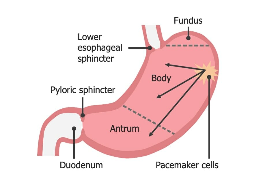
Regions of the stomach and their functions:
In the body of the stomach, the interstitial cells of Cajal are the pacemaker cells triggering regular peristaltic movements.

Image by Lecturio.

Intestinal Motility and Defecation

Segmentation

Mixes chyme Chyme Small Intestine: Anatomy:

  • With bile Bile An emulsifying agent produced in the liver and secreted into the duodenum. Its composition includes bile acids and salts; cholesterol; and electrolytes. It aids digestion of fats in the duodenum. Gallbladder and Biliary Tract: Anatomy and intestinal and pancreatic juices
  • To ensure adequate contact with the intestinal wall and its membrane-bound proteins Proteins Linear polypeptides that are synthesized on ribosomes and may be further modified, crosslinked, cleaved, or assembled into complex proteins with several subunits. The specific sequence of amino acids determines the shape the polypeptide will take, during protein folding, and the function of the protein. Energy Homeostasis:
    • Brush-border digestive enzymes Enzymes Enzymes are complex protein biocatalysts that accelerate chemical reactions without being consumed by them. Due to the body’s constant metabolic needs, the absence of enzymes would make life unsustainable, as reactions would occur too slowly without these molecules. Basics of Enzymes 
    • Absorptive transport proteins Transport proteins Proteins and Peptides

Migrating motor Motor Neurons which send impulses peripherally to activate muscles or secretory cells. Nervous System: Histology complex (MMC)

  • Describes a pattern of internal electrical stimulation through the intestines that triggers large peristaltic waves
  • These waves propel residual material down the tract:
    • The 1st wave begins in the stomach Stomach The stomach is a muscular sac in the upper left portion of the abdomen that plays a critical role in digestion. The stomach develops from the foregut and connects the esophagus with the duodenum. Structurally, the stomach is C-shaped and forms a greater and lesser curvature and is divided grossly into regions: the cardia, fundus, body, and pylorus. Stomach: Anatomy or duodenum Duodenum The shortest and widest portion of the small intestine adjacent to the pylorus of the stomach. It is named for having the length equal to about the width of 12 fingers. Small Intestine: Anatomy → travels approximately 10–70 cm before dying out
    • The 2nd wave begins slightly farther down the tract from where the 1st one started.
    • The 3rd wave begins even farther down than the 2nd one, and so on.
  • “Milks” remaining content toward the colon Colon The large intestines constitute the last portion of the digestive system. The large intestine consists of the cecum, appendix, colon (with ascending, transverse, descending, and sigmoid segments), rectum, and anal canal. The primary function of the colon is to remove water and compact the stool prior to expulsion from the body via the rectum and anal canal. Colon, Cecum, and Appendix: Anatomy
  • Takes about 2 hours to move through the entire intestinal tract
  • Occurs during fasting (known as the interdigestive phase)
  • A 2nd MMC may occur:
    • Moves bacteria Bacteria Bacteria are prokaryotic single-celled microorganisms that are metabolically active and divide by binary fission. Some of these organisms play a significant role in the pathogenesis of diseases. Bacteriology from the small intestine Small intestine The small intestine is the longest part of the GI tract, extending from the pyloric orifice of the stomach to the ileocecal junction. The small intestine is the major organ responsible for chemical digestion and absorption of nutrients. It is divided into 3 segments: the duodenum, the jejunum, and the ileum. Small Intestine: Anatomy large intestine Large intestine The large intestines constitute the last portion of the digestive system. The large intestine consists of the cecum, appendix, colon (with ascending, transverse, descending, and sigmoid segments), rectum, and anal canal. The primary function of the colon is to remove water and compact the stool prior to expulsion from the body via the rectum and anal canal. Colon, Cecum, and Appendix: Anatomy
    • MMC dysfunction can lead to small intestinal bacterial overgrowth Bacterial overgrowth Lactose Intolerance
  • Phases of the MMC:
  • Motilin Motilin A peptide of about 22-amino acids isolated from the duodenum. At low ph it inhibits gastric motor activity, whereas at high ph it has a stimulating effect. Gastrointestinal Secretions: hormone thought to initiate an MMC
Waves of the migrating motor complex

A graph showing the waves of the migrating motor complex during fasting:
The red dotted line shows the phase 3 waves.

Image by Lecturio.

Defecation

The rectum Rectum The rectum and anal canal are the most terminal parts of the lower GI tract/large intestine that form a functional unit and control defecation. Fecal continence is maintained by several important anatomic structures including rectal folds, anal valves, the sling-like puborectalis muscle, and internal and external anal sphincters. Rectum and Anal Canal: Anatomy contains stretch receptors Receptors Receptors are proteins located either on the surface of or within a cell that can bind to signaling molecules known as ligands (e.g., hormones) and cause some type of response within the cell. Receptors that stimulate the defecation reflex when the rectum Rectum The rectum and anal canal are the most terminal parts of the lower GI tract/large intestine that form a functional unit and control defecation. Fecal continence is maintained by several important anatomic structures including rectal folds, anal valves, the sling-like puborectalis muscle, and internal and external anal sphincters. Rectum and Anal Canal: Anatomy begins to fill with feces.

  • Stretch receptors Receptors Receptors are proteins located either on the surface of or within a cell that can bind to signaling molecules known as ligands (e.g., hormones) and cause some type of response within the cell. Receptors activated in rectum Rectum The rectum and anal canal are the most terminal parts of the lower GI tract/large intestine that form a functional unit and control defecation. Fecal continence is maintained by several important anatomic structures including rectal folds, anal valves, the sling-like puborectalis muscle, and internal and external anal sphincters. Rectum and Anal Canal: Anatomy → 
  • Sensory Sensory Neurons which conduct nerve impulses to the central nervous system. Nervous System: Histology nerve fibers Nerve Fibers Slender processes of neurons, including the axons and their glial envelopes (myelin sheath). Nerve fibers conduct nerve impulses to and from the central nervous system. Nervous System: Histology carry signal to the sacral spinal cord Spinal cord The spinal cord is the major conduction pathway connecting the brain to the body; it is part of the CNS. In cross section, the spinal cord is divided into an H-shaped area of gray matter (consisting of synapsing neuronal cell bodies) and a surrounding area of white matter (consisting of ascending and descending tracts of myelinated axons). Spinal Cord: Anatomy → 
  • Synapse Synapse The junction between 2 neurons is called a synapse. The synapse allows a neuron to pass an electrical or chemical signal to another neuron or target effector cell. Synapses and Neurotransmission with parasympathetic motor Motor Neurons which send impulses peripherally to activate muscles or secretory cells. Nervous System: Histology fibers → 
  • Send signals for peristaltic waves to the myenteric nerve plexus in the muscular layers of:
    • Descending colon Descending colon The segment of large intestine between transverse colon and the sigmoid colon. Colon, Cecum, and Appendix: Anatomy
    • Sigmoid Sigmoid A segment of the colon between the rectum and the descending colon. Volvulus colon Colon The large intestines constitute the last portion of the digestive system. The large intestine consists of the cecum, appendix, colon (with ascending, transverse, descending, and sigmoid segments), rectum, and anal canal. The primary function of the colon is to remove water and compact the stool prior to expulsion from the body via the rectum and anal canal. Colon, Cecum, and Appendix: Anatomy
    • Rectum Rectum The rectum and anal canal are the most terminal parts of the lower GI tract/large intestine that form a functional unit and control defecation. Fecal continence is maintained by several important anatomic structures including rectal folds, anal valves, the sling-like puborectalis muscle, and internal and external anal sphincters. Rectum and Anal Canal: Anatomy
    • Internal anal sphincter Internal anal sphincter Rectum and Anal Canal: Anatomy
  • Peristaltic contractions in the colon Colon The large intestines constitute the last portion of the digestive system. The large intestine consists of the cecum, appendix, colon (with ascending, transverse, descending, and sigmoid segments), rectum, and anal canal. The primary function of the colon is to remove water and compact the stool prior to expulsion from the body via the rectum and anal canal. Colon, Cecum, and Appendix: Anatomy and rectum Rectum The rectum and anal canal are the most terminal parts of the lower GI tract/large intestine that form a functional unit and control defecation. Fecal continence is maintained by several important anatomic structures including rectal folds, anal valves, the sling-like puborectalis muscle, and internal and external anal sphincters. Rectum and Anal Canal: Anatomy move feces downward.
  • Internal anal sphincter Internal anal sphincter Rectum and Anal Canal: Anatomy relaxes.
  • Defecation occurs only if the external anal sphincter External anal sphincter Rectum and Anal Canal: Anatomy is voluntarily relaxed at the same time via motor Motor Neurons which send impulses peripherally to activate muscles or secretory cells. Nervous System: Histology impulses from the cerebral cortex Cerebral cortex The cerebral cortex is the largest and most developed part of the human brain and CNS. Occupying the upper part of the cranial cavity, the cerebral cortex has 4 lobes and is divided into 2 hemispheres that are joined centrally by the corpus callosum. Cerebral Cortex: Anatomy
  • If defecation is consciously suppressed, peristaltic contractions cease within a few minutes.
The defecation reflex

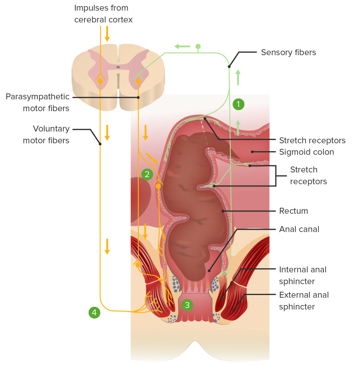
The defecation reflex:
1. Feces stretch the rectum and stimulate the stretch receptors, transmitting the signal to the spinal cord.
2. A spinal reflex sends parasympathetic motor signals to the myenteric nerve plexus, resulting in contraction of the smooth muscles within the rectum and pushing feces downward.
3. The same spinal reflex also sends parasympathetic motor signals to relax the internal anal sphincter.
4. Voluntary impulses from the brain prevent defecation by keeping the external anal sphincter contracted. Defecation will occur if voluntary signals allow the external anal sphincter to relax.

Image by Lecturio.

Clinical Relevance

  • Achalasia Achalasia Achalasia is a primary esophageal motility disorder that develops from the degeneration of the myenteric plexus. This condition results in impaired lower esophageal sphincter relaxation and absence of normal esophageal peristalsis. Patients typically present with dysphagia to solids and liquids along with regurgitation. Achalasia: primary esophageal motility disorder that develops from degeneration of the myenteric plexus Myenteric plexus One of two ganglionated neural networks which together form the enteric nervous system. The myenteric (Auerbach’s) plexus is located between the longitudinal and circular muscle layers of the gut. Its neurons project to the circular muscle, to other myenteric ganglia, to submucosal ganglia, or directly to the epithelium, and play an important role in regulating and patterning gut motility. Gastrointestinal Neural and Hormonal Signaling. This degeneration results in impaired lower esophageal sphincter Lower Esophageal Sphincter Esophagus: Anatomy relaxation and absence of normal esophageal peristalsis. Presentation is typically with dysphagia Dysphagia Dysphagia is the subjective sensation of difficulty swallowing. Symptoms can range from a complete inability to swallow, to the sensation of solids or liquids becoming “stuck.” Dysphagia is classified as either oropharyngeal or esophageal, with esophageal dysphagia having 2 sub-types: functional and mechanical. Dysphagia to solids and liquids along with regurgitation Regurgitation Gastroesophageal Reflux Disease (GERD)
  • Gastroparesis Gastroparesis Chronic delayed gastric emptying. Gastroparesis may be caused by motor dysfunction or paralysis of stomach muscles or may be associated with other systemic diseases such as diabetes mellitus. Malabsorption and Maldigestion: impaired or delayed gastric emptying without evidence of obstruction. Gastroparesis Gastroparesis Chronic delayed gastric emptying. Gastroparesis may be caused by motor dysfunction or paralysis of stomach muscles or may be associated with other systemic diseases such as diabetes mellitus. Malabsorption and Maldigestion can be idiopathic Idiopathic Dermatomyositis or due to systemic causes, such as diabetes Diabetes Diabetes mellitus (DM) is a metabolic disease characterized by hyperglycemia and dysfunction of the regulation of glucose metabolism by insulin. Type 1 DM is diagnosed mostly in children and young adults as the result of autoimmune destruction of β cells in the pancreas and the resulting lack of insulin. Type 2 DM has a significant association with obesity and is characterized by insulin resistance. Diabetes Mellitus mellitus or scleroderma Scleroderma Scleroderma (systemic sclerosis) is an autoimmune condition characterized by diffuse collagen deposition and fibrosis. The clinical presentation varies from limited skin involvement to diffuse involvement of internal organs. Scleroderma
  • Irritable bowel syndrome Irritable bowel syndrome Irritable bowel syndrome (IBS) is a functional bowel disease characterized by chronic abdominal pain and altered bowel habits without an identifiable organic cause. The etiology and pathophysiology of this disease are not well understood, and there are many factors that may contribute. Irritable Bowel Syndrome: group of diseases consisting of various GI symptoms (e.g., bloating Bloating Constipation, diarrhea Diarrhea Diarrhea is defined as ≥ 3 watery or loose stools in a 24-hour period. There are a multitude of etiologies, which can be classified based on the underlying mechanism of disease. The duration of symptoms (acute or chronic) and characteristics of the stools (e.g., watery, bloody, steatorrheic, mucoid) can help guide further diagnostic evaluation. Diarrhea, abdominal pain Abdominal Pain Acute Abdomen) without an identifiable organic cause. Some individuals have GI motility abnormalities.
  • Constipation Constipation Constipation is common and may be due to a variety of causes. Constipation is generally defined as bowel movement frequency < 3 times per week. Patients who are constipated often strain to pass hard stools. The condition is classified as primary (also known as idiopathic or functional constipation) or secondary, and as acute or chronic. Constipation: symptom that is generally defined as bowel movement frequency < 3 times per week. Constipated individuals often pass hard stools with straining.
  • Hirschsprung disease Hirschsprung Disease Hirschsprung disease (HD), also known as congenital aganglionosis or congenital megacolon, is a congenital anomaly of the colon caused by the failure of neural crest-derived ganglion cells to migrate into the distal colon. The lack of innervation always involves the rectum and extends proximally and contiguously over variable distances. M Hirschsprung Disease: also called congenital Congenital Chorioretinitis megacolon Megacolon Megacolon is a severe, abnormal dilatation of the colon, and is classified as acute or chronic. There are many etiologies of megacolon, including neuropathic and dysmotility conditions, severe infections, ischemia, and inflammatory bowel disease. Megacolon. Hirschsprung disease Hirschsprung Disease Hirschsprung disease (HD), also known as congenital aganglionosis or congenital megacolon, is a congenital anomaly of the colon caused by the failure of neural crest-derived ganglion cells to migrate into the distal colon. The lack of innervation always involves the rectum and extends proximally and contiguously over variable distances. M Hirschsprung Disease is a condition that occurs as a consequence of the lack of ganglionic cells and ICCs in the last segment of the large intestine Large intestine The large intestines constitute the last portion of the digestive system. The large intestine consists of the cecum, appendix, colon (with ascending, transverse, descending, and sigmoid segments), rectum, and anal canal. The primary function of the colon is to remove water and compact the stool prior to expulsion from the body via the rectum and anal canal. Colon, Cecum, and Appendix: Anatomy, producing permanent contraction of the internal anal sphincter Internal anal sphincter Rectum and Anal Canal: Anatomy and dilation of the segment that precedes it.
  • Fecal incontinence Fecal incontinence Failure of voluntary control of the anal sphincters, with involuntary passage of feces and flatus. Pediatric Constipation: unintentional release of fecal mater due to an inability of the anal sphincter complex to maintain adequate tone.

References

  1. Boland, M. (2016). Human digestion—a processing perspective. J Sci Food Agric 96:2275–2283. https://pubmed.ncbi.nlm.nih.gov/26711173/
  2. Cheng, L.K., et al. (2010). Gastrointestinal system. Wiley Interdiscip Rev Syst Biol Med 2:65–79. https://wires.onlinelibrary.wiley.com/doi/abs/10.1002/wsbm.19
  3. Konturek, P.C., Brzozowski, T., Konturek, S.J. (2011). Stress and the gut: pathophysiology, clinical consequences, diagnostic approach and treatment options. J Physiol Pharmacol 62:591–599. https://pubmed.ncbi.nlm.nih.gov/22314561/
  4. Kusano, M., et al. (2014). Gastrointestinal motility and functional gastrointestinal diseases. Curr Pharm Des 20:2775–2282. https://pubmed.ncbi.nlm.nih.gov/23886379/
  5. Sanders, K.M., Koh, S.D., Ro, S., Ward, S.M. (2012). Regulation of gastrointestinal motility—insights from smooth muscle biology. Nat Rev Gastroenterol Hepatol 9:633–645. https://www.nature.com/articles/nrgastro.2012.168

Create your free account or log in to continue reading!

Sign up now and get free access to Lecturio with concept pages, medical videos, and questions for your medical education.

User Reviews

Details