Achieve Mastery of Medical Concepts

Study for medical school and boards with Lecturio

Gastrointestinal Neural and Hormonal Signaling

Function of the GI system is highly regulated through neural and hormonal signaling. Much of this regulation comes from the ANS. Parasympathetic stimulation is excitatory, triggering digestive secretions, an increase in GI blood flow, and movement of material through the tract, while sympathetic stimulation is inhibitory and has the opposite effects. The GI tract also has its own enteric nervous system, which controls numerous local GI reflexes. For example, the presence of a particular nutrient may be detected by chemoreceptors and may trigger release of a particular digestive enzyme, which is coordinated entirely by the enteric nervous system (without input from the brain). In addition, there are numerous hormones and paracrine signaling molecules released by GI organs that similarly affect GI function.

Last updated: Dec 12, 2022

Editorial responsibility: Stanley Oiseth, Lindsay Jones, Evelin Maza

Overview of GI Tract Physiology

Functions of the GI tract

The GI system is constantly working to meet human energy demands. To achieve this end, the GI system has several major functions:

  • Digestion Digestion Digestion refers to the process of the mechanical and chemical breakdown of food into smaller particles, which can then be absorbed and utilized by the body. Digestion and Absorption of complex nutrients:
    • Mechanical 
    • Chemical
  • Absorption Absorption Absorption involves the uptake of nutrient molecules and their transfer from the lumen of the GI tract across the enterocytes and into the interstitial space, where they can be taken up in the venous or lymphatic circulation. Digestion and Absorption of nutrients and water (its critical function)
  • Secretion Secretion Coagulation Studies of:
    • Digestive enzymes Enzymes Enzymes are complex protein biocatalysts that accelerate chemical reactions without being consumed by them. Due to the body’s constant metabolic needs, the absence of enzymes would make life unsustainable, as reactions would occur too slowly without these molecules. Basics of Enzymes
    • Lubricating fluid
    • Signaling molecules Signaling molecules Second Messengers
  • Motility Motility The motor activity of the gastrointestinal tract. Gastrointestinal Motility and storage: moves material through the tract at the appropriate times
  • Excretion of waste

Anatomical structures and their associated functions

  • Pharynx Pharynx The pharynx is a component of the digestive system that lies posterior to the nasal cavity, oral cavity, and larynx. The pharynx can be divided into the oropharynx, nasopharynx, and laryngopharynx. Pharyngeal muscles play an integral role in vital processes such as breathing, swallowing, and speaking. Pharynx: Anatomy:
    • Mouth and teeth Teeth Normally, an adult has 32 teeth: 16 maxillary and 16 mandibular. These teeth are divided into 4 quadrants with 8 teeth each. Each quadrant consists of 2 incisors (dentes incisivi), 1 canine (dens caninus), 2 premolars (dentes premolares), and 3 molars (dentes molares). Teeth are composed of enamel, dentin, and dental cement. Teeth: Anatomy: mechanical digestion Digestion Digestion refers to the process of the mechanical and chemical breakdown of food into smaller particles, which can then be absorbed and utilized by the body. Digestion and Absorption
    • Salivary glands Salivary glands The salivary glands are exocrine glands positioned in and around the oral cavity. These glands are responsible for secreting saliva into the mouth, which aids in digestion. There are 3 major paired salivary glands: the sublingual, submandibular, and parotid glands. Salivary Glands: Anatomy: chemical digestion Digestion Digestion refers to the process of the mechanical and chemical breakdown of food into smaller particles, which can then be absorbed and utilized by the body. Digestion and Absorption, lubrication
  • Esophagus Esophagus The esophagus is a muscular tube-shaped organ of around 25 centimeters in length that connects the pharynx to the stomach. The organ extends from approximately the 6th cervical vertebra to the 11th thoracic vertebra and can be divided grossly into 3 parts: the cervical part, the thoracic part, and the abdominal part. Esophagus: Anatomy: motility Motility The motor activity of the gastrointestinal tract. Gastrointestinal Motility/transport to the stomach Stomach The stomach is a muscular sac in the upper left portion of the abdomen that plays a critical role in digestion. The stomach develops from the foregut and connects the esophagus with the duodenum. Structurally, the stomach is C-shaped and forms a greater and lesser curvature and is divided grossly into regions: the cardia, fundus, body, and pylorus. Stomach: Anatomy
  • Stomach Stomach The stomach is a muscular sac in the upper left portion of the abdomen that plays a critical role in digestion. The stomach develops from the foregut and connects the esophagus with the duodenum. Structurally, the stomach is C-shaped and forms a greater and lesser curvature and is divided grossly into regions: the cardia, fundus, body, and pylorus. Stomach: Anatomy:
    • Blends food (mechanical digestion Digestion Digestion refers to the process of the mechanical and chemical breakdown of food into smaller particles, which can then be absorbed and utilized by the body. Digestion and Absorption)
    • Secretes acid and enzymes Enzymes Enzymes are complex protein biocatalysts that accelerate chemical reactions without being consumed by them. Due to the body’s constant metabolic needs, the absence of enzymes would make life unsustainable, as reactions would occur too slowly without these molecules. Basics of Enzymes to assist in chemical digestion Digestion Digestion refers to the process of the mechanical and chemical breakdown of food into smaller particles, which can then be absorbed and utilized by the body. Digestion and Absorption and sterilization Sterilization Procedures to block or remove all or part of the genital tract for the purpose of rendering individuals sterile, incapable of reproduction. Surgical sterilization procedures are the most commonly used. There are also sterilization procedures involving chemical or physical means. Reproductive Ethical Issues
    • Acts as a reservoir Reservoir Animate or inanimate sources which normally harbor disease-causing organisms and thus serve as potential sources of disease outbreaks. Reservoirs are distinguished from vectors (disease vectors) and carriers, which are agents of disease transmission rather than continuing sources of potential disease outbreaks. Humans may serve both as disease reservoirs and carriers. Escherichia coli (storage); releases chyme Chyme Small Intestine: Anatomy into the small intestine Small intestine The small intestine is the longest part of the GI tract, extending from the pyloric orifice of the stomach to the ileocecal junction. The small intestine is the major organ responsible for chemical digestion and absorption of nutrients. It is divided into 3 segments: the duodenum, the jejunum, and the ileum. Small Intestine: Anatomy at a controlled rate
  • Small intestine Small intestine The small intestine is the longest part of the GI tract, extending from the pyloric orifice of the stomach to the ileocecal junction. The small intestine is the major organ responsible for chemical digestion and absorption of nutrients. It is divided into 3 segments: the duodenum, the jejunum, and the ileum. Small Intestine: Anatomy ( duodenum Duodenum The shortest and widest portion of the small intestine adjacent to the pylorus of the stomach. It is named for having the length equal to about the width of 12 fingers. Small Intestine: Anatomy, jejunum Jejunum The middle portion of the small intestine, between duodenum and ileum. It represents about 2/5 of the remaining portion of the small intestine below duodenum. Small Intestine: Anatomy, and iIeum):
    • Receives contents from the stomach Stomach The stomach is a muscular sac in the upper left portion of the abdomen that plays a critical role in digestion. The stomach develops from the foregut and connects the esophagus with the duodenum. Structurally, the stomach is C-shaped and forms a greater and lesser curvature and is divided grossly into regions: the cardia, fundus, body, and pylorus. Stomach: Anatomy and neutralizes acidity
    • Receives secretions from the liver Liver The liver is the largest gland in the human body. The liver is found in the superior right quadrant of the abdomen and weighs approximately 1.5 kilograms. Its main functions are detoxification, metabolism, nutrient storage (e.g., iron and vitamins), synthesis of coagulation factors, formation of bile, filtration, and storage of blood. Liver: Anatomy, gallbladder Gallbladder The gallbladder is a pear-shaped sac, located directly beneath the liver, that sits on top of the superior part of the duodenum. The primary functions of the gallbladder include concentrating and storing up to 50 mL of bile. Gallbladder and Biliary Tract: Anatomy, and pancreas Pancreas The pancreas lies mostly posterior to the stomach and extends across the posterior abdominal wall from the duodenum on the right to the spleen on the left. This organ has both exocrine and endocrine tissue. Pancreas: Anatomy to aid in chemical digestion Digestion Digestion refers to the process of the mechanical and chemical breakdown of food into smaller particles, which can then be absorbed and utilized by the body. Digestion and Absorption
    • Nutrient absorption Absorption Absorption involves the uptake of nutrient molecules and their transfer from the lumen of the GI tract across the enterocytes and into the interstitial space, where they can be taken up in the venous or lymphatic circulation. Digestion and Absorption
    • Motility Motility The motor activity of the gastrointestinal tract. Gastrointestinal Motility
  • Large intestine Large intestine The large intestines constitute the last portion of the digestive system. The large intestine consists of the cecum, appendix, colon (with ascending, transverse, descending, and sigmoid segments), rectum, and anal canal. The primary function of the colon is to remove water and compact the stool prior to expulsion from the body via the rectum and anal canal. Colon, Cecum, and Appendix: Anatomy ( cecum Cecum The blind sac or outpouching area of the large intestine that is below the entrance of the small intestine. It has a worm-like extension, the vermiform appendix. Colon, Cecum, and Appendix: Anatomy, colon Colon The large intestines constitute the last portion of the digestive system. The large intestine consists of the cecum, appendix, colon (with ascending, transverse, descending, and sigmoid segments), rectum, and anal canal. The primary function of the colon is to remove water and compact the stool prior to expulsion from the body via the rectum and anal canal. Colon, Cecum, and Appendix: Anatomy):
    • Water absorption Absorption Absorption involves the uptake of nutrient molecules and their transfer from the lumen of the GI tract across the enterocytes and into the interstitial space, where they can be taken up in the venous or lymphatic circulation. Digestion and Absorption
    • Motility Motility The motor activity of the gastrointestinal tract. Gastrointestinal Motility
  • Rectum Rectum The rectum and anal canal are the most terminal parts of the lower GI tract/large intestine that form a functional unit and control defecation. Fecal continence is maintained by several important anatomic structures including rectal folds, anal valves, the sling-like puborectalis muscle, and internal and external anal sphincters. Rectum and Anal Canal: Anatomy and anal canal: excretion
  • Associated structures:
    • Liver Liver The liver is the largest gland in the human body. The liver is found in the superior right quadrant of the abdomen and weighs approximately 1.5 kilograms. Its main functions are detoxification, metabolism, nutrient storage (e.g., iron and vitamins), synthesis of coagulation factors, formation of bile, filtration, and storage of blood. Liver: Anatomy: metabolism, synthesizes bile Bile An emulsifying agent produced in the liver and secreted into the duodenum. Its composition includes bile acids and salts; cholesterol; and electrolytes. It aids digestion of fats in the duodenum. Gallbladder and Biliary Tract: Anatomy (“detergent” used for digesting fats Fats The glyceryl esters of a fatty acid, or of a mixture of fatty acids. They are generally odorless, colorless, and tasteless if pure, but they may be flavored according to origin. Fats are insoluble in water, soluble in most organic solvents. They occur in animal and vegetable tissue and are generally obtained by boiling or by extraction under pressure. They are important in the diet (dietary fats) as a source of energy. Energy Homeostasis)
    • Gallbladder Gallbladder The gallbladder is a pear-shaped sac, located directly beneath the liver, that sits on top of the superior part of the duodenum. The primary functions of the gallbladder include concentrating and storing up to 50 mL of bile. Gallbladder and Biliary Tract: Anatomy and biliary duct system: stores and secretes bile Bile An emulsifying agent produced in the liver and secreted into the duodenum. Its composition includes bile acids and salts; cholesterol; and electrolytes. It aids digestion of fats in the duodenum. Gallbladder and Biliary Tract: Anatomy
    • Pancreas Pancreas The pancreas lies mostly posterior to the stomach and extends across the posterior abdominal wall from the duodenum on the right to the spleen on the left. This organ has both exocrine and endocrine tissue. Pancreas: Anatomy: secretes digestive enzymes Enzymes Enzymes are complex protein biocatalysts that accelerate chemical reactions without being consumed by them. Due to the body’s constant metabolic needs, the absence of enzymes would make life unsustainable, as reactions would occur too slowly without these molecules. Basics of Enzymes
The gi tract as engineering tube

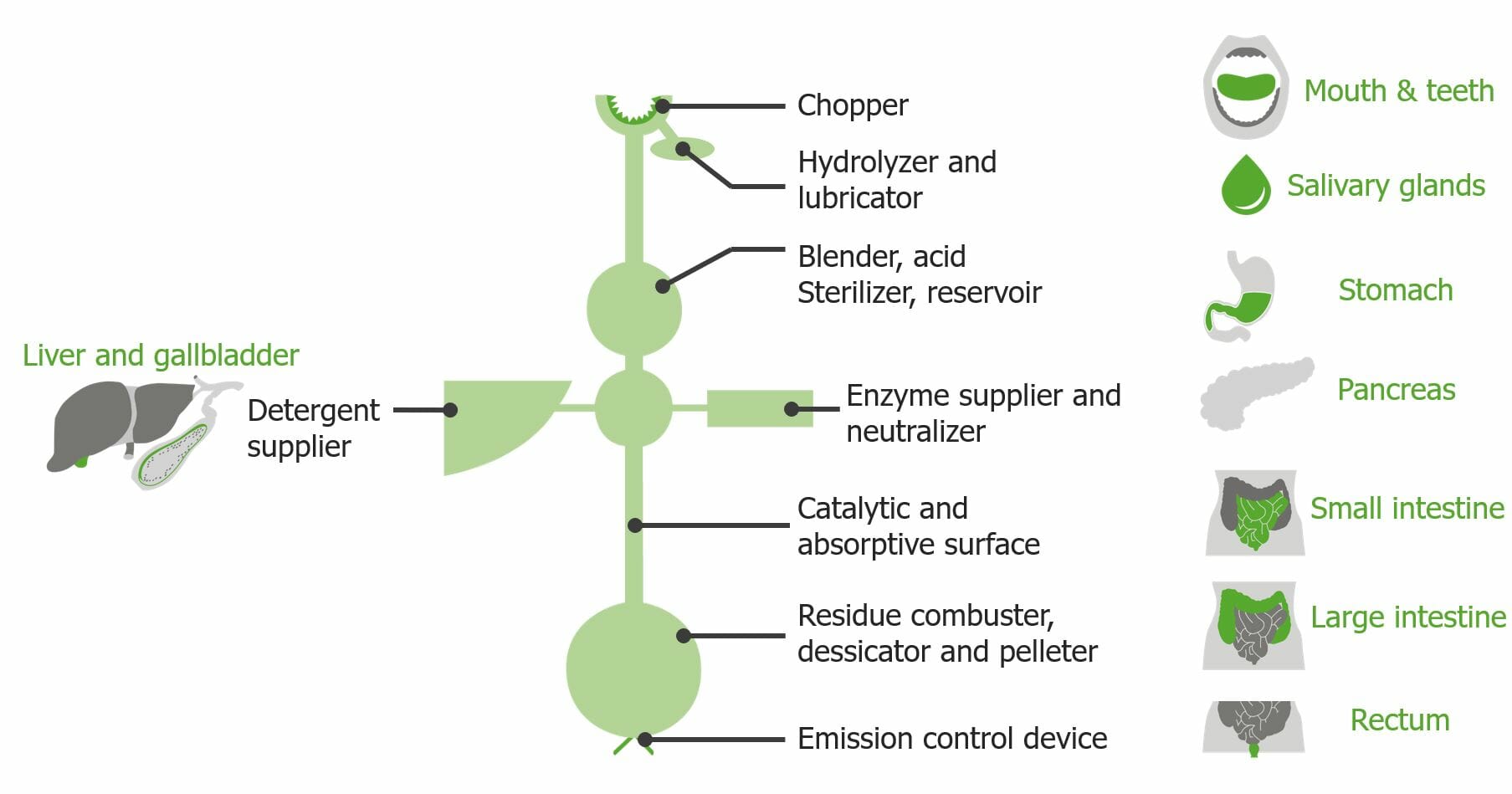
As a way of understanding the GI system, the GI tract can be envisioned as an “engineering tube,” as shown here, with different segments having specialized functions.

Image by Lecturio.

Autonomic Control of the GI Tract

Autonomic control of the GI tract relies on the enteric, parasympathetic, and sympathetic nervous systems.

Enteric nervous system Enteric nervous system Two ganglionated neural plexuses in the gut wall which form one of the three major divisions of the autonomic nervous system. The enteric nervous system innervates the gastrointestinal tract, the pancreas, and the gallbladder. It contains sensory neurons, interneurons, and motor neurons. Thus the circuitry can autonomously sense the tension and the chemical environment in the gut and regulate blood vessel tone, motility, secretions, and fluid transport. The system is itself governed by the central nervous system and receives both parasympathetic and sympathetic innervation. Autonomic Nervous System: Anatomy (ENS)

A specialized portion of the autonomic nervous system Autonomic nervous system The ANS is a component of the peripheral nervous system that uses both afferent (sensory) and efferent (effector) neurons, which control the functioning of the internal organs and involuntary processes via connections with the CNS. The ANS consists of the sympathetic and parasympathetic nervous systems. Autonomic Nervous System: Anatomy located within the walls of the GI tract.

  • Consists of:
    • Meissner plexus (also called the submucosal plexus):
      • Located in the submucosa
      • Controls the muscularis mucosa (independent of the muscular layer of the intestines)
    • Auerbach plexus (also called the myenteric plexus):
      • Located in the muscularis, between the circular and longitudinal layers 
      • Includes both motor Motor Neurons which send impulses peripherally to activate muscles or secretory cells. Nervous System: Histology and sensory Sensory Neurons which conduct nerve impulses to the central nervous system. Nervous System: Histology neurons Neurons The basic cellular units of nervous tissue. Each neuron consists of a body, an axon, and dendrites. Their purpose is to receive, conduct, and transmit impulses in the nervous system. Nervous System: Histology
      • Provides motor Motor Neurons which send impulses peripherally to activate muscles or secretory cells. Nervous System: Histology input to the smooth muscle → generates contractions
      • Receives sensory Sensory Neurons which conduct nerve impulses to the central nervous system. Nervous System: Histology input from chemoreceptors and mechanoreceptors and coordinates appropriate responses (often as a reflex) 
    • Interstitial cells of Cajal (ICCs):
      • Specialized pacemaker Pacemaker A device designed to stimulate, by electric impulses, contraction of the heart muscles. It may be temporary (external) or permanent (internal or internal-external). Bradyarrhythmias cells located in the muscularis
      • Generate the slow wave of smooth muscle contraction Smooth muscle contraction Smooth muscle is primarily found in the walls of hollow structures and some visceral organs, including the walls of the vasculature, GI, respiratory, and genitourinary tracts. Smooth muscle contracts more slowly and is regulated differently than skeletal muscle. Smooth muscle can be stimulated by nerve impulses, hormones, metabolic factors (like pH, CO2 or O2 levels), its own intrinsic pacemaker ability, or even mechanical stretch. Smooth Muscle Contraction responsible for baseline GI motility GI Motility The primary functions of the GI tract are digestion and absorption, which require coordinated contractions of the smooth muscles present in the GI tract. Peristaltic waves, segmentation contractions, and the migrating motor complex are all important contraction patterns that help to mix contents, get them in contact with the intestinal walls, and propel material down the tract at appropriate times and in appropriate amounts. Gastrointestinal Motility
  • Function: local control of GI function
    • Controls peristalsis Peristalsis A movement, caused by sequential muscle contraction, that pushes the contents of the intestines or other tubular organs in one direction. Gastrointestinal Motility: coordinated, wave-like contractions that move material through the tract, ultimately toward the rectum Rectum The rectum and anal canal are the most terminal parts of the lower GI tract/large intestine that form a functional unit and control defecation. Fecal continence is maintained by several important anatomic structures including rectal folds, anal valves, the sling-like puborectalis muscle, and internal and external anal sphincters. Rectum and Anal Canal: Anatomy
    • Segmentation Segmentation Gastrointestinal Motility: circular muscle contraction allows for mixing of food material to aid in absorption Absorption Absorption involves the uptake of nutrient molecules and their transfer from the lumen of the GI tract across the enterocytes and into the interstitial space, where they can be taken up in the venous or lymphatic circulation. Digestion and Absorption
    • Stimulates the secretion Secretion Coagulation Studies of regulatory hormones Hormones Hormones are messenger molecules that are synthesized in one part of the body and move through the bloodstream to exert specific regulatory effects on another part of the body. Hormones play critical roles in coordinating cellular activities throughout the body in response to the constant changes in both the internal and external environments. Hormones: Overview and Types and neurotransmitters (see sections below)
  • The ENS does not routinely act on its own; it receives input from:
    • Vagus nerve Vagus nerve The 10th cranial nerve. The vagus is a mixed nerve which contains somatic afferents (from skin in back of the ear and the external auditory meatus), visceral afferents (from the pharynx, larynx, thorax, and abdomen), parasympathetic efferents (to the thorax and abdomen), and efferents to striated muscle (of the larynx and pharynx). Pharynx: Anatomy (parasympathetic, stimulatory)
    • Prevertebral ganglia (sympathetic, inhibitory)
  • Mechanisms of control:
    • Parasympathetic or sympathetic fibers synapse Synapse The junction between 2 neurons is called a synapse. The synapse allows a neuron to pass an electrical or chemical signal to another neuron or target effector cell. Synapses and Neurotransmission with myenteric motor Motor Neurons which send impulses peripherally to activate muscles or secretory cells. Nervous System: Histology neurons Neurons The basic cellular units of nervous tissue. Each neuron consists of a body, an axon, and dendrites. Their purpose is to receive, conduct, and transmit impulses in the nervous system. Nervous System: Histology → adjust their activity
    • Myenteric motor Motor Neurons which send impulses peripherally to activate muscles or secretory cells. Nervous System: Histology neurons Neurons The basic cellular units of nervous tissue. Each neuron consists of a body, an axon, and dendrites. Their purpose is to receive, conduct, and transmit impulses in the nervous system. Nervous System: Histology synapse Synapse The junction between 2 neurons is called a synapse. The synapse allows a neuron to pass an electrical or chemical signal to another neuron or target effector cell. Synapses and Neurotransmission with ICCs → ICCs adjust rate of firing/contraction waves
    • Smooth muscle cells communicate the signal via gap junctions Gap Junctions Connections between cells which allow passage of small molecules and electric current. Gap junctions were first described anatomically as regions of close apposition between cells with a narrow (1-2 nm) gap between cell membranes. The variety in the properties of gap junctions is reflected in the number of connexins, the family of proteins which form the junctions. The Cell: Cell Junctions
Layers and folds in intestinal walls

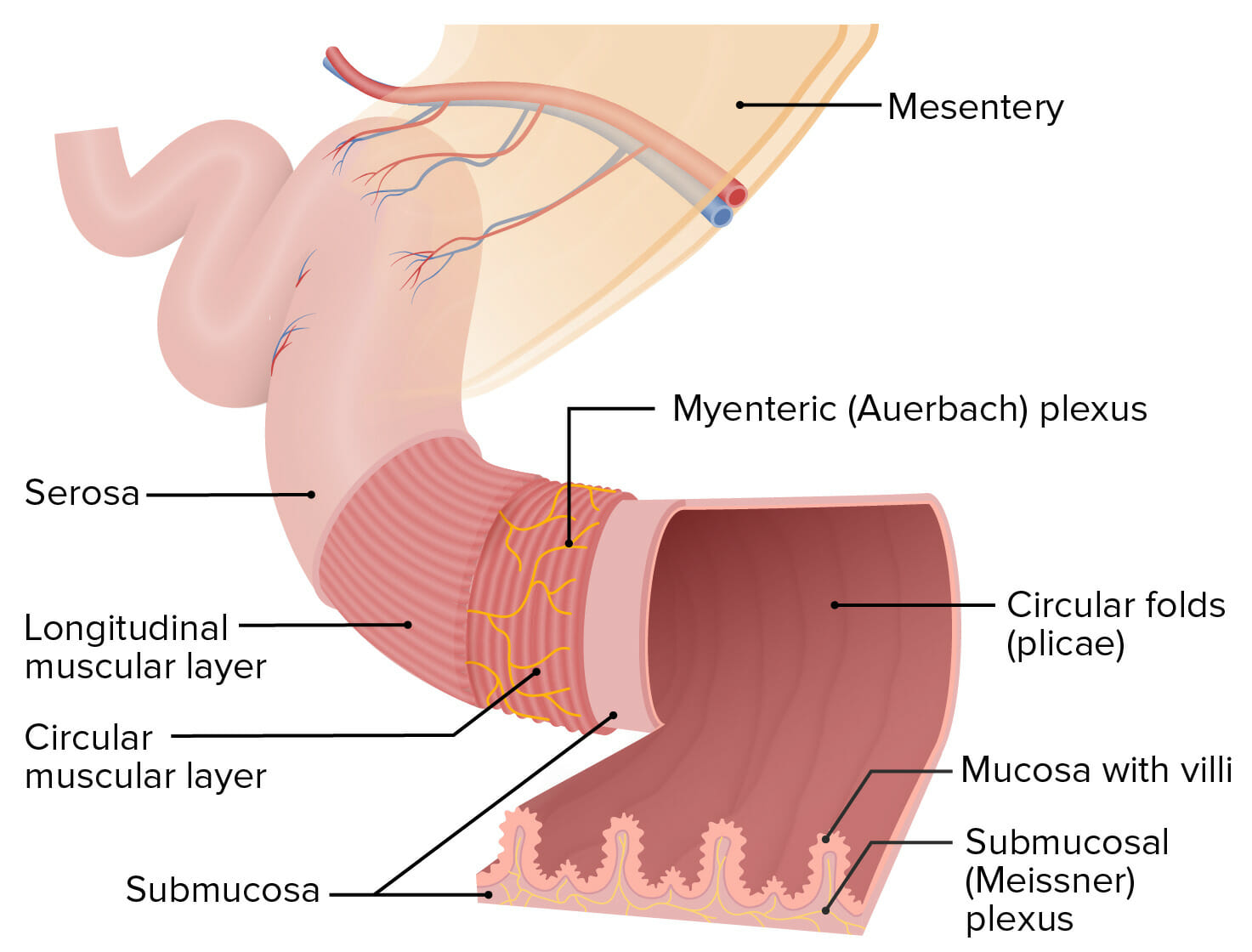
Structure of the intestinal walls:
The Meissner plexus is located within the submucosa, and the Auerbach plexus (also known as the myenteric plexus) is located between the circular and longitudinal muscular layers.

Image by Lecturio.

Parasympathetic nervous system Nervous system The nervous system is a small and complex system that consists of an intricate network of neural cells (or neurons) and even more glial cells (for support and insulation). It is divided according to its anatomical components as well as its functional characteristics. The brain and spinal cord are referred to as the central nervous system, and the branches of nerves from these structures are referred to as the peripheral nervous system. Nervous System: Anatomy, Structure, and Classification (PNS)

  • Activates GI function
  • Effects of parasympathetic signaling:
    • GI motility GI Motility The primary functions of the GI tract are digestion and absorption, which require coordinated contractions of the smooth muscles present in the GI tract. Peristaltic waves, segmentation contractions, and the migrating motor complex are all important contraction patterns that help to mix contents, get them in contact with the intestinal walls, and propel material down the tract at appropriate times and in appropriate amounts. Gastrointestinal Motility 
    • ↑ GI secretions
    • Blood flow Blood flow Blood flow refers to the movement of a certain volume of blood through the vasculature over a given unit of time (e.g., mL per minute). Vascular Resistance, Flow, and Mean Arterial Pressure
    • ↓ Constriction of GI sphincters (allows food to move through the tract)
  • Nerves:
    • Facial nerve Facial nerve The 7th cranial nerve. The facial nerve has two parts, the larger motor root which may be called the facial nerve proper, and the smaller intermediate or sensory root. Together they provide efferent innervation to the muscles of facial expression and to the lacrimal and salivary glands, and convey afferent information for taste from the anterior two-thirds of the tongue and for touch from the external ear. The 12 Cranial Nerves: Overview and Functions (cranial nerve (CN) VII): controls the salivary glands Salivary glands The salivary glands are exocrine glands positioned in and around the oral cavity. These glands are responsible for secreting saliva into the mouth, which aids in digestion. There are 3 major paired salivary glands: the sublingual, submandibular, and parotid glands. Salivary Glands: Anatomy
    • Glossopharyngeal nerve Glossopharyngeal nerve The 9th cranial nerve. The glossopharyngeal nerve is a mixed motor and sensory nerve; it conveys somatic and autonomic efferents as well as general, special, and visceral afferents. Among the connections are motor fibers to the stylopharyngeus muscle, parasympathetic fibers to the parotid glands, general and taste afferents from the posterior third of the tongue, the nasopharynx, and the palate, and afferents from baroreceptors and chemoreceptor cells of the carotid sinus. Pharynx: Anatomy (CN IX) controls the:
      • Tongue Tongue The tongue, on the other hand, is a complex muscular structure that permits tasting and facilitates the process of mastication and communication. The blood supply of the tongue originates from the external carotid artery, and the innervation is through cranial nerves. Lips and Tongue: Anatomy
      • Oropharynx Oropharynx The middle portion of the pharynx that lies posterior to the mouth, inferior to the soft palate, and superior to the base of the tongue and epiglottis. It has a digestive function as food passes from the mouth into the oropharynx before entering esophagus. Pharynx: Anatomy 
      • Upper esophagus Esophagus The esophagus is a muscular tube-shaped organ of around 25 centimeters in length that connects the pharynx to the stomach. The organ extends from approximately the 6th cervical vertebra to the 11th thoracic vertebra and can be divided grossly into 3 parts: the cervical part, the thoracic part, and the abdominal part. Esophagus: Anatomy
    • Vagus nerve Vagus nerve The 10th cranial nerve. The vagus is a mixed nerve which contains somatic afferents (from skin in back of the ear and the external auditory meatus), visceral afferents (from the pharynx, larynx, thorax, and abdomen), parasympathetic efferents (to the thorax and abdomen), and efferents to striated muscle (of the larynx and pharynx). Pharynx: Anatomy (CN X) controls the:
      • Lower esophagus Esophagus The esophagus is a muscular tube-shaped organ of around 25 centimeters in length that connects the pharynx to the stomach. The organ extends from approximately the 6th cervical vertebra to the 11th thoracic vertebra and can be divided grossly into 3 parts: the cervical part, the thoracic part, and the abdominal part. Esophagus: Anatomy
      • Stomach Stomach The stomach is a muscular sac in the upper left portion of the abdomen that plays a critical role in digestion. The stomach develops from the foregut and connects the esophagus with the duodenum. Structurally, the stomach is C-shaped and forms a greater and lesser curvature and is divided grossly into regions: the cardia, fundus, body, and pylorus. Stomach: Anatomy 
      • Pancreas Pancreas The pancreas lies mostly posterior to the stomach and extends across the posterior abdominal wall from the duodenum on the right to the spleen on the left. This organ has both exocrine and endocrine tissue. Pancreas: Anatomy
      • Small intestines
      • Part of the large intestines
    • Pelvic nerves:
      • Large intestine Large intestine The large intestines constitute the last portion of the digestive system. The large intestine consists of the cecum, appendix, colon (with ascending, transverse, descending, and sigmoid segments), rectum, and anal canal. The primary function of the colon is to remove water and compact the stool prior to expulsion from the body via the rectum and anal canal. Colon, Cecum, and Appendix: Anatomy
      • Rectum Rectum The rectum and anal canal are the most terminal parts of the lower GI tract/large intestine that form a functional unit and control defecation. Fecal continence is maintained by several important anatomic structures including rectal folds, anal valves, the sling-like puborectalis muscle, and internal and external anal sphincters. Rectum and Anal Canal: Anatomy
      • Anus
  • Primary neural signaling molecules Signaling molecules Second Messengers:
    • Acetylcholine Acetylcholine A neurotransmitter found at neuromuscular junctions, autonomic ganglia, parasympathetic effector junctions, a subset of sympathetic effector junctions, and at many sites in the central nervous system. Receptors and Neurotransmitters of the CNS
    • Vasoactive intestinal peptide (VIP)
Parasympathetic innervation of the gi tract

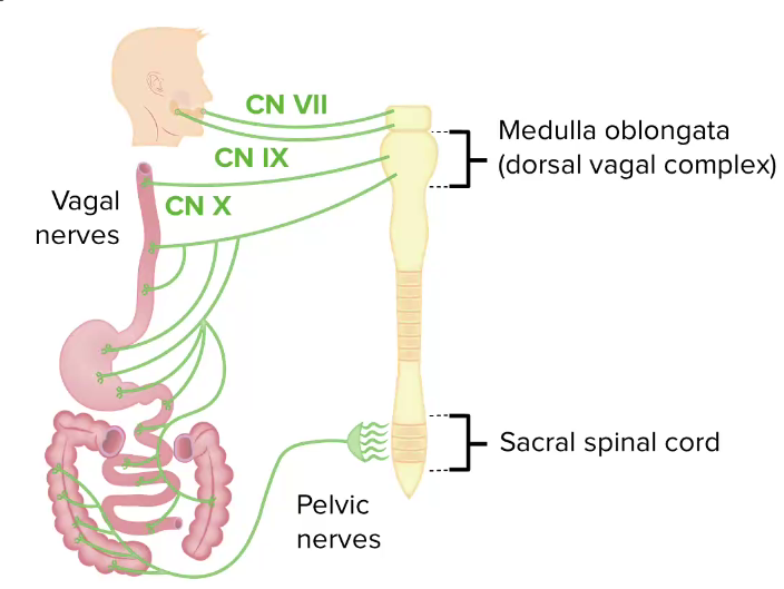
Parasympathetic influence on the GI tract:
Note the vast reach of the vagus nerve.
CN: cranial nerve

Image by Lecturio.

Sympathetic nervous system Nervous system The nervous system is a small and complex system that consists of an intricate network of neural cells (or neurons) and even more glial cells (for support and insulation). It is divided according to its anatomical components as well as its functional characteristics. The brain and spinal cord are referred to as the central nervous system, and the branches of nerves from these structures are referred to as the peripheral nervous system. Nervous System: Anatomy, Structure, and Classification (SNS)

  • Restricts GI function during the fight-or-flight response (so that energy can be diverted to skeletal muscle)
  • Effects of sympathetic signaling (opposite to parasympathetic signaling):
  • Nerves: sympathetic ganglia in the thoracolumbar region (primarily T8‒L2):
    • Celiac ganglia
    • Superior mesenteric ganglia
    • Inferior mesenteric ganglia
  • Primary neural signaling molecules Signaling molecules Second Messengers:
    • Norepinephrine Norepinephrine Precursor of epinephrine that is secreted by the adrenal medulla and is a widespread central and autonomic neurotransmitter. Norepinephrine is the principal transmitter of most postganglionic sympathetic fibers, and of the diffuse projection system in the brain that arises from the locus ceruleus. Receptors and Neurotransmitters of the CNS (NE)
    • ATP
Sympathetic innervation of the gi tract

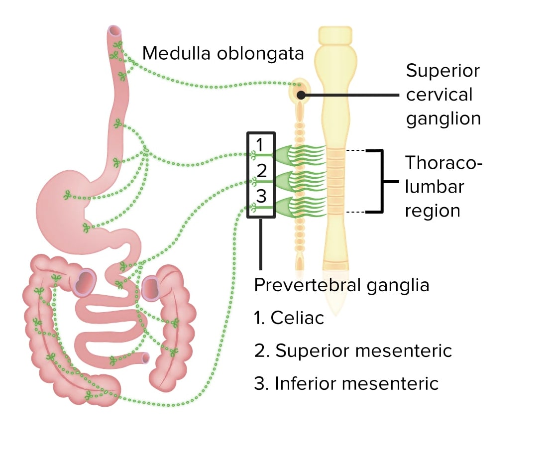
Sympathetic nervous system influence on the GI tract

Image by Lecturio.

Regulation of blood flow Blood flow Blood flow refers to the movement of a certain volume of blood through the vasculature over a given unit of time (e.g., mL per minute). Vascular Resistance, Flow, and Mean Arterial Pressure

Vasodilation Vasodilation The physiological widening of blood vessels by relaxing the underlying vascular smooth muscle. Pulmonary Hypertension Drugs:

  • Increases blood flow Blood flow Blood flow refers to the movement of a certain volume of blood through the vasculature over a given unit of time (e.g., mL per minute). Vascular Resistance, Flow, and Mean Arterial Pressure
  • Stimulated by:
    • Parasympathetic nerves, via acetylcholine Acetylcholine A neurotransmitter found at neuromuscular junctions, autonomic ganglia, parasympathetic effector junctions, a subset of sympathetic effector junctions, and at many sites in the central nervous system. Receptors and Neurotransmitters of the CNS ( ACh ACh A neurotransmitter found at neuromuscular junctions, autonomic ganglia, parasympathetic effector junctions, a subset of sympathetic effector junctions, and at many sites in the central nervous system. Receptors and Neurotransmitters of the CNS) and VIP
    • Local reflexes Local reflexes Gastrointestinal Secretions in the enteric nervous system Enteric nervous system Two ganglionated neural plexuses in the gut wall which form one of the three major divisions of the autonomic nervous system. The enteric nervous system innervates the gastrointestinal tract, the pancreas, and the gallbladder. It contains sensory neurons, interneurons, and motor neurons. Thus the circuitry can autonomously sense the tension and the chemical environment in the gut and regulate blood vessel tone, motility, secretions, and fluid transport. The system is itself governed by the central nervous system and receives both parasympathetic and sympathetic innervation. Autonomic Nervous System: Anatomy via ACh ACh A neurotransmitter found at neuromuscular junctions, autonomic ganglia, parasympathetic effector junctions, a subset of sympathetic effector junctions, and at many sites in the central nervous system. Receptors and Neurotransmitters of the CNS and VIP
    • Sensory Sensory Neurons which conduct nerve impulses to the central nervous system. Nervous System: Histology afferents (lesser degree) via:
      • Calcitonin Calcitonin A peptide hormone that lowers calcium concentration in the blood. In humans, it is released by thyroid cells and acts to decrease the formation and absorptive activity of osteoclasts. Its role in regulating plasma calcium is much greater in children and in certain diseases than in normal adults. Other Antiresorptive Drugs gene Gene A category of nucleic acid sequences that function as units of heredity and which code for the basic instructions for the development, reproduction, and maintenance of organisms. Basic Terms of Genetics–related peptide (CGRP)
      • Substance P

Vasoconstriction Vasoconstriction The physiological narrowing of blood vessels by contraction of the vascular smooth muscle. Vascular Resistance, Flow, and Mean Arterial Pressure:

Regulation of the blood flow to the gi tract

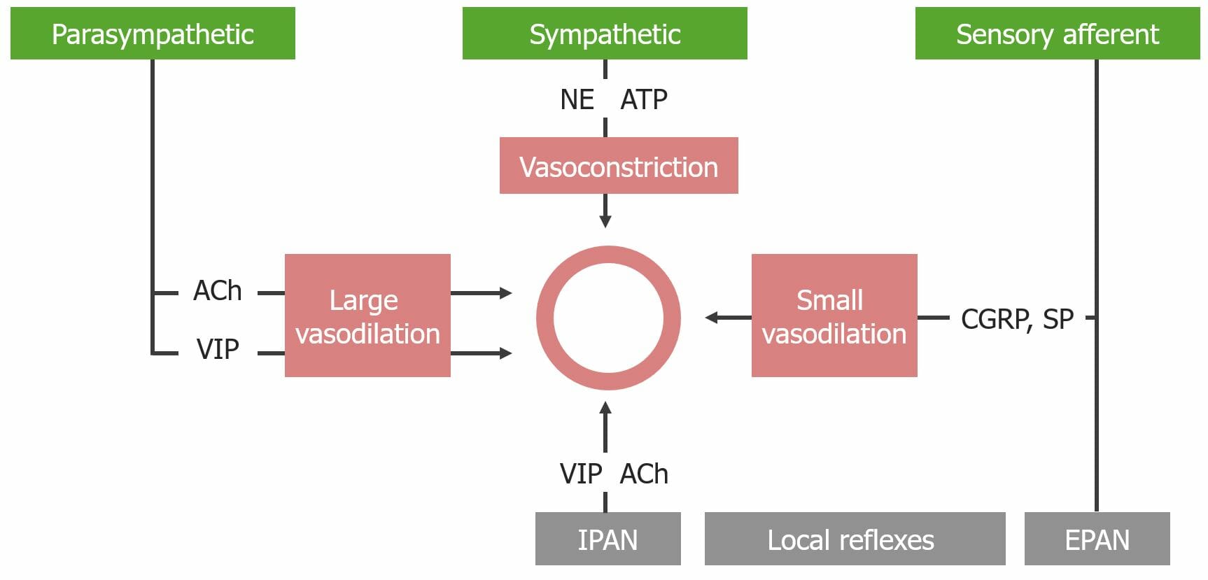
Flowchart detailing the regulation of blood flow to the GI tract
ACh: acetylcholine
CGRP: calcitonin gene–related peptide
EPAN: extrinsic primary afferent neuron
IPAN : intrinsic primary afferent
NE: norepinephrine
SP: substance P
VIP: vasoactive intestinal

Image by Lecturio.

Neural Signaling Molecules

The nervous system Nervous system The nervous system is a small and complex system that consists of an intricate network of neural cells (or neurons) and even more glial cells (for support and insulation). It is divided according to its anatomical components as well as its functional characteristics. The brain and spinal cord are referred to as the central nervous system, and the branches of nerves from these structures are referred to as the peripheral nervous system. Nervous System: Anatomy, Structure, and Classification sends signals to mediate activity in the GI tract. Nerves in the PNS, SNS, ENS, and sensory Sensory Neurons which conduct nerve impulses to the central nervous system. Nervous System: Histology afferent Afferent Neurons which conduct nerve impulses to the central nervous system. Nervous System: Histology nerves release different types of signaling molecules Signaling molecules Second Messengers, which have different effects on their target tissue.

Table: Neural signaling molecules Signaling molecules Second Messengers
Molecule Primary releasing nerve(s) Structure(s) acted on Primary function
Released by the PNS and/or ENS
Acetylcholine Acetylcholine A neurotransmitter found at neuromuscular junctions, autonomic ganglia, parasympathetic effector junctions, a subset of sympathetic effector junctions, and at many sites in the central nervous system. Receptors and Neurotransmitters of the CNS PNS
  • Glands
  • Smooth muscle
  • Blood vessels
Increases secretions, motility Motility The motor activity of the gastrointestinal tract. Gastrointestinal Motility, and blood flow Blood flow Blood flow refers to the movement of a certain volume of blood through the vasculature over a given unit of time (e.g., mL per minute). Vascular Resistance, Flow, and Mean Arterial Pressure
VIP PNS and ENS
  • Glands
  • Smooth muscle
  • Blood vessels
Increase motility Motility The motor activity of the gastrointestinal tract. Gastrointestinal Motility and blood flow Blood flow Blood flow refers to the movement of a certain volume of blood through the vasculature over a given unit of time (e.g., mL per minute). Vascular Resistance, Flow, and Mean Arterial Pressure
GRP PNS and ENS Glands Increase gastrin Gastrin A family of gastrointestinal peptide hormones that excite the secretion of gastric juice. They may also occur in the central nervous system where they are presumed to be neurotransmitters. Gastrointestinal Secretions secretion Secretion Coagulation Studies
Enkephalins Enkephalins One of the three major families of endogenous opioid peptides. The enkephalins are pentapeptides that are widespread in the central and peripheral nervous systems and in the adrenal medulla. Receptors and Neurotransmitters of the CNS (related to opioids Opioids Opiates are drugs that are derived from the sap of the opium poppy. Opiates have been used since antiquity for the relief of acute severe pain. Opioids are synthetic opiates with properties that are substantially similar to those of opiates. Opioid Analgesics) ENS Smooth muscle Constrict sphincters (prevents forward movement)
Released by sensory Sensory Neurons which conduct nerve impulses to the central nervous system. Nervous System: Histology afferents
CGRP Sensory Sensory Neurons which conduct nerve impulses to the central nervous system. Nervous System: Histology afferents Blood vessels Increase blood flow Blood flow Blood flow refers to the movement of a certain volume of blood through the vasculature over a given unit of time (e.g., mL per minute). Vascular Resistance, Flow, and Mean Arterial Pressure via vasodilation Vasodilation The physiological widening of blood vessels by relaxing the underlying vascular smooth muscle. Pulmonary Hypertension Drugs
Substance P Sensory Sensory Neurons which conduct nerve impulses to the central nervous system. Nervous System: Histology afferents and PNS
  • Blood vessels
  • Glands
Increase blood flow Blood flow Blood flow refers to the movement of a certain volume of blood through the vasculature over a given unit of time (e.g., mL per minute). Vascular Resistance, Flow, and Mean Arterial Pressure
Released primarily by the SNS
Norepinephrine Norepinephrine Precursor of epinephrine that is secreted by the adrenal medulla and is a widespread central and autonomic neurotransmitter. Norepinephrine is the principal transmitter of most postganglionic sympathetic fibers, and of the diffuse projection system in the brain that arises from the locus ceruleus. Receptors and Neurotransmitters of the CNS SNS
  • Glands
  • Smooth muscle
  • Blood vessels
Decrease secretions, motility Motility The motor activity of the gastrointestinal tract. Gastrointestinal Motility, and blood flow Blood flow Blood flow refers to the movement of a certain volume of blood through the vasculature over a given unit of time (e.g., mL per minute). Vascular Resistance, Flow, and Mean Arterial Pressure
ATP SNS Blood vessels Decrease blood flow Blood flow Blood flow refers to the movement of a certain volume of blood through the vasculature over a given unit of time (e.g., mL per minute). Vascular Resistance, Flow, and Mean Arterial Pressure via vasoconstriction Vasoconstriction The physiological narrowing of blood vessels by contraction of the vascular smooth muscle. Vascular Resistance, Flow, and Mean Arterial Pressure
CGRP: calcitonin gene–related peptide
ENS: enteric nervous system
GRP: gastric-releasing peptide
PNS: parasympathetic nervous system
SNS: sympathetic nervous system
VIP: vasoactive intestinal peptide

GI Hormones

The GI hormones GI Hormones Hormones secreted by the gastrointestinal mucosa that affect the timing or the quality of secretion of digestive enzymes, and regulate the motor activity of the digestive system organs. Gastrointestinal Secretions are secreted by enteroendocrine cells Enteroendocrine cells Cells found throughout the lining of the gastrointestinal tract that contain and secrete regulatory peptide hormones and/or biogenic amines. Gastrointestinal Secretions and modulate function of the GI tract. Enteroendocrine cells Enteroendocrine cells Cells found throughout the lining of the gastrointestinal tract that contain and secrete regulatory peptide hormones and/or biogenic amines. Gastrointestinal Secretions are spread throughout the GI tract.

Classification by chemical structure

  • Gastrin Gastrin A family of gastrointestinal peptide hormones that excite the secretion of gastric juice. They may also occur in the central nervous system where they are presumed to be neurotransmitters. Gastrointestinal Secretions
  • Cholecystokinin Cholecystokinin A peptide, of about 33 amino acids, secreted by the upper intestinal mucosa and also found in the central nervous system. It causes gallbladder contraction, release of pancreatic exocrine (or digestive) enzymes, and affects other gastrointestinal functions. Cholecystokinin may be the mediator of satiety. Gastrointestinal Secretions
  • Secretin Secretin A peptide hormone of about 27 amino acids from the duodenal mucosa that activates pancreatic secretion and lowers the blood sugar level. Gastrointestinal Secretions family:
    • Secretin Secretin A peptide hormone of about 27 amino acids from the duodenal mucosa that activates pancreatic secretion and lowers the blood sugar level. Gastrointestinal Secretions
    • Glucagon Glucagon A 29-amino acid pancreatic peptide derived from proglucagon which is also the precursor of intestinal glucagon-like peptides. Glucagon is secreted by pancreatic alpha cells and plays an important role in regulation of blood glucose concentration, ketone metabolism, and several other biochemical and physiological processes. Gastrointestinal Secretions
    • VIP
  • Other:
    • Somatostatin Somatostatin A 14-amino acid peptide named for its ability to inhibit pituitary growth hormone release, also called somatotropin release-inhibiting factor. It is expressed in the central and peripheral nervous systems, the gut, and other organs. SRIF can also inhibit the release of thyroid-stimulating hormone; prolactin; insulin; and glucagon besides acting as a neurotransmitter and neuromodulator. In a number of species including humans, there is an additional form of somatostatin, srif-28 with a 14-amino acid extension at the n-terminal. Gastrointestinal Secretions 
    • Motilin Motilin A peptide of about 22-amino acids isolated from the duodenum. At low ph it inhibits gastric motor activity, whereas at high ph it has a stimulating effect. Gastrointestinal Secretions 

Functions

The major GI hormones GI Hormones Hormones secreted by the gastrointestinal mucosa that affect the timing or the quality of secretion of digestive enzymes, and regulate the motor activity of the digestive system organs. Gastrointestinal Secretions

Table: GI hormones GI Hormones Hormones secreted by the gastrointestinal mucosa that affect the timing or the quality of secretion of digestive enzymes, and regulate the motor activity of the digestive system organs. Gastrointestinal Secretions outlined with releasing cell, structure affected, and primary function
Molecule Primarily released from Structure(s) acted on Primary function
Gastrin Gastrin A family of gastrointestinal peptide hormones that excite the secretion of gastric juice. They may also occur in the central nervous system where they are presumed to be neurotransmitters. Gastrointestinal Secretions G cells in the:
  • Stomach Stomach The stomach is a muscular sac in the upper left portion of the abdomen that plays a critical role in digestion. The stomach develops from the foregut and connects the esophagus with the duodenum. Structurally, the stomach is C-shaped and forms a greater and lesser curvature and is divided grossly into regions: the cardia, fundus, body, and pylorus. Stomach: Anatomy
  • Duodenum Duodenum The shortest and widest portion of the small intestine adjacent to the pylorus of the stomach. It is named for having the length equal to about the width of 12 fingers. Small Intestine: Anatomy
Stomach Stomach The stomach is a muscular sac in the upper left portion of the abdomen that plays a critical role in digestion. The stomach develops from the foregut and connects the esophagus with the duodenum. Structurally, the stomach is C-shaped and forms a greater and lesser curvature and is divided grossly into regions: the cardia, fundus, body, and pylorus. Stomach: Anatomy
Cholecystokinin Cholecystokinin A peptide, of about 33 amino acids, secreted by the upper intestinal mucosa and also found in the central nervous system. It causes gallbladder contraction, release of pancreatic exocrine (or digestive) enzymes, and affects other gastrointestinal functions. Cholecystokinin may be the mediator of satiety. Gastrointestinal Secretions I cells in the:
  • Duodenum Duodenum The shortest and widest portion of the small intestine adjacent to the pylorus of the stomach. It is named for having the length equal to about the width of 12 fingers. Small Intestine: Anatomy
  • Jejunum Jejunum The middle portion of the small intestine, between duodenum and ileum. It represents about 2/5 of the remaining portion of the small intestine below duodenum. Small Intestine: Anatomy
Exocrine pancreas Exocrine pancreas The major component (about 80%) of the pancreas composed of acinar functional units of tubular and spherical cells. The acinar cells synthesize and secrete several digestive enzymes such as trypsinogen; lipase; amylase; and ribonuclease. Secretion from the exocrine pancreas drains into the pancreatic ductal system and empties into the duodenum. Pancreas: Anatomy, gallbladder Gallbladder The gallbladder is a pear-shaped sac, located directly beneath the liver, that sits on top of the superior part of the duodenum. The primary functions of the gallbladder include concentrating and storing up to 50 mL of bile. Gallbladder and Biliary Tract: Anatomy, and stomach Stomach The stomach is a muscular sac in the upper left portion of the abdomen that plays a critical role in digestion. The stomach develops from the foregut and connects the esophagus with the duodenum. Structurally, the stomach is C-shaped and forms a greater and lesser curvature and is divided grossly into regions: the cardia, fundus, body, and pylorus. Stomach: Anatomy
  • Increase pancreatic enzyme and bicarbonate Bicarbonate Inorganic salts that contain the -HCO3 radical. They are an important factor in determining the ph of the blood and the concentration of bicarbonate ions is regulated by the kidney. Levels in the blood are an index of the alkali reserve or buffering capacity. Electrolytes secretion Secretion Coagulation Studies
  • Contracts the gallbladder Gallbladder The gallbladder is a pear-shaped sac, located directly beneath the liver, that sits on top of the superior part of the duodenum. The primary functions of the gallbladder include concentrating and storing up to 50 mL of bile. Gallbladder and Biliary Tract: Anatomy
  • Relaxes the sphincter of Oddi
  • Contracts the pyloric sphincter and decreases gastric emptying Gastric emptying The evacuation of food from the stomach into the duodenum. Gastrointestinal Motility
Gastric inhibitory peptide (GIP) K cells in the:
  • Duodenum Duodenum The shortest and widest portion of the small intestine adjacent to the pylorus of the stomach. It is named for having the length equal to about the width of 12 fingers. Small Intestine: Anatomy
  • Jejunum Jejunum The middle portion of the small intestine, between duodenum and ileum. It represents about 2/5 of the remaining portion of the small intestine below duodenum. Small Intestine: Anatomy
Stomach Stomach The stomach is a muscular sac in the upper left portion of the abdomen that plays a critical role in digestion. The stomach develops from the foregut and connects the esophagus with the duodenum. Structurally, the stomach is C-shaped and forms a greater and lesser curvature and is divided grossly into regions: the cardia, fundus, body, and pylorus. Stomach: Anatomy, endocrine pancreas Endocrine pancreas Pancreas: Anatomy
  • Stimulates insulin Insulin Insulin is a peptide hormone that is produced by the beta cells of the pancreas. Insulin plays a role in metabolic functions such as glucose uptake, glycolysis, glycogenesis, lipogenesis, and protein synthesis. Exogenous insulin may be needed for individuals with diabetes mellitus, in whom there is a deficiency in endogenous insulin or increased insulin resistance. Insulin release
  • Inhibits acid secretion Secretion Coagulation Studies
  • Induces satiety
Motilin Motilin A peptide of about 22-amino acids isolated from the duodenum. At low ph it inhibits gastric motor activity, whereas at high ph it has a stimulating effect. Gastrointestinal Secretions M cells M cells Salmonella in the:
  • Duodenum Duodenum The shortest and widest portion of the small intestine adjacent to the pylorus of the stomach. It is named for having the length equal to about the width of 12 fingers. Small Intestine: Anatomy
  • Jejunum Jejunum The middle portion of the small intestine, between duodenum and ileum. It represents about 2/5 of the remaining portion of the small intestine below duodenum. Small Intestine: Anatomy
Smooth muscle
  • GI motility GI Motility The primary functions of the GI tract are digestion and absorption, which require coordinated contractions of the smooth muscles present in the GI tract. Peristaltic waves, segmentation contractions, and the migrating motor complex are all important contraction patterns that help to mix contents, get them in contact with the intestinal walls, and propel material down the tract at appropriate times and in appropriate amounts. Gastrointestinal Motility by stimulating the migrating motor Motor Neurons which send impulses peripherally to activate muscles or secretory cells. Nervous System: Histology complex ( MMC MMC A pattern of gastrointestinal muscle contraction and depolarizing myoelectric activity that moves from the stomach to the ileocecal valve at regular frequency during the interdigestive period. The complex and its accompanying motor activity periodically cleanse the bowel of interdigestive secretion and debris in preparation for the next meal. Gastrointestinal Motility)
Secretin Secretin A peptide hormone of about 27 amino acids from the duodenal mucosa that activates pancreatic secretion and lowers the blood sugar level. Gastrointestinal Secretions S cells in the:
  • Duodenum Duodenum The shortest and widest portion of the small intestine adjacent to the pylorus of the stomach. It is named for having the length equal to about the width of 12 fingers. Small Intestine: Anatomy
  • Jejunum Jejunum The middle portion of the small intestine, between duodenum and ileum. It represents about 2/5 of the remaining portion of the small intestine below duodenum. Small Intestine: Anatomy
Pancreas Pancreas The pancreas lies mostly posterior to the stomach and extends across the posterior abdominal wall from the duodenum on the right to the spleen on the left. This organ has both exocrine and endocrine tissue. Pancreas: Anatomy and stomach Stomach The stomach is a muscular sac in the upper left portion of the abdomen that plays a critical role in digestion. The stomach develops from the foregut and connects the esophagus with the duodenum. Structurally, the stomach is C-shaped and forms a greater and lesser curvature and is divided grossly into regions: the cardia, fundus, body, and pylorus. Stomach: Anatomy
  • Bicarbonate Bicarbonate Inorganic salts that contain the -HCO3 radical. They are an important factor in determining the ph of the blood and the concentration of bicarbonate ions is regulated by the kidney. Levels in the blood are an index of the alkali reserve or buffering capacity. Electrolytes secretion Secretion Coagulation Studies ( pancreas Pancreas The pancreas lies mostly posterior to the stomach and extends across the posterior abdominal wall from the duodenum on the right to the spleen on the left. This organ has both exocrine and endocrine tissue. Pancreas: Anatomy)
  • Pepsin Pepsin Pepsin breaks down proteins into proteoses, peptones, and large polypeptides. Proteins and Peptides secretion Secretion Coagulation Studies ( stomach Stomach The stomach is a muscular sac in the upper left portion of the abdomen that plays a critical role in digestion. The stomach develops from the foregut and connects the esophagus with the duodenum. Structurally, the stomach is C-shaped and forms a greater and lesser curvature and is divided grossly into regions: the cardia, fundus, body, and pylorus. Stomach: Anatomy)
  • Inhibits gastrin Gastrin A family of gastrointestinal peptide hormones that excite the secretion of gastric juice. They may also occur in the central nervous system where they are presumed to be neurotransmitters. Gastrointestinal Secretions and acid secretion Secretion Coagulation Studies
Location of gi hormones

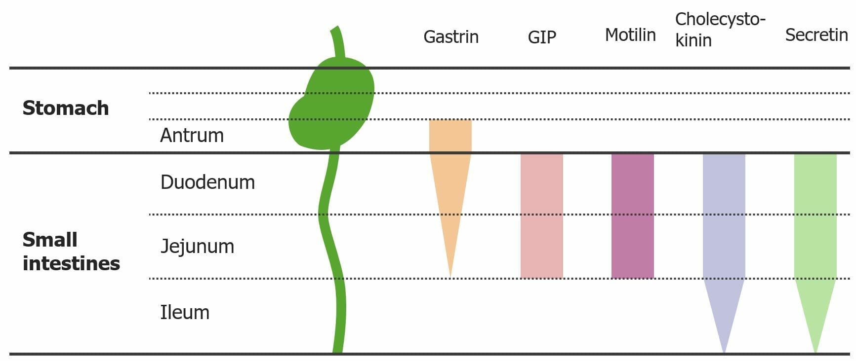
Diagram showing the locations from which GI hormones are released:
The tapered ends on the gastrin, cholecystokinin, and secretin bars represent the decreasing amounts of hormone secretion in these locations.
GIP: gastric inhibitory peptide

Image by Lecturio.

GI Paracrine Signaling Molecules

Paracrine signaling molecules Signaling molecules Second Messengers are similar to hormones Hormones Hormones are messenger molecules that are synthesized in one part of the body and move through the bloodstream to exert specific regulatory effects on another part of the body. Hormones play critical roles in coordinating cellular activities throughout the body in response to the constant changes in both the internal and external environments. Hormones: Overview and Types; however, instead of traveling to different tissues via the blood, paracrine signaling molecules Signaling molecules Second Messengers affect cells close to the site of their release. The major paracrine signaling molecules Signaling molecules Second Messengers in the GI system are histamine, NO, prostaglandins Prostaglandins A group of compounds derived from unsaturated 20-carbon fatty acids, primarily arachidonic acid, via the cyclooxygenase pathway. They are extremely potent mediators of a diverse group of physiological processes. Eicosanoids, and somatostatin Somatostatin A 14-amino acid peptide named for its ability to inhibit pituitary growth hormone release, also called somatotropin release-inhibiting factor. It is expressed in the central and peripheral nervous systems, the gut, and other organs. SRIF can also inhibit the release of thyroid-stimulating hormone; prolactin; insulin; and glucagon besides acting as a neurotransmitter and neuromodulator. In a number of species including humans, there is an additional form of somatostatin, srif-28 with a 14-amino acid extension at the n-terminal. Gastrointestinal Secretions.

Table: Paracrine signaling molecules Signaling molecules Second Messengers of the GI tract
Molecule Released from Structure(s) acted on Primary function
Histamine
  • Enterochromaffin like cells
  • Mast cells Mast cells Granulated cells that are found in almost all tissues, most abundantly in the skin and the gastrointestinal tract. Like the basophils, mast cells contain large amounts of histamine and heparin. Unlike basophils, mast cells normally remain in the tissues and do not circulate in the blood. Mast cells, derived from the bone marrow stem cells, are regulated by the stem cell factor. Innate Immunity: Phagocytes and Antigen Presentation
Stomach Stomach The stomach is a muscular sac in the upper left portion of the abdomen that plays a critical role in digestion. The stomach develops from the foregut and connects the esophagus with the duodenum. Structurally, the stomach is C-shaped and forms a greater and lesser curvature and is divided grossly into regions: the cardia, fundus, body, and pylorus. Stomach: Anatomy Increase acid secretion Secretion Coagulation Studies
Somatostatin Somatostatin A 14-amino acid peptide named for its ability to inhibit pituitary growth hormone release, also called somatotropin release-inhibiting factor. It is expressed in the central and peripheral nervous systems, the gut, and other organs. SRIF can also inhibit the release of thyroid-stimulating hormone; prolactin; insulin; and glucagon besides acting as a neurotransmitter and neuromodulator. In a number of species including humans, there is an additional form of somatostatin, srif-28 with a 14-amino acid extension at the n-terminal. Gastrointestinal Secretions D cells Stomach Stomach The stomach is a muscular sac in the upper left portion of the abdomen that plays a critical role in digestion. The stomach develops from the foregut and connects the esophagus with the duodenum. Structurally, the stomach is C-shaped and forms a greater and lesser curvature and is divided grossly into regions: the cardia, fundus, body, and pylorus. Stomach: Anatomy and pancreas Pancreas The pancreas lies mostly posterior to the stomach and extends across the posterior abdominal wall from the duodenum on the right to the spleen on the left. This organ has both exocrine and endocrine tissue. Pancreas: Anatomy Inhibit secretion Secretion Coagulation Studies
Prostaglandins Prostaglandins A group of compounds derived from unsaturated 20-carbon fatty acids, primarily arachidonic acid, via the cyclooxygenase pathway. They are extremely potent mediators of a diverse group of physiological processes. Eicosanoids (PGs) Numerous Mucosa Increase mucus and bicarbonate Bicarbonate Inorganic salts that contain the -HCO3 radical. They are an important factor in determining the ph of the blood and the concentration of bicarbonate ions is regulated by the kidney. Levels in the blood are an index of the alkali reserve or buffering capacity. Electrolytes secretion Secretion Coagulation Studies
NO Numerous Smooth muscle, blood vessels Relax smooth muscle and increase blood flow Blood flow Blood flow refers to the movement of a certain volume of blood through the vasculature over a given unit of time (e.g., mL per minute). Vascular Resistance, Flow, and Mean Arterial Pressure

Clinical Relevance

  • Zollinger-Ellison Syndrome Zollinger-ellison syndrome A syndrome that is characterized by the triad of severe peptic ulcer, hypersecretion of gastric acid, and gastrin-producing tumors of the pancreas or other tissue (gastrinoma). This syndrome may be sporadic or be associated with multiple endocrine neoplasia type 1. Esophagitis (ZES): an often-malignant gastrin-secreting tumor Tumor Inflammation arising from the pancreas Pancreas The pancreas lies mostly posterior to the stomach and extends across the posterior abdominal wall from the duodenum on the right to the spleen on the left. This organ has both exocrine and endocrine tissue. Pancreas: Anatomy, stomach Stomach The stomach is a muscular sac in the upper left portion of the abdomen that plays a critical role in digestion. The stomach develops from the foregut and connects the esophagus with the duodenum. Structurally, the stomach is C-shaped and forms a greater and lesser curvature and is divided grossly into regions: the cardia, fundus, body, and pylorus. Stomach: Anatomy, duodenum Duodenum The shortest and widest portion of the small intestine adjacent to the pylorus of the stomach. It is named for having the length equal to about the width of 12 fingers. Small Intestine: Anatomy, jejunum Jejunum The middle portion of the small intestine, between duodenum and ileum. It represents about 2/5 of the remaining portion of the small intestine below duodenum. Small Intestine: Anatomy, and/or lymph nodes Lymph Nodes They are oval or bean shaped bodies (1 – 30 mm in diameter) located along the lymphatic system. Lymphatic Drainage System: Anatomy. The syndrome is characterized by recurrent/refractory peptic ulcers, gastroesophageal reflux, and diarrhea Diarrhea Diarrhea is defined as ≥ 3 watery or loose stools in a 24-hour period. There are a multitude of etiologies, which can be classified based on the underlying mechanism of disease. The duration of symptoms (acute or chronic) and characteristics of the stools (e.g., watery, bloody, steatorrheic, mucoid) can help guide further diagnostic evaluation. Diarrhea. Diagnosis is based on elevated fasting serum gastrin Gastrin A family of gastrointestinal peptide hormones that excite the secretion of gastric juice. They may also occur in the central nervous system where they are presumed to be neurotransmitters. Gastrointestinal Secretions levels. Management consists of surgical resection of the tumor Tumor Inflammation and/or symptom management.
  • Achalasia Achalasia Achalasia is a primary esophageal motility disorder that develops from the degeneration of the myenteric plexus. This condition results in impaired lower esophageal sphincter relaxation and absence of normal esophageal peristalsis. Patients typically present with dysphagia to solids and liquids along with regurgitation. Achalasia: a primary esophageal motility Esophageal Motility Gastrointestinal Motility disorder that develops from degeneration of the myenteric plexus. This degeneration results in impaired lower esophageal sphincter Lower Esophageal Sphincter Esophagus: Anatomy relaxation and absence of normal esophageal peristalsis Peristalsis A movement, caused by sequential muscle contraction, that pushes the contents of the intestines or other tubular organs in one direction. Gastrointestinal Motility. Presentation is typically with dysphagia Dysphagia Dysphagia is the subjective sensation of difficulty swallowing. Symptoms can range from a complete inability to swallow, to the sensation of solids or liquids becoming “stuck.” Dysphagia is classified as either oropharyngeal or esophageal, with esophageal dysphagia having 2 sub-types: functional and mechanical. Dysphagia for solids and liquids, along with regurgitation Regurgitation Gastroesophageal Reflux Disease (GERD). Management options include pneumatic balloon dilation, surgical myotomy, and botulinum toxin Botulinum toxin Toxic proteins produced from the species Clostridium botulinum. The toxins are synthesized as a single peptide chain which is processed into a mature protein consisting of a heavy chain and light chain joined via a disulfide bond. The botulinum toxin light chain is a zinc-dependent protease which is released from the heavy chain upon endocytosis into presynaptic nerve endings. Once inside the cell the botulinum toxin light chain cleaves specific snare proteins which are essential for secretion of acetylcholine by synaptic vesicles. This inhibition of acetylcholine release results in muscular paralysis. Botulism injection. Medications are available for those for whom initial intervention failed, but they provide the least benefit.
  • Hirschsprung disease Hirschsprung Disease Hirschsprung disease (HD), also known as congenital aganglionosis or congenital megacolon, is a congenital anomaly of the colon caused by the failure of neural crest-derived ganglion cells to migrate into the distal colon. The lack of innervation always involves the rectum and extends proximally and contiguously over variable distances. M Hirschsprung Disease: congenital Congenital Chorioretinitis anomaly of the colon Colon The large intestines constitute the last portion of the digestive system. The large intestine consists of the cecum, appendix, colon (with ascending, transverse, descending, and sigmoid segments), rectum, and anal canal. The primary function of the colon is to remove water and compact the stool prior to expulsion from the body via the rectum and anal canal. Colon, Cecum, and Appendix: Anatomy caused by the failure of neural crest Neural crest The two longitudinal ridges along the primitive streak appearing near the end of gastrulation during development of nervous system (neurulation). The ridges are formed by folding of neural plate. Between the ridges is a neural groove which deepens as the fold become elevated. When the folds meet at midline, the groove becomes a closed tube, the neural tube. Hirschsprung Disease–derived ganglion cells Ganglion Cells The Visual Pathway and Related Disorders to migrate into the distal colon Colon The large intestines constitute the last portion of the digestive system. The large intestine consists of the cecum, appendix, colon (with ascending, transverse, descending, and sigmoid segments), rectum, and anal canal. The primary function of the colon is to remove water and compact the stool prior to expulsion from the body via the rectum and anal canal. Colon, Cecum, and Appendix: Anatomy. This lack of innervation always involves the rectum Rectum The rectum and anal canal are the most terminal parts of the lower GI tract/large intestine that form a functional unit and control defecation. Fecal continence is maintained by several important anatomic structures including rectal folds, anal valves, the sling-like puborectalis muscle, and internal and external anal sphincters. Rectum and Anal Canal: Anatomy and extends proximally and contiguously for variable Variable Variables represent information about something that can change. The design of the measurement scales, or of the methods for obtaining information, will determine the data gathered and the characteristics of that data. As a result, a variable can be qualitative or quantitative, and may be further classified into subgroups. Types of Variables distances. Most cases are diagnosed in the neonatal period, with a classic triad of symptoms: delayed passage of meconium Meconium The thick green-to-black mucilaginous material found in the intestines of a full-term fetus. It consists of secretions of the intestinal glands; bile pigments; fatty acids; amniotic fluid; and intrauterine debris. It constitutes the first stools passed by a newborn. Prenatal and Postnatal Physiology of the Neonate, abdominal distention Abdominal distention Megacolon, and bilious vomiting Bilious Vomiting Congenital Duodenal Obstruction. Surgical resection of the aganglionic segment is the standard treatment.

References

  1. Cummings, D.E., Overduin, J. (2007). Gastrointestinal regulation of food intake. J Clin Invest 117:13–23. https://doi.org/10.1172/JCI30227
  2. Antunes, L.C., Davies, J.E., Finlay, B.B. (2011). Chemical signaling in the gastrointestinal tract. F1000 Biol Rep 3:4. https://doi.org/10.3410/B3-4
  3. Okonkwo, O., Zezoff, D., Adeyinka, A. (2021). Biochemistry, cholecystokinin. StatPearls. Retrieved December 7, 2021, from: https://www.ncbi.nlm.nih.gov/books/NBK534204/
  4. O’Toole, T.J., Sharma, S. (2021). Physiology, somatostatin. StatPearls. Retrieved
  5. December 7, 2021, from: https://www.ncbi.nlm.nih.gov/books/NBK538327/
  6. Purves, D., Augustine, G.J., Fitzpatrick, D., et al. (Ed.) (2001). In: Neuroscience, 2nd ed. Part I, Neural Signaling. Sinauer Associates. https://www.ncbi.nlm.nih.gov/books/NBK10882/
  7. Sullivan, A.M. (2016). Neuronal signaling: An introduction. Neuronal Signal 1(1):NS20160025. https://doi.org/10.1042/NS20160025
  8. Liddle, R. A. (2020). Overview of gastrointestinal peptides in health and disease. UpToDate. Retrieved November 29, 2021, from https://www.uptodate.com/contents/overview-of-gastrointestinal-peptides-in-health-and-disease 
  9. Parikh, A. (2021). Physiology, gastrointestinal hormonal control. StatPearls. Retrieved November 29, 2021, from https://www.statpearls.com/articlelibrary/viewarticle/806/ 
  10. Tobias, A. (2021). Physiology, gastrointestinal nervous control. StatPearls. Retrieved November 29, 2021, from https://www.statpearls.com/articlelibrary/viewarticle/805/ 
  11. Saladin, K.S., Miller, L. (2004). Anatomy and physiology, 3rd ed. pp. 957–958. Mc Graw-Hill.

Create your free account or log in to continue reading!

Sign up now and get free access to Lecturio with concept pages, medical videos, and questions for your medical education.

User Reviews

Details