Normally, an adult has 32 teeth: 16 maxillary and 16 mandibular. These teeth are divided into 4 quadrants with 8 teeth each. Each quadrant consists of 2 incisors (dentes incisivi), 1 canine (dens caninus), 2 premolars (dentes premolares), and 3 molars (dentes molares). Teeth are located within the alveolar processes and are held in position by the periodontal ligament. Teeth are composed of enamel, dentin, and dental cement.
Last updated: Dec 15, 2025
The teeth are derived from the 1st pharyngeal arch.
Stages:
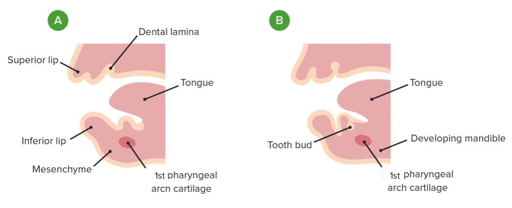
Formation of the dental lamina arch (A) and tooth bud (B)
Image by Lecturio.
A and B: Formation of the dental lamina and tooth bud
C and D: Formation of the enamel organ
E: Formation of the dental papilla and dental sac
F: Formation and development of the enamel and dentine layers
G: Early stage of tooth eruption
H: Fully erupted deciduous tooth
I: Cross-section of a developing tooth showing its composition
Teeth begin to erupt within 6 months to 1 year of life, usually beginning with the lower incisors.
| Teeth | Expected age at eruption | Expected age at shedding |
|---|---|---|
| Central incisor | 8–12 months | 6–7 years |
| Lateral incisor | 9–13 months | 7–8 years |
| Canine | 16–22 months | 10–12 years |
| 1st molar | 12–19 months | 9–11 years |
| 2nd molar | 25–33 months | 10–12 years |
| Teeth | Expected age at eruption | Expected age at shedding |
|---|---|---|
| 2nd molar | 23–31 months | 10–12 years |
| 1st molar | 14–18 months | 9–11 years |
| Canine | 12–23 months | 9–12 years |
| Lateral incisor | 10–16 months | 7–8 years |
| Central incisor | 6–10 months | 6–7 years |
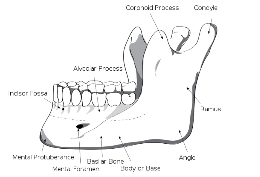
Mandible with teeth in place within the alveolar processes
Image: “Human mandible left” by Djexplo. License: CC0 1.0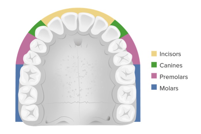
Location of the incisors, canines, premolars, and molars within the jawline
Image by Lecturio.Blood supply:
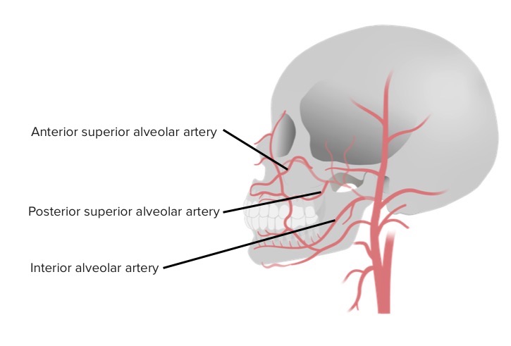
Blood supply of the teeth
Image by Lecturio.Innervation:
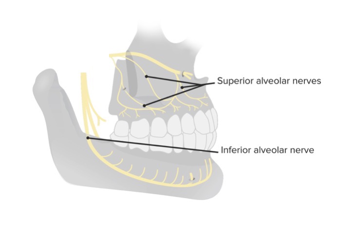
Innervation of the teeth
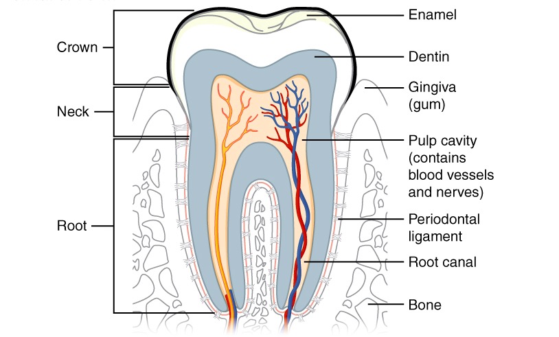
Image by Lecturio.Each tooth comprises 4 components:
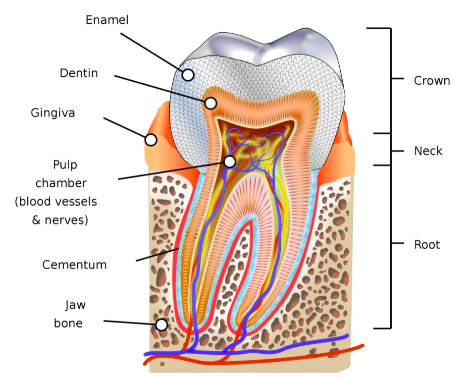
The tooth and its parts
Image: “Diagram of a healthy human molar” by KDS4444. License: CC BY-SA 4.0