Achieve Mastery of Medical Concepts

Study for medical school and boards with Lecturio

Electrolytes

Electrolytes are mineral salts that dissolve in water and dissociate into charged particles called ions, which can be either be positively (cations) or negatively (anions) charged. Electrolytes are distributed in the extracellular and intracellular compartments in different concentrations. Electrolytes are essential for various basic life-sustaining functions such as maintaining electrical neutrality in cells, generating action potentials in nerves and muscles, and maintaining normal blood pH pH The quantitative measurement of the acidity or basicity of a solution. Acid-Base Balance. The most important electrolytes are sodium Sodium A member of the alkali group of metals. It has the atomic symbol na, atomic number 11, and atomic weight 23. Hyponatremia, potassium Potassium An element in the alkali group of metals with an atomic symbol k, atomic number 19, and atomic weight 39. 10. It is the chief cation in the intracellular fluid of muscle and other cells. Potassium ion is a strong electrolyte that plays a significant role in the regulation of fluid volume and maintenance of the water-electrolyte balance. Hyperkalemia, chloride, magnesium, calcium, phosphate, and bicarbonate. In order for these electrolytes to participate in biochemical reactions and cellular processes, regulatory mechanisms are in place, which help maintain homeostasis Homeostasis The processes whereby the internal environment of an organism tends to remain balanced and stable. Cell Injury and Death.

Last updated: Dec 6, 2022

Editorial responsibility: Stanley Oiseth, Lindsay Jones, Evelin Maza

Introduction

Body fluids and electrolytes

Electrolyte balance

The ICF ICF The fluid inside cells. Body Fluid Compartments and ECF compartments have different and unequal electrolyte distribution to maintain physiological function.

Intracellular fluid Intracellular fluid The fluid inside cells. Body Fluid Compartments:

  • K⁺: main intracellular cation
  • Mg2
  • Phosphates (HPO₄²/H₂PO₄) electrically balance the intracellular cations along with the negatively charged proteins Proteins Linear polypeptides that are synthesized on ribosomes and may be further modified, crosslinked, cleaved, or assembled into complex proteins with several subunits. The specific sequence of amino acids determines the shape the polypeptide will take, during protein folding, and the function of the protein. Energy Homeostasis.

Extracellular fluid Extracellular fluid The fluid of the body that is outside of cells. It is the external environment for the cells. Body Fluid Compartments:

  • Na⁺:
    • Main extracellular cation
    • Important in determining serum osmolarity Osmolarity The concentration of osmotically active particles in solution expressed in terms of osmoles of solute per liter of solution. Osmolality is expressed in terms of osmoles of solute per kilogram of solvent. Hypernatremia (solutes/L)
    • Controls ECF volume and water distribution in the body
  • Calcium (Ca²⁺)
  • Chloride and HCO₃:
    • Chloride is the most abundant anion in the ECF.
    • Anions balance the extracellular cations.

Sodium

Overview

  • Normal range: 135–145 mEq/L
  • 95% of the ingested Na+ is absorbed by the gut.
  • Excretion:
    • 90%–95% excreted by the kidneys Kidneys The kidneys are a pair of bean-shaped organs located retroperitoneally against the posterior wall of the abdomen on either side of the spine. As part of the urinary tract, the kidneys are responsible for blood filtration and excretion of water-soluble waste in the urine. Kidneys: Anatomy
    • Remainder through feces and sweat
  • Functions:
    • Establishes osmotic pressure Osmotic pressure The pressure required to prevent the passage of solvent through a semipermeable membrane that separates a pure solvent from a solution of the solvent and solute or that separates different concentrations of a solution. It is proportional to the osmolality of the solution. Intravenous Fluids
    • Na+ transcellular Transcellular The movement of one cell into, through, and out of another cell. Tubular System gradient:
    • Transient influx of Na+ generates action potential Action Potential Abrupt changes in the membrane potential that sweep along the cell membrane of excitable cells in response to excitation stimuli. Membrane Potential that leads to:
      • Nerve conduction
      • Muscle contraction
      • Cardiac conduction
      • Activation of intracellular signaling pathways

Regulation

  • Na+ homeostasis Homeostasis The processes whereby the internal environment of an organism tends to remain balanced and stable. Cell Injury and Death:
    • Regulated through the kidneys Kidneys The kidneys are a pair of bean-shaped organs located retroperitoneally against the posterior wall of the abdomen on either side of the spine. As part of the urinary tract, the kidneys are responsible for blood filtration and excretion of water-soluble waste in the urine. Kidneys: Anatomy (majority in the proximal tubules)
    • Occurs by sensing changes in the effective circulating volume Effective circulating volume Renal Sodium and Water Regulation ( ECV ECV A procedure in which the physician attempts to manually rotate the fetus from a breech to a cephalic presentation by pushing on the maternal abdomen Fetal Malpresentation and Malposition):
      • ↑ Na+ causes ↑ ECV ECV A procedure in which the physician attempts to manually rotate the fetus from a breech to a cephalic presentation by pushing on the maternal abdomen Fetal Malpresentation and Malposition
      • ↓ Na+ causes ↓ ECV ECV A procedure in which the physician attempts to manually rotate the fetus from a breech to a cephalic presentation by pushing on the maternal abdomen Fetal Malpresentation and Malposition
  • RAAS RAAS A blood pressure regulating system of interacting components that include renin; angiotensinogen; angiotensin converting enzyme; angiotensin i; angiotensin ii; and angiotensinase. Renin, an enzyme produced in the kidney, acts on angiotensinogen, an alpha-2 globulin produced by the liver, forming angiotensin I. Angiotensin-converting enzyme, contained in the lung, acts on angiotensin I in the plasma converting it to angiotensin II, an extremely powerful vasoconstrictor. Angiotensin II causes contraction of the arteriolar and renal vascular smooth muscle, leading to retention of salt and water in the kidney and increased arterial blood pressure. In addition, angiotensin II stimulates the release of aldosterone from the adrenal cortex, which in turn also increases salt and water retention in the kidney. Angiotensin-converting enzyme also breaks down bradykinin, a powerful vasodilator and component of the kallikrein-kinin system. Adrenal Hormones:
    • Juxtaglomerular apparatus and carotid sinus Carotid sinus The dilated portion of the common carotid artery at its bifurcation into external and internal carotids. It contains baroreceptors which, when stimulated, cause slowing of the heart, vasodilatation, and a fall in blood pressure. Carotid Arterial System: Anatomy/ aortic arch Aortic arch Mediastinum and Great Vessels: Anatomy baroreceptors Baroreceptors Receptors in the vascular system, particularly the aorta and carotid sinus, which are sensitive to stretch of the vessel walls. Diabetes Insipidus trigger Trigger The type of signal that initiates the inspiratory phase by the ventilator Invasive Mechanical Ventilation renin Renin A highly specific (leu-leu) endopeptidase that generates angiotensin I from its precursor angiotensinogen, leading to a cascade of reactions which elevate blood pressure and increase sodium retention by the kidney in the renin-angiotensin system. Renal Sodium and Water Regulation release from kidneys Kidneys The kidneys are a pair of bean-shaped organs located retroperitoneally against the posterior wall of the abdomen on either side of the spine. As part of the urinary tract, the kidneys are responsible for blood filtration and excretion of water-soluble waste in the urine. Kidneys: Anatomy when ECV ECV A procedure in which the physician attempts to manually rotate the fetus from a breech to a cephalic presentation by pushing on the maternal abdomen Fetal Malpresentation and Malposition
    • If Na+ is low: ↓ ECV ECV A procedure in which the physician attempts to manually rotate the fetus from a breech to a cephalic presentation by pushing on the maternal abdomen Fetal Malpresentation and Malposition renin Renin A highly specific (leu-leu) endopeptidase that generates angiotensin I from its precursor angiotensinogen, leading to a cascade of reactions which elevate blood pressure and increase sodium retention by the kidney in the renin-angiotensin system. Renal Sodium and Water Regulation → angiotensin (causing vasoconstriction Vasoconstriction The physiological narrowing of blood vessels by contraction of the vascular smooth muscle. Vascular Resistance, Flow, and Mean Arterial Pressure) → aldosterone Aldosterone A hormone secreted by the adrenal cortex that regulates electrolyte and water balance by increasing the renal retention of sodium and the excretion of potassium. Hyperkalemia is secreted:
  • Natriuretic peptides Natriuretic peptides Peptides that regulate the water-electrolyte balance in the body, also known as natriuretic peptide hormones. Several have been sequenced (atrial natriuretic factor; brain natriuretic peptide; c-type natriuretic peptide). Arterial Pressure Regulation:
    • Includes atrial natriuretic peptide Atrial natriuretic peptide A potent natriuretic and vasodilatory peptide or mixture of different-sized low molecular weight peptides derived from a common precursor and secreted mainly by the heart atrium. All these peptides share a sequence of about 20 amino acids. Renal Sodium and Water Regulation and BNP BNP A peptide that is secreted by the brain and the heart atria, stored mainly in cardiac ventricular myocardium. It can cause natriuresis; diuresis; vasodilation; and inhibits secretion of renin and aldosterone. It improves heart function. It contains 32 amino acids. Renal Sodium and Water Regulation
    • If Na+ is high: ↑ ECV ECV A procedure in which the physician attempts to manually rotate the fetus from a breech to a cephalic presentation by pushing on the maternal abdomen Fetal Malpresentation and Malposition → cardiac baroreceptors Baroreceptors Receptors in the vascular system, particularly the aorta and carotid sinus, which are sensitive to stretch of the vessel walls. Diabetes Insipidus sense an ↑ in ECV ECV A procedure in which the physician attempts to manually rotate the fetus from a breech to a cephalic presentation by pushing on the maternal abdomen Fetal Malpresentation and Malpositionnatriuretic peptide is secreted
      • Stimulates urinary Na+ excretion (natriuresis)
      • Also promotes excretion of water
      • Inhibits angiotensin II Angiotensin II An octapeptide that is a potent but labile vasoconstrictor. It is produced from angiotensin I after the removal of two amino acids at the c-terminal by angiotensin converting enzyme. The amino acid in position 5 varies in different species. To block vasoconstriction and hypertension effect of angiotensin II, patients are often treated with ace inhibitors or with angiotensin II type 1 receptor blockers. Renal Sodium and Water Regulation production

Related disorders

  • Hypernatremia Hypernatremia Hypernatremia is an elevated serum sodium concentration > 145 mmol/L. Serum sodium is the greatest contributor to plasma osmolality, which is very tightly controlled by the hypothalamus via the thirst mechanism and antidiuretic hormone (ADH) release. Hypernatremia occurs either from a lack of access to water or an excessive intake of sodium. Hypernatremia: Na+ > 145 meq/L
  • Hyponatremia Hyponatremia Hyponatremia is defined as a decreased serum sodium (sNa+) concentration less than 135 mmol/L. Serum sodium is the greatest contributor to plasma osmolality, which is very tightly controlled via antidiuretic hormone (ADH) release from the hypothalamus and by the thirst mechanism. Hyponatremia: Na+ < 135 meq/L

Potassium

Overview

  • Normal range: 3.5–5.2 mEq/L
  • Excretion: 90% is excreted in the urine, 10% in feces
  • Functions:
    • ٌResting cellular membrane potential Membrane potential The membrane potential is the difference in electric charge between the interior and the exterior of a cell. All living cells maintain a potential difference across the membrane thanks to the insulating properties of their plasma membranes (PMs) and the selective transport of ions across this membrane by transporters. Membrane Potential and the propagation Propagation Propagation refers to how the electrical signal spreads to every myocyte in the heart. Cardiac Physiology of action potentials
    • Hormone secretion Secretion Coagulation Studies and action
    • Vascular tone
    • Systemic BP control
    • GI motility GI Motility The primary functions of the GI tract are digestion and absorption, which require coordinated contractions of the smooth muscles present in the GI tract. Peristaltic waves, segmentation contractions, and the migrating motor complex are all important contraction patterns that help to mix contents, get them in contact with the intestinal walls, and propel material down the tract at appropriate times and in appropriate amounts. Gastrointestinal Motility
    • Glucose Glucose A primary source of energy for living organisms. It is naturally occurring and is found in fruits and other parts of plants in its free state. It is used therapeutically in fluid and nutrient replacement. Lactose Intolerance and insulin Insulin Insulin is a peptide hormone that is produced by the beta cells of the pancreas. Insulin plays a role in metabolic functions such as glucose uptake, glycolysis, glycogenesis, lipogenesis, and protein synthesis. Exogenous insulin may be needed for individuals with diabetes mellitus, in whom there is a deficiency in endogenous insulin or increased insulin resistance. Insulin metabolism
    • Concentrating ability of kidneys Kidneys The kidneys are a pair of bean-shaped organs located retroperitoneally against the posterior wall of the abdomen on either side of the spine. As part of the urinary tract, the kidneys are responsible for blood filtration and excretion of water-soluble waste in the urine. Kidneys: Anatomy and pH pH The quantitative measurement of the acidity or basicity of a solution. Acid-Base Balance regulation

Regulation

  • Kidneys Kidneys The kidneys are a pair of bean-shaped organs located retroperitoneally against the posterior wall of the abdomen on either side of the spine. As part of the urinary tract, the kidneys are responsible for blood filtration and excretion of water-soluble waste in the urine. Kidneys: Anatomy are responsible for 90%–95% of the overall K+ regulation.
    • ↑ ECF K+ level triggers mechanisms for renal K+ excretion
    • Aldosterone Aldosterone A hormone secreted by the adrenal cortex that regulates electrolyte and water balance by increasing the renal retention of sodium and the excretion of potassium. Hyperkalemia:
      • Stimulates Na+-K⁺ ATPase
      • Increases K+ excretion into distal tubules and collecting ducts 
  • Transcellular Transcellular The movement of one cell into, through, and out of another cell. Tubular System shifting (mediated by insulin Insulin Insulin is a peptide hormone that is produced by the beta cells of the pancreas. Insulin plays a role in metabolic functions such as glucose uptake, glycolysis, glycogenesis, lipogenesis, and protein synthesis. Exogenous insulin may be needed for individuals with diabetes mellitus, in whom there is a deficiency in endogenous insulin or increased insulin resistance. Insulin and sympathetic nervous system Nervous system The nervous system is a small and complex system that consists of an intricate network of neural cells (or neurons) and even more glial cells (for support and insulation). It is divided according to its anatomical components as well as its functional characteristics. The brain and spinal cord are referred to as the central nervous system, and the branches of nerves from these structures are referred to as the peripheral nervous system. Nervous System: Anatomy, Structure, and Classification) prevents the excessive increase in ECF K+ levels.
    • β2 receptor Receptor Receptors are proteins located either on the surface of or within a cell that can bind to signaling molecules known as ligands (e.g., hormones) and cause some type of response within the cell. Receptors activation:
      • Stimulates K+ uptake into cells (primarily into muscle and liver Liver The liver is the largest gland in the human body. The liver is found in the superior right quadrant of the abdomen and weighs approximately 1.5 kilograms. Its main functions are detoxification, metabolism, nutrient storage (e.g., iron and vitamins), synthesis of coagulation factors, formation of bile, filtration, and storage of blood. Liver: Anatomy cells)
      • β2 antagonists block K+ uptake and cause hyperkalemia Hyperkalemia Hyperkalemia is defined as a serum potassium (K+) concentration >5.2 mEq/L. Homeostatic mechanisms maintain the serum K+ concentration between 3.5 and 5.2 mEq/L, despite marked variation in dietary intake. Hyperkalemia can be due to a variety of causes, which include transcellular shifts, tissue breakdown, inadequate renal excretion, and drugs. Hyperkalemia.
      • α1 receptor Receptor Receptors are proteins located either on the surface of or within a cell that can bind to signaling molecules known as ligands (e.g., hormones) and cause some type of response within the cell. Receptors activation causes a shift of K+ out of the cells. 
    • Insulin Insulin Insulin is a peptide hormone that is produced by the beta cells of the pancreas. Insulin plays a role in metabolic functions such as glucose uptake, glycolysis, glycogenesis, lipogenesis, and protein synthesis. Exogenous insulin may be needed for individuals with diabetes mellitus, in whom there is a deficiency in endogenous insulin or increased insulin resistance. Insulin:
      • Stimulates Na+-K⁺ ATPase
      • Increases K+ uptake into cells
      • Responsible for dietary uptake of K+ into cells after a meal
  • Other factors:
    • Acid-base status:
      • Acidosis Acidosis A pathologic condition of acid accumulation or depletion of base in the body. The two main types are respiratory acidosis and metabolic acidosis, due to metabolic acid build up. Respiratory Acidosis causes K+ to move out of cells.
      • Alkalosis Alkalosis A pathological condition that removes acid or adds base to the body fluids. Respiratory Alkalosis causes K+ to move into cells.
    • Exercise: moves K+ out of muscle cells
Sodium-potassium pump

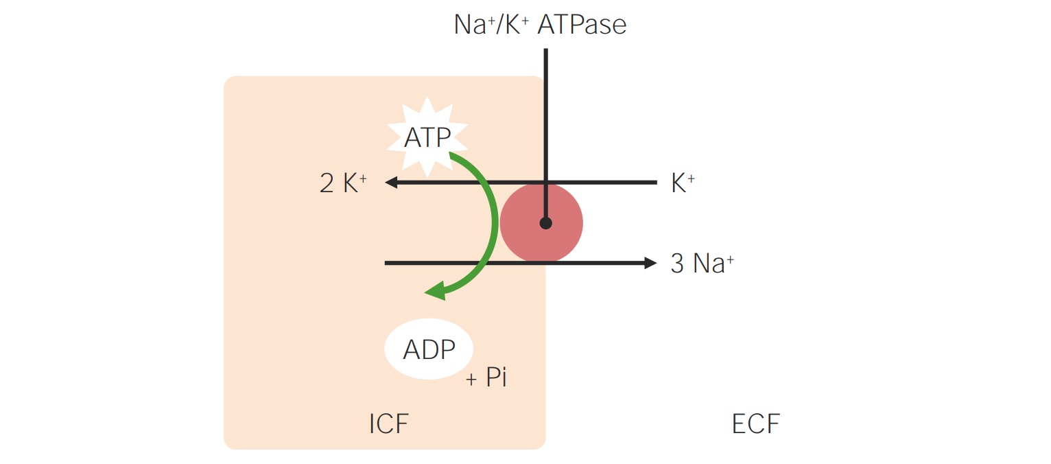
Sodium-potassium pump:
Transmembrane ATPase maintains a gradient of higher Na+ concentration in the ECF and a higher K+ concentration in the ICF. For every ATP consumed, the ATPase pumps 3 Na+ out of the cell and 2 K+ into the cell, which stabilizes the cellular resting membrane potential and cell volume.
Pi: inorganic phosphate
ECF: extracellular fluid
ICF: intracellular fluid

Image by Lecturio.

Related disorders

  • Hyperkalemia Hyperkalemia Hyperkalemia is defined as a serum potassium (K+) concentration >5.2 mEq/L. Homeostatic mechanisms maintain the serum K+ concentration between 3.5 and 5.2 mEq/L, despite marked variation in dietary intake. Hyperkalemia can be due to a variety of causes, which include transcellular shifts, tissue breakdown, inadequate renal excretion, and drugs. Hyperkalemia: K+ > 5.2 mEq/L
  • Hypokalemia Hypokalemia Hypokalemia is defined as plasma potassium (K+) concentration < 3.5 mEq/L. Homeostatic mechanisms maintain plasma concentration between 3.5-5.2 mEq/L despite marked variation in dietary intake. Hypokalemia can be due to renal losses, GI losses, transcellular shifts, or poor dietary intake. Hypokalemia: K+ < 3.5 mEq/L

Chloride

Overview

  • Normal range: 96–106 mEq/L
  • Rapidly and almost totally absorbed by the GI tract
  • Excretion: through GI tract, urinary tract Urinary tract The urinary tract is located in the abdomen and pelvis and consists of the kidneys, ureters, urinary bladder, and urethra. The structures permit the excretion of urine from the body. Urine flows from the kidneys through the ureters to the urinary bladder and out through the urethra. Urinary Tract: Anatomy, and skin Skin The skin, also referred to as the integumentary system, is the largest organ of the body. The skin is primarily composed of the epidermis (outer layer) and dermis (deep layer). The epidermis is primarily composed of keratinocytes that undergo rapid turnover, while the dermis contains dense layers of connective tissue. Skin: Structure and Functions
  • Functions:
    • Follows Na+ across cellular membranes to maintain charge neutrality
    • Maintenance of cell homeostasis Homeostasis The processes whereby the internal environment of an organism tends to remain balanced and stable. Cell Injury and Death 
    • Transmission of action potentials in neurons Neurons The basic cellular units of nervous tissue. Each neuron consists of a body, an axon, and dendrites. Their purpose is to receive, conduct, and transmit impulses in the nervous system. Nervous System: Histology
    • Maintenance of ECF osmolarity Osmolarity The concentration of osmotically active particles in solution expressed in terms of osmoles of solute per liter of solution. Osmolality is expressed in terms of osmoles of solute per kilogram of solvent. Hypernatremia (together with Na+), thus regulating:
      • Blood volume
      • BP 
    • Acid-base balance
    • Synthesis Synthesis Polymerase Chain Reaction (PCR) of gastric hydrochloric acid Hydrochloric acid A strong corrosive acid that is commonly used as a laboratory reagent. It is formed by dissolving hydrogen chloride in water. Gastric acid is the hydrochloric acid component of gastric juice. Caustic Ingestion (Cleaning Products) 
    • Control of epithelial fluid transport

Regulation

Chloride homeostasis Homeostasis The processes whereby the internal environment of an organism tends to remain balanced and stable. Cell Injury and Death:

  • Mainly by the kidneys Kidneys The kidneys are a pair of bean-shaped organs located retroperitoneally against the posterior wall of the abdomen on either side of the spine. As part of the urinary tract, the kidneys are responsible for blood filtration and excretion of water-soluble waste in the urine. Kidneys: Anatomy (through reabsorption in the tubules)
  • Affected by acid-base disorders:
    • ↑ Chloride: associated with acidosis Acidosis A pathologic condition of acid accumulation or depletion of base in the body. The two main types are respiratory acidosis and metabolic acidosis, due to metabolic acid build up. Respiratory Acidosis
    • ↓ Chloride: associated with alkalosis Alkalosis A pathological condition that removes acid or adds base to the body fluids. Respiratory Alkalosis 
  • Affected by changes in Na⁺ levels. Reabsorption is mediated by cation cotransporters in combination with Na⁺.

Related disorders

  • Hyperchloremia (chloride > 106 mEq/L):
    • From pure water loss (electrolyte-free fluid), such as hypotonic Hypotonic Solutions that have a lesser osmotic pressure than a reference solution such as blood, plasma, or interstitial fluid. Renal Sodium and Water Regulation dehydration Dehydration The condition that results from excessive loss of water from a living organism. Volume Depletion and Dehydration 
    • Administration of NaCl-containing fluids or hypertonic Hypertonic Solutions that have a greater osmotic pressure than a reference solution such as blood, plasma, or interstitial fluid. Renal Sodium and Water Regulation feeding
    • Acidosis Acidosis A pathologic condition of acid accumulation or depletion of base in the body. The two main types are respiratory acidosis and metabolic acidosis, due to metabolic acid build up. Respiratory Acidosis:
      • Renal tubular acidosis Acidosis A pathologic condition of acid accumulation or depletion of base in the body. The two main types are respiratory acidosis and metabolic acidosis, due to metabolic acid build up. Respiratory Acidosis
      • Small bowel Small bowel The small intestine is the longest part of the GI tract, extending from the pyloric orifice of the stomach to the ileocecal junction. The small intestine is the major organ responsible for chemical digestion and absorption of nutrients. It is divided into 3 segments: the duodenum, the jejunum, and the ileum. Small Intestine: Anatomy diarrhea Diarrhea Diarrhea is defined as ≥ 3 watery or loose stools in a 24-hour period. There are a multitude of etiologies, which can be classified based on the underlying mechanism of disease. The duration of symptoms (acute or chronic) and characteristics of the stools (e.g., watery, bloody, steatorrheic, mucoid) can help guide further diagnostic evaluation. Diarrhea 
      • Pancreatic fistula Fistula Abnormal communication most commonly seen between two internal organs, or between an internal organ and the surface of the body. Anal Fistula
    • Drugs ( acetazolamide Acetazolamide One of the carbonic anhydrase inhibitors that is sometimes effective against absence seizures. It is sometimes useful also as an adjunct in the treatment of tonic-clonic, myoclonic, and atonic seizures, particularly in women whose seizures occur or are exacerbated at specific times in the menstrual cycle. However, its usefulness is transient often because of rapid development of tolerance. Its antiepileptic effect may be due to its inhibitory effect on brain carbonic anhydrase, which leads to an increased transneuronal chloride gradient, increased chloride current, and increased inhibition. Carbonic Anhydrase Inhibitors)
  • Hypochloremia (< 96 mEq/L):
    • Inadequate NaCl intake
    • Loss of chloride:
      • Vomiting Vomiting The forcible expulsion of the contents of the stomach through the mouth. Hypokalemia: loss of hydrochloric acid Hydrochloric acid A strong corrosive acid that is commonly used as a laboratory reagent. It is formed by dissolving hydrogen chloride in water. Gastric acid is the hydrochloric acid component of gastric juice. Caustic Ingestion (Cleaning Products) in the stomach Stomach The stomach is a muscular sac in the upper left portion of the abdomen that plays a critical role in digestion. The stomach develops from the foregut and connects the esophagus with the duodenum. Structurally, the stomach is C-shaped and forms a greater and lesser curvature and is divided grossly into regions: the cardia, fundus, body, and pylorus. Stomach: Anatomy, leading to hypochloremic hypokalemic metabolic alkalosis Alkalosis A pathological condition that removes acid or adds base to the body fluids. Respiratory Alkalosis
      • Gastric lavage
      • Burns Burns A burn is a type of injury to the skin and deeper tissues caused by exposure to heat, electricity, chemicals, friction, or radiation. Burns are classified according to their depth as superficial (1st-degree), partial-thickness (2nd-degree), full-thickness (3rd-degree), and 4th-degree burns. Burns
      • Excessive use of diuretics Diuretics Agents that promote the excretion of urine through their effects on kidney function. Heart Failure and Angina Medication or osmotic diuresis Osmotic diuresis Volume Depletion and Dehydration
      • In combination with hyponatremia Hyponatremia Hyponatremia is defined as a decreased serum sodium (sNa+) concentration less than 135 mmol/L. Serum sodium is the greatest contributor to plasma osmolality, which is very tightly controlled via antidiuretic hormone (ADH) release from the hypothalamus and by the thirst mechanism. Hyponatremia

Calcium

Overview

  • Normal range: 8.5–10.5 mg/dL
  • Ca²⁺: metabolically active form (normal range 4.65–5.25 mg/dL)
  • Most abundant mineral in the human body with 99% found in the skeleton
  • Functions:
    • Enzyme activity
    • Cellular functions related to cell division Cell Division A type of cell nucleus division by means of which the two daughter nuclei normally receive identical complements of the number of chromosomes of the somatic cells of the species. Cell Cycle, exocytosis Exocytosis Cellular release of material within membrane-limited vesicles by fusion of the vesicles with the cell membrane. The Cell: Cell Membrane, communication Communication The exchange or transmission of ideas, attitudes, or beliefs between individuals or groups. Decision-making Capacity and Legal Competence
    • Muscle contraction
    • Cardiac contractility
    • Nerve conduction
    • Blood coagulation
    • Bone mineralization Bone mineralization Calcium (Ca2+) and phosphate (PO43–) combine to form hydroxyapatite crystals on the bone matrix. Bones: Development and Ossification

Regulation

  • Bone Bone Bone is a compact type of hardened connective tissue composed of bone cells, membranes, an extracellular mineralized matrix, and central bone marrow. The 2 primary types of bone are compact and spongy. Bones: Structure and Types, intestine, and kidneys Kidneys The kidneys are a pair of bean-shaped organs located retroperitoneally against the posterior wall of the abdomen on either side of the spine. As part of the urinary tract, the kidneys are responsible for blood filtration and excretion of water-soluble waste in the urine. Kidneys: Anatomy are involved in homeostasis Homeostasis The processes whereby the internal environment of an organism tends to remain balanced and stable. Cell Injury and Death.
  • Key elements of Ca²⁺ regulation:
    • Parathyroid Parathyroid The parathyroid glands are 2 pairs of small endocrine glands found in close proximity to the thyroid gland. The superior parathyroid glands are lodged within the parenchyma of the upper poles of the right and left thyroid lobes; the inferior parathyroid glands are close to the inferior tips or poles of the lobes. Parathyroid Glands: Anatomy hormone (PTH) from parathyroid Parathyroid The parathyroid glands are 2 pairs of small endocrine glands found in close proximity to the thyroid gland. The superior parathyroid glands are lodged within the parenchyma of the upper poles of the right and left thyroid lobes; the inferior parathyroid glands are close to the inferior tips or poles of the lobes. Parathyroid Glands: Anatomy glands:
      • Vitamin D Vitamin D A vitamin that includes both cholecalciferols and ergocalciferols, which have the common effect of preventing or curing rickets in animals. It can also be viewed as a hormone since it can be formed in skin by action of ultraviolet rays upon the precursors, 7-dehydrocholesterol and ergosterol, and acts on vitamin D receptors to regulate calcium in opposition to parathyroid hormone. Fat-soluble Vitamins and their Deficiencies production in the kidneys Kidneys The kidneys are a pair of bean-shaped organs located retroperitoneally against the posterior wall of the abdomen on either side of the spine. As part of the urinary tract, the kidneys are responsible for blood filtration and excretion of water-soluble waste in the urine. Kidneys: Anatomy 
      • ↑ Ca²⁺ reabsorption in the distal tubules and ↑ Ca²⁺ absorption Absorption Absorption involves the uptake of nutrient molecules and their transfer from the lumen of the GI tract across the enterocytes and into the interstitial space, where they can be taken up in the venous or lymphatic circulation. Digestion and Absorption in the intestines
      • Bone resorption Bone resorption Bone loss due to osteoclastic activity. Bones: Remodeling and Healing (release of Ca²⁺ and HPO₄²/H₂PO₄ from bones)
    • Vitamin D Vitamin D A vitamin that includes both cholecalciferols and ergocalciferols, which have the common effect of preventing or curing rickets in animals. It can also be viewed as a hormone since it can be formed in skin by action of ultraviolet rays upon the precursors, 7-dehydrocholesterol and ergosterol, and acts on vitamin D receptors to regulate calcium in opposition to parathyroid hormone. Fat-soluble Vitamins and their Deficiencies:
      • Activates osteoclasts Osteoclasts A large multinuclear cell associated with the bone resorption. An odontoclast, also called cementoclast, is cytomorphologically the same as an osteoclast and is involved in cementum resorption. Bones: Development and Ossification to release Ca²⁺ and phosphorus
      • ↑ Intestinal Ca²⁺ and HPO₄²/H₂PO₄ absorption Absorption Absorption involves the uptake of nutrient molecules and their transfer from the lumen of the GI tract across the enterocytes and into the interstitial space, where they can be taken up in the venous or lymphatic circulation. Digestion and Absorption
    • pH pH The quantitative measurement of the acidity or basicity of a solution. Acid-Base Balance:
      • pH pH The quantitative measurement of the acidity or basicity of a solution. Acid-Base Balance ( alkalosis Alkalosis A pathological condition that removes acid or adds base to the body fluids. Respiratory Alkalosis) → ↑ binding to albumin Albumin Serum albumin from humans. It is an essential carrier of both endogenous substances, such as fatty acids and bilirubin, and of xenobiotics in the blood. Liver Function Tests = ↓ ionized Ca²⁺
      • pH pH The quantitative measurement of the acidity or basicity of a solution. Acid-Base Balance ( acidosis Acidosis A pathologic condition of acid accumulation or depletion of base in the body. The two main types are respiratory acidosis and metabolic acidosis, due to metabolic acid build up. Respiratory Acidosis) → ↓ binding to albumin Albumin Serum albumin from humans. It is an essential carrier of both endogenous substances, such as fatty acids and bilirubin, and of xenobiotics in the blood. Liver Function Tests = ↑ ionized Ca²⁺
    • Albumin Albumin Serum albumin from humans. It is an essential carrier of both endogenous substances, such as fatty acids and bilirubin, and of xenobiotics in the blood. Liver Function Tests:
      • Every 1 g/dL ↓ in albumin Albumin Serum albumin from humans. It is an essential carrier of both endogenous substances, such as fatty acids and bilirubin, and of xenobiotics in the blood. Liver Function Tests → ↓ in Ca²⁺ by 0.8 mg/dL
      • Corrected Ca²⁺ (mg/dL) = measured total Ca²⁺ (mg/dL) + [0.8 x (4.0 – albumin Albumin Serum albumin from humans. It is an essential carrier of both endogenous substances, such as fatty acids and bilirubin, and of xenobiotics in the blood. Liver Function Tests concentration (g/dL))]
    • Other factors:
      • Calcitonin Calcitonin A peptide hormone that lowers calcium concentration in the blood. In humans, it is released by thyroid cells and acts to decrease the formation and absorptive activity of osteoclasts. Its role in regulating plasma calcium is much greater in children and in certain diseases than in normal adults. Other Antiresorptive Drugs from the thyroid Thyroid The thyroid gland is one of the largest endocrine glands in the human body. The thyroid gland is a highly vascular, brownish-red gland located in the visceral compartment of the anterior region of the neck. Thyroid Gland: Anatomy gland (opposes PTH →↓ Ca²⁺)
      • Hyperphosphatemia Hyperphosphatemia A condition of abnormally high level of phosphates in the blood, usually significantly above the normal range of 0. 84-1. 58 mmol per liter of serum. Hypocalcemia (↑ HPO₄²/H₂PO₄ binding, ↓ Ca²⁺)
      • Hypomagnesemia (↓ PTH release → ↓ Ca²⁺)
Calcium metabolism

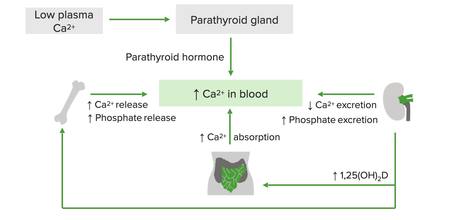
Schematic diagram of calcium (Ca²⁺) regulation:
Low plasma Ca²⁺ stimulates the release of parathyroid hormone (PTH), which increases Ca²⁺ and phosphate release from the bone, Ca²⁺ absorption in the GI tract, and vitamin D production in the kidneys. Active vitamin D, in turn, increases Ca²⁺ release from the bones and Ca²⁺ absorption in the small intestine.

1,25 (OH)₂D: 1,25-dihydroxyvitamin D

Image by Lecturio.

Related disorders

  • Hypercalcemia Hypercalcemia Hypercalcemia (serum calcium > 10.5 mg/dL) can result from various conditions, the majority of which are due to hyperparathyroidism and malignancy. Other causes include disorders leading to vitamin D elevation, granulomatous diseases, and the use of certain pharmacological agents. Symptoms vary depending on calcium levels and the onset of hypercalcemia. Hypercalcemia: Ca²⁺ > 10.5 mg/dL
  • Hypocalcemia Hypocalcemia Hypocalcemia, a serum calcium < 8.5 mg/dL, can result from various conditions. The causes may include hypoparathyroidism, drugs, disorders leading to vitamin D deficiency, and more. Calcium levels are regulated and affected by different elements such as dietary intake, parathyroid hormone (PTH), vitamin D, pH, and albumin. Presentation can range from an asymptomatic (mild deficiency) to a life-threatening condition (acute, significant deficiency). Hypocalcemia: Ca²⁺ < 8.5 mg/dL

Magnesium

Overview

  • Normal range: 1.5–2.2 mg/dL
  • 4th most abundant cation in the body
  • Mg²⁺ distribution in the body:
  • Functions:
    • Cofactor for enzymatic reactions involved in ATP and/or DNA DNA A deoxyribonucleotide polymer that is the primary genetic material of all cells. Eukaryotic and prokaryotic organisms normally contain DNA in a double-stranded state, yet several important biological processes transiently involve single-stranded regions. DNA, which consists of a polysugar-phosphate backbone possessing projections of purines (adenine and guanine) and pyrimidines (thymine and cytosine), forms a double helix that is held together by hydrogen bonds between these purines and pyrimidines (adenine to thymine and guanine to cytosine). DNA Types and Structure and RNA RNA A polynucleotide consisting essentially of chains with a repeating backbone of phosphate and ribose units to which nitrogenous bases are attached. RNA is unique among biological macromolecules in that it can encode genetic information, serve as an abundant structural component of cells, and also possesses catalytic activity. RNA Types and Structure synthesis Synthesis Polymerase Chain Reaction (PCR)
    • Cellular replication and biochemical processes (i.e., glycolysis Glycolysis Glycolysis is a central metabolic pathway responsible for the breakdown of glucose and plays a vital role in generating free energy for the cell and metabolites for further oxidative degradation. Glucose primarily becomes available in the blood as a result of glycogen breakdown or from its synthesis from noncarbohydrate precursors (gluconeogenesis) and is imported into cells by specific transport proteins. Glycolysis)
    • Linked with Ca²⁺ and K⁺ homeostasis Homeostasis The processes whereby the internal environment of an organism tends to remain balanced and stable. Cell Injury and Death
    • Neuromuscular excitability Excitability Skeletal Muscle Contraction:
      • Smooth muscle contraction Smooth muscle contraction Smooth muscle is primarily found in the walls of hollow structures and some visceral organs, including the walls of the vasculature, GI, respiratory, and genitourinary tracts. Smooth muscle contracts more slowly and is regulated differently than skeletal muscle. Smooth muscle can be stimulated by nerve impulses, hormones, metabolic factors (like pH, CO2 or O2 levels), its own intrinsic pacemaker ability, or even mechanical stretch. Smooth Muscle Contraction and relaxation
      • Stabilization of cardiac muscle Cardiac muscle The muscle tissue of the heart. It is composed of striated, involuntary muscle cells connected to form the contractile pump to generate blood flow. Muscle Tissue: Histology
    • Coagulation

Regulation

  • Maintaining Mg²⁺ levels in the serum varies and depends on:
    • Intake
    • GI absorption Absorption Absorption involves the uptake of nutrient molecules and their transfer from the lumen of the GI tract across the enterocytes and into the interstitial space, where they can be taken up in the venous or lymphatic circulation. Digestion and Absorption
    • Renal reabsorption and excretion
  • Kidneys Kidneys The kidneys are a pair of bean-shaped organs located retroperitoneally against the posterior wall of the abdomen on either side of the spine. As part of the urinary tract, the kidneys are responsible for blood filtration and excretion of water-soluble waste in the urine. Kidneys: Anatomy will conserve Mg²⁺ when the levels are low and excrete the excess when the intake is high.
  • Key elements:
    • Increased Mg²⁺ absorption Absorption Absorption involves the uptake of nutrient molecules and their transfer from the lumen of the GI tract across the enterocytes and into the interstitial space, where they can be taken up in the venous or lymphatic circulation. Digestion and Absorption in the presence of:
      • PTH
      • Vitamin D Vitamin D A vitamin that includes both cholecalciferols and ergocalciferols, which have the common effect of preventing or curing rickets in animals. It can also be viewed as a hormone since it can be formed in skin by action of ultraviolet rays upon the precursors, 7-dehydrocholesterol and ergosterol, and acts on vitamin D receptors to regulate calcium in opposition to parathyroid hormone. Fat-soluble Vitamins and their Deficiencies
      • Na⁺ in the diet
      • Thyroid Thyroid The thyroid gland is one of the largest endocrine glands in the human body. The thyroid gland is a highly vascular, brownish-red gland located in the visceral compartment of the anterior region of the neck. Thyroid Gland: Anatomy hormone
    • Decreased Mg²⁺ absorption Absorption Absorption involves the uptake of nutrient molecules and their transfer from the lumen of the GI tract across the enterocytes and into the interstitial space, where they can be taken up in the venous or lymphatic circulation. Digestion and Absorption in the presence of:
      • Ca²⁺
      • Aldosterone Aldosterone A hormone secreted by the adrenal cortex that regulates electrolyte and water balance by increasing the renal retention of sodium and the excretion of potassium. Hyperkalemia (promotes excretion of Mg²⁺ with K⁺, and retention of Na⁺)

Related disorders

  • Hypermagnesemia (Mg²⁺ > 2.2 mg/dL):
    • Overuse of Mg²⁺-containing laxatives Laxatives Laxatives are medications used to promote defecation. Most often, laxatives are used to treat constipation or for bowel preparation for certain procedures. There are 4 main classes of laxatives: bulk-forming, stimulant, osmotic, and emollient. Laxatives or antacids
    • Renal failure Renal failure Conditions in which the kidneys perform below the normal level in the ability to remove wastes, concentrate urine, and maintain electrolyte balance; blood pressure; and calcium metabolism. Renal insufficiency can be classified by the degree of kidney damage (as measured by the level of proteinuria) and reduction in glomerular filtration rate. Crush Syndrome
    • Features:
      • Generalized muscle weakness, nausea Nausea An unpleasant sensation in the stomach usually accompanied by the urge to vomit. Common causes are early pregnancy, sea and motion sickness, emotional stress, intense pain, food poisoning, and various enteroviruses. Antiemetics
      • Hypotension Hypotension Hypotension is defined as low blood pressure, specifically < 90/60 mm Hg, and is most commonly a physiologic response. Hypotension may be mild, serious, or life threatening, depending on the cause. Hypotension 
      • Cardiac rhythm abnormalities (especially if Mg²⁺ > 6 mg/dL): prolonged PR, wide QRS, peaked T waves
    • Management includes calcium gluconate Calcium gluconate The calcium salt of gluconic acid. The compound has a variety of uses, including its use as a calcium replenisher in hypocalcemic states. Hypocalcemia, diuresis, and dialysis Dialysis Renal replacement therapy refers to dialysis and/or kidney transplantation. Dialysis is a procedure by which toxins and excess water are removed from the circulation. Hemodialysis and peritoneal dialysis (PD) are the two types of dialysis, and their primary difference is the location of the filtration process (external to the body in hemodialysis versus inside the body for PD). Peritoneal Dialysis and Hemodialysis.
  • Hypomagnesemia (Mg²⁺ < 1.5 mg/dL):
    • Alcoholism Alcoholism A primary, chronic disease with genetic, psychosocial, and environmental factors influencing its development and manifestations. The disease is often progressive and fatal. It is characterized by impaired control over drinking, preoccupation with the drug alcohol, use of alcohol despite adverse consequences, and distortions in thinking, most notably denial. Each of these symptoms may be continuous or periodic. Wernicke Encephalopathy and Korsakoff Syndrome (most common electrolyte change is ↓ Mg²⁺)
    • Diarrhea Diarrhea Diarrhea is defined as ≥ 3 watery or loose stools in a 24-hour period. There are a multitude of etiologies, which can be classified based on the underlying mechanism of disease. The duration of symptoms (acute or chronic) and characteristics of the stools (e.g., watery, bloody, steatorrheic, mucoid) can help guide further diagnostic evaluation. Diarrhea and GI disease
    • Diuretic use
    • Proton pump Pump ACES and RUSH: Resuscitation Ultrasound Protocols inhibitor therapy
    • Common cause of refractory hypokalemia Hypokalemia Hypokalemia is defined as plasma potassium (K+) concentration < 3.5 mEq/L. Homeostatic mechanisms maintain plasma concentration between 3.5-5.2 mEq/L despite marked variation in dietary intake. Hypokalemia can be due to renal losses, GI losses, transcellular shifts, or poor dietary intake. Hypokalemia

Phosphate

Overview

  • Normal range: 2.5–4.5 mg/dL (higher in children)
  • 2 forms in the serum (depending on the acid-base status):
    •  Dihydrogen phosphate (H₂PO₄
    • Monohydrogen phosphate (HPO₄²)
  • 85% of the total body phosphate is in the bones and teeth Teeth Normally, an adult has 32 teeth: 16 maxillary and 16 mandibular. These teeth are divided into 4 quadrants with 8 teeth each. Each quadrant consists of 2 incisors (dentes incisivi), 1 canine (dens caninus), 2 premolars (dentes premolares), and 3 molars (dentes molares). Teeth are composed of enamel, dentin, and dental cement. Teeth: Anatomy in the form of hydroxyapatite.
  • Only 1% in the ECF
  • Functions:
    • Required for cellular function and skeletal mineralization
    • Component of many metabolic intermediates (ATP) and nucleotides Nucleotides The monomeric units from which DNA or RNA polymers are constructed. They consist of a purine or pyrimidine base, a pentose sugar, and a phosphate group. Nucleic Acids
    • Aerobic and anaerobic energy metabolism
    • O2 delivery to tissues

Regulation

  • Maintained by dietary intake, mobilization from the bone Bone Bone is a compact type of hardened connective tissue composed of bone cells, membranes, an extracellular mineralized matrix, and central bone marrow. The 2 primary types of bone are compact and spongy. Bones: Structure and Types, and renal excretion
  • Key elements:
    • Vitamin D Vitamin D A vitamin that includes both cholecalciferols and ergocalciferols, which have the common effect of preventing or curing rickets in animals. It can also be viewed as a hormone since it can be formed in skin by action of ultraviolet rays upon the precursors, 7-dehydrocholesterol and ergosterol, and acts on vitamin D receptors to regulate calcium in opposition to parathyroid hormone. Fat-soluble Vitamins and their Deficiencies:
      • Increases HPO₄²/H₂PO₄ reabsorption and release from bones
      • Renal tubular reabsorption: in proximal tubules through Na⁺-dependent phosphate (Na/Pi) cotransporter
    • PTH: increased HPO₄²/H₂PO₄ excretion by the kidneys Kidneys The kidneys are a pair of bean-shaped organs located retroperitoneally against the posterior wall of the abdomen on either side of the spine. As part of the urinary tract, the kidneys are responsible for blood filtration and excretion of water-soluble waste in the urine. Kidneys: Anatomy
    • Fibroblast growth factor Fibroblast growth factor A family of small polypeptide growth factors that share several common features including a strong affinity for heparin, and a central barrel-shaped core region of 140 amino acids that is highly homologous between family members. Although originally studied as proteins that stimulate the growth of fibroblasts this distinction is no longer a requirement for membership in the fibroblast growth factor family. X-linked Hypophosphatemic Rickets (FGF)-23:
      • Produced by osteocytes Osteocytes Mature osteoblasts that have become embedded in the bone matrix. They occupy a small cavity, called lacuna, in the matrix and are connected to adjacent osteocytes via protoplasmic projections called canaliculi. Bones: Development and Ossification
      • Inhibits renal production of vitamin D Vitamin D A vitamin that includes both cholecalciferols and ergocalciferols, which have the common effect of preventing or curing rickets in animals. It can also be viewed as a hormone since it can be formed in skin by action of ultraviolet rays upon the precursors, 7-dehydrocholesterol and ergosterol, and acts on vitamin D receptors to regulate calcium in opposition to parathyroid hormone. Fat-soluble Vitamins and their Deficiencies →↓ HPO₄²/H₂PO₄ reabsorption
      • Inhibits GI HPO₄²/H₂PO₄ absorption Absorption Absorption involves the uptake of nutrient molecules and their transfer from the lumen of the GI tract across the enterocytes and into the interstitial space, where they can be taken up in the venous or lymphatic circulation. Digestion and Absorption

Related disorders

  • Hyperphosphatemia Hyperphosphatemia A condition of abnormally high level of phosphates in the blood, usually significantly above the normal range of 0. 84-1. 58 mmol per liter of serum. Hypocalcemia (HPO₄²/H₂PO₄ > 4.5 mg/dL):
    • Renal failure Renal failure Conditions in which the kidneys perform below the normal level in the ability to remove wastes, concentrate urine, and maintain electrolyte balance; blood pressure; and calcium metabolism. Renal insufficiency can be classified by the degree of kidney damage (as measured by the level of proteinuria) and reduction in glomerular filtration rate. Crush Syndrome
    • Hypoparathyroidism Hypoparathyroidism Hypoparathyroidism is defined as reduced parathyroid hormone (PTH) levels due to poor function of the parathyroid glands. The cause of hypoparathyroidism is most commonly iatrogenic following neck surgery, but it can also be associated with genetic or autoimmune disorders as well as infiltrative diseases causing destruction of the normal parathyroid tissue. Hypoparathyroidism
    • Tumor lysis Tumor Lysis Tumor Lysis Syndrome syndrome
    • Rhabdomyolysis Rhabdomyolysis Rhabdomyolysis is characterized by muscle necrosis and the release of toxic intracellular contents, especially myoglobin, into the circulation. Rhabdomyolysis
    • Vitamin D toxicity Vitamin D Toxicity Hypercalcemia
  • Hypophosphatemia Hypophosphatemia A condition of an abnormally low level of phosphates in the blood. Bartter Syndrome (HPO₄²/H₂PO₄ < 2.5 mg/dL):
    • Decreased intake ( malabsorption Malabsorption General term for a group of malnutrition syndromes caused by failure of normal intestinal absorption of nutrients. Malabsorption and Maldigestion, alcoholism Alcoholism A primary, chronic disease with genetic, psychosocial, and environmental factors influencing its development and manifestations. The disease is often progressive and fatal. It is characterized by impaired control over drinking, preoccupation with the drug alcohol, use of alcohol despite adverse consequences, and distortions in thinking, most notably denial. Each of these symptoms may be continuous or periodic. Wernicke Encephalopathy and Korsakoff Syndrome)
    • Hyperparathyroidism Hyperparathyroidism Hyperparathyroidism is a condition associated with elevated blood levels of parathyroid hormone (PTH). Depending on the pathogenesis of this condition, hyperparathyroidism can be defined as primary, secondary or tertiary. Hyperparathyroidism
    • Refeeding syndrome Refeeding syndrome A condition of metabolic imbalance that is caused by complications of initially feeding a severely malnourished patient too aggressively. Usually occurring within the first 5 days of refeeding, this syndrome is characterized by water-electrolyte imbalance; glucose intolerance; cardiac arrhythmias; and diarrhea. Failure to Thrive ( patients Patients Individuals participating in the health care system for the purpose of receiving therapeutic, diagnostic, or preventive procedures. Clinician–Patient Relationship who are malnourished experience sudden insulin Insulin Insulin is a peptide hormone that is produced by the beta cells of the pancreas. Insulin plays a role in metabolic functions such as glucose uptake, glycolysis, glycogenesis, lipogenesis, and protein synthesis. Exogenous insulin may be needed for individuals with diabetes mellitus, in whom there is a deficiency in endogenous insulin or increased insulin resistance. Insulin release with parenteral nutrition Parenteral nutrition The administering of nutrients for assimilation and utilization by a patient who cannot maintain adequate nutrition by enteral feeding alone. Nutrients are administered by a route other than the alimentary canal (e.g., intravenously, subcutaneously). Central Venous Catheter, shifting HPO₄²/H₂PO₄ intracellularly)

Bicarbonate

Overview

  • Normal range: 22–28 mEq/L
  • Functions:
    • Indirect indicator Indicator Methods for assessing flow through a system by injection of a known quantity of an indicator, such as a dye, radionuclide, or chilled liquid, into the system and monitoring its concentration over time at a specific point in the system. Body Fluid Compartments of total CO₂ level in serum
    • Vital component of pH pH The quantitative measurement of the acidity or basicity of a solution. Acid-Base Balance regulation
    • HCO₃ buffer system: the balance between carbonic acid (H2CO3), HCO₃, and CO2, which maintains blood pH pH The quantitative measurement of the acidity or basicity of a solution. Acid-Base Balance:

                                      CO2 + H2O ⇆ H2CO3 ⇆ H+ + HCO3

Regulation

To maintain homeostasis Homeostasis The processes whereby the internal environment of an organism tends to remain balanced and stable. Cell Injury and Death, the following mechanisms are triggered to keep the pH pH The quantitative measurement of the acidity or basicity of a solution. Acid-Base Balance in the physiological range (7.35–7.45):

  • Respiratory compensation Respiratory compensation Metabolic Alkalosis:
    • Changes in ventilation Ventilation The total volume of gas inspired or expired per unit of time, usually measured in liters per minute. Ventilation: Mechanics of Breathing compensate for changes in the blood HCO₃ levels.
    • Lungs Lungs Lungs are the main organs of the respiratory system. Lungs are paired viscera located in the thoracic cavity and are composed of spongy tissue. The primary function of the lungs is to oxygenate blood and eliminate CO2. Lungs: Anatomy respond to metabolic acidosis Acidosis A pathologic condition of acid accumulation or depletion of base in the body. The two main types are respiratory acidosis and metabolic acidosis, due to metabolic acid build up. Respiratory Acidosis by ventilation Ventilation The total volume of gas inspired or expired per unit of time, usually measured in liters per minute. Ventilation: Mechanics of Breathing
    • Lungs Lungs Lungs are the main organs of the respiratory system. Lungs are paired viscera located in the thoracic cavity and are composed of spongy tissue. The primary function of the lungs is to oxygenate blood and eliminate CO2. Lungs: Anatomy respond to metabolic alkalosis Alkalosis A pathological condition that removes acid or adds base to the body fluids. Respiratory Alkalosis by ventilation Ventilation The total volume of gas inspired or expired per unit of time, usually measured in liters per minute. Ventilation: Mechanics of Breathing
  • Renal compensation Renal compensation Respiratory Alkalosis:
    • Kidneys Kidneys The kidneys are a pair of bean-shaped organs located retroperitoneally against the posterior wall of the abdomen on either side of the spine. As part of the urinary tract, the kidneys are responsible for blood filtration and excretion of water-soluble waste in the urine. Kidneys: Anatomy regulate the secretion Secretion Coagulation Studies of H+ into the urine, and, at the same time, reabsorb HCO₃ (normally 100% is absorbed).
    • Kidneys Kidneys The kidneys are a pair of bean-shaped organs located retroperitoneally against the posterior wall of the abdomen on either side of the spine. As part of the urinary tract, the kidneys are responsible for blood filtration and excretion of water-soluble waste in the urine. Kidneys: Anatomy respond to respiratory acidosis Acidosis A pathologic condition of acid accumulation or depletion of base in the body. The two main types are respiratory acidosis and metabolic acidosis, due to metabolic acid build up. Respiratory Acidosis by increasing serum HCO3 levels through ↑ secretion Secretion Coagulation Studies of H+
    • Kidneys Kidneys The kidneys are a pair of bean-shaped organs located retroperitoneally against the posterior wall of the abdomen on either side of the spine. As part of the urinary tract, the kidneys are responsible for blood filtration and excretion of water-soluble waste in the urine. Kidneys: Anatomy respond to respiratory alkalosis Alkalosis A pathological condition that removes acid or adds base to the body fluids. Respiratory Alkalosis by decreasing serum HCO3 through ↓ secretion Secretion Coagulation Studies of Hand urinary excretion of HCO3

Related disorders

  • Metabolic disorders: primarily caused by abnormal HCO3 levels
    • Metabolic acidosis Acidosis A pathologic condition of acid accumulation or depletion of base in the body. The two main types are respiratory acidosis and metabolic acidosis, due to metabolic acid build up. Respiratory Acidosis
    • Metabolic alkalosis Alkalosis A pathological condition that removes acid or adds base to the body fluids. Respiratory Alkalosis
  • Respiratory disorders: primarily caused by abnormal CO2 levels
    • Respiratory acidosis Acidosis A pathologic condition of acid accumulation or depletion of base in the body. The two main types are respiratory acidosis and metabolic acidosis, due to metabolic acid build up. Respiratory Acidosis
    • Respiratory alkalosis Alkalosis A pathological condition that removes acid or adds base to the body fluids. Respiratory Alkalosis

Clinical Relevance

Disorders related to sodium Sodium A member of the alkali group of metals. It has the atomic symbol na, atomic number 11, and atomic weight 23. Hyponatremia levels

  • Hyponatremia Hyponatremia Hyponatremia is defined as a decreased serum sodium (sNa+) concentration less than 135 mmol/L. Serum sodium is the greatest contributor to plasma osmolality, which is very tightly controlled via antidiuretic hormone (ADH) release from the hypothalamus and by the thirst mechanism. Hyponatremia: a condition defined as Na+ < 135 mmol/L. Severe hyponatremia Hyponatremia Hyponatremia is defined as a decreased serum sodium (sNa+) concentration less than 135 mmol/L. Serum sodium is the greatest contributor to plasma osmolality, which is very tightly controlled via antidiuretic hormone (ADH) release from the hypothalamus and by the thirst mechanism. Hyponatremia is defined as Na+ < 120 mEq/L. Hyponatremia Hyponatremia Hyponatremia is defined as a decreased serum sodium (sNa+) concentration less than 135 mmol/L. Serum sodium is the greatest contributor to plasma osmolality, which is very tightly controlled via antidiuretic hormone (ADH) release from the hypothalamus and by the thirst mechanism. Hyponatremia occurs in the case of severe diarrhea Diarrhea Diarrhea is defined as ≥ 3 watery or loose stools in a 24-hour period. There are a multitude of etiologies, which can be classified based on the underlying mechanism of disease. The duration of symptoms (acute or chronic) and characteristics of the stools (e.g., watery, bloody, steatorrheic, mucoid) can help guide further diagnostic evaluation. Diarrhea (with both Na+ and water losses) and SIADH SIADH Syndrome of inappropriate antidiuretic hormone secretion (SIADH) is a disorder of impaired water excretion due to the inability to suppress the secretion of antidiuretic hormone (ADH). SIADH is characterized by impaired water excretion leading to dilutional hyponatremia, which is mainly asymptomatic but may cause neurologic symptoms. S Syndrome of Inappropriate Antidiuretic Hormone Secretion (SIADH) (increased total body water Total body water Body Fluid Compartments). Symptoms can be absent, mild, or severe (confusion, seizures Seizures A seizure is abnormal electrical activity of the neurons in the cerebral cortex that can manifest in numerous ways depending on the region of the brain affected. Seizures consist of a sudden imbalance that occurs between the excitatory and inhibitory signals in cortical neurons, creating a net excitation. The 2 major classes of seizures are focal and generalized. Seizures, coma Coma Coma is defined as a deep state of unarousable unresponsiveness, characterized by a score of 3 points on the GCS. A comatose state can be caused by a multitude of conditions, making the precise epidemiology and prognosis of coma difficult to determine. Coma). Acute severe hyponatremia Hyponatremia Hyponatremia is defined as a decreased serum sodium (sNa+) concentration less than 135 mmol/L. Serum sodium is the greatest contributor to plasma osmolality, which is very tightly controlled via antidiuretic hormone (ADH) release from the hypothalamus and by the thirst mechanism. Hyponatremia and neurologic or hemodynamic compromise require an urgent supplementation of serum Na+, which is best accomplished using hypertonic Hypertonic Solutions that have a greater osmotic pressure than a reference solution such as blood, plasma, or interstitial fluid. Renal Sodium and Water Regulation (3%) NaCl. If untreated, the complications include acute cerebral edema Cerebral edema Increased intracellular or extracellular fluid in brain tissue. Cytotoxic brain edema (swelling due to increased intracellular fluid) is indicative of a disturbance in cell metabolism, and is commonly associated with hypoxic or ischemic injuries. An increase in extracellular fluid may be caused by increased brain capillary permeability (vasogenic edema), an osmotic gradient, local blockages in interstitial fluid pathways, or by obstruction of CSF flow (e.g., obstructive hydrocephalus). Increased Intracranial Pressure (ICP) and osmotic demyelination Demyelination Multiple Sclerosis syndrome. In all other cases, the gradual correction of Na+ levels is preferred.
  • Hypernatremia Hypernatremia Hypernatremia is an elevated serum sodium concentration > 145 mmol/L. Serum sodium is the greatest contributor to plasma osmolality, which is very tightly controlled by the hypothalamus via the thirst mechanism and antidiuretic hormone (ADH) release. Hypernatremia occurs either from a lack of access to water or an excessive intake of sodium. Hypernatremia: a condition defined by serum Na+ > 145 mEq/L. The vast majority of cases are chronic where there is a slow rise in plasma Plasma The residual portion of blood that is left after removal of blood cells by centrifugation without prior blood coagulation. Transfusion Products tonicity Tonicity Plasma tonicity refers to the concentration of only the osmotically active solutes in blood Renal Sodium and Water Regulation. Increased Na+ levels are seen in diarrhea Diarrhea Diarrhea is defined as ≥ 3 watery or loose stools in a 24-hour period. There are a multitude of etiologies, which can be classified based on the underlying mechanism of disease. The duration of symptoms (acute or chronic) and characteristics of the stools (e.g., watery, bloody, steatorrheic, mucoid) can help guide further diagnostic evaluation. Diarrhea (hypovolemic) and diabetes Diabetes Diabetes mellitus (DM) is a metabolic disease characterized by hyperglycemia and dysfunction of the regulation of glucose metabolism by insulin. Type 1 DM is diagnosed mostly in children and young adults as the result of autoimmune destruction of β cells in the pancreas and the resulting lack of insulin. Type 2 DM has a significant association with obesity and is characterized by insulin resistance. Diabetes Mellitus insipidus ( euvolemic Euvolemic Syndrome of Inappropriate Antidiuretic Hormone Secretion (SIADH)). Neurologic ( lethargy Lethargy A general state of sluggishness, listless, or uninterested, with being tired, and having difficulty concentrating and doing simple tasks. It may be related to depression or drug addiction. Hyponatremia, altered mental status Altered Mental Status Sepsis in Children, irritability, and seizures Seizures A seizure is abnormal electrical activity of the neurons in the cerebral cortex that can manifest in numerous ways depending on the region of the brain affected. Seizures consist of a sudden imbalance that occurs between the excitatory and inhibitory signals in cortical neurons, creating a net excitation. The 2 major classes of seizures are focal and generalized. Seizures) features are mainly observed. Hypernatremia Hypernatremia Hypernatremia is an elevated serum sodium concentration > 145 mmol/L. Serum sodium is the greatest contributor to plasma osmolality, which is very tightly controlled by the hypothalamus via the thirst mechanism and antidiuretic hormone (ADH) release. Hypernatremia occurs either from a lack of access to water or an excessive intake of sodium. Hypernatremia can also cause muscle cramps Cramps Ion Channel Myopathy and decreased deep tendon reflexes Deep Tendon Reflexes Neurological Examination. The initial treatment for severe hypovolemic hypernatremia Hypovolemic Hypernatremia Osmotic Diuretics is isotonic Isotonic Solutions having the same osmotic pressure as blood serum, or another solution with which they are compared. Renal Sodium and Water Regulation 0.9% saline. Once the volume deficit has been restored, patients Patients Individuals participating in the health care system for the purpose of receiving therapeutic, diagnostic, or preventive procedures. Clinician–Patient Relationship are switched to half-normal (0.45%) saline.

Disorders related to potassium Potassium An element in the alkali group of metals with an atomic symbol k, atomic number 19, and atomic weight 39. 10. It is the chief cation in the intracellular fluid of muscle and other cells. Potassium ion is a strong electrolyte that plays a significant role in the regulation of fluid volume and maintenance of the water-electrolyte balance. Hyperkalemia levels

  • Hyperkalemia Hyperkalemia Hyperkalemia is defined as a serum potassium (K+) concentration >5.2 mEq/L. Homeostatic mechanisms maintain the serum K+ concentration between 3.5 and 5.2 mEq/L, despite marked variation in dietary intake. Hyperkalemia can be due to a variety of causes, which include transcellular shifts, tissue breakdown, inadequate renal excretion, and drugs. Hyperkalemia: a condition defined by serum K+ > 5.2 mEq/L. Severe hyperkalemia Hyperkalemia Hyperkalemia is defined as a serum potassium (K+) concentration >5.2 mEq/L. Homeostatic mechanisms maintain the serum K+ concentration between 3.5 and 5.2 mEq/L, despite marked variation in dietary intake. Hyperkalemia can be due to a variety of causes, which include transcellular shifts, tissue breakdown, inadequate renal excretion, and drugs. Hyperkalemia is seen in acute renal failure Renal failure Conditions in which the kidneys perform below the normal level in the ability to remove wastes, concentrate urine, and maintain electrolyte balance; blood pressure; and calcium metabolism. Renal insufficiency can be classified by the degree of kidney damage (as measured by the level of proteinuria) and reduction in glomerular filtration rate. Crush Syndrome and can be catastrophic as it causes respiratory paralysis, generalized muscle paralysis, and cardiac arrest Cardiac arrest Cardiac arrest is the sudden, complete cessation of cardiac output with hemodynamic collapse. Patients present as pulseless, unresponsive, and apneic. Rhythms associated with cardiac arrest are ventricular fibrillation/tachycardia, asystole, or pulseless electrical activity. Cardiac Arrest. Drugs such as ACEi ACEi A class of drugs whose main indications are the treatment of hypertension and heart failure. They exert their hemodynamic effect mainly by inhibiting the renin-angiotensin system. They also modulate sympathetic nervous system activity and increase prostaglandin synthesis. They cause mainly vasodilation and mild natriuresis without affecting heart rate and contractility. Renin-Angiotensin-Aldosterone System Inhibitors are a common cause of hyperkalemia Hyperkalemia Hyperkalemia is defined as a serum potassium (K+) concentration >5.2 mEq/L. Homeostatic mechanisms maintain the serum K+ concentration between 3.5 and 5.2 mEq/L, despite marked variation in dietary intake. Hyperkalemia can be due to a variety of causes, which include transcellular shifts, tissue breakdown, inadequate renal excretion, and drugs. Hyperkalemia.
  • Hypokalemia Hypokalemia Hypokalemia is defined as plasma potassium (K+) concentration < 3.5 mEq/L. Homeostatic mechanisms maintain plasma concentration between 3.5-5.2 mEq/L despite marked variation in dietary intake. Hypokalemia can be due to renal losses, GI losses, transcellular shifts, or poor dietary intake. Hypokalemia: a condition defined by plasma Plasma The residual portion of blood that is left after removal of blood cells by centrifugation without prior blood coagulation. Transfusion Products K+ < 3.5 mEq/L. Features of hypokalemia Hypokalemia Hypokalemia is defined as plasma potassium (K+) concentration < 3.5 mEq/L. Homeostatic mechanisms maintain plasma concentration between 3.5-5.2 mEq/L despite marked variation in dietary intake. Hypokalemia can be due to renal losses, GI losses, transcellular shifts, or poor dietary intake. Hypokalemia include muscle weakness and general fatigue Fatigue The state of weariness following a period of exertion, mental or physical, characterized by a decreased capacity for work and reduced efficiency to respond to stimuli. Fibromyalgia. Hyperpolarization Hyperpolarization Membrane Potential affects excitability Excitability Skeletal Muscle Contraction and delays the repolarization Repolarization Membrane Potential of cardiac muscles. The ECG ECG An electrocardiogram (ECG) is a graphic representation of the electrical activity of the heart plotted against time. Adhesive electrodes are affixed to the skin surface allowing measurement of cardiac impulses from many angles. The ECG provides 3-dimensional information about the conduction system of the heart, the myocardium, and other cardiac structures. Electrocardiogram (ECG) findings include low T wave T wave Electrocardiogram (ECG) and presence of U wave. Diuretic use, thyrotoxicosis Thyrotoxicosis A hypermetabolic syndrome caused by excess thyroid hormones which may come from endogenous or exogenous sources. The endogenous source of hormone may be thyroid hyperplasia; thyroid neoplasms; or hormone-producing extrathyroidal tissue. Thyrotoxicosis is characterized by nervousness; tachycardia; fatigue; weight loss; heat intolerance; and excessive sweating. Thyrotoxicosis and Hyperthyroidism, and other conditions (Liddle’s syndrome, Bartter’s syndrome) lead to low K+.

Disorders related to chloride levels

  • Cystic Cystic Fibrocystic Change fibrosis Fibrosis Any pathological condition where fibrous connective tissue invades any organ, usually as a consequence of inflammation or other injury. Bronchiolitis Obliterans: an autosomal recessive Autosomal recessive Autosomal inheritance, both dominant and recessive, refers to the transmission of genes from the 22 autosomal chromosomes. Autosomal recessive diseases are only expressed when 2 copies of the recessive allele are inherited. Autosomal Recessive and Autosomal Dominant Inheritance hereditary disease of the exocrine glands Exocrine glands Glands of external secretion that release its secretions to the body’s cavities, organs, or surface, through a duct. Glandular Epithelium: Histology that primarily affects the lungs Lungs Lungs are the main organs of the respiratory system. Lungs are paired viscera located in the thoracic cavity and are composed of spongy tissue. The primary function of the lungs is to oxygenate blood and eliminate CO2. Lungs: Anatomy and digestive system.  Cystic Cystic Fibrocystic Change fibrosis Fibrosis Any pathological condition where fibrous connective tissue invades any organ, usually as a consequence of inflammation or other injury. Bronchiolitis Obliterans is due to a spectrum of defects in the cystic Cystic Fibrocystic Change fibrosis Fibrosis Any pathological condition where fibrous connective tissue invades any organ, usually as a consequence of inflammation or other injury. Bronchiolitis Obliterans transmembrane conductance regulator (CFTR) gene Gene A category of nucleic acid sequences that function as units of heredity and which code for the basic instructions for the development, reproduction, and maintenance of organisms. Basic Terms of Genetics. A mutation Mutation Genetic mutations are errors in DNA that can cause protein misfolding and dysfunction. There are various types of mutations, including chromosomal, point, frameshift, and expansion mutations. Types of Mutations in this gene Gene A category of nucleic acid sequences that function as units of heredity and which code for the basic instructions for the development, reproduction, and maintenance of organisms. Basic Terms of Genetics leads to an inability to properly transport chloride. Given the interlinking functions of chloride with Na+, there is a subsequent impairment in Na+ and water absorption Absorption Absorption involves the uptake of nutrient molecules and their transfer from the lumen of the GI tract across the enterocytes and into the interstitial space, where they can be taken up in the venous or lymphatic circulation. Digestion and Absorption. A characteristic feature is the abnormally high (> 60 mEq/L) chloride concentration in sweat. Hyperviscous mucus is found in the other glands.

Disorders related to calcium levels

  • Hypocalcemia Hypocalcemia Hypocalcemia, a serum calcium < 8.5 mg/dL, can result from various conditions. The causes may include hypoparathyroidism, drugs, disorders leading to vitamin D deficiency, and more. Calcium levels are regulated and affected by different elements such as dietary intake, parathyroid hormone (PTH), vitamin D, pH, and albumin. Presentation can range from an asymptomatic (mild deficiency) to a life-threatening condition (acute, significant deficiency). Hypocalcemia: Calcium levels are regulated by the PTH secreted by the parathyroid Parathyroid The parathyroid glands are 2 pairs of small endocrine glands found in close proximity to the thyroid gland. The superior parathyroid glands are lodged within the parenchyma of the upper poles of the right and left thyroid lobes; the inferior parathyroid glands are close to the inferior tips or poles of the lobes. Parathyroid Glands: Anatomy gland. Hypercalcemia Hypercalcemia Hypercalcemia (serum calcium > 10.5 mg/dL) can result from various conditions, the majority of which are due to hyperparathyroidism and malignancy. Other causes include disorders leading to vitamin D elevation, granulomatous diseases, and the use of certain pharmacological agents. Symptoms vary depending on calcium levels and the onset of hypercalcemia. Hypercalcemia or hypocalcemia Hypocalcemia Hypocalcemia, a serum calcium < 8.5 mg/dL, can result from various conditions. The causes may include hypoparathyroidism, drugs, disorders leading to vitamin D deficiency, and more. Calcium levels are regulated and affected by different elements such as dietary intake, parathyroid hormone (PTH), vitamin D, pH, and albumin. Presentation can range from an asymptomatic (mild deficiency) to a life-threatening condition (acute, significant deficiency). Hypocalcemia result if the body fails to maintain Ca²⁺ levels within the normal range. The presentation of patients Patients Individuals participating in the health care system for the purpose of receiving therapeutic, diagnostic, or preventive procedures. Clinician–Patient Relationship with hypocalcemia Hypocalcemia Hypocalcemia, a serum calcium < 8.5 mg/dL, can result from various conditions. The causes may include hypoparathyroidism, drugs, disorders leading to vitamin D deficiency, and more. Calcium levels are regulated and affected by different elements such as dietary intake, parathyroid hormone (PTH), vitamin D, pH, and albumin. Presentation can range from an asymptomatic (mild deficiency) to a life-threatening condition (acute, significant deficiency). Hypocalcemia can vary from asymptomatic to life-threatening hemodynamic instability. Conditions related to low Ca²⁺ include hypoparathyroidism Hypoparathyroidism Hypoparathyroidism is defined as reduced parathyroid hormone (PTH) levels due to poor function of the parathyroid glands. The cause of hypoparathyroidism is most commonly iatrogenic following neck surgery, but it can also be associated with genetic or autoimmune disorders as well as infiltrative diseases causing destruction of the normal parathyroid tissue. Hypoparathyroidism, CKD CKD Chronic kidney disease (CKD) is kidney impairment that lasts for ≥ 3 months, implying that it is irreversible. Hypertension and diabetes are the most common causes; however, there are a multitude of other etiologies. In the early to moderate stages, CKD is usually asymptomatic and is primarily diagnosed by laboratory abnormalities. Chronic Kidney Disease, and acute pancreatitis Pancreatitis Inflammation of the pancreas. Pancreatitis is classified as acute unless there are computed tomographic or endoscopic retrograde cholangiopancreatographic findings of chronic pancreatitis. The two most common forms of acute pancreatitis are alcoholic pancreatitis and gallstone pancreatitis. Acute Pancreatitis.
  • Hypercalcemia Hypercalcemia Hypercalcemia (serum calcium > 10.5 mg/dL) can result from various conditions, the majority of which are due to hyperparathyroidism and malignancy. Other causes include disorders leading to vitamin D elevation, granulomatous diseases, and the use of certain pharmacological agents. Symptoms vary depending on calcium levels and the onset of hypercalcemia. Hypercalcemia: might be mild, moderate, or severe. Principal causes include hyperparathyroidism Hyperparathyroidism Hyperparathyroidism is a condition associated with elevated blood levels of parathyroid hormone (PTH). Depending on the pathogenesis of this condition, hyperparathyroidism can be defined as primary, secondary or tertiary. Hyperparathyroidism, thyrotoxicosis Thyrotoxicosis A hypermetabolic syndrome caused by excess thyroid hormones which may come from endogenous or exogenous sources. The endogenous source of hormone may be thyroid hyperplasia; thyroid neoplasms; or hormone-producing extrathyroidal tissue. Thyrotoxicosis is characterized by nervousness; tachycardia; fatigue; weight loss; heat intolerance; and excessive sweating. Thyrotoxicosis and Hyperthyroidism, and cancer. Clinical features include constipation Constipation Constipation is common and may be due to a variety of causes. Constipation is generally defined as bowel movement frequency < 3 times per week. Patients who are constipated often strain to pass hard stools. The condition is classified as primary (also known as idiopathic or functional constipation) or secondary, and as acute or chronic. Constipation, weakness, confusion, and coma Coma Coma is defined as a deep state of unarousable unresponsiveness, characterized by a score of 3 points on the GCS. A comatose state can be caused by a multitude of conditions, making the precise epidemiology and prognosis of coma difficult to determine. Coma.
  • Osteoporosis Osteoporosis Osteoporosis refers to a decrease in bone mass and density leading to an increased number of fractures. There are 2 forms of osteoporosis: primary, which is commonly postmenopausal or senile; and secondary, which is a manifestation of immobilization, underlying medical disorders, or long-term use of certain medications. Osteoporosis: a chronic progressive metabolic bone Bone Bone is a compact type of hardened connective tissue composed of bone cells, membranes, an extracellular mineralized matrix, and central bone marrow. The 2 primary types of bone are compact and spongy. Bones: Structure and Types disease characterized by decreased bone Bone Bone is a compact type of hardened connective tissue composed of bone cells, membranes, an extracellular mineralized matrix, and central bone marrow. The 2 primary types of bone are compact and spongy. Bones: Structure and Types density and deterioration of bone Bone Bone is a compact type of hardened connective tissue composed of bone cells, membranes, an extracellular mineralized matrix, and central bone marrow. The 2 primary types of bone are compact and spongy. Bones: Structure and Types strength and integrity. Osteoporosis Osteoporosis Osteoporosis refers to a decrease in bone mass and density leading to an increased number of fractures. There are 2 forms of osteoporosis: primary, which is commonly postmenopausal or senile; and secondary, which is a manifestation of immobilization, underlying medical disorders, or long-term use of certain medications. Osteoporosis is common in post-menopausal women and older men and is also associated with many chronic conditions.

Disorders related to magnesium levels

  • Alcoholism Alcoholism A primary, chronic disease with genetic, psychosocial, and environmental factors influencing its development and manifestations. The disease is often progressive and fatal. It is characterized by impaired control over drinking, preoccupation with the drug alcohol, use of alcohol despite adverse consequences, and distortions in thinking, most notably denial. Each of these symptoms may be continuous or periodic. Wernicke Encephalopathy and Korsakoff Syndrome: a chronic (> 12 months), problematic pattern of alcohol use causing significant distress. Hypomagnesemia is the most common electrolyte change in alcoholism Alcoholism A primary, chronic disease with genetic, psychosocial, and environmental factors influencing its development and manifestations. The disease is often progressive and fatal. It is characterized by impaired control over drinking, preoccupation with the drug alcohol, use of alcohol despite adverse consequences, and distortions in thinking, most notably denial. Each of these symptoms may be continuous or periodic. Wernicke Encephalopathy and Korsakoff Syndrome (decrease intake, increased renal losses, and diarrhea Diarrhea Diarrhea is defined as ≥ 3 watery or loose stools in a 24-hour period. There are a multitude of etiologies, which can be classified based on the underlying mechanism of disease. The duration of symptoms (acute or chronic) and characteristics of the stools (e.g., watery, bloody, steatorrheic, mucoid) can help guide further diagnostic evaluation. Diarrhea).

Disorders related to phosphate levels

  • Hyperparathyroidism Hyperparathyroidism Hyperparathyroidism is a condition associated with elevated blood levels of parathyroid hormone (PTH). Depending on the pathogenesis of this condition, hyperparathyroidism can be defined as primary, secondary or tertiary. Hyperparathyroidism: a condition resulting from elevated PTH levels. The most common cause is a parathyroid adenoma Parathyroid Adenoma Hyperparathyroidism, and other causes include hyperplasia Hyperplasia An increase in the number of cells in a tissue or organ without tumor formation. It differs from hypertrophy, which is an increase in bulk without an increase in the number of cells. Cellular Adaptation or carcinomas. The presenting features of hyperparathyroidism Hyperparathyroidism Hyperparathyroidism is a condition associated with elevated blood levels of parathyroid hormone (PTH). Depending on the pathogenesis of this condition, hyperparathyroidism can be defined as primary, secondary or tertiary. Hyperparathyroidism include nonspecific symptoms ( fatigue Fatigue The state of weariness following a period of exertion, mental or physical, characterized by a decreased capacity for work and reduced efficiency to respond to stimuli. Fibromyalgia, constipation Constipation Constipation is common and may be due to a variety of causes. Constipation is generally defined as bowel movement frequency < 3 times per week. Patients who are constipated often strain to pass hard stools. The condition is classified as primary (also known as idiopathic or functional constipation) or secondary, and as acute or chronic. Constipation), abdominal pain Abdominal Pain Acute Abdomen, renal stones, bone Bone Bone is a compact type of hardened connective tissue composed of bone cells, membranes, an extracellular mineralized matrix, and central bone marrow. The 2 primary types of bone are compact and spongy. Bones: Structure and Types pain Pain An unpleasant sensation induced by noxious stimuli which are detected by nerve endings of nociceptive neurons. Pain: Types and Pathways, and neuropsychiatric symptoms.
  • Hypoparathyroidism Hypoparathyroidism Hypoparathyroidism is defined as reduced parathyroid hormone (PTH) levels due to poor function of the parathyroid glands. The cause of hypoparathyroidism is most commonly iatrogenic following neck surgery, but it can also be associated with genetic or autoimmune disorders as well as infiltrative diseases causing destruction of the normal parathyroid tissue. Hypoparathyroidism: decreased secretion Secretion Coagulation Studies or activity of the PTH. The most common cause of hypoparathyroidism Hypoparathyroidism Hypoparathyroidism is defined as reduced parathyroid hormone (PTH) levels due to poor function of the parathyroid glands. The cause of hypoparathyroidism is most commonly iatrogenic following neck surgery, but it can also be associated with genetic or autoimmune disorders as well as infiltrative diseases causing destruction of the normal parathyroid tissue. Hypoparathyroidism is the iatrogenic Iatrogenic Any adverse condition in a patient occurring as the result of treatment by a physician, surgeon, or other health professional, especially infections acquired by a patient during the course of treatment. Anterior Cord Syndrome removal of the parathyroid Parathyroid The parathyroid glands are 2 pairs of small endocrine glands found in close proximity to the thyroid gland. The superior parathyroid glands are lodged within the parenchyma of the upper poles of the right and left thyroid lobes; the inferior parathyroid glands are close to the inferior tips or poles of the lobes. Parathyroid Glands: Anatomy glands during thyroid Thyroid The thyroid gland is one of the largest endocrine glands in the human body. The thyroid gland is a highly vascular, brownish-red gland located in the visceral compartment of the anterior region of the neck. Thyroid Gland: Anatomy surgeries. Other causes include autoimmune conditions, congenital Congenital Chorioretinitis absence of the parathyroid Parathyroid The parathyroid glands are 2 pairs of small endocrine glands found in close proximity to the thyroid gland. The superior parathyroid glands are lodged within the parenchyma of the upper poles of the right and left thyroid lobes; the inferior parathyroid glands are close to the inferior tips or poles of the lobes. Parathyroid Glands: Anatomy glands, or defective Ca²⁺ sensing. Features include perioral tingling Tingling Posterior Cord Syndrome and numbness, muscle cramps Cramps Ion Channel Myopathy, tetany Tetany A disorder characterized by muscle twitches, cramps, and carpopedal spasm, and when severe, laryngospasm and seizures. This condition is associated with unstable depolarization of axonal membranes, primarily in the peripheral nervous system. Tetany usually results from hypocalcemia or reduced serum levels of magnesium that may be associated with hyperventilation; hypoparathyroidism; rickets; uremia; or other conditions. Hypocalcemia, carpopedal spasms Spasms An involuntary contraction of a muscle or group of muscles. Spasms may involve skeletal muscle or smooth muscle. Ion Channel Myopathy, and seizures Seizures A seizure is abnormal electrical activity of the neurons in the cerebral cortex that can manifest in numerous ways depending on the region of the brain affected. Seizures consist of a sudden imbalance that occurs between the excitatory and inhibitory signals in cortical neurons, creating a net excitation. The 2 major classes of seizures are focal and generalized. Seizures.

Disorders related to bicarbonate levels

  • Renal tubular acidosis Acidosis A pathologic condition of acid accumulation or depletion of base in the body. The two main types are respiratory acidosis and metabolic acidosis, due to metabolic acid build up. Respiratory Acidosis ( RTA RTA Renal tubular acidosis (RTA) is an imbalance in physiologic pH caused by the kidney’s inability to acidify urine to maintain blood pH at physiologic levels. Renal tubular acidosis exist in multiple types, including distal RTA (type 1), proximal RTA (type 2), and hyperkalemic RTA (type 4). Renal Tubular Acidosis) II: a condition characterized by impaired proximal tubular acidification mechanism in the kidneys Kidneys The kidneys are a pair of bean-shaped organs located retroperitoneally against the posterior wall of the abdomen on either side of the spine. As part of the urinary tract, the kidneys are responsible for blood filtration and excretion of water-soluble waste in the urine. Kidneys: Anatomy caused by the reduced reabsorption of HCO3 in the proximal tubules. Renal tubular acidosis Acidosis A pathologic condition of acid accumulation or depletion of base in the body. The two main types are respiratory acidosis and metabolic acidosis, due to metabolic acid build up. Respiratory Acidosis II is seen in multiple myeloma Multiple myeloma Multiple myeloma (MM) is a malignant condition of plasma cells (activated B lymphocytes) primarily seen in the elderly. Monoclonal proliferation of plasma cells results in cytokine-driven osteoclastic activity and excessive secretion of IgG antibodies. Multiple Myeloma, where the excreted light chains Light chains Polypeptide chains, consisting of 211 to 217 amino acid residues and having a molecular weight of approximately 22 kda. There are two major types of light chains, kappa and lambda. Two ig light chains and two ig heavy chains (immunoglobulin heavy chains) make one immunoglobulin molecule. Immunoglobulins: Types and Functions produce proximal tubular dysfunction.

References

  1. Allen, M.J., Sharma, S. (2020). Magnesium. StatPearls (Internet). StatPearls Publishing. https://www.ncbi.nlm.nih.gov/books/NBK519036/
  2. Barrett, K.E., Barman, S.M., Brooks, H.L., Yuan, J.J. (Eds.). (2019). General Principles & Energy Production in Medical Physiology. Ganong’s Review of Medical Physiology, 26e. McGraw-Hill. https://accessmedicine.mhmedical.com/content.aspx?bookid=2525&sectionid=204290215
  3. Goyal, R., Jialal, I. (2020). Hyperphosphatemia. StatPearls (Internet). StatPearls Publishing. https://www.ncbi.nlm.nih.gov/books/NBK551586/
  4. Lewis, J.L. (2020). Overview of Disorders of Calcium Concentration. MSD Manual. Professional Version. Retrieved May 2, 2021, from https://www.msdmanuals.com/professional/endocrine-and-metabolic-disorders/electrolyte-disorders/overview-of-disorders-of-calcium-concentration
  5. Lewis, J.L. (2020). Overview of Disorders of Magnesium Concentration. MSD Manual. Professional Version. Retrieved May 2, 2021, from https://www.msdmanuals.com/professional/endocrine-and-metabolic-disorders/electrolyte-disorders/overview-of-disorders-of-magnesium-concentration
  6. Lewis, J.L. (2020). Overview of Sodium’s Role in the Body. MSD Manual. Professional Version. Retrieved May 2, 2021, from https://www.msdmanuals.com/home/hormonal-and-metabolic-disorders/electrolyte-balance/overview-of-sodium-s-role-in-the-body
  7. Lewis, J.L. (2020). Overview of Disorders of Potassium Concentration. MSD Manual. Professional Version. Retrieved May 2, 2021, from https://www.msdmanuals.com/professional/endocrine-and-metabolic-disorders/electrolyte-disorders/overview-of-disorders-of-potassium-concentration
  8. Lewis, J.L. (2020). Overview of Disorders of Phosphate Concentration. MSD Manual. Professional Version. Retrieved May 2, 2021, from https://www.msdmanuals.com/professional/endocrine-and-metabolic-disorders/electrolyte-disorders/overview-of-disorders-of-phosphate-concentration
  9. Morrison, G. (1990). Serum Chloride. In Walker, H.K., Hall, W.D., Hurst, J.W. (Eds). Clinical Methods: The History, Physical, and Laboratory Examinations. 3rd edition. Butterworths. Chapter 197. https://www.ncbi.nlm.nih.gov/books/NBK309/
  10. Patel, S., Sharma, S. (2020). Respiratory Acidosis. StatPearls (Internet). StatPearls Publishing. Available from: https://www.ncbi.nlm.nih.gov/books/NBK482430/
  11. Petrino, R, Marino, R. (2020). Fluids and Electrolytes. Tintinalli, J.E., Ma, O., Yealy, D.M., Meckler, G.D., Stapczynski, J., Cline, D.M., Thomas, S.H. (Eds.). Tintinalli’s Emergency Medicine: A Comprehensive Study Guide, 9e. McGraw-Hill. https://accessmedicine.mhmedical.com/content.aspx?bookid=2353&sectionid=218687466
  12. Poulson, M., Aly, S., Dechert, T. (2020). Fluid, Electrolyte, & Acid–Base Disorders. Doherty G.M.(Ed.). Current Diagnosis & Treatment: Surgery, 15e. McGraw-Hill. https://accessmedicine.mhmedical.com/content.aspx?bookid=2859&sectionid=242154006
  13. Qadeer, H., Bashir, K. (2020) Physiology, Phosphate. StatPearls. Retrieved May 2, 2021, from https://www.statpearls.com/ArticleLibrary/viewarticle/27136
  14. Shrimanker, I., Bhattarai, S. (2020). Electrolytes. StatPearls (Internet). StatPearls Publishing. Retrieved May 2, 2021, from https://www.ncbi.nlm.nih.gov/books/NBK541123/

Create your free account or log in to continue reading!

Sign up now and get free access to Lecturio with concept pages, medical videos, and questions for your medical education.

User Reviews

Details