Achieve Mastery of Medical Concepts

Study for medical school and boards with Lecturio

Bronchiolitis Obliterans

Bronchiolitis Bronchiolitis Inflammation of the bronchioles. Pediatric Chest Abnormalities obliterans is an obstructive lung disease triggered by a bronchiolar injury, which leads to inflammatory fibrosis and narrowing of the distal bronchioles Bronchioles The small airways branching off the tertiary bronchi. Terminal bronchioles lead into several orders of respiratory bronchioles which in turn lead into alveolar ducts and then into pulmonary alveoli. Bronchial Tree: Anatomy. The triggering bronchiolar injury is often due to inhalation of a noxious substance, infection, or drug toxicity Toxicity Dosage Calculation. Bronchiolitis Bronchiolitis Inflammation of the bronchioles. Pediatric Chest Abnormalities obliterans is also associated with rheumatic disease and is an important complication to recognize following a lung or hematopoietic stem-cell transplant. Following bronchiolar injury, there is an abnormal fibroproliferation within the bronchioles Bronchioles The small airways branching off the tertiary bronchi. Terminal bronchioles lead into several orders of respiratory bronchioles which in turn lead into alveolar ducts and then into pulmonary alveoli. Bronchial Tree: Anatomy, which results in small-airway obstruction. Patients Patients Individuals participating in the health care system for the purpose of receiving therapeutic, diagnostic, or preventive procedures. Clinician–Patient Relationship present with a persistent progressive cough and dyspnea Dyspnea Dyspnea is the subjective sensation of breathing discomfort. Dyspnea is a normal manifestation of heavy physical or psychological exertion, but also may be caused by underlying conditions (both pulmonary and extrapulmonary). Dyspnea. Diagnosis is usually made based on pulmonary function tests (showing a non-reversible obstructive pattern, air trapping, and decreased gas exchange Gas exchange Human cells are primarily reliant on aerobic metabolism. The respiratory system is involved in pulmonary ventilation and external respiration, while the circulatory system is responsible for transport and internal respiration. Pulmonary ventilation (breathing) represents movement of air into and out of the lungs. External respiration, or gas exchange, is represented by the O2 and CO2 exchange between the lungs and the blood. Gas Exchange) and high-resolution CT High-resolution CT Imaging of the Lungs and Pleura (showing air trapping and bronchial-wall thickening). Management involves supportive care, bronchodilators Bronchodilators Asthma Drugs, glucocorticoids Glucocorticoids Glucocorticoids are a class within the corticosteroid family. Glucocorticoids are chemically and functionally similar to endogenous cortisol. There are a wide array of indications, which primarily benefit from the antiinflammatory and immunosuppressive effects of this class of drugs. Glucocorticoids, and/or macrolide antibiotics. Immunosuppressive therapy is usually increased in patients Patients Individuals participating in the health care system for the purpose of receiving therapeutic, diagnostic, or preventive procedures. Clinician–Patient Relationship who have undergone transplantation, and retransplantation may be required if the condition worsens.

Last updated: Nov 17, 2022

Editorial responsibility: Stanley Oiseth, Lindsay Jones, Evelin Maza

Definition and Epidemiology

Definitions

  • Bronchiolitis Bronchiolitis Inflammation of the bronchioles. Pediatric Chest Abnormalities obliterans:
    • An obstructive lung disease triggered by bronchiolar injury, leading to inflammatory fibrosis and narrowing of the distal bronchioles Bronchioles The small airways branching off the tertiary bronchi. Terminal bronchioles lead into several orders of respiratory bronchioles which in turn lead into alveolar ducts and then into pulmonary alveoli. Bronchial Tree: Anatomy
    • Leads to dyspnea Dyspnea Dyspnea is the subjective sensation of breathing discomfort. Dyspnea is a normal manifestation of heavy physical or psychological exertion, but also may be caused by underlying conditions (both pulmonary and extrapulmonary). Dyspnea and ↓ airflow that is not reversible by bronchodilators Bronchodilators Asthma Drugs
    • Bronchiolitis Bronchiolitis Inflammation of the bronchioles. Pediatric Chest Abnormalities obliterans syndrome (BOS):
      • When the condition occurs after a lung transplant or hematopoietic stem-cell transplant
      • Considered a form of allograft rejection
  • Conditions representing entities separate from bronchiolitis Bronchiolitis Inflammation of the bronchioles. Pediatric Chest Abnormalities obliterans:
    • Acute bronchiolitis Bronchiolitis Inflammation of the bronchioles. Pediatric Chest Abnormalities:
      • An intense inflammatory process in the small bronchioles Bronchioles The small airways branching off the tertiary bronchi. Terminal bronchioles lead into several orders of respiratory bronchioles which in turn lead into alveolar ducts and then into pulmonary alveoli. Bronchial Tree: Anatomy
      • Common in young children, rare in adults
      • Most often caused by the respiratory syncytial virus Respiratory Syncytial Virus Respiratory syncytial virus (RSV) is an enveloped, single-stranded, linear, negative-sense RNA virus of the family Paramyxoviridae and the genus Orthopneumovirus. Two subtypes (A and B) are present in outbreaks, but type A causes more severe disease. Respiratory syncytial virus causes infections of the lungs and respiratory tract. Respiratory Syncytial Virus ( RSV RSV Respiratory syncytial virus (RSV) is an enveloped, single-stranded, linear, negative-sense RNA virus of the family Paramyxoviridae and the genus Orthopneumovirus. Two subtypes (A and B) are present in outbreaks, but type A causes more severe disease. Respiratory syncytial virus causes infections of the lungs and respiratory tract. Respiratory Syncytial Virus)
    • Cryptogenic organizing pneumonia Pneumonia Pneumonia or pulmonary inflammation is an acute or chronic inflammation of lung tissue. Causes include infection with bacteria, viruses, or fungi. In more rare cases, pneumonia can also be caused through toxic triggers through inhalation of toxic substances, immunological processes, or in the course of radiotherapy. Pneumonia:
      • Previously referred to as bronchiolitis Bronchiolitis Inflammation of the bronchioles. Pediatric Chest Abnormalities obliterans organizing pneumonia Pneumonia Pneumonia or pulmonary inflammation is an acute or chronic inflammation of lung tissue. Causes include infection with bacteria, viruses, or fungi. In more rare cases, pneumonia can also be caused through toxic triggers through inhalation of toxic substances, immunological processes, or in the course of radiotherapy. Pneumonia
      • A diffuse interstitial lung disease affecting the bronchioles Bronchioles The small airways branching off the tertiary bronchi. Terminal bronchioles lead into several orders of respiratory bronchioles which in turn lead into alveolar ducts and then into pulmonary alveoli. Bronchial Tree: Anatomy and alveoli Alveoli Small polyhedral outpouchings along the walls of the alveolar sacs, alveolar ducts and terminal bronchioles through the walls of which gas exchange between alveolar air and pulmonary capillary blood takes place. Acute Respiratory Distress Syndrome (ARDS)

Epidemiology

  • Rare in individuals who have not undergone a transplant
  • > 50% of patients Patients Individuals participating in the health care system for the purpose of receiving therapeutic, diagnostic, or preventive procedures. Clinician–Patient Relationship who have undergone lung transplant develop BOS by 5 years

Etiology

Small airway Airway ABCDE Assessment injuries that trigger Trigger The type of signal that initiates the inspiratory phase by the ventilator Invasive Mechanical Ventilation bronchiolitis Bronchiolitis Inflammation of the bronchioles. Pediatric Chest Abnormalities obliterans can result from inhalation, infections Infections Invasion of the host organism by microorganisms or their toxins or by parasites that can cause pathological conditions or diseases. Chronic Granulomatous Disease, drug exposure, lung inflammation Inflammation Inflammation is a complex set of responses to infection and injury involving leukocytes as the principal cellular mediators in the body’s defense against pathogenic organisms. Inflammation is also seen as a response to tissue injury in the process of wound healing. The 5 cardinal signs of inflammation are pain, heat, redness, swelling, and loss of function. Inflammation due to a rheumatic process, or chronic transplant rejection.

  • Inhalational injury:
    • Toxic fumes and gases:
      • Nitrogen Nitrogen An element with the atomic symbol n, atomic number 7, and atomic weight [14. 00643; 14. 00728]. Nitrogen exists as a diatomic gas and makes up about 78% of the earth’s atmosphere by volume. It is a constituent of proteins and nucleic acids and found in all living cells. Urea Cycle oxides
      • Ammonia Ammonia A colorless alkaline gas. It is formed in the body during decomposition of organic materials during a large number of metabolically important reactions. Note that the aqueous form of ammonia is referred to as ammonium hydroxide. Acid-Base Balance
      • Chlorine
      • Welding fumes
      • Sulfur mustard gas
    • Electronic cigarettes
    • Nicotine Nicotine Nicotine is highly toxic alkaloid. It is the prototypical agonist at nicotinic cholinergic receptors where it dramatically stimulates neurons and ultimately blocks synaptic transmission. Nicotine is also important medically because of its presence in tobacco smoke. Stimulants aerosols Aerosols Colloids with a gaseous dispersing phase and either liquid (fog) or solid (smoke) dispersed phase; used in fumigation or in inhalation therapy; may contain propellant agents. Coxiella/Q Fever
    • Volatile organic compounds
    • Heavy metals and mineral dusts
    • Popcorn flavoring (diacetyl)
  • Infections Infections Invasion of the host organism by microorganisms or their toxins or by parasites that can cause pathological conditions or diseases. Chronic Granulomatous Disease:
    • RSV RSV Respiratory syncytial virus (RSV) is an enveloped, single-stranded, linear, negative-sense RNA virus of the family Paramyxoviridae and the genus Orthopneumovirus. Two subtypes (A and B) are present in outbreaks, but type A causes more severe disease. Respiratory syncytial virus causes infections of the lungs and respiratory tract. Respiratory Syncytial Virus
    • Adenovirus Adenovirus Adenovirus (member of the family Adenoviridae) is a nonenveloped, double-stranded DNA virus. Adenovirus is transmitted in a variety of ways, and it can have various presentations based on the site of entry. Presentation can include febrile pharyngitis, conjunctivitis, acute respiratory disease, atypical pneumonia, and gastroenteritis. Adenovirus
    • Mycoplasma pneumoniae Mycoplasma pneumoniae Short filamentous organism of the genus mycoplasma, which binds firmly to the cells of the respiratory epithelium. It is one of the etiologic agents of non-viral primary atypical pneumonia in man. Mycoplasma
    • HIV HIV Anti-HIV Drugs
  • Drug exposure:
    • Busulfan
    • Gold
    • Penicillamine Penicillamine 3-mercapto-d-valine. The most characteristic degradation product of the penicillin antibiotics. It is used as an antirheumatic and as a chelating agent in wilson’s disease. Wilson Disease
  • Rheumatic disease → lung inflammation Inflammation Inflammation is a complex set of responses to infection and injury involving leukocytes as the principal cellular mediators in the body’s defense against pathogenic organisms. Inflammation is also seen as a response to tissue injury in the process of wound healing. The 5 cardinal signs of inflammation are pain, heat, redness, swelling, and loss of function. Inflammation:
    • Rheumatoid arthritis Arthritis Acute or chronic inflammation of joints. Osteoarthritis
    • Systemic lupus erythematosus Systemic lupus erythematosus Systemic lupus erythematosus (SLE) is a chronic autoimmune, inflammatory condition that causes immune-complex deposition in organs, resulting in systemic manifestations. Women, particularly those of African American descent, are more commonly affected. Systemic Lupus Erythematosus
  • Transplant → graft-versus-host disease Graft-versus-host disease The clinical entity characterized by anorexia, diarrhea, loss of hair, leukopenia, thrombocytopenia, growth retardation, and eventual death brought about by the graft vs host reaction. Transfusion Reactions:
    • Lung transplant
    • Hematopoietic stem-cell transplant

Pathophysiology and Clinical Presentation

Pathogenesis

Injury to the distal bronchioles Bronchioles The small airways branching off the tertiary bronchi. Terminal bronchioles lead into several orders of respiratory bronchioles which in turn lead into alveolar ducts and then into pulmonary alveoli. Bronchial Tree: Anatomy results in inflammation Inflammation Inflammation is a complex set of responses to infection and injury involving leukocytes as the principal cellular mediators in the body’s defense against pathogenic organisms. Inflammation is also seen as a response to tissue injury in the process of wound healing. The 5 cardinal signs of inflammation are pain, heat, redness, swelling, and loss of function. Inflammation. A dysregulated immune response then leads to fibroproliferation and narrowing of these small airways and decreases airflow.

  • Process:
    • Injury to the bronchiolar epithelium Epithelium The epithelium is a complex of specialized cellular organizations arranged into sheets and lining cavities and covering the surfaces of the body. The cells exhibit polarity, having an apical and a basal pole. Structures important for the epithelial integrity and function involve the basement membrane, the semipermeable sheet on which the cells rest, and interdigitations, as well as cellular junctions. Surface Epithelium: Histology (terminal and respiratory bronchioles Bronchioles The small airways branching off the tertiary bronchi. Terminal bronchioles lead into several orders of respiratory bronchioles which in turn lead into alveolar ducts and then into pulmonary alveoli. Bronchial Tree: Anatomy) → inflammation Inflammation Inflammation is a complex set of responses to infection and injury involving leukocytes as the principal cellular mediators in the body’s defense against pathogenic organisms. Inflammation is also seen as a response to tissue injury in the process of wound healing. The 5 cardinal signs of inflammation are pain, heat, redness, swelling, and loss of function. Inflammation of the bronchioles Bronchioles The small airways branching off the tertiary bronchi. Terminal bronchioles lead into several orders of respiratory bronchioles which in turn lead into alveolar ducts and then into pulmonary alveoli. Bronchial Tree: Anatomy and adjacent alveoli Alveoli Small polyhedral outpouchings along the walls of the alveolar sacs, alveolar ducts and terminal bronchioles through the walls of which gas exchange between alveolar air and pulmonary capillary blood takes place. Acute Respiratory Distress Syndrome (ARDS) → fibrosis (abnormal) instead of normal repair
    • Overall effect is narrowing or obliteration of the small airways leading to:
      • Obstruction
      • Air trapping
      • Gas exchange Gas exchange Human cells are primarily reliant on aerobic metabolism. The respiratory system is involved in pulmonary ventilation and external respiration, while the circulatory system is responsible for transport and internal respiration. Pulmonary ventilation (breathing) represents movement of air into and out of the lungs. External respiration, or gas exchange, is represented by the O2 and CO2 exchange between the lungs and the blood. Gas Exchange
  • In patients Patients Individuals participating in the health care system for the purpose of receiving therapeutic, diagnostic, or preventive procedures. Clinician–Patient Relationship who have undergone a transplant, airway Airway ABCDE Assessment injury is caused by:
    • Microvascular insufficiency
    • Alloimmune responses
    • GERD GERD Gastroesophageal reflux disease (GERD) occurs when the stomach acid frequently flows back into the esophagus. This backwash (acid reflux) can irritate the lining of the esophagus, causing symptoms such as retrosternal burning pain (heartburn). Gastroesophageal Reflux Disease (GERD) with microaspiration
Bronchiolitis pathophysiology

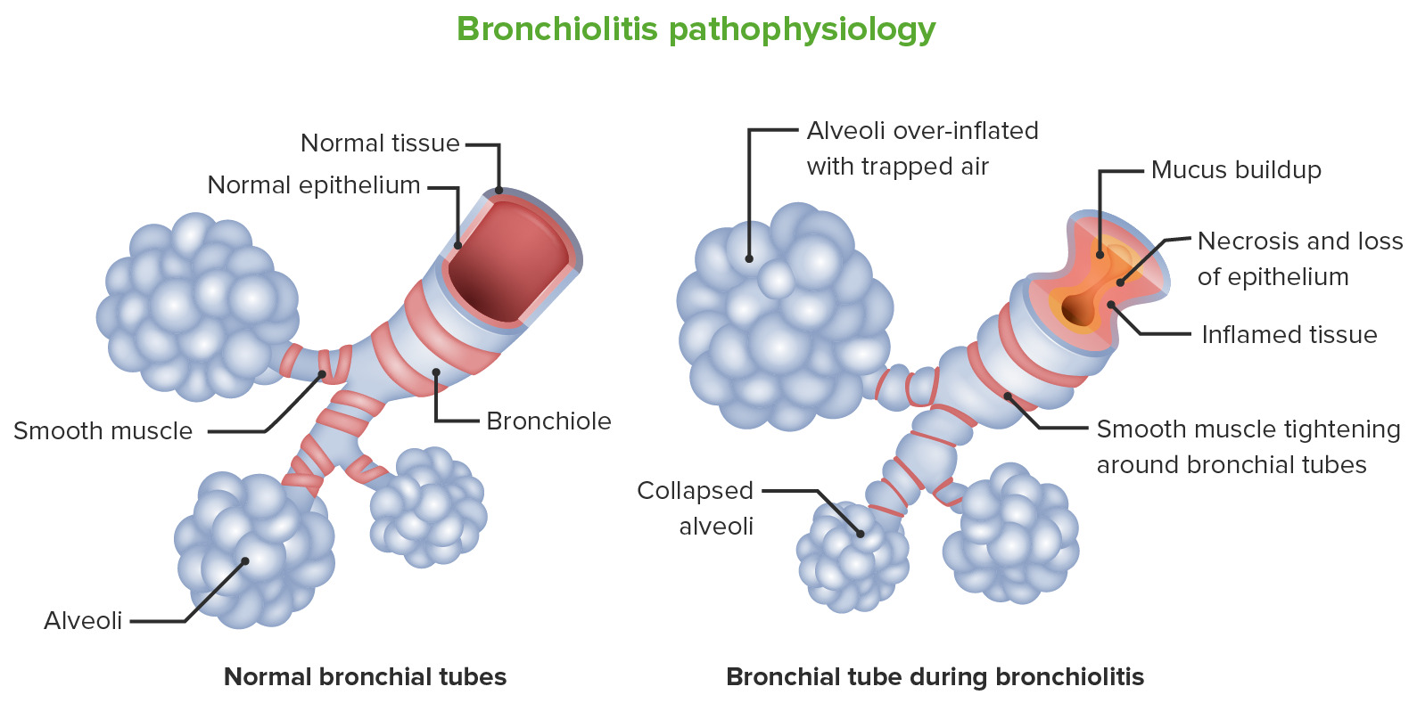
Bronchiolitis pathophysiology:
The image on the left represents the structure of a normal bronchiole. The image on the right represents changes occurring in bronchiolitis obliterans. Injury to the distal bronchioles results in inflammation of the bronchioles and adjacent alveoli. Fibrosis occurs (which is abnormal) instead of normal repair. There is mucus buildup, tightening and hypertrophy of the bronchial smooth muscles followed by narrowing of the small airways.

Image by Lecturio.

Clinical presentation

Bronchiolitis Bronchiolitis Inflammation of the bronchioles. Pediatric Chest Abnormalities obliterans should be suspected in patients Patients Individuals participating in the health care system for the purpose of receiving therapeutic, diagnostic, or preventive procedures. Clinician–Patient Relationship with slowly progressive dyspnea Dyspnea Dyspnea is the subjective sensation of breathing discomfort. Dyspnea is a normal manifestation of heavy physical or psychological exertion, but also may be caused by underlying conditions (both pulmonary and extrapulmonary). Dyspnea and cough, especially when the presentation is otherwise atypical for asthma Asthma Asthma is a chronic inflammatory respiratory condition characterized by bronchial hyperresponsiveness and airflow obstruction. The disease is believed to result from the complex interaction of host and environmental factors that increase disease predisposition, with inflammation causing symptoms and structural changes. Patients typically present with wheezing, cough, and dyspnea. Asthma or chronic obstructive pulmonary disease Pulmonary disease Diseases involving the respiratory system. Blastomyces/Blastomycosis ( COPD COPD Chronic obstructive pulmonary disease (COPD) is a lung disease characterized by progressive, largely irreversible airflow obstruction. The condition usually presents in middle-aged or elderly persons with a history of cigarette smoking. Signs and symptoms include prolonged expiration, wheezing, diminished breath sounds, progressive dyspnea, and chronic cough. Chronic Obstructive Pulmonary Disease (COPD)).

  • Dyspnea Dyspnea Dyspnea is the subjective sensation of breathing discomfort. Dyspnea is a normal manifestation of heavy physical or psychological exertion, but also may be caused by underlying conditions (both pulmonary and extrapulmonary). Dyspnea
  • Cough:
    • Persistent (as opposed to episodic-like asthma Asthma Asthma is a chronic inflammatory respiratory condition characterized by bronchial hyperresponsiveness and airflow obstruction. The disease is believed to result from the complex interaction of host and environmental factors that increase disease predisposition, with inflammation causing symptoms and structural changes. Patients typically present with wheezing, cough, and dyspnea. Asthma)
    • Progressive, slowly developing over weeks to months
    • Sputum may or may not be present
  • Potential exam findings:
    • Tachypnea Tachypnea Increased respiratory rate. Pulmonary Examination
    • ↓ Breath sounds
    • Prolonged expiratory phase while breathing
    • Crackles and/or wheezing Wheezing Wheezing is an abnormal breath sound characterized by a whistling noise that can be relatively high-pitched and shrill (more common) or coarse. Wheezing is produced by the movement of air through narrowed or compressed small (intrathoracic) airways. Wheezing

Diagnosis

The diagnosis of bronchiolitis Bronchiolitis Inflammation of the bronchioles. Pediatric Chest Abnormalities obliterans is usually made based on history, pulmonary function test Pulmonary function test Pulmonary function tests are a group of diagnostic procedures yielding useful, quantifiable information about the rate of the flow of air through the individual’s airways, lung capacity, and the efficiency of gas exchange in relation to time. The most commonly utilized tests include spirometry (before and after bronchodilator use), lung volumes, and quantitation of diffusing capacity for carbon monoxide (CO). The tests can be influenced by the individual’s effort/fatigue, disease state, or anatomical malformation. Pulmonary Function Tests, imaging, and biopsy Biopsy Removal and pathologic examination of specimens from the living body. Ewing Sarcoma findings.

Pulmonary tests

  • Lung tests include:
    • Spirometry Spirometry Measurement of volume of air inhaled or exhaled by the lung. Pulmonary Function Tests:
      • Measures the volume of inhaled and exhaled air
      • Usually performed before and after administration of bronchodilators Bronchodilators Asthma Drugs → determines the degree of reversible airflow obstruction
    • Diffusion Diffusion The tendency of a gas or solute to pass from a point of higher pressure or concentration to a point of lower pressure or concentration and to distribute itself throughout the available space. Diffusion, especially facilitated diffusion, is a major mechanism of biological transport. Peritoneal Dialysis and Hemodialysis capacity:
      • Measures the transfer of gas into the blood
      • A low concentration of carbon monoxide Carbon monoxide Carbon monoxide (CO). A poisonous colorless, odorless, tasteless gas. It combines with hemoglobin to form carboxyhemoglobin, which has no oxygen carrying capacity. The resultant oxygen deprivation causes headache, dizziness, decreased pulse and respiratory rates, unconsciousness, and death. Carbon Monoxide Poisoning in the inhaled air is absorbed by hemoglobin in RBCs RBCs Erythrocytes, or red blood cells (RBCs), are the most abundant cells in the blood. While erythrocytes in the fetus are initially produced in the yolk sac then the liver, the bone marrow eventually becomes the main site of production. Erythrocytes: Histology.
    • Helpful in diagnosing multiple lung conditions
  • Important definitions and acronyms:
    • Total lung capacity Total lung capacity The volume of air contained in the lungs at the end of a maximal inspiration. It is the equivalent to each of the following sums: vital capacity plus residual volume; inspiratory capacity plus functional residual capacity; tidal volume plus inspiratory reserve volume plus functional residual capacity; or tidal volume plus inspiratory reserve volume plus expiratory reserve volume plus residual volume. Ventilation: Mechanics of Breathing (TLC): total volume of air in lungs Lungs Lungs are the main organs of the respiratory system. Lungs are paired viscera located in the thoracic cavity and are composed of spongy tissue. The primary function of the lungs is to oxygenate blood and eliminate CO2. Lungs: Anatomy at the end of a maximal inspiration Inspiration Ventilation: Mechanics of Breathing
    • Forced expiratory volume in the 1st second (FEV1)
    • Forced vital capacity Vital capacity The volume of air that is exhaled by a maximal expiration following a maximal inspiration. Ventilation: Mechanics of Breathing (FVC): total forced exhaled volume
    • Residual volume Residual volume The volume of air remaining in the lungs at the end of a maximal expiration. Common abbreviation is rv. Ventilation: Mechanics of Breathing (RV): volume remaining after maximal exhalation
    • Functional residual capacity Functional residual capacity The volume of air remaining in the lungs at the end of a normal, quiet expiration. It is the sum of the residual volume and the expiratory reserve volume. Common abbreviation is frc. Ventilation: Mechanics of Breathing (FRC): volume remaining at the end of a tidal volume Tidal volume The volume of air inspired or expired during each normal, quiet respiratory cycle. Common abbreviations are tv or V with subscript t. Ventilation: Mechanics of Breathing breath
    • Diffusing capacity of the lungs Lungs Lungs are the main organs of the respiratory system. Lungs are paired viscera located in the thoracic cavity and are composed of spongy tissue. The primary function of the lungs is to oxygenate blood and eliminate CO2. Lungs: Anatomy for carbon monoxide Carbon monoxide Carbon monoxide (CO). A poisonous colorless, odorless, tasteless gas. It combines with hemoglobin to form carboxyhemoglobin, which has no oxygen carrying capacity. The resultant oxygen deprivation causes headache, dizziness, decreased pulse and respiratory rates, unconsciousness, and death. Carbon Monoxide Poisoning ( DLCO DLCO Pulmonary Function Tests): reflects the lung’s ability to transfer inhaled gas across the alveolar-capillary membrane 
  • Obstruction versus restriction:
    • Restriction pattern:
      • ↓ TLC and FVC
      • Think: “the lungs Lungs Lungs are the main organs of the respiratory system. Lungs are paired viscera located in the thoracic cavity and are composed of spongy tissue. The primary function of the lungs is to oxygenate blood and eliminate CO2. Lungs: Anatomy can’t expand (restriction) → ↓ volumes moving in and out”
      • Example: interstitial lung disease
    • Obstruction pattern:
      • ↓ FEV1/FVC ratio
      • Think: “The lungs Lungs Lungs are the main organs of the respiratory system. Lungs are paired viscera located in the thoracic cavity and are composed of spongy tissue. The primary function of the lungs is to oxygenate blood and eliminate CO2. Lungs: Anatomy can expand and contract, but the air moves slowly (obstruction).”
      • Examples: asthma Asthma Asthma is a chronic inflammatory respiratory condition characterized by bronchial hyperresponsiveness and airflow obstruction. The disease is believed to result from the complex interaction of host and environmental factors that increase disease predisposition, with inflammation causing symptoms and structural changes. Patients typically present with wheezing, cough, and dyspnea. Asthma, COPD COPD Chronic obstructive pulmonary disease (COPD) is a lung disease characterized by progressive, largely irreversible airflow obstruction. The condition usually presents in middle-aged or elderly persons with a history of cigarette smoking. Signs and symptoms include prolonged expiration, wheezing, diminished breath sounds, progressive dyspnea, and chronic cough. Chronic Obstructive Pulmonary Disease (COPD), and bronchiolitis Bronchiolitis Inflammation of the bronchioles. Pediatric Chest Abnormalities
Interpretation of pulmonary function tests

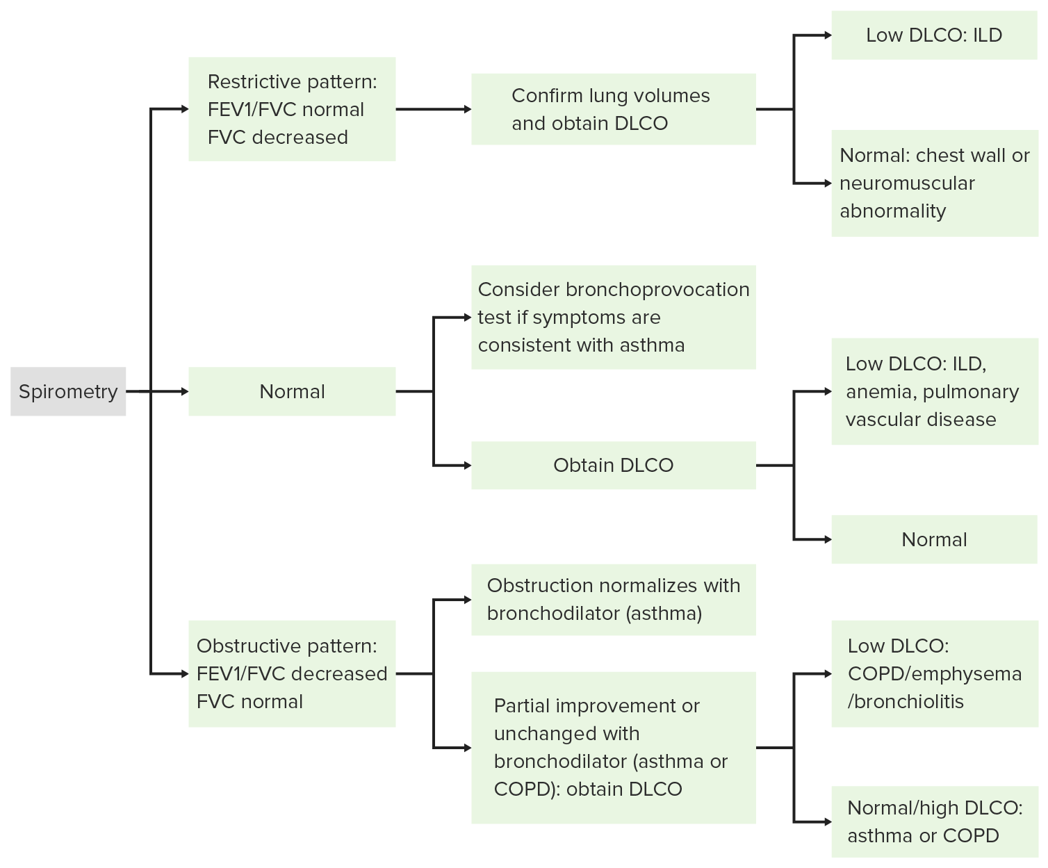
Algorithm for interpretation of pulmonary function tests:
FEV1: forced expiratory volume in the 1st second
FVC: forced vital capacity (total forced exhaled volume)
DLCO: diffusing capacity of the lungs for carbon monoxide (reflects the lung’s ability to transfer inhaled gas across the alveolar-capillary membrane)
COPD: chronic obstructive pulmonary disease
ILD: interstitial lung disease

Image by Lecturio.

Bronchiolitis Bronchiolitis Inflammation of the bronchioles. Pediatric Chest Abnormalities obliterans

  • Non-reversible airflow obstruction: ↓ FEV1
  • Evidence of air trapping: ↑ RV and FRC
  • Possible hyperinflation Hyperinflation Imaging of the Lungs and Pleura: ↑ TLC
  • Impaired gas exchange Gas exchange Human cells are primarily reliant on aerobic metabolism. The respiratory system is involved in pulmonary ventilation and external respiration, while the circulatory system is responsible for transport and internal respiration. Pulmonary ventilation (breathing) represents movement of air into and out of the lungs. External respiration, or gas exchange, is represented by the O2 and CO2 exchange between the lungs and the blood. Gas Exchange: ↓ DLCO DLCO Pulmonary Function Tests
  • May be normal:
    • There are a large number of bronchioles Bronchioles The small airways branching off the tertiary bronchi. Terminal bronchioles lead into several orders of respiratory bronchioles which in turn lead into alveolar ducts and then into pulmonary alveoli. Bronchial Tree: Anatomy.
    • A high percentage needs to be affected before spirometry Spirometry Measurement of volume of air inhaled or exhaled by the lung. Pulmonary Function Tests changes are seen.

Other diagnostic tests Diagnostic tests Diagnostic tests are important aspects in making a diagnosis. Some of the most important epidemiological values of diagnostic tests include sensitivity and specificity, false positives and false negatives, positive and negative predictive values, likelihood ratios, and pre-test and post-test probabilities. Epidemiological Values of Diagnostic Tests

  • Chest X-ray X-ray Penetrating electromagnetic radiation emitted when the inner orbital electrons of an atom are excited and release radiant energy. X-ray wavelengths range from 1 pm to 10 nm. Hard x-rays are the higher energy, shorter wavelength x-rays. Soft x-rays or grenz rays are less energetic and longer in wavelength. The short wavelength end of the x-ray spectrum overlaps the gamma rays wavelength range. The distinction between gamma rays and x-rays is based on their radiation source. Pulmonary Function Tests:
  • High-resolution CT High-resolution CT Imaging of the Lungs and Pleura ( HRCT HRCT Pulmonary Function Tests):
  • Bronchoscopy Bronchoscopy Endoscopic examination, therapy or surgery of the bronchi. Laryngomalacia and Tracheomalacia:
    • Rarely required for clinical diagnosis
    • Findings are usually nonspecific.
    • Used when other causes of airflow obstruction are suspected:
      • Endobronchial tumors
      • Sarcoidosis Sarcoidosis Sarcoidosis is a multisystem inflammatory disease that causes noncaseating granulomas. The exact etiology is unknown. Sarcoidosis usually affects the lungs and thoracic lymph nodes, but it can also affect almost every system in the body, including the skin, heart, and eyes, most commonly. Sarcoidosis
      • Infections Infections Invasion of the host organism by microorganisms or their toxins or by parasites that can cause pathological conditions or diseases. Chronic Granulomatous Disease leading to lung damage
  • Lung biopsy Lung Biopsy Antineutrophil Cytoplasmic Antibody (ANCA)-Associated Vasculitis:
    • Required for definitive diagnosis (although a clinical diagnosis can often be made without a biopsy Biopsy Removal and pathologic examination of specimens from the living body. Ewing Sarcoma)
    • Surgical (over transbronchial) specimen preferred
    • Histopathology:
      • Hypertrophy Hypertrophy General increase in bulk of a part or organ due to cell enlargement and accumulation of fluids and secretions, not due to tumor formation, nor to an increase in the number of cells (hyperplasia). Cellular Adaptation of the bronchiolar smooth muscle
      • Peribronchiolar inflammatory infiltrates
      • Bronchiolar mucus accumulation 
      • Fibrosis and obliteration of bronchioles Bronchioles The small airways branching off the tertiary bronchi. Terminal bronchioles lead into several orders of respiratory bronchioles which in turn lead into alveolar ducts and then into pulmonary alveoli. Bronchial Tree: Anatomy (by submucosal scarring Scarring Inflammation)
Hrct in bronchiolitis obliterans

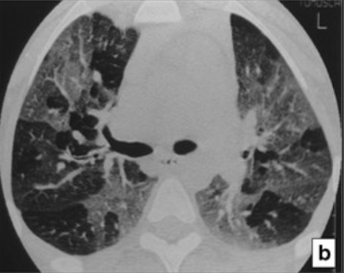
High-resolution CT (HRCT) in bronchiolitis obliterans:
The HRCT at expiration demonstrates a mosaic-attenuation pattern that results from air trapping.

Image: “Mimics in chest disease: interstitial opacities” by Oikonomou A, Prassopoulos P. License: Public Domain, cropped by Lecturio.

Management

Bronchiolitis Bronchiolitis Inflammation of the bronchioles. Pediatric Chest Abnormalities obliterans is often progressive and refractory to therapy; thus, management is often supportive, symptom driven, and based on the etiology.

General management

  • Remove any offending agents.
  • Smoking Smoking Willful or deliberate act of inhaling and exhaling smoke from burning substances or agents held by hand. Interstitial Lung Diseases cessation
  • Updated vaccination Vaccination Vaccination is the administration of a substance to induce the immune system to develop protection against a disease. Unlike passive immunization, which involves the administration of pre-performed antibodies, active immunization constitutes the administration of a vaccine to stimulate the body to produce its own antibodies. Vaccination against influenza Influenza Influenza viruses are members of the Orthomyxoviridae family and the causative organisms of influenza, a highly contagious febrile respiratory disease. There are 3 primary influenza viruses (A, B, and C) and various subtypes, which are classified based on their virulent surface antigens, hemagglutinin (HA) and neuraminidase (NA). Influenza typically presents with a fever, myalgia, headache, and symptoms of an upper respiratory infection. Influenza Viruses/Influenza and pneumococcus
  • Treat GERD GERD Gastroesophageal reflux disease (GERD) occurs when the stomach acid frequently flows back into the esophagus. This backwash (acid reflux) can irritate the lining of the esophagus, causing symptoms such as retrosternal burning pain (heartburn). Gastroesophageal Reflux Disease (GERD):
  • Symptom relief:
    • Cough suppressants:
      • Dextromethorphan
      • Codeine Codeine An opioid analgesic related to morphine but with less potent analgesic properties and mild sedative effects. It also acts centrally to suppress cough. Opioid Analgesics
    • Inhaled bronchodilators Bronchodilators Asthma Drugs
  • Supplemental oxygen Supplemental Oxygen Respiratory Failure if needed to maintain oxygen saturation Oxygen Saturation Basic Procedures ≥ 89%
  • Consider a trial (3‒6 months) of macrolide antibiotics:
    • Options:
      • Azithromycin Azithromycin A semi-synthetic macrolide antibiotic structurally related to erythromycin. It has been used in the treatment of Mycobacterium avium intracellulare infections, toxoplasmosis, and cryptosporidiosis. Macrolides and Ketolides
      • Erythromycin Erythromycin A bacteriostatic antibiotic macrolide produced by streptomyces erythreus. Erythromycin a is considered its major active component. In sensitive organisms, it inhibits protein synthesis by binding to 50s ribosomal subunits. This binding process inhibits peptidyl transferase activity and interferes with translocation of amino acids during translation and assembly of proteins. Macrolides and Ketolides
      • Clarithromycin Clarithromycin A semisynthetic macrolide antibiotic derived from erythromycin that is active against a variety of microorganisms. It can inhibit protein synthesis in bacteria by reversibly binding to the 50s ribosomal subunits. This inhibits the translocation of aminoacyl transfer-RNA and prevents peptide chain elongation. Macrolides and Ketolides
    • Not formally studied in patients Patients Individuals participating in the health care system for the purpose of receiving therapeutic, diagnostic, or preventive procedures. Clinician–Patient Relationship who have not undergone a transplant
    • Limited evidence suggests that there may be some benefits.
  • Consider a trial (4‒6 weeks) of glucocorticoids Glucocorticoids Glucocorticoids are a class within the corticosteroid family. Glucocorticoids are chemically and functionally similar to endogenous cortisol. There are a wide array of indications, which primarily benefit from the antiinflammatory and immunosuppressive effects of this class of drugs. Glucocorticoids in patients Patients Individuals participating in the health care system for the purpose of receiving therapeutic, diagnostic, or preventive procedures. Clinician–Patient Relationship with:
    • Rheumatic disease
    • Coexisting asthma Asthma Asthma is a chronic inflammatory respiratory condition characterized by bronchial hyperresponsiveness and airflow obstruction. The disease is believed to result from the complex interaction of host and environmental factors that increase disease predisposition, with inflammation causing symptoms and structural changes. Patients typically present with wheezing, cough, and dyspnea. Asthma

Management in patients Patients Individuals participating in the health care system for the purpose of receiving therapeutic, diagnostic, or preventive procedures. Clinician–Patient Relationship who have undergone transplant

  • Increase immune suppression Suppression Defense Mechanisms:
    • Tacrolimus Tacrolimus A macrolide isolated from the culture broth of a strain of streptomyces tsukubaensis that has strong immunosuppressive activity in vivo and prevents the activation of T-lymphocytes in response to antigenic or mitogenic stimulation in vitro. Immunosuppressants
    • Cyclosporine Cyclosporine A cyclic undecapeptide from an extract of soil fungi. It is a powerful immunosupressant with a specific action on T-lymphocytes. It is used for the prophylaxis of graft rejection in organ and tissue transplantation. Immunosuppressants
    • Mycophenolate Mycophenolate Immunosuppressants mofetil
    • Prednisone Prednisone A synthetic anti-inflammatory glucocorticoid derived from cortisone. It is biologically inert and converted to prednisolone in the liver. Immunosuppressants
  • Evidence shows improvement in lung function (in patients Patients Individuals participating in the health care system for the purpose of receiving therapeutic, diagnostic, or preventive procedures. Clinician–Patient Relationship who have undergone post-hematopoietic stem-cell transplant) and are on the following therapy:
    • Fluticasone Fluticasone A steroid with glucocorticoid receptor activity that is used to manage the symptoms of asthma; allergic rhinitis, and atopic dermatitis. Glucocorticoids (inhaled corticosteroid)
    • Montelukast Montelukast Asthma Drugs (oral leukotriene Leukotriene Asthma Drugs receptor Receptor Receptors are proteins located either on the surface of or within a cell that can bind to signaling molecules known as ligands (e.g., hormones) and cause some type of response within the cell. Receptors antagonist)
    • Azithromycin Azithromycin A semi-synthetic macrolide antibiotic structurally related to erythromycin. It has been used in the treatment of Mycobacterium avium intracellulare infections, toxoplasmosis, and cryptosporidiosis. Macrolides and Ketolides (oral macrolide antibiotic)
  • Consider retransplantation when BOS is severe.

Differential Diagnosis

  • Asthma Asthma Asthma is a chronic inflammatory respiratory condition characterized by bronchial hyperresponsiveness and airflow obstruction. The disease is believed to result from the complex interaction of host and environmental factors that increase disease predisposition, with inflammation causing symptoms and structural changes. Patients typically present with wheezing, cough, and dyspnea. Asthma: a condition of bronchial hyperresponsiveness and chronic airway Airway ABCDE Assessment inflammation Inflammation Inflammation is a complex set of responses to infection and injury involving leukocytes as the principal cellular mediators in the body’s defense against pathogenic organisms. Inflammation is also seen as a response to tissue injury in the process of wound healing. The 5 cardinal signs of inflammation are pain, heat, redness, swelling, and loss of function. Inflammation that leads to intermittent airflow obstruction. Asthma Asthma Asthma is a chronic inflammatory respiratory condition characterized by bronchial hyperresponsiveness and airflow obstruction. The disease is believed to result from the complex interaction of host and environmental factors that increase disease predisposition, with inflammation causing symptoms and structural changes. Patients typically present with wheezing, cough, and dyspnea. Asthma typically presents with intermittent dyspnea Dyspnea Dyspnea is the subjective sensation of breathing discomfort. Dyspnea is a normal manifestation of heavy physical or psychological exertion, but also may be caused by underlying conditions (both pulmonary and extrapulmonary). Dyspnea, cough, and wheezing Wheezing Wheezing is an abnormal breath sound characterized by a whistling noise that can be relatively high-pitched and shrill (more common) or coarse. Wheezing is produced by the movement of air through narrowed or compressed small (intrathoracic) airways. Wheezing in response to a variety of stimuli. Asthma Asthma Asthma is a chronic inflammatory respiratory condition characterized by bronchial hyperresponsiveness and airflow obstruction. The disease is believed to result from the complex interaction of host and environmental factors that increase disease predisposition, with inflammation causing symptoms and structural changes. Patients typically present with wheezing, cough, and dyspnea. Asthma is diagnosed based on history and spirometry Spirometry Measurement of volume of air inhaled or exhaled by the lung. Pulmonary Function Tests, which shows a reduced FEV1/FVC ratio (obstruction) that is reversible with bronchodilators Bronchodilators Asthma Drugs (unlike bronchiolitis Bronchiolitis Inflammation of the bronchioles. Pediatric Chest Abnormalities obliterans which is not reversible). Management involves avoiding triggers, and the use of bronchodilators Bronchodilators Asthma Drugs and glucocorticoids Glucocorticoids Glucocorticoids are a class within the corticosteroid family. Glucocorticoids are chemically and functionally similar to endogenous cortisol. There are a wide array of indications, which primarily benefit from the antiinflammatory and immunosuppressive effects of this class of drugs. Glucocorticoids.
  • COPD COPD Chronic obstructive pulmonary disease (COPD) is a lung disease characterized by progressive, largely irreversible airflow obstruction. The condition usually presents in middle-aged or elderly persons with a history of cigarette smoking. Signs and symptoms include prolonged expiration, wheezing, diminished breath sounds, progressive dyspnea, and chronic cough. Chronic Obstructive Pulmonary Disease (COPD): an obstructive lung disease due to inflammation Inflammation Inflammation is a complex set of responses to infection and injury involving leukocytes as the principal cellular mediators in the body’s defense against pathogenic organisms. Inflammation is also seen as a response to tissue injury in the process of wound healing. The 5 cardinal signs of inflammation are pain, heat, redness, swelling, and loss of function. Inflammation resulting from chronic exposure to noxious particles or gas (usually smoking Smoking Willful or deliberate act of inhaling and exhaling smoke from burning substances or agents held by hand. Interstitial Lung Diseases). Subtypes include emphysema Emphysema Enlargement of air spaces distal to the terminal bronchioles where gas-exchange normally takes place. This is usually due to destruction of the alveolar wall. Pulmonary emphysema can be classified by the location and distribution of the lesions. Chronic Obstructive Pulmonary Disease (COPD), chronic bronchitis Chronic bronchitis A subcategory of chronic obstructive pulmonary disease. The disease is characterized by hypersecretion of mucus accompanied by a chronic (more than 3 months in 2 consecutive years) productive cough. Infectious agents are a major cause of chronic bronchitis. Rhinovirus, and chronic obstructive asthma Asthma Asthma is a chronic inflammatory respiratory condition characterized by bronchial hyperresponsiveness and airflow obstruction. The disease is believed to result from the complex interaction of host and environmental factors that increase disease predisposition, with inflammation causing symptoms and structural changes. Patients typically present with wheezing, cough, and dyspnea. Asthma. Both small airways and parenchyma can be affected. Spirometry Spirometry Measurement of volume of air inhaled or exhaled by the lung. Pulmonary Function Tests usually shows non-reversible or only partially reversible obstruction (reduced FEV1/FVC ratio). Management involves smoking Smoking Willful or deliberate act of inhaling and exhaling smoke from burning substances or agents held by hand. Interstitial Lung Diseases cessation and treatment with long-acting bronchodilators Bronchodilators Asthma Drugs.
  • Bronchiectasis Bronchiectasis Bronchiectasis is a chronic disease of the airways that results from permanent bronchial distortion. This results from a continuous cycle of inflammation, bronchial damage and dilation, impaired clearance of secretions, and recurrent infections. Bronchiectasis: an abnormal dilatation of the bronchi Bronchi The larger air passages of the lungs arising from the terminal bifurcation of the trachea. They include the largest two primary bronchi which branch out into secondary bronchi, and tertiary bronchi which extend into bronchioles and pulmonary alveoli. Bronchial Tree: Anatomy with destruction and thickening of the bronchial walls. Bronchiectasis Bronchiectasis Bronchiectasis is a chronic disease of the airways that results from permanent bronchial distortion. This results from a continuous cycle of inflammation, bronchial damage and dilation, impaired clearance of secretions, and recurrent infections. Bronchiectasis is triggered by chronic or recurrent infections Recurrent infections Common Variable Immunodeficiency (CVID) in the setting of impaired airway Airway ABCDE Assessment drainage or obstruction, and is characterized by a chronic cough, dyspnea Dyspnea Dyspnea is the subjective sensation of breathing discomfort. Dyspnea is a normal manifestation of heavy physical or psychological exertion, but also may be caused by underlying conditions (both pulmonary and extrapulmonary). Dyspnea, and daily sputum production. Bronchiectasis Bronchiectasis Bronchiectasis is a chronic disease of the airways that results from permanent bronchial distortion. This results from a continuous cycle of inflammation, bronchial damage and dilation, impaired clearance of secretions, and recurrent infections. Bronchiectasis is a condition affecting the large airways, whereas bronchiolitis Bronchiolitis Inflammation of the bronchioles. Pediatric Chest Abnormalities obliterans affects the small airways. Diagnosis is usually confirmed with a chest CT showing bronchial-wall thickening and large-airway dilatation.
  • Hypersensitivity pneumonitis Pneumonitis Human Herpesvirus 6 and 7: an interstitial lung disease characterized by an abnormal immune reaction within the pulmonary parenchyma in response to an inhaled substance. The inciting agents are often related to agricultural dust or microorganisms associated with farming, animal handling, and ventilation Ventilation The total volume of gas inspired or expired per unit of time, usually measured in liters per minute. Ventilation: Mechanics of Breathing and construction work. Patients Patients Individuals participating in the health care system for the purpose of receiving therapeutic, diagnostic, or preventive procedures. Clinician–Patient Relationship present with a productive cough, dyspnea Dyspnea Dyspnea is the subjective sensation of breathing discomfort. Dyspnea is a normal manifestation of heavy physical or psychological exertion, but also may be caused by underlying conditions (both pulmonary and extrapulmonary). Dyspnea, fatigue Fatigue The state of weariness following a period of exertion, mental or physical, characterized by a decreased capacity for work and reduced efficiency to respond to stimuli. Fibromyalgia, and weight loss Weight loss Decrease in existing body weight. Bariatric Surgery. Diagnosis involves characteristic findings on HRCT HRCT Pulmonary Function Tests, and lymphocytosis Lymphocytosis WBCs develop from stem cells in the bone marrow and are called leukocytes when circulating in the bloodstream. Lymphocytes are 1 of the 5 subclasses of WBCs. Lymphocytosis is an increase in the number or proportion of the lymphocyte subclass of WBCs, often as a result of an immune response to infection (known as reactive lymphocytosis). Lymphocytosis on broncho-alveolar lavage.

References

  1. Bronchiolitis: Causes, symptoms and treatments (n.d.). Retrieved March 19, 2021, from https://my.clevelandclinic.org/health/diseases/8272-bronchiolitis
  2. King, T. (2020). Overview of bronchiolar disorders in adults. In Hollingsworth, H. (Ed.). UpToDate. Retrieved February 11, 2021, from https://www.uptodate.com/contents/overview-of-bronchiolar-disorders-in-adults
  3. McCormack, M. (2020). Overview of pulmonary function testing in adults. In Hollingsworth, H. (Ed.). UpToDate. Retrieved February 11, 2021, from https://www.uptodate.com/contents/overview-of-pulmonary-function-testing-in-adults
  4. Krishna, R., Anjum, F. (2020). Bronchiolitis obliterans. In Oliver, T. (Ed.). StatPearls. Retrieved February 11, 2021, from https://www.statpearls.com/articlelibrary/viewarticle/18642/

Create your free account or log in to continue reading!

Sign up now and get free access to Lecturio with concept pages, medical videos, and questions for your medical education.

User Reviews

Details