Achieve Mastery of Medical Concepts

Study for medical school and boards with Lecturio

Human Herpesvirus 6 and 7

Human herpesvirus (HHV)-6 and HHV-7 are similar double-stranded DNA viruses DNA Viruses Viruses whose nucleic acid is DNA. Virology belonging to the Herpesviridae Herpesviridae A family of enveloped, linear, double-stranded DNA viruses infecting a wide variety of animals. Subfamilies, based on biological characteristics, include: alphaherpesvirinae; betaherpesvirinae; and gammaherpesvirinae. Herpes Simplex Virus 1 and 2 family. Human herpesviruses are ubiquitous and infections Infections Invasion of the host organism by microorganisms or their toxins or by parasites that can cause pathological conditions or diseases. Chronic Granulomatous Disease are commonly contracted during childhood. Both HHV-6B and HHV-7 cause a common childhood illness known as roseola infantum (also known as roseola or 6th disease). Roseola is a self-limiting Self-Limiting Meningitis in Children disease that presents with high fever Fever Fever is defined as a measured body temperature of at least 38°C (100.4°F). Fever is caused by circulating endogenous and/or exogenous pyrogens that increase levels of prostaglandin E2 in the hypothalamus. Fever is commonly associated with chills, rigors, sweating, and flushing of the skin. Fever, followed by a diffuse, rose-pink maculopapular Maculopapular Dermatologic Examination rash Rash Rocky Mountain Spotted Fever. The diagnosis is clinical, and management is supportive.

Last updated: Mar 27, 2023

Editorial responsibility: Stanley Oiseth, Lindsay Jones, Evelin Maza

Classification

Dna virus classification flowchart

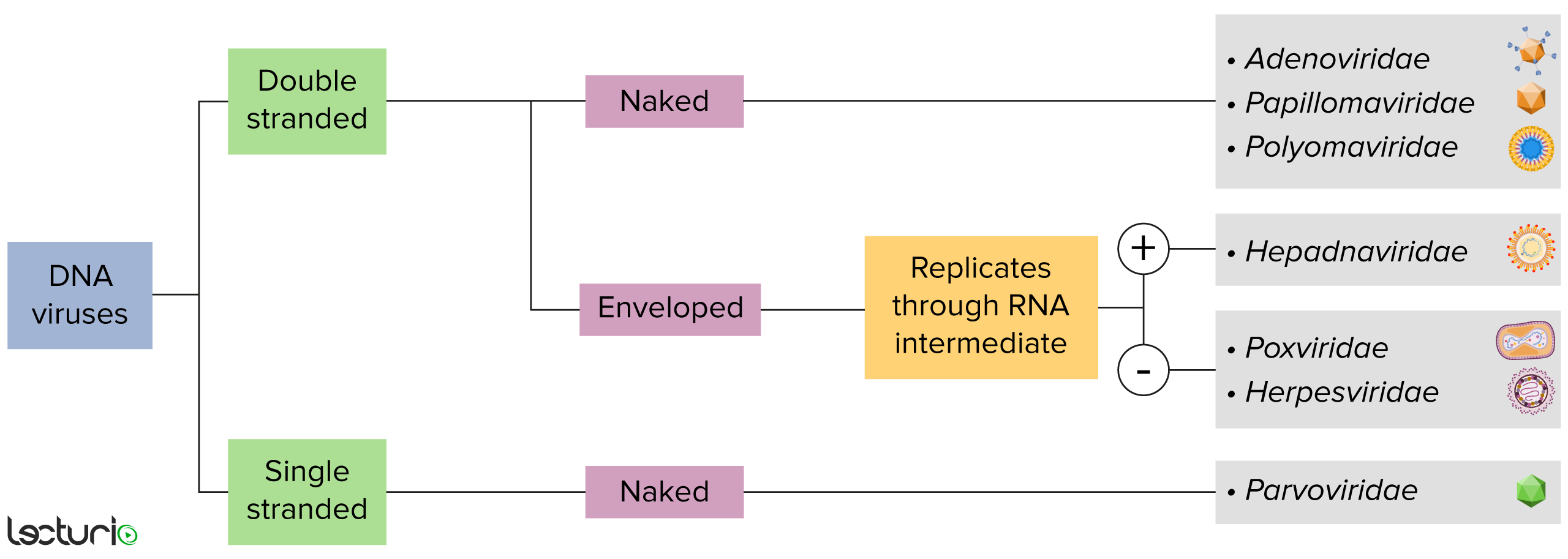
Identification of DNA viruses:
Viruses can be classified in many ways. Most viruses, however, will have a genome formed by either DNA or RNA. Viruses with a DNA genome can be further characterized as single or double stranded. “Enveloped” viruses are covered by a thin coat of cell membrane, which is usually taken from the host cell. If the coat is absent, however, the viruses are called “naked” viruses. Some enveloped viruses translate DNA into RNA before incorporating into the genome of the host cell.

Image by Lecturio. License: CC BY-NC-SA 4.0

General Characteristics and Epidemiology

Basic features of HHV-6 and HHV-7

  • Taxonomy:
    • Family: Herpesviridae Herpesviridae A family of enveloped, linear, double-stranded DNA viruses infecting a wide variety of animals. Subfamilies, based on biological characteristics, include: alphaherpesvirinae; betaherpesvirinae; and gammaherpesvirinae. Herpes Simplex Virus 1 and 2
    • Subfamily: Betaherpesvirinae Betaherpesvirinae A subfamily of herpesviridae characterized by a relatively long replication cycle. Genera include: cytomegalovirus; muromegalovirus; and roseolovirus. Cytomegalovirus
    • Genus: Roseolovirus
    • Species:
      • Human betaherpesvirus 6A
      • Human betaherpesvirus 6B
      • Human betaherpesvirus 7
  • DNA viruses DNA Viruses Viruses whose nucleic acid is DNA. Virology:
    • Linear
    • Double stranded
  • Structure:
    • DNA DNA A deoxyribonucleotide polymer that is the primary genetic material of all cells. Eukaryotic and prokaryotic organisms normally contain DNA in a double-stranded state, yet several important biological processes transiently involve single-stranded regions. DNA, which consists of a polysugar-phosphate backbone possessing projections of purines (adenine and guanine) and pyrimidines (thymine and cytosine), forms a double helix that is held together by hydrogen bonds between these purines and pyrimidines (adenine to thymine and guanine to cytosine). DNA Types and Structure core
    • Icosahedral capsid Capsid The outer protein protective shell of a virus, which protects the viral nucleic acid. Capsids are composed of repeating units (capsomers or capsomeres) of capsid proteins which when assembled together form either an icosahedral or helical shape. Virology
    • Tegument Tegument Herpes Simplex Virus 1 and 2
    • Lipid envelope Envelope Bilayer lipid membrane acquired by viral particles during viral morphogenesis. Although the lipids of the viral envelope are host derived, various virus-encoded integral membrane proteins, i.e. Viral envelope proteins are incorporated there. Virology with glycoprotein spikes
Human herpesvirus 6

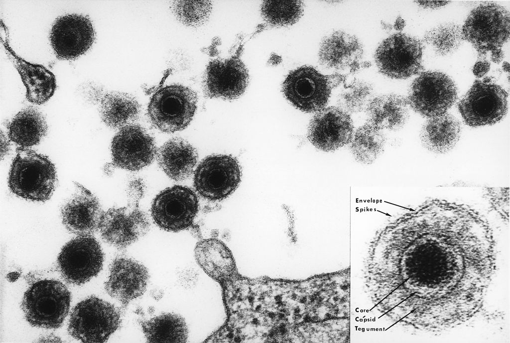
Electron micrograph of HHV-6 virions

Image: “Electron micrograph of one of the HHV6 species” by Bernard Kramarsky. License: Public Domain

Associated diseases

The clinical relevance of HHV-6A is not entirely known.

  • HHV-6B and HHV-7 are associated with:
    • Roseola infantum (also known as exanthem Exanthem Diseases in which skin eruptions or rashes are a prominent manifestation. Classically, six such diseases were described with similar rashes; they were numbered in the order in which they were reported. Only the fourth (Duke’s disease), fifth (erythema infectiosum), and sixth (exanthema subitum) numeric designations survive as occasional synonyms in current terminology. Varicella-Zoster Virus/Chickenpox subitum, roseola, or 6th disease)
    • Acute febrile respiratory disease
    • Febrile seizures Febrile seizures Seizures that occur during a febrile episode. It is a common condition, affecting 2-5% of children aged 3 months to five years. An autosomal dominant pattern of inheritance has been identified in some families. The majority are simple febrile seizures (generally defined as generalized onset, single seizures with a duration of less than 30 minutes). Complex febrile seizures are characterized by focal onset, duration greater than 30 minutes, and/or more than one seizure in a 24 hour period. The likelihood of developing epilepsy (i.e., a nonfebrile seizure disorder) following simple febrile seizures is low. Complex febrile seizures are associated with a moderately increased incidence of epilepsy. Seizures in Children
    • Reactivation Reactivation Herpes Simplex Virus 1 and 2 syndromes in transplant recipients:
      • Viremia Viremia The presence of viruses in the blood. Erythema Infectiosum
      • Encephalitis Encephalitis Encephalitis is inflammation of the brain parenchyma caused by an infection, usually viral. Encephalitis may present with mild symptoms such as headache, fever, fatigue, and muscle and joint pain or with severe symptoms such as seizures, altered consciousness, and paralysis. Encephalitis
      • Pneumonitis
      • Myocarditis Myocarditis Myocarditis is an inflammatory disease of the myocardium, which may occur alone or in association with a systemic process. There are numerous etiologies of myocarditis, but all lead to inflammation and myocyte injury, most often leading to signs and symptoms of heart failure. Myocarditis
      • Hepatitis
    • Possible:
      • Pityriasis rosea Pityriasis rosea Pityriasis rosea is an acute, self-limited skin disease. The etiology is not known, and it commonly occurs in young adults. Patients initially present with a single, ovoid “herald patch.” This is followed by diffuse, pruritic, scaly, oval lesions over the trunk (often in a “Christmas tree” distribution on the back) and extremities. Pityriasis Rosea
      • Lichen planus Lichen planus Lichen planus (LP) is an idiopathic, cell-mediated inflammatory skin disease. It is characterized by pruritic, flat-topped, papular, purple skin lesions commonly found on the flexural surfaces of the extremities. Other areas affected include genitalia, nails, scalp, and mucous membranes. Lichen Planus (HHV-7)

Epidemiology

  • HHV-6:
    • > 70% of adults are seropositive.
    • Infection generally occurs by the age of 2 years.
  • HHV-7:
    • > 95% of adults are seropositive.
    • Infection often occurs in childhood.

Pathogenesis

Reservoir Reservoir Animate or inanimate sources which normally harbor disease-causing organisms and thus serve as potential sources of disease outbreaks. Reservoirs are distinguished from vectors (disease vectors) and carriers, which are agents of disease transmission rather than continuing sources of potential disease outbreaks. Humans may serve both as disease reservoirs and carriers. Escherichia coli

Humans are the reservoir Reservoir Animate or inanimate sources which normally harbor disease-causing organisms and thus serve as potential sources of disease outbreaks. Reservoirs are distinguished from vectors (disease vectors) and carriers, which are agents of disease transmission rather than continuing sources of potential disease outbreaks. Humans may serve both as disease reservoirs and carriers. Escherichia coli for HHV-6 and HHV-7.

Transmission

HHV-6 and HHV-7 are transmitted through contact with saliva Saliva The clear, viscous fluid secreted by the salivary glands and mucous glands of the mouth. It contains mucins, water, organic salts, and ptyalin. Salivary Glands: Anatomy.

Pathophysiology

  • Replication occurs in T lymphocytes T lymphocytes Lymphocytes responsible for cell-mediated immunity. Two types have been identified – cytotoxic (t-lymphocytes, cytotoxic) and helper T-lymphocytes (t-lymphocytes, helper-inducer). They are formed when lymphocytes circulate through the thymus gland and differentiate to thymocytes. When exposed to an antigen, they divide rapidly and produce large numbers of new T cells sensitized to that antigen. T cells: Types and Functions.
  • Viral entry through:
    • HHV-6: CD46 receptor Receptor Receptors are proteins located either on the surface of or within a cell that can bind to signaling molecules known as ligands (e.g., hormones) and cause some type of response within the cell. Receptors
    • HHV-7: CD4 receptor Receptor Receptors are proteins located either on the surface of or within a cell that can bind to signaling molecules known as ligands (e.g., hormones) and cause some type of response within the cell. Receptors
  • Viral DNA DNA A deoxyribonucleotide polymer that is the primary genetic material of all cells. Eukaryotic and prokaryotic organisms normally contain DNA in a double-stranded state, yet several important biological processes transiently involve single-stranded regions. DNA, which consists of a polysugar-phosphate backbone possessing projections of purines (adenine and guanine) and pyrimidines (thymine and cytosine), forms a double helix that is held together by hydrogen bonds between these purines and pyrimidines (adenine to thymine and guanine to cytosine). DNA Types and Structure is released into the nucleoplasm → production of viral proteins Proteins Linear polypeptides that are synthesized on ribosomes and may be further modified, crosslinked, cleaved, or assembled into complex proteins with several subunits. The specific sequence of amino acids determines the shape the polypeptide will take, during protein folding, and the function of the protein. Energy Homeostasis and replication
  • Lytic replication → cell death Cell death Injurious stimuli trigger the process of cellular adaptation, whereby cells respond to withstand the harmful changes in their environment. Overwhelmed adaptive mechanisms lead to cell injury. Mild stimuli produce reversible injury. If the stimulus is severe or persistent, injury becomes irreversible. Apoptosis is programmed cell death, a mechanism with both physiologic and pathologic effects. Cell Injury and Death
  • Low-level persistent infection occurs in the salivary glands Salivary glands The salivary glands are exocrine glands positioned in and around the oral cavity. These glands are responsible for secreting saliva into the mouth, which aids in digestion. There are 3 major paired salivary glands: the sublingual, submandibular, and parotid glands. Salivary Glands: Anatomy.
  • Latency:
    • HHV-6: occurs in monocytes Monocytes Large, phagocytic mononuclear leukocytes produced in the vertebrate bone marrow and released into the blood; contain a large, oval or somewhat indented nucleus surrounded by voluminous cytoplasm and numerous organelles. Innate Immunity: Phagocytes and Antigen Presentation
    • HHV-7: occurs in T lymphocytes T lymphocytes Lymphocytes responsible for cell-mediated immunity. Two types have been identified – cytotoxic (t-lymphocytes, cytotoxic) and helper T-lymphocytes (t-lymphocytes, helper-inducer). They are formed when lymphocytes circulate through the thymus gland and differentiate to thymocytes. When exposed to an antigen, they divide rapidly and produce large numbers of new T cells sensitized to that antigen. T cells: Types and Functions

Clinical Presentation

Human herpesvirus-6 infections Infections Invasion of the host organism by microorganisms or their toxins or by parasites that can cause pathological conditions or diseases. Chronic Granulomatous Disease are usually mild and occur during childhood. Most HHV-7 infections Infections Invasion of the host organism by microorganisms or their toxins or by parasites that can cause pathological conditions or diseases. Chronic Granulomatous Disease are asymptomatic.
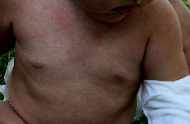
Roseola infantum

  • High fever Fever Fever is defined as a measured body temperature of at least 38°C (100.4°F). Fever is caused by circulating endogenous and/or exogenous pyrogens that increase levels of prostaglandin E2 in the hypothalamus. Fever is commonly associated with chills, rigors, sweating, and flushing of the skin. Fever:
    • Lasts approximately 3–5 days
    • Resolves abruptly
  • Rash Rash Rocky Mountain Spotted Fever:
    • Timing:
      • Follows resolution of fever Fever Fever is defined as a measured body temperature of at least 38°C (100.4°F). Fever is caused by circulating endogenous and/or exogenous pyrogens that increase levels of prostaglandin E2 in the hypothalamus. Fever is commonly associated with chills, rigors, sweating, and flushing of the skin. Fever
      • Lasts approximately 1–2 days
    • Appearance:
    • Nonpruritic
    • Distribution:
      • Starts on neck Neck The part of a human or animal body connecting the head to the rest of the body. Peritonsillar Abscess and trunk
      • Spreads to face and extremities
  • Other manifestations:
    • Conjunctivitis Conjunctivitis Conjunctivitis is a common inflammation of the bulbar and/or palpebral conjunctiva. It can be classified into infectious (mostly viral) and noninfectious conjunctivitis, which includes allergic causes. Patients commonly present with red eyes, increased tearing, burning, foreign body sensation, and photophobia. Conjunctivitis
    • Edematous eyelids Eyelids Each of the upper and lower folds of skin which cover the eye when closed. Blepharitis
    • Otitis media
    • Nagayama spots (erythematous uvulopalatoglossal papules)
    • Rhinorrhea Rhinorrhea Excess nasal drainage. Respiratory Syncytial Virus
    • Cough
    • Diarrhea Diarrhea Diarrhea is defined as ≥ 3 watery or loose stools in a 24-hour period. There are a multitude of etiologies, which can be classified based on the underlying mechanism of disease. The duration of symptoms (acute or chronic) and characteristics of the stools (e.g., watery, bloody, steatorrheic, mucoid) can help guide further diagnostic evaluation. Diarrhea
    • Vomiting Vomiting The forcible expulsion of the contents of the stomach through the mouth. Hypokalemia
    • Bulting fontanelle Fontanelle Any of six membrane-covered openings between the cranial sutures in the incompletely ossified skull of the fetus or newborn infant. The fontanelles normally close sometime after birth. Skull: Anatomy
    • Lymphadenopathy Lymphadenopathy Lymphadenopathy is lymph node enlargement (> 1 cm) and is benign and self-limited in most patients. Etiologies include malignancy, infection, and autoimmune disorders, as well as iatrogenic causes such as the use of certain medications. Generalized lymphadenopathy often indicates underlying systemic disease. Lymphadenopathy:
      • Cervical
      • Postauricular
      • Occipital Occipital Part of the back and base of the cranium that encloses the foramen magnum. Skull: Anatomy

Diagnosis and Management

Diagnosis

The diagnosis is clinical, and testing is rarely indicated. An exception is made in the case of severe disease in immunocompromised immunocompromised A human or animal whose immunologic mechanism is deficient because of an immunodeficiency disorder or other disease or as the result of the administration of immunosuppressive drugs or radiation. Gastroenteritis individuals (e.g., encephalitis Encephalitis Encephalitis is inflammation of the brain parenchyma caused by an infection, usually viral. Encephalitis may present with mild symptoms such as headache, fever, fatigue, and muscle and joint pain or with severe symptoms such as seizures, altered consciousness, and paralysis. Encephalitis, myocarditis Myocarditis Myocarditis is an inflammatory disease of the myocardium, which may occur alone or in association with a systemic process. There are numerous etiologies of myocarditis, but all lead to inflammation and myocyte injury, most often leading to signs and symptoms of heart failure. Myocarditis).

  • PCR PCR Polymerase chain reaction (PCR) is a technique that amplifies DNA fragments exponentially for analysis. The process is highly specific, allowing for the targeting of specific genomic sequences, even with minuscule sample amounts. The PCR cycles multiple times through 3 phases: denaturation of the template DNA, annealing of a specific primer to the individual DNA strands, and synthesis/elongation of new DNA molecules. Polymerase Chain Reaction (PCR) to detect viral DNA DNA A deoxyribonucleotide polymer that is the primary genetic material of all cells. Eukaryotic and prokaryotic organisms normally contain DNA in a double-stranded state, yet several important biological processes transiently involve single-stranded regions. DNA, which consists of a polysugar-phosphate backbone possessing projections of purines (adenine and guanine) and pyrimidines (thymine and cytosine), forms a double helix that is held together by hydrogen bonds between these purines and pyrimidines (adenine to thymine and guanine to cytosine). DNA Types and Structure
  • Serology Serology The study of serum, especially of antigen-antibody reactions in vitro. Yellow Fever Virus
    • Indirect immunofluorescence
    • ELISA ELISA An immunoassay utilizing an antibody labeled with an enzyme marker such as horseradish peroxidase. While either the enzyme or the antibody is bound to an immunosorbent substrate, they both retain their biologic activity; the change in enzyme activity as a result of the enzyme-antibody-antigen reaction is proportional to the concentration of the antigen and can be measured spectrophotometrically or with the naked eye. Many variations of the method have been developed. St. Louis Encephalitis Virus

Management

Treatment is generally not necessary, as symptoms are self-limiting Self-Limiting Meningitis in Children. Patients Patients Individuals participating in the health care system for the purpose of receiving therapeutic, diagnostic, or preventive procedures. Clinician–Patient Relationship with severe disease may be given:

  • Ganciclovir Ganciclovir An acyclovir analog that is a potent inhibitor of the herpesvirus family including cytomegalovirus. Ganciclovir is used to treat complications from aids-associated cytomegalovirus infections. Antivirals for Herpes Virus
  • Cidofovir Cidofovir An acyclic nucleoside phosphonate that acts as a competitive inhibitor of viral DNA polymerases. It is used in the treatment of retinitis caused by cytomegalovirus infections and may also be useful for treating herpesvirus infections. Antivirals for Herpes Virus
  • Foscarnet Foscarnet An antiviral agent used in the treatment of cytomegalovirus retinitis. Foscarnet also shows activity against human herpesviruses and HIV. Antivirals for Herpes Virus
  • Note: Data for these treatments are limited.

Comparison of Herpesviruses

The following table compares the 9 herpesviruses considered endemic in humans; there are 115 different total known species of herpesviruses, grouped into 3 families: 

  • Alpha (infect epithelial cells and produce latent infection in post-mitotic neurons Neurons The basic cellular units of nervous tissue. Each neuron consists of a body, an axon, and dendrites. Their purpose is to receive, conduct, and transmit impulses in the nervous system. Nervous System: Histology)
  • Beta (infect and produce latent infection in a variety of cell types)
  • Gamma (produce latent infection, mainly in lymphoid cells)
Table: Comparison of the 9 herpesviruses considered endemic in humans
HHV Common name Primary target cells Latency site Clinical presentation*
1
(alpha group)
HSV-1 Mucoepithelial cells Dorsal root ganglia
  • Gingivostomatitis Gingivostomatitis Herpes Simplex Virus 1 and 2
  • Keratitis Keratitis Inflammation of the cornea. Herpes Simplex Virus 1 and 2
  • Herpetic whitlow Herpetic whitlow Infection of the finger from inoculation of the virus through a break in the skin Herpes Simplex Virus 1 and 2
  • Encephalitis Encephalitis Encephalitis is inflammation of the brain parenchyma caused by an infection, usually viral. Encephalitis may present with mild symptoms such as headache, fever, fatigue, and muscle and joint pain or with severe symptoms such as seizures, altered consciousness, and paralysis. Encephalitis
  • Hepatitis
  • Esophagitis Esophagitis Esophagitis is the inflammation or irritation of the esophagus. The major types of esophagitis are medication-induced, infectious, eosinophilic, corrosive, and acid reflux. Patients typically present with odynophagia, dysphagia, and retrosternal chest pain. Esophagitis
  • Pneumonitis
2
(alpha group)
HSV-2
  • Genital herpes Genital Herpes Genital herpes infections are common sexually transmitted infections caused by herpes simplex virus (HSV) type 1 or 2. Primary infection often presents with systemic, prodromal symptoms followed by clusters of painful, fluid-filled vesicles on an erythematous base, dysuria, and painful lymphadenopathy. Labial and Genital Herpes
  • Meningitis Meningitis Meningitis is inflammation of the meninges, the protective membranes of the brain, and spinal cord. The causes of meningitis are varied, with the most common being bacterial or viral infection. The classic presentation of meningitis is a triad of fever, altered mental status, and nuchal rigidity. Meningitis
  • Proctitis Proctitis Inflammation of the mucous membrane of the rectum, the distal end of the large intestine. Chronic Granulomatous Disease
3
(alpha group)
VZV
  • Chickenpox Chickenpox A highly contagious infectious disease caused by the varicella-zoster virus. It usually affects children, is spread by direct contact or respiratory route via droplet nuclei, and is characterized by the appearance on the skin and mucous membranes of successive crops of typical pruritic vesicular lesions that are easily broken and become scabbed. Chickenpox is relatively benign in children, but may be complicated by pneumonia and encephalitis in adults. Varicella-Zoster Virus/Chickenpox
  • Herpes zoster Herpes Zoster Varicella-zoster virus (VZV) is a linear, double-stranded DNA virus in the Herpesviridae family. Shingles (also known as herpes zoster) is more common in adults and occurs due to the reactivation of VZV. Varicella-Zoster Virus/Chickenpox
4
(gamma group)
EBV EBV Epstein-barr virus (EBV) is a linear, double-stranded DNA virus belonging to the herpesviridae family. This highly prevalent virus is mostly transmitted through contact with oropharyngeal secretions from an infected individual. The virus can infect epithelial cells and B lymphocytes, where it can undergo lytic replication or latency. Epstein-Barr Virus
  • Epithelial cells
  • B cells B cells Lymphoid cells concerned with humoral immunity. They are short-lived cells resembling bursa-derived lymphocytes of birds in their production of immunoglobulin upon appropriate stimulation. B cells: Types and Functions
Memory Memory Complex mental function having four distinct phases: (1) memorizing or learning, (2) retention, (3) recall, and (4) recognition. Clinically, it is usually subdivided into immediate, recent, and remote memory. Psychiatric Assessment B cells B cells Lymphoid cells concerned with humoral immunity. They are short-lived cells resembling bursa-derived lymphocytes of birds in their production of immunoglobulin upon appropriate stimulation. B cells: Types and Functions
  • Infectious mononucleosis Mononucleosis Infectious mononucleosis (IM), also known as “the kissing disease,” is a highly contagious viral infection caused by the Epstein-Barr virus. Its common name is derived from its main method of transmission: the spread of infected saliva via kissing. Clinical manifestations of IM include fever, tonsillar pharyngitis, and lymphadenopathy. Mononucleosis
  • Hodgkin lymphoma Lymphoma A general term for various neoplastic diseases of the lymphoid tissue. Imaging of the Mediastinum
  • Burkitt lymphoma Lymphoma A general term for various neoplastic diseases of the lymphoid tissue. Imaging of the Mediastinum
  • Oral hairy leukoplakia Hairy leukoplakia Epithelial hyperplasia of the oral mucosa associated with epstein-barr virus and found almost exclusively in persons with HIV infection. The lesion consists of a white patch that is often corrugated or hairy. Epstein-Barr Virus
  • EBV-associated gastric cancer Gastric cancer Gastric cancer is the 3rd-most common cause of cancer-related deaths worldwide. The majority of cases are from adenocarcinoma. The modifiable risk factors include Helicobacter pylori infection, smoking, and nitrate-rich diets. Gastric Cancer
5
(beta group)
CMV
  • Monocytes Monocytes Large, phagocytic mononuclear leukocytes produced in the vertebrate bone marrow and released into the blood; contain a large, oval or somewhat indented nucleus surrounded by voluminous cytoplasm and numerous organelles. Innate Immunity: Phagocytes and Antigen Presentation
  • Lymphocytes Lymphocytes Lymphocytes are heterogeneous WBCs involved in immune response. Lymphocytes develop from the bone marrow, starting from hematopoietic stem cells (HSCs) and progressing to common lymphoid progenitors (CLPs). B and T lymphocytes and natural killer (NK) cells arise from the lineage. Lymphocytes: Histology
  • Epithelial cells
Hematopoietic progenitor cells in bone marrow Bone marrow The soft tissue filling the cavities of bones. Bone marrow exists in two types, yellow and red. Yellow marrow is found in the large cavities of large bones and consists mostly of fat cells and a few primitive blood cells. Red marrow is a hematopoietic tissue and is the site of production of erythrocytes and granular leukocytes. Bone marrow is made up of a framework of connective tissue containing branching fibers with the frame being filled with marrow cells. Bone Marrow: Composition and Hematopoiesis
  • CMV mononucleosis CMV mononucleosis Cytomegalovirus
  • CMV retinitis CMV retinitis Infection of the retina by cytomegalovirus characterized by retinal necrosis, hemorrhage, vessel sheathing, and retinal edema. Cytomegalovirus retinitis is a major opportunistic infection in AIDS patients and can cause blindness. Retinal Detachment
  • CMV colitis Colitis Inflammation of the colon section of the large intestine, usually with symptoms such as diarrhea (often with blood and mucus), abdominal pain, and fever. Pseudomembranous Colitis
  • CMV encephalitis Encephalitis Encephalitis is inflammation of the brain parenchyma caused by an infection, usually viral. Encephalitis may present with mild symptoms such as headache, fever, fatigue, and muscle and joint pain or with severe symptoms such as seizures, altered consciousness, and paralysis. Encephalitis
6A, 6B
(beta group)
HHV-6 T cells T cells Lymphocytes responsible for cell-mediated immunity. Two types have been identified – cytotoxic (t-lymphocytes, cytotoxic) and helper T-lymphocytes (t-lymphocytes, helper-inducer). They are formed when lymphocytes circulate through the thymus gland and differentiate to thymocytes. When exposed to an antigen, they divide rapidly and produce large numbers of new T cells sensitized to that antigen. T cells: Types and Functions Monocytes Monocytes Large, phagocytic mononuclear leukocytes produced in the vertebrate bone marrow and released into the blood; contain a large, oval or somewhat indented nucleus surrounded by voluminous cytoplasm and numerous organelles. Innate Immunity: Phagocytes and Antigen Presentation Roseola
7
(beta group)
HHV-7 T cells T cells Lymphocytes responsible for cell-mediated immunity. Two types have been identified – cytotoxic (t-lymphocytes, cytotoxic) and helper T-lymphocytes (t-lymphocytes, helper-inducer). They are formed when lymphocytes circulate through the thymus gland and differentiate to thymocytes. When exposed to an antigen, they divide rapidly and produce large numbers of new T cells sensitized to that antigen. T cells: Types and Functions
8
(gamma group)
Kaposi sarcoma–associated herpesvirus Kaposi sarcoma–associated herpesvirus Human herpesvirus 8, also known as kaposi sarcoma-associated herpesvirus, is a double-stranded DNA virus belonging to the herpesviridae family. This uncommon oncogenic virus causes kaposi sarcoma (an AIDS-defining condition), primary effusion lymphomas, and multicentric castleman disease mainly in immunocompromised patients. Human Herpesvirus 8
  • Lymphocytes Lymphocytes Lymphocytes are heterogeneous WBCs involved in immune response. Lymphocytes develop from the bone marrow, starting from hematopoietic stem cells (HSCs) and progressing to common lymphoid progenitors (CLPs). B and T lymphocytes and natural killer (NK) cells arise from the lineage. Lymphocytes: Histology
  • Epithelial cells
B cells B cells Lymphoid cells concerned with humoral immunity. They are short-lived cells resembling bursa-derived lymphocytes of birds in their production of immunoglobulin upon appropriate stimulation. B cells: Types and Functions Kaposi Kaposi A multicentric, malignant neoplastic vascular proliferation characterized by the development of bluish-red cutaneous nodules, usually on the lower extremities, most often on the toes or feet, and slowly increasing in size and number and spreading to more proximal areas. The tumors have endothelium-lined channels and vascular spaces admixed with variably sized aggregates of spindle-shaped cells, and often remain confined to the skin and subcutaneous tissue, but widespread visceral involvement may occur. Hhv-8 is the suspected cause. There is also a high incidence in AIDS patients. AIDS-defining Conditions sarcoma
* Bold in “clinical presentation” column denotes an AIDS-defining illness.
CMV: cytomegalovirus
EBV: Epstein-Barr virus
HHV: Human herpesvirus
HSV: herpes simplex virus
KSHV: Kaposi sarcoma-associated herpesvirus
VZV: varicella-zoster virus

Comparison of Common Childhood Rashes

Table: Comparison of common childhood rashes Rashes Rashes are a group of diseases that cause abnormal coloration and texture to the skin. The etiologies are numerous but can include irritation, allergens, infections, or inflammatory conditions. Rashes that present in only 1 area of the body are called localized rashes. Generalized rashes occur diffusely throughout the body. Generalized and Localized Rashes
Number Other names for the disease Etiology Description
1st disease 1st disease Measles (also known as rubeola) is caused by a single-stranded, linear, negative-sense RNA virus of the family Paramyxoviridae. It is highly contagious and spreads by respiratory droplets or direct-contact transmission from an infected person. Typically a disease of childhood, measles classically starts with cough, coryza, and conjunctivitis, followed by a maculopapular rash. Measles Virus
  • Measles Measles Measles (also known as rubeola) is caused by a single-stranded, linear, negative-sense RNA virus of the family Paramyxoviridae. It is highly contagious and spreads by respiratory droplets or direct-contact transmission from an infected person. Typically a disease of childhood, measles classically starts with cough, coryza, and conjunctivitis, followed by a maculopapular rash. Measles Virus
  • Rubeola Rubeola Measles (also known as rubeola) is caused by a single-stranded, linear, negative-sense RNA virus of the family Paramyxoviridae. It is highly contagious and spreads by respiratory droplets or direct-contact transmission from an infected person. Typically a disease of childhood, measles classically starts with cough, coryza, and conjunctivitis, followed by a maculopapular rash. Measles Virus
  • 14-day measles 14-day measles Measles (also known as rubeola) is caused by a single-stranded, linear, negative-sense RNA virus of the family Paramyxoviridae. It is highly contagious and spreads by respiratory droplets or direct-contact transmission from an infected person. Typically a disease of childhood, measles classically starts with cough, coryza, and conjunctivitis, followed by a maculopapular rash. Measles Virus
  • Morbilli Morbilli Measles (also known as rubeola) is caused by a single-stranded, linear, negative-sense RNA virus of the family Paramyxoviridae. It is highly contagious and spreads by respiratory droplets or direct-contact transmission from an infected person. Typically a disease of childhood, measles classically starts with cough, coryza, and conjunctivitis, followed by a maculopapular rash. Measles Virus
Measles Measles Measles (also known as rubeola) is caused by a single-stranded, linear, negative-sense RNA virus of the family Paramyxoviridae. It is highly contagious and spreads by respiratory droplets or direct-contact transmission from an infected person. Typically a disease of childhood, measles classically starts with cough, coryza, and conjunctivitis, followed by a maculopapular rash. Measles Virus morbillivirus Morbillivirus A genus of the family paramyxoviridae (subfamily paramyxovirinae) where the virions of most members have hemagglutinin but not neuraminidase activity. All members produce both cytoplasmic and intranuclear inclusion bodies. Measles virus is the type species. Measles Virus
  • Cough, coryza Coryza Inflammation of the nasal mucosa, the mucous membrane lining the nasal cavities. Rhinitis, conjunctivitis Conjunctivitis Conjunctivitis is a common inflammation of the bulbar and/or palpebral conjunctiva. It can be classified into infectious (mostly viral) and noninfectious conjunctivitis, which includes allergic causes. Patients commonly present with red eyes, increased tearing, burning, foreign body sensation, and photophobia. Conjunctivitis
  • Koplik’s spots (blue-white spots with a red halo) on the buccal membrane
  • Maculopapular Maculopapular Dermatologic Examination rash Rash Rocky Mountain Spotted Fever begins on the face and behind the ears → spreads to trunk/extremities
2nd disease
  • Scarlet Fever Scarlet fever Infection with group a Streptococci that is characterized by tonsillitis and pharyngitis. An erythematous rash is commonly present. Scarlet Fever
  • Scarlatina Scarlatina Infection with group a Streptococci that is characterized by tonsillitis and pharyngitis. An erythematous rash is commonly present. Scarlet Fever
Streptococcus Streptococcus Streptococcus is one of the two medically important genera of gram-positive cocci, the other being Staphylococcus. Streptococci are identified as different species on blood agar on the basis of their hemolytic pattern and sensitivity to optochin and bacitracin. There are many pathogenic species of streptococci, including S. pyogenes, S. agalactiae, S. pneumoniae, and the viridans streptococci. Streptococcus pyogenes
3rd disease 3rd disease An acute infectious disease caused by the rubella virus. The virus enters the respiratory tract via airborne droplet and spreads to the lymphatic system. Rubella Virus
  • Rubella Rubella An acute infectious disease caused by the rubella virus. The virus enters the respiratory tract via airborne droplet and spreads to the lymphatic system. Rubella Virus
  • German measles German measles An acute infectious disease caused by the rubella virus. The virus enters the respiratory tract via airborne droplet and spreads to the lymphatic system. Rubella Virus
  • 3-day measles 3-day measles An acute infectious disease caused by the rubella virus. The virus enters the respiratory tract via airborne droplet and spreads to the lymphatic system. Rubella Virus
Rubella Rubella An acute infectious disease caused by the rubella virus. The virus enters the respiratory tract via airborne droplet and spreads to the lymphatic system. Rubella Virus virus Virus Viruses are infectious, obligate intracellular parasites composed of a nucleic acid core surrounded by a protein capsid. Viruses can be either naked (non-enveloped) or enveloped. The classification of viruses is complex and based on many factors, including type and structure of the nucleoid and capsid, the presence of an envelope, the replication cycle, and the host range. Virology
  • Asymptomatic in 50% of cases
  • Fine macular rash Rash Rocky Mountain Spotted Fever on the face (behind the ears) → spreads to the neck Neck The part of a human or animal body connecting the head to the rest of the body. Peritonsillar Abscess, trunk, and extremities (spares palms/soles)
  • Forscheimer’s spots: Pinpoint red macules and petechiae Petechiae Primary Skin Lesions can be seen over the soft palate Soft palate A movable fold suspended from the posterior border of the hard palate. The uvula hangs from the middle of the lower border. Palate: Anatomy/ uvula Uvula A fleshy extension at the back of the soft palate that hangs above the opening of the throat. Peritonsillar Abscess.
  • Generalized tender lymphadenopathy Lymphadenopathy Lymphadenopathy is lymph node enlargement (> 1 cm) and is benign and self-limited in most patients. Etiologies include malignancy, infection, and autoimmune disorders, as well as iatrogenic causes such as the use of certain medications. Generalized lymphadenopathy often indicates underlying systemic disease. Lymphadenopathy
4th disease
  • Staphylococcal scalded skin syndrome Staphylococcal Scalded Skin Syndrome Staphylococcal scalded skin syndrome (SSSS), also known as Ritter disease and staphylococcal epidermal necrolysis, is a toxin-mediated condition caused by Staphylococcus aureus. The exfoliative toxin produced disseminates and cleaves desmoglein 1 in the epidermis, causing separation and detachment of the skin. Staphylococcal Scalded Skin Syndrome (SSSS)
  • Filatow-Dukes’ disease
  • Ritter’s disease
Due to Staphylococcus aureus Staphylococcus aureus Potentially pathogenic bacteria found in nasal membranes, skin, hair follicles, and perineum of warm-blooded animals. They may cause a wide range of infections and intoxications. Brain Abscess strains that make epidermolytic (exfoliative) toxin
  • Some believe that 4th disease is a misdiagnosis and, thus, nonexistent.
  • The term was dropped in the 1960s and is only used for medical trivia today.
  • Begins with a diffuse erythematous rash Rash Rocky Mountain Spotted Fever that usually starts around the mouth → fluid-filled bullae Bullae Erythema Multiforme or cutaneous blisters → rupture and desquamate
  • Nikolsky’s sign: Applying pressure on the skin Skin The skin, also referred to as the integumentary system, is the largest organ of the body. The skin is primarily composed of the epidermis (outer layer) and dermis (deep layer). The epidermis is primarily composed of keratinocytes that undergo rapid turnover, while the dermis contains dense layers of connective tissue. Skin: Structure and Functions with a finger (stroking) results in sloughing off of upper layers.
5th disease Erythema Erythema Redness of the skin produced by congestion of the capillaries. This condition may result from a variety of disease processes. Chalazion infectiosum Erythrovirus or parvovirus B19 Parvovirus B19 Primate erythroparvovirus 1 (generally referred to as parvovirus B19, B19 virus, or sometimes erythrovirus B19) ranks among the smallest DNA viruses. Parvovirus B19 is of the family Parvoviridae and genus Erythrovirus. In immunocompetent humans, parvovirus B19 classically results in erythema infectiosum (5th disease) or “slapped cheek syndrome.” Parvovirus B19 (Primate erythroparvovirus Erythroparvovirus Parvovirus B19 1)
  • Facial erythema (“slapped-cheek” rash Rash Rocky Mountain Spotted Fever) that consist of red papules on the cheeks Cheeks The part of the face that is below the eye and to the side of the nose and mouth. Melasma
  • Begins on the face → spreads to the extremities → extends to trunk/buttocks
  • Initially confluent, then becomes net-like or reticular as it clears
6th disease
  • Exanthem Exanthem Diseases in which skin eruptions or rashes are a prominent manifestation. Classically, six such diseases were described with similar rashes; they were numbered in the order in which they were reported. Only the fourth (Duke’s disease), fifth (erythema infectiosum), and sixth (exanthema subitum) numeric designations survive as occasional synonyms in current terminology. Varicella-Zoster Virus/Chickenpox subitum
  • Roseola infantum
  • Rose rash Rash Rocky Mountain Spotted Fever of infants
  • 3-day fever Fever Fever is defined as a measured body temperature of at least 38°C (100.4°F). Fever is caused by circulating endogenous and/or exogenous pyrogens that increase levels of prostaglandin E2 in the hypothalamus. Fever is commonly associated with chills, rigors, sweating, and flushing of the skin. Fever
Human herpesvirus 6B or 7
  • Sudden onset of high fever Fever Fever is defined as a measured body temperature of at least 38°C (100.4°F). Fever is caused by circulating endogenous and/or exogenous pyrogens that increase levels of prostaglandin E2 in the hypothalamus. Fever is commonly associated with chills, rigors, sweating, and flushing of the skin. Fever
  • Nagayama spots: papular spots on the soft palate Soft palate A movable fold suspended from the posterior border of the hard palate. The uvula hangs from the middle of the lower border. Palate: Anatomy/ uvula Uvula A fleshy extension at the back of the soft palate that hangs above the opening of the throat. Peritonsillar Abscess
  • Rash Rash Rocky Mountain Spotted Fever begins as fever Fever Fever is defined as a measured body temperature of at least 38°C (100.4°F). Fever is caused by circulating endogenous and/or exogenous pyrogens that increase levels of prostaglandin E2 in the hypothalamus. Fever is commonly associated with chills, rigors, sweating, and flushing of the skin. Fever resolves (the term “ exanthem Exanthem Diseases in which skin eruptions or rashes are a prominent manifestation. Classically, six such diseases were described with similar rashes; they were numbered in the order in which they were reported. Only the fourth (Duke’s disease), fifth (erythema infectiosum), and sixth (exanthema subitum) numeric designations survive as occasional synonyms in current terminology. Varicella-Zoster Virus/Chickenpox subitum” describes “surprise” of rash Rash Rocky Mountain Spotted Fever after the fever Fever Fever is defined as a measured body temperature of at least 38°C (100.4°F). Fever is caused by circulating endogenous and/or exogenous pyrogens that increase levels of prostaglandin E2 in the hypothalamus. Fever is commonly associated with chills, rigors, sweating, and flushing of the skin. Fever subsides).
  • Numerous rose-pink, almond-shaped macules on the trunk and neck Neck The part of a human or animal body connecting the head to the rest of the body. Peritonsillar Abscess → sometimes spreads to face/extremities

References

  1. Tremblay, C. (2020). Virology, pathogenesis, and epidemiology of human herpesvirus 6 infection. In Bond, S. (Ed.), UpToDate. Retrieved June 24, 2021, from https://www.uptodate.com/contents/virology-pathogenesis-and-epidemiology-of-human-herpesvirus-6-infection
  2. Tremblay, C., Brady, M.T. (2020). Human herpesvirus 6 infection in children: Clinical manifestations, diagnosis, and treatment. In Torchia, M.M. (Ed.), UpToDate. Retrieved June 24, 2021, from https://www.uptodate.com/contents/human-herpesvirus-6-infection-in-children-clinical-manifestations-diagnosis-and-treatment
  3. Tremblay, C. (2020). Clinical manifestations, diagnosis, and treatment of human herpesvirus 6 infection in adults. In Bond, S. (Ed.), UpToDate. Retrieved June 24, 2021, from https://www.uptodate.com/contents/clinical-manifestations-diagnosis-and-treatment-of-human-herpesvirus-6-infection-in-adults
  4. Tremblay, C. (2019). Human herpesvirus 7 infection. In Bond, S. (Ed.), UpToDate. Retrieved June 24, 2021, from https://www.uptodate.com/contents/human-herpesvirus-7-infection
  5. Tesini, B.L. (2019). Roseola infantum. [online] MSD Manual Professional Version. Retrieved June 24, 2021, from https://www.msdmanuals.com/professional/pediatrics/miscellaneous-viral-infections-in-infants-and-children/roseola-infantum
  6. King, O., Al Khalili, Y. (2020). Herpes virus type 6. [online] StatPearls. Retrieved June 24, 2021, from https://www.ncbi.nlm.nih.gov/books/NBK540998/
  7. Mullins, T.B., Krishnamurthy, K. (2020). Roseola infantum. [online] StatPearls. Retrieved June 24, 2021, from https://www.ncbi.nlm.nih.gov/books/NBK448190/
  8. Kiley, J.L., Blyth, D.M. (2019). Human herpesvirus 6 (HHV-6) infection. In Chandrasekar, P.H. (Ed.), Medscape. Retrieved June 24, 2021, from https://emedicine.medscape.com/article/219019-overview
  9. Wolz, M.M., Sciallis, G.F., Pittelkow, M.K. (2012). Human herpesviruses 6, 7, and 8 from a dermatologic perspective. In Mayo Clinical Proceedings. 87(10):1004-1014. https://www.ncbi.nlm.nih.gov/pmc/articles/PMC3538396/

Create your free account or log in to continue reading!

Sign up now and get free access to Lecturio with concept pages, medical videos, and questions for your medical education.

User Reviews

Details