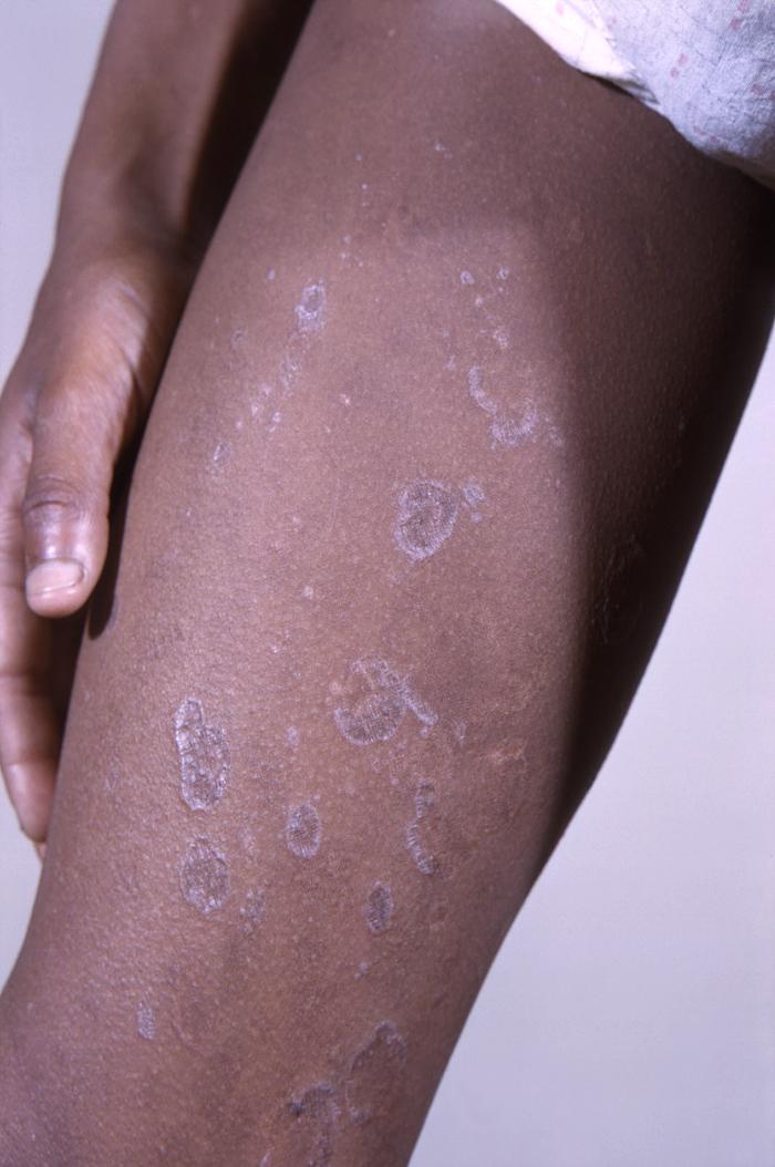
Epidemiology and Etiology
Epidemiology
- Lichen planus (LP) affects < 1% of the population in the United States.
- Can occur at any age, but most commonly found in patients 30–60 years of age
- Rare in children
Etiology
- Unknown
- Associated risk factors:
- Genetic predisposition: ↑ frequency of human leukocyte antigen B7 (HLA-B7), HLA-DR1, and HLA-DR10 in those affected
- Hepatitis C virus infection
- Drug exposure:
- Angiotensin-converting enzyme (ACE) inhibitors
- Beta-blockers
- Methyldopa
- Quinidine
- Chloroquine
- Hydroxychloroquine
- Thiazide diuretics
- Gold salts
- Primary biliary cirrhosis
- Human herpesvirus type 7
- Mercury (in dental amalgam)
- Radiotherapy
- Psychogenic factors, such as anxiety, depression, and stress
Pathophysiology
- Proposed mechanism involves cell-mediated immunity.
- Exposure to exogenous agent (drug, virus, other risk factors) → alters epidermal self-antigens
- CD4+ and CD8+ (more predominant) lymphocytes are recruited to dermal-epidermal junction → lead a T-cell–mediated attack → apoptosis of keratinocytes
- Basal layer is injured → release melanin into dermis → hyperpigmentation
- Interaction of T lymphocytes and basal keratinocytes enhanced by:
- Intercellular adhesion molecule-1 (ICAM-1) upregulation by basal keratinocytes
- Other cytokines: interferon (IFN)-gamma, tumor necrosis factor (TNF)-alpha, interleukin (IL)-1 alpha, IL-6, and IL-8
Related videos
Clinical Presentation
Cutaneous LP
- Clinical features:
- Primary lesions can be characterized by the 6 Ps:
- Polygonal
- Purple (violaceous)
- Pruritic (often severe)
- Planar Papules (flat-topped)
- May coalesce into larger Plaques
- Wickham’s striae: lace-like white lines/dots, can be seen on the surface of the lesions
- Koebner phenomenon:
- Development of lesions in previously healthy sites
- Due to trauma from scratching
- Hyperpigmentation occurs with healed lesions.
- May be painful depending on location
- Primary lesions can be characterized by the 6 Ps:
- Distribution and location:
- Symmetric and bilateral
- Most commonly found on flexural surfaces of extremities (ankles and wrists)
- Clinical course:
- Most resolve within 1–2 years
- May leave permanent hyperpigmented macules after active disease is cleared
Cutaneous variants by morphology
- Hypertrophic:
- Lichen planus verrucosus
- Most pruritic form; associated with residual scarring
- Affects the anterior distal lower extremities
- Annular:
- Plaques with central clearing
- Bullous:
- Vesicles or bullae develop within existing lesions.
- Legs usually affected
- Actinic:
- Plaques develop in sun-exposed areas.
- Seen in patients of Middle Eastern descent
- Pigmentosus:
- Gray-brown or dark-brown patches
- Located in sun-exposed areas
- Inverse:
- Plaques develop in uncommon sites (axillae, inguinal creases, inframammary area, limb flexures).
- Atrophic:
- Plaques are well-demarcated pale or violaceous papules with superficial atrophy.
Image: “12188” by CDC/ Wallace N. McLeod, M.D.; Bob Craig. License: Public Domain 
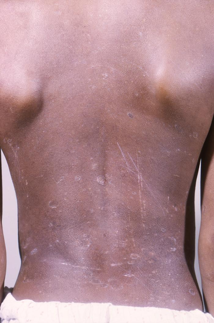
Lichen planus of the back of an 11-year-old African American boy with a 6-year history of generalized LP. Note the presence of the Koebner phenomenon: the spread of LP lesions along areas of trauma, including scratches on the skin.
Image: “12186” by CDC. License: Public Domain
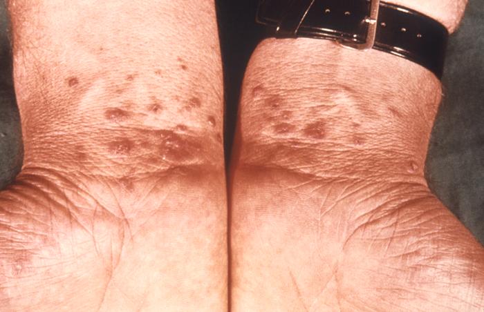
Lichen planus on bilateral wrists
Image: “6545” by CDC/ Susan Lindsley. License: Public Domain
Oral LP
- Clinical features:
- Can occur independently or with cutaneous LP
- Can present as:
- Wickham’s striae or reticular form (lacy, web-like white threads)
- Atrophic lesions
- Bullous lesions
- Painful erosions or ulcers (erosive mucosal lichen planus) that can lead to secondary Candida infection and/or loss of appetite due to pain
- Location:
- Most common in buccal mucosa
- Also affects alveolar mucosa and tongue
- Clinical course: a chronic form of LP
- Mucosal and paramucosal lesions, especially at oral and vulvar sites (but not cutaneous lesions) have an increased risk of squamous cell carcinoma.
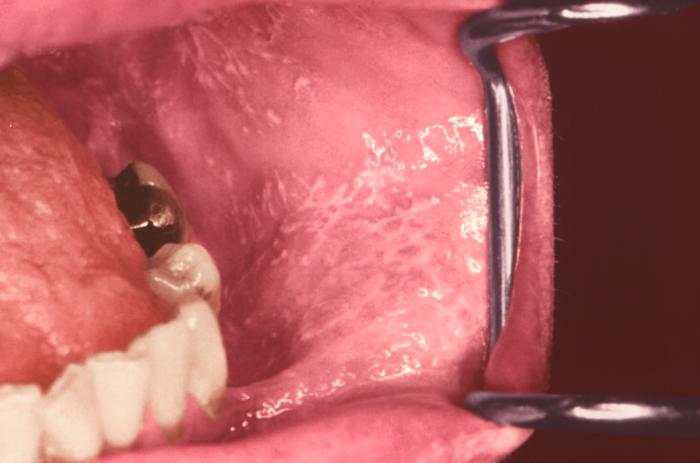
Lichen planus presenting as Wickham’s striae on the oral mucosa
Image: “12632” by CDC/ Robert E. Sumpter. License: Public Domain
Genital LP
- More common in men; lesions are typically on the glans penis.
- In women, vulva and vagina are affected.
- Associated with pruritus and dyspareunia
- Erosive lesions can be complicated by urethral stenosis.
- Increased risk of squamous cell carcinoma
- Clinical course: chronic or persistent
Other forms of LP
- Nail:
- Can be minor dystrophy to total nail loss
- Dorsal pterygium (raised wedge-shaped deformity of nailbed): most specific LP nail abnormality
- Lichen planopilaris:
- LP of the scalp that leads to keratotic follicular papules
- Affects women more than men
- Can progress to scarring alopecia if left untreated
- Esophageal:
- Can be asymptomatic or present with dysphagia or odynophagia
- Endoscopic findings: pseudomembranes, friable and inflamed mucosa, submucosal papules, lacy-white plaques, erosions
- Can lead to esophageal stricture
- Otic:
- Affects external auditory canals and tympanic membranes
- Presents with erythema, pruritus, and induration. but can cause persistent otorrhea and hearing loss
Diagnosis
History and clinical findings
- Review of medical history, medications, and risk factors (hepatitis C)
- Physical examination of entire cutaneous surface (including the scalp, oral cavity, and external genitalia)
Mucocutaneous biopsy
- Deep shave or punch biopsy of the most prominent lesion
- Histopathological findings include:
- Hyperkeratosis (hyperplasia of the stratum corneum)
- Civatte bodies (anucleate, necrotic hypereosinophilic keratinocytes) in the lower epidermis
- Wedge-shaped hypergranulosis (hyperplasia of the stratum granulosum)
- “Saw-tooth” rete ridges
- Vacuolar alteration of basal layer of epidermis
- Band-like lymphocytic infiltrate at the dermal–epidermal junction
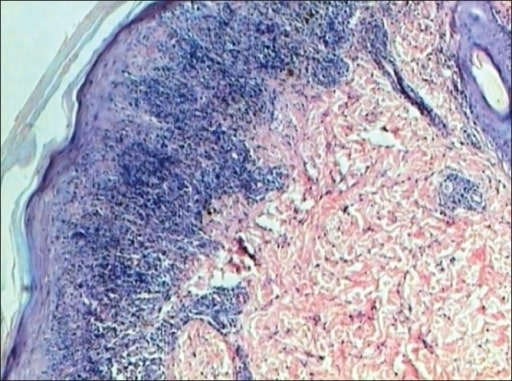
Lichen planus: epidermis shows hyperkeratosis, hypergranulosis, acanthosis, and saw-tooth rete ridges. Dermis shows a band-like lymphocytic infiltrate touching the epidermis (H&E stain, ×40).
Image: “Lichen planus” by Department of Pathology, JJ Hospital, Mumbai, India. License: CC BY 2.0
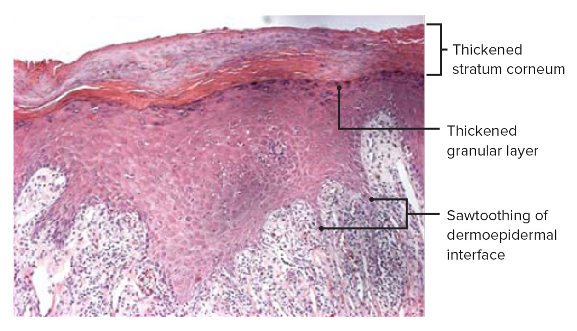
Histopathology of a skin biopsy of an LP plaque showing dense lichenoid inflammatory infiltrates underneath the lamina propria. Note the inflammation at the dermal–epidermal junction and an angulated zigzag lower contour, which gives it the saw-tooth appearance characteristic of LP. Note also the thickening of the granular cell layer (hypergranulosis) and the stratum corneum (hyperkeratosis), which are more pronounced in the white lines of Wickham’s striae.
Image: “Histopathology of lichen planus” by Shi G, Sohn KC et al. License: CC BY 4.0, edited by Lecturio.
Other tests
- Dermoscopy: aids in visualizing Wickham’s striae in cutaneous lesions
- Laboratory test(s): check for hepatitis C in patients with risk factors
Management
Supportive therapy
- Reduce pruritus with antihistamines (hydroxyzine)
- Avoid dry skin with the use of emollients
First-line pharmacologic therapy
- High-potency topical corticosteroids:
- For localized lesions including oral lesions
- Risks: cutaneous atrophy, striae, changes in pigmentation
- Betamethasone or clobetasol cream/ointment twice daily
- Reassess response in 2–3 weeks.
- Intralesional corticosteroids:
- For hypertrophic lesions
- Risks: cutaneous atrophy, changes in pigmentation
- Triamcinolone injection
Second-line pharmacologic therapy
- Systemic corticosteroids
- For extensive disease
- Prednisone 30–60 mg/day for 4–6 weeks, followed by taper
- Topical calcineurin inhibitors
- Treatment option for genital LP, resistant oral LP
- Pimecrolimus, tacrolimus
- Phototherapy
- Narrowband ultraviolet B (UVB) therapy: preferred
- Psoralen plus ultraviolet A (PUVA)
- Oral retinoids
- Acitrecin 30 mg/day for 8 weeks
- Side effects: hypertriglyceridemia, visual changes, hair loss
- Teratogenic (avoid pregnancy up to 3 years after discontinuation)
Differential Diagnosis
- Irritant contact dermatitis: skin condition that results from exposure to a physical or chemical irritant. Patients present with erythema, edema, vesicles, and oozing that are limited to areas where the offending substance was present.
- Discoid lupus erythematosus: a chronic autoimmune skin condition characterized by erythematous scaly plaques that often result in inflammation, scarring, and atrophy on the face, neck, ears, and scalp. The condition may occur with or without concurrent systemic lupus erythematosus.
- Lichenoid drug eruption: the symmetric cutaneous eruption that appears as erythematous papules on the trunk and extremities after taking a drug. Wickham’s striae is usually not present. In most cases, the condition will spontaneously resolve with discontinuation of the offending drug.
- Eczema (seen in atopic dermatitis): a chronic pruritic skin condition commonly found in children. Eczema is associated with asthma. Lesions are dry, pruritic, red papules mainly located on the cheeks or scalp (in infants) and flexural areas of extremities (older children).
- Seborrheic dermatitis: a chronic inflammatory dermatosis characterized by the presence of greasy, red (inflamed) skin lesions affecting the scalp (as dandruff), posterior aspect of the neck, and the forehead. The condition is linked to increased sebum production and possible colonization of the skin by fungi of the genus Malassezia.
- Pityriasis rosea: skin condition affecting mostly older children and young adults. Pityriasis rosea begins with a single “herald” patch on the chest, neck, or back. This is followed by smaller pruritic salmon-colored oval plaques with scaling located on the trunk and extremities. The characteristic morphology is a “Christmas tree” pattern on the back.
- Plaque psoriasis: lesions are characterized by well-demarcated, erythematous, symmetrically distributed plaques with overlying silver scales involving the scalp and extensor surfaces (elbows and knees).
- Lichen simplex chronicus: a form of neurodermatitis caused by the itch-scratch cycle, leading to plaques and thickened skin. Locations affected are areas accessible to scratching. Management includes potent topical steroids.
References
- Arnold, D., Krishnamurthy, K. (2020). Lichen Planus. https://www.ncbi.nlm.nih.gov/books/NBK526126/
- Chuang, T., James, W. (2020). Lichen planus. Medscape. Retrieved September 14, 2020, from https://emedicine.medscape.com/article/1123213-overview.
- Goldstein, B., Goldstein, A., Mostow, E., Dellavalle, R. Callen, J., Ofori, A. (2019). UpToDate. Retrieved September 14, 2020, from https://www.uptodate.com/contents/lichen-planus
- Lazar, A. (2020). The Skin in Kumar, V., Abbas, A., Aster, J. (Eds.), Robbins and Cotran Pathologic Basis of Disease (10th ed., pp. 1133-1170). Elsevier, Inc
- Lehman, J., Tollefson, M.,Gibson, L. (2009). Lichen Planus. International Journal of Dermatology. https://doi.org/10.1111/j.1365-4632.2009.04062.x
- Usatine, R., Tinitigan, M. (2011). Diagnosis and treatment of Lichen Planus. Am Fam Physician. 1;84(1):53-60. https://www.aafp.org/afp/2011/0701/p53.html
- Dinulos, J.G.H. (2020). In Habif’s Clinical Dermatology (7th ed. pp. 313-322).