Achieve Mastery of Medical Concepts

Study for medical school and boards with Lecturio

Hearing Loss

Hearing loss, also known as hearing impairment, is any degree of impairment in the ability to apprehend sound as determined by audiometry Audiometry The testing of the acuity of the sense of hearing to determine the thresholds of the loWest intensity levels at which an individual can hear a set of tones. The frequencies between 125 and 8000 hz are used to test air conduction thresholds and the frequencies between 250 and 4000 hz are used to test bone conduction thresholds. Ménière Disease to be below normal hearing thresholds. Clinical presentation may occur at birth or as a gradual loss of hearing with age, including a short-term or sudden loss at any point. Diagnostic evaluation relies on history, physical examination (including otoscopic and tuning fork examinations), and audiology testing. Management is directed toward the underlying cause of the hearing loss to choose the appropriate course of treatment.

Last updated: Mar 6, 2023

Editorial responsibility: Stanley Oiseth, Lindsay Jones, Evelin Maza

Overview

Definition

Hearing loss, also known as hearing impairment, is any degree of impairment in the ability to apprehend sound as determined by audiometry Audiometry The testing of the acuity of the sense of hearing to determine the thresholds of the loWest intensity levels at which an individual can hear a set of tones. The frequencies between 125 and 8000 hz are used to test air conduction thresholds and the frequencies between 250 and 4000 hz are used to test bone conduction thresholds. Ménière Disease to be below normal hearing thresholds.

Anatomy

  • External ear: auricle and external auditory canal External Auditory Canal Otitis Externa
  • Middle ear Middle ear The space and structures directly internal to the tympanic membrane and external to the inner ear (labyrinth). Its major components include the auditory ossicles and the eustachian tube that connects the cavity of middle ear (tympanic cavity) to the upper part of the throat. Acute Otitis Media:
    • Tympanic membrane Tympanic membrane An oval semitransparent membrane separating the external ear canal from the tympanic cavity. It contains three layers: the skin of the external ear canal; the core of radially and circularly arranged collagen fibers; and the mucosa of the middle ear. Ear: Anatomy
    • Ossicles: malleus, incus, and stapes
  • Inner ear Inner ear The essential part of the hearing organ consists of two labyrinthine compartments: the bony labyrinthine and the membranous labyrinth. Ear: Anatomy:
    • Cochlea Cochlea The part of the inner ear (labyrinth) that is concerned with hearing. It forms the anterior part of the labyrinth, as a snail-like structure that is situated almost horizontally anterior to the vestibular labyrinth. Ear: Anatomy
    • Semicircular canals Semicircular canals Three long canals (anterior, posterior, and lateral) of the bony labyrinth. They are set at right angles to each other and are situated posterosuperior to the vestibule of the bony labyrinth (vestibular labyrinth). The semicircular canals have five openings into the vestibule with one shared by the anterior and the posterior canals. Within the canals are the semicircular ducts. Auditory and Vestibular Pathways: Anatomy
    • Vestibulocochlear nerve Vestibulocochlear nerve The 8th cranial nerve. The vestibulocochlear nerve has a cochlear part (cochlear nerve) which is concerned with hearing and a vestibular part (vestibular nerve) which mediates the sense of balance and head position. The fibers of the cochlear nerve originate from neurons of the spiral ganglion and project to the cochlear nuclei (cochlear nucleus). The fibers of the vestibular nerve arise from neurons of scarpa’s ganglion and project to the vestibular nuclei. The 12 Cranial Nerves: Overview and Functions (CN VIII) 
  • CNS:
Anatomy of the external, middle, and inner ear

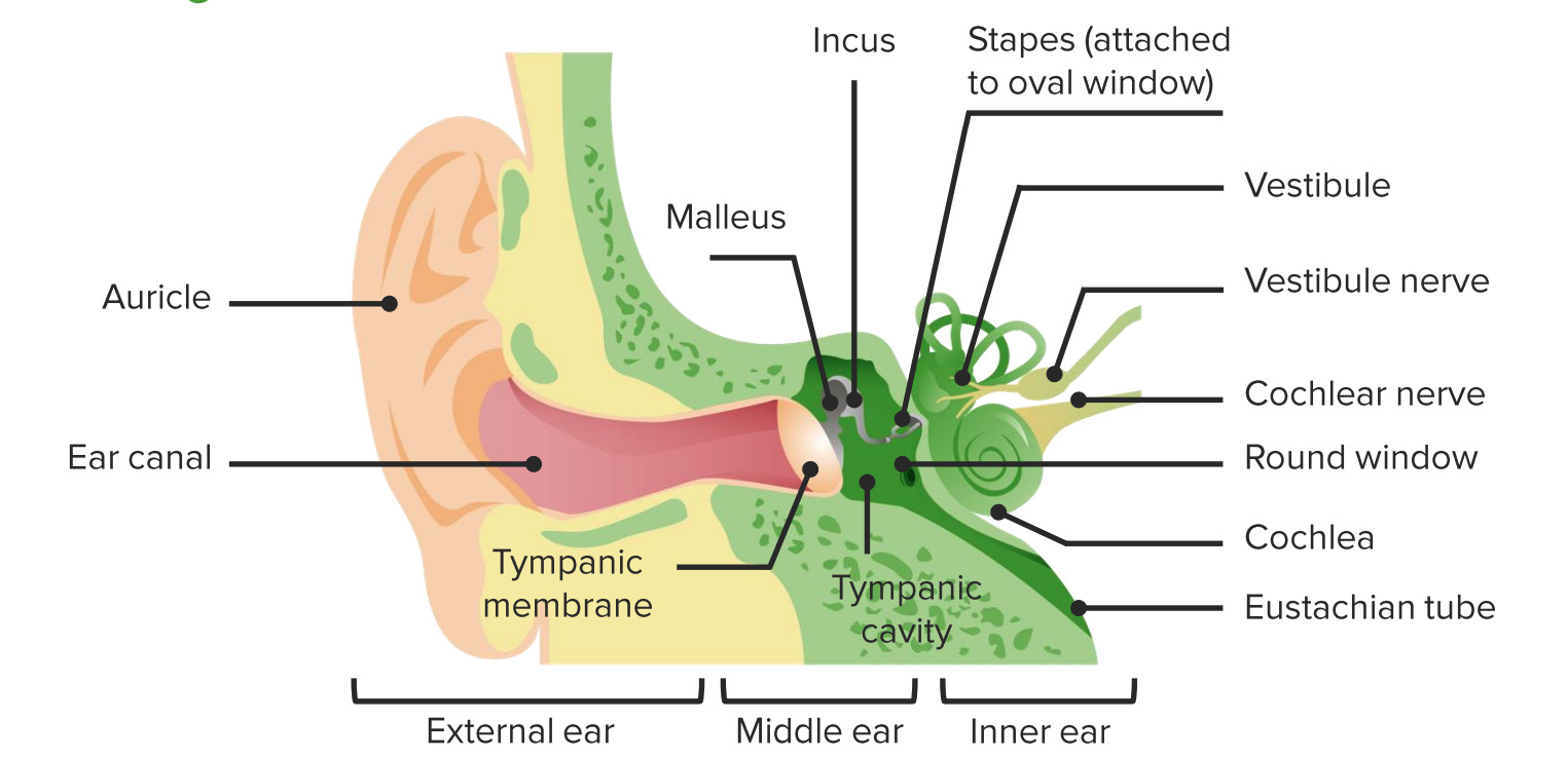
Diagram of the anatomy of the external, middle, and inner ear

Image by Lecturio.

Epidemiology

  • Incidence Incidence The number of new cases of a given disease during a given period in a specified population. It also is used for the rate at which new events occur in a defined population. It is differentiated from prevalence, which refers to all cases in the population at a given time. Measures of Disease Frequency of hearing loss: 2 per 1,000 in young children
  • Prevalence Prevalence The total number of cases of a given disease in a specified population at a designated time. It is differentiated from incidence, which refers to the number of new cases in the population at a given time. Measures of Disease Frequency in adults:
    • 21–34 years of age: 3%
    • 35–44 years of age: 6%
    • 44–54 years of age: 11%
    • 55–64 years of age: 25%
    • 65–84 years of age: 43%

Classification of hearing loss

  • Conductive hearing loss (CHL): inability of the ear to conduct sound through the structures of the ear (ossicular chain and tympanic membrane Tympanic membrane An oval semitransparent membrane separating the external ear canal from the tympanic cavity. It contains three layers: the skin of the external ear canal; the core of radially and circularly arranged collagen fibers; and the mucosa of the middle ear. Ear: Anatomy)
  • Sensorineural hearing loss (SNHL): impact on the cochlea Cochlea The part of the inner ear (labyrinth) that is concerned with hearing. It forms the anterior part of the labyrinth, as a snail-like structure that is situated almost horizontally anterior to the vestibular labyrinth. Ear: Anatomy, the CN VIII, or the central auditory pathway
  • Mixed: both conductive and sensorineural components 
  • Central auditory dysfunction: damage at the level of the CN VIII, the auditory brain Brain The part of central nervous system that is contained within the skull (cranium). Arising from the neural tube, the embryonic brain is comprised of three major parts including prosencephalon (the forebrain); mesencephalon (the midbrain); and rhombencephalon (the hindbrain). The developed brain consists of cerebrum; cerebellum; and other structures in the brain stem. Nervous System: Anatomy, Structure, and Classification stem, or the cerebral cortex Cerebral cortex The cerebral cortex is the largest and most developed part of the human brain and CNS. Occupying the upper part of the cranial cavity, the cerebral cortex has 4 lobes and is divided into 2 hemispheres that are joined centrally by the corpus callosum. Cerebral Cortex: Anatomy

Etiology

Conductive hearing loss (CHL):

  • Otitis media (acute or chronic)
  • Otitis externa Otitis externa Otitis externa (also known as external otitis or swimmer’s ear) is an infection of the external auditory canal that is most often caused by acute bacterial infection and is frequently associated with hot, humid weather and water exposure. Patients commonly present with ear pain, pruritus, discharge, and hearing loss. Otitis Externa (acute or chronic)
  • Eustachian tube dysfunction Eustachian Tube Dysfunction Acute Otitis Media/fluid retention in the middle ear Middle ear The space and structures directly internal to the tympanic membrane and external to the inner ear (labyrinth). Its major components include the auditory ossicles and the eustachian tube that connects the cavity of middle ear (tympanic cavity) to the upper part of the throat. Acute Otitis Media
  • Cholesteatoma
  • Otosclerosis Otosclerosis Formation of spongy bone in the labyrinth capsule which can progress toward the stapes (stapedial fixation) or anteriorly toward the cochlea leading to conductive, sensorineural, or mixed hearing loss. Several genes are associated with familial otosclerosis with varied clinical signs. Vertigo
  • Cerumen Cerumen The yellow or brown waxy secretions produced by vestigial apocrine sweat glands in the external ear canal. Otitis Externa impaction
  • Neoplasm:
    • Tumor Tumor Inflammation of the external auditory canal External Auditory Canal Otitis Externa
    • Paraganglioma
    • Adenoma of the middle ear Middle ear The space and structures directly internal to the tympanic membrane and external to the inner ear (labyrinth). Its major components include the auditory ossicles and the eustachian tube that connects the cavity of middle ear (tympanic cavity) to the upper part of the throat. Acute Otitis Media
  • Tympanosclerosis Tympanosclerosis Acute Otitis Media
  • Trauma:
    • Ossicular fracture Fracture A fracture is a disruption of the cortex of any bone and periosteum and is commonly due to mechanical stress after an injury or accident. Open fractures due to trauma can be a medical emergency. Fractures are frequently associated with automobile accidents, workplace injuries, and trauma. Overview of Bone Fractures
    • Tympanic membrane perforation Tympanic membrane perforation A temporary or persistent opening in the eardrum (tympanic membrane). Clinical signs depend on the size, location, and associated pathological condition. Acute Otitis Media
  • Congenital Congenital Chorioretinitis malformations:

Sensorineural hearing loss (SNHL):

  • Presbycusis Presbycusis Gradual bilateral hearing loss associated with aging that is due to progressive degeneration of cochlear structures and central auditory pathways. Hearing loss usually begins with the high frequencies then progresses to sounds of middle and low frequencies. Geriatric Changes
  • Prolonged exposure to loud noise
  • Prenatal toxoplasmosis Toxoplasmosis Toxoplasmosis is an infectious disease caused by Toxoplasma gondii, an obligate intracellular protozoan parasite. Felines are the definitive host, but transmission to humans can occur through contact with cat feces or the consumption of contaminated foods. The clinical presentation and complications depend on the host’s immune status. Toxoplasma/Toxoplasmosis, other agents, rubella Rubella An acute infectious disease caused by the rubella virus. The virus enters the respiratory tract via airborne droplet and spreads to the lymphatic system. Rubella Virus, cytomegalovirus Cytomegalovirus CMV is a ubiquitous double-stranded DNA virus belonging to the Herpesviridae family. CMV infections can be transmitted in bodily fluids, such as blood, saliva, urine, semen, and breast milk. The initial infection is usually asymptomatic in the immunocompetent host, or it can present with symptoms of mononucleosis. Cytomegalovirus, and herpes simplex Herpes Simplex A group of acute infections caused by herpes simplex virus type 1 or type 2 that is characterized by the development of one or more small fluid-filled vesicles with a raised erythematous base on the skin or mucous membrane. It occurs as a primary infection or recurs due to a reactivation of a latent infection. Congenital TORCH Infections (TORCH) infections Infections Invasion of the host organism by microorganisms or their toxins or by parasites that can cause pathological conditions or diseases. Chronic Granulomatous Disease:
    • Toxoplasmosis Toxoplasmosis Toxoplasmosis is an infectious disease caused by Toxoplasma gondii, an obligate intracellular protozoan parasite. Felines are the definitive host, but transmission to humans can occur through contact with cat feces or the consumption of contaminated foods. The clinical presentation and complications depend on the host’s immune status. Toxoplasma/Toxoplasmosis
    • Rubella Rubella An acute infectious disease caused by the rubella virus. The virus enters the respiratory tract via airborne droplet and spreads to the lymphatic system. Rubella Virus
    • Cytomegalovirus Cytomegalovirus CMV is a ubiquitous double-stranded DNA virus belonging to the Herpesviridae family. CMV infections can be transmitted in bodily fluids, such as blood, saliva, urine, semen, and breast milk. The initial infection is usually asymptomatic in the immunocompetent host, or it can present with symptoms of mononucleosis. Cytomegalovirus (can cause asymptomatic hearing loss)
    • Herpes simplex Herpes Simplex A group of acute infections caused by herpes simplex virus type 1 or type 2 that is characterized by the development of one or more small fluid-filled vesicles with a raised erythematous base on the skin or mucous membrane. It occurs as a primary infection or recurs due to a reactivation of a latent infection. Congenital TORCH Infections
  • Postnatal infections Infections Invasion of the host organism by microorganisms or their toxins or by parasites that can cause pathological conditions or diseases. Chronic Granulomatous Disease:
    • Meningitis Meningitis Meningitis is inflammation of the meninges, the protective membranes of the brain, and spinal cord. The causes of meningitis are varied, with the most common being bacterial or viral infection. The classic presentation of meningitis is a triad of fever, altered mental status, and nuchal rigidity. Meningitis due to Neisseria meningitidis Neisseria meningitidis A species of gram-negative, aerobic bacteria. It is a commensal and pathogen only of humans, and can be carried asymptomatically in the nasopharynx. When found in cerebrospinal fluid it is the causative agent of cerebrospinal meningitis. It is also found in venereal discharges and blood. There are at least 13 serogroups based on antigenic differences in the capsular polysaccharides; the ones causing most meningitis infections being a, b, c, y, and w-135. Each serogroup can be further classified by serotype, serosubtype, and immunotype. Neisseria, Haemophilus Haemophilus Haemophilus is a genus of Gram-negative coccobacilli, all of whose strains require at least 1 of 2 factors for growth (factor V [NAD] and factor X [heme]); therefore, it is most often isolated on chocolate agar, which can supply both factors. The pathogenic species are H. influenzae and H. ducreyi. Haemophilus influenza Influenza Influenza viruses are members of the Orthomyxoviridae family and the causative organisms of influenza, a highly contagious febrile respiratory disease. There are 3 primary influenza viruses (A, B, and C) and various subtypes, which are classified based on their virulent surface antigens, hemagglutinin (HA) and neuraminidase (NA). Influenza typically presents with a fever, myalgia, headache, and symptoms of an upper respiratory infection. Influenza Viruses/Influenza, or Streptococcus Streptococcus Streptococcus is one of the two medically important genera of gram-positive cocci, the other being Staphylococcus. Streptococci are identified as different species on blood agar on the basis of their hemolytic pattern and sensitivity to optochin and bacitracin. There are many pathogenic species of streptococci, including S. pyogenes, S. agalactiae, S. pneumoniae, and the viridans streptococci. Streptococcus pneumoniae
    • Meningitis Meningitis Meningitis is inflammation of the meninges, the protective membranes of the brain, and spinal cord. The causes of meningitis are varied, with the most common being bacterial or viral infection. The classic presentation of meningitis is a triad of fever, altered mental status, and nuchal rigidity. Meningitis from many organisms can cause hearing loss.
  • Additional infectious causes of hearing loss:
    • Mumps Mumps Mumps is caused by a single-stranded, linear, negative-sense RNA virus of the family Paramyxoviridae. Mumps is typically a disease of childhood, which manifests initially with fever, muscle pain, headache, poor appetite, and a general feeling of malaise, and is classically followed by parotitis. Mumps Virus/Mumps
    • Syphilis Syphilis Syphilis is a bacterial infection caused by the spirochete Treponema pallidum pallidum (T. p. pallidum), which is usually spread through sexual contact. Syphilis has 4 clinical stages: primary, secondary, latent, and tertiary. Syphilis
  • Genetic causes:
    • > 400 genetic syndromes (e.g., Alport syndrome Alport Syndrome Alport syndrome, also called hereditary nephritis, is a genetic disorder caused by a mutation in the genes encoding for the alpha chains of type IV collagen, resulting in the production of abnormal type IV collagen strands. Patients present with glomerulonephritis, hypertension, edema, hematuria, and proteinuria, as well as with ocular and auditory findings. Alport Syndrome)
    • Many autosomal dominant Autosomal dominant Autosomal inheritance, both dominant and recessive, refers to the transmission of genes from the 22 autosomal chromosomes. Autosomal dominant diseases are expressed when only 1 copy of the dominant allele is inherited. Autosomal Recessive and Autosomal Dominant Inheritance, autosomal recessive Autosomal recessive Autosomal inheritance, both dominant and recessive, refers to the transmission of genes from the 22 autosomal chromosomes. Autosomal recessive diseases are only expressed when 2 copies of the recessive allele are inherited. Autosomal Recessive and Autosomal Dominant Inheritance, and X-linked X-linked Genetic diseases that are linked to gene mutations on the X chromosome in humans or the X chromosome in other species. Included here are animal models of human X-linked diseases. Common Variable Immunodeficiency (CVID) nonsyndromic genetic causes exist.
  • Neoplasms Neoplasms New abnormal growth of tissue. Malignant neoplasms show a greater degree of anaplasia and have the properties of invasion and metastasis, compared to benign neoplasms. Benign Bone Tumors:
    • Acoustic neuroma Acoustic neuroma Acoustic neuroma, also referred to as vestibular schwannoma, is a benign tumor arising from Schwann cells of the vestibular component of the cranial nerve VIII. Acoustic neuroma forms within the internal auditory meatus and extends into the cerebellopontine angle. Acoustic Neuroma
    • Meningioma Meningioma Meningiomas are slow-growing tumors that arise from the meninges of the brain and spinal cord. The vast majority are benign. These tumors commonly occur in individuals with a history of high doses of skull radiation, head trauma, and neurofibromatosis 2. Meningioma
    • Tumor Tumor Inflammation of the endolymphatic sac
  • Trauma:
    • Blunt or penetrating trauma to the ear
    • Blast injury or temporal bone Temporal bone Either of a pair of compound bones forming the lateral (left and right) surfaces and base of the skull which contains the organs of hearing. It is a large bone formed by the fusion of parts: the squamous (the flattened anterior-superior part), the tympanic (the curved anterior-inferior part), the mastoid (the irregular posterior portion), and the petrous (the part at the base of the skull). Jaw and Temporomandibular Joint: Anatomy fracture Fracture A fracture is a disruption of the cortex of any bone and periosteum and is commonly due to mechanical stress after an injury or accident. Open fractures due to trauma can be a medical emergency. Fractures are frequently associated with automobile accidents, workplace injuries, and trauma. Overview of Bone Fractures
    • Barotrauma Barotrauma Injury following pressure changes; includes injury to the eustachian tube, ear drum, lung and stomach. Invasive Mechanical Ventilation from diving or flying
  • Ototoxic medications:
    • Aminoglycoside antibiotics
    • Loop diuretics Diuretics Agents that promote the excretion of urine through their effects on kidney function. Heart Failure and Angina Medication
    • Aspirin Aspirin The prototypical analgesic used in the treatment of mild to moderate pain. It has anti-inflammatory and antipyretic properties and acts as an inhibitor of cyclooxygenase which results in the inhibition of the biosynthesis of prostaglandins. Aspirin also inhibits platelet aggregation and is used in the prevention of arterial and venous thrombosis. Nonsteroidal Antiinflammatory Drugs (NSAIDs)
    • Platinum-based chemotherapy Chemotherapy Osteosarcoma agents
  • Radiation Radiation Emission or propagation of acoustic waves (sound), electromagnetic energy waves (such as light; radio waves; gamma rays; or x-rays), or a stream of subatomic particles (such as electrons; neutrons; protons; or alpha particles). Osteosarcoma therapy
  • Meniere disease

Physiology of hearing

  1. Sound is received in the external auditory canal External Auditory Canal Otitis Externa and funneled to the middle ear Middle ear The space and structures directly internal to the tympanic membrane and external to the inner ear (labyrinth). Its major components include the auditory ossicles and the eustachian tube that connects the cavity of middle ear (tympanic cavity) to the upper part of the throat. Acute Otitis Media.
  2. The tympanic membrane Tympanic membrane An oval semitransparent membrane separating the external ear canal from the tympanic cavity. It contains three layers: the skin of the external ear canal; the core of radially and circularly arranged collagen fibers; and the mucosa of the middle ear. Ear: Anatomy vibrates → conducts sound vibrations through the ossicles → oval window → inner ear Inner ear The essential part of the hearing organ consists of two labyrinthine compartments: the bony labyrinthine and the membranous labyrinth. Ear: Anatomy
  3. Sound travels through the inner ear Inner ear The essential part of the hearing organ consists of two labyrinthine compartments: the bony labyrinthine and the membranous labyrinth. Ear: Anatomy perilymph (in the upper and lower chambers of the cochlea Cochlea The part of the inner ear (labyrinth) that is concerned with hearing. It forms the anterior part of the labyrinth, as a snail-like structure that is situated almost horizontally anterior to the vestibular labyrinth. Ear: Anatomy) → sensed by the hair cells Hair cells Auditory sensory cells of organ of corti, usually placed in one row medially to the core of spongy bone (the modiolus). Inner hair cells are in fewer numbers than the outer auditory hair cells, and their stereocilia are approximately twice as thick as those of the outer hair cells. Auditory and Vestibular Pathways: Anatomy on the basilar membrane in the organ of Corti Organ of Corti The spiral epithelium containing sensory auditory hair cells and supporting cells in the cochlea. Organ of corti, situated on the basilar membrane and overlaid by a gelatinous tectorial membrane, converts sound-induced mechanical waves to neural impulses to the brain. Auditory and Vestibular Pathways: Anatomy (middle chamber of the cochlea Cochlea The part of the inner ear (labyrinth) that is concerned with hearing. It forms the anterior part of the labyrinth, as a snail-like structure that is situated almost horizontally anterior to the vestibular labyrinth. Ear: Anatomy), which is filled with endolymph Endolymph The lymph fluid found in the membranous labyrinth of the ear. Vertigo
  4. Signals are carried through the cochlear division of CN VIII → processed in the CN VIII nucleus Nucleus Within a eukaryotic cell, a membrane-limited body which contains chromosomes and one or more nucleoli (cell nucleolus). The nuclear membrane consists of a double unit-type membrane which is perforated by a number of pores; the outermost membrane is continuous with the endoplasmic reticulum. A cell may contain more than one nucleus. The Cell: Organelles and auditory cortex Auditory cortex The region of the cerebral cortex that receives the auditory radiation from the medial geniculate body. Auditory and Vestibular Pathways: Anatomy perception Perception The process by which the nature and meaning of sensory stimuli are recognized and interpreted. Psychiatric Assessment of sound (audition)

Cultural relevance

  • Members of the Deaf community in the United States are deaf, which implies profound hearing loss.
  • Members use American Sign Language. 
  • Members are not considered to have a hearing impairment or hearing loss.
  • Deafness is not a disease to be treated or cured.

History of Present Illness

Information to gather from the individual or caregiver regarding hearing loss:

  • Onset and duration of hearing loss:
    • Prelingual or postlingual (before or after a child learns to speak)
    • Sudden or chronic 
    • Fluctuating or progressive 
  • Laterality: unilateral or bilateral
  • Severity:
    • Milder forms may be unnoticed by the individual.
    • More severe forms may require assistive devices for amplification.
  • Alarm symptoms:
    • Sudden or rapid onset 
    • Rapidly progressive (possibly from ototoxic medications)
    • Tinnitus Tinnitus A nonspecific symptom of hearing disorder characterized by the sensation of buzzing, ringing, clicking, pulsations, and other noises in the ear. Objective tinnitus refers to noises generated from within the ear or adjacent structures that can be heard by other individuals. The term subjective tinnitus is used when the sound is audible only to the affected individual. Tinnitus may occur as a manifestation of cochlear diseases; vestibulocochlear nerve diseases; intracranial hypertension; craniocerebral trauma; and other conditions. Cranial Nerve Palsies associated with acute hearing loss
    • Vertigo Vertigo Vertigo is defined as the perceived sensation of rotational motion while remaining still. A very common complaint in primary care and the ER, vertigo is more frequently experienced by women and its prevalence increases with age. Vertigo is classified into peripheral or central based on its etiology. Vertigo may indicate CN VIII pathology:
      • Meniere disease
      • Labyrinthitis
      • Neoplasm
      • Brainstem ischemia Ischemia A hypoperfusion of the blood through an organ or tissue caused by a pathologic constriction or obstruction of its blood vessels, or an absence of blood circulation. Ischemic Cell Damage/ infarct Infarct Area of necrotic cells in an organ, arising mainly from hypoxia and ischemia Ischemic Cell Damage
      • Perilymph fistula Fistula Abnormal communication most commonly seen between two internal organs, or between an internal organ and the surface of the body. Anal Fistula
  • Recent history:
    • Upper respiratory infection Upper respiratory infection Rhinitis 
    • Use of aminoglycoside antibiotics
    • Recent ear surgery:
      • Pressure equalization tubes
      • Tympanoplasty
      • Mastoidectomy

Physical Examination

An otoscope Otoscope Instruments designed to inspect or auscultate the ear. They are designed primarily to examine the outer ear canal and tympanic membrane by means of light and air under moderate pressure, as with a pneumatic otoscope. Head and Neck Examination is used to examine the outer and middle ear Middle ear The space and structures directly internal to the tympanic membrane and external to the inner ear (labyrinth). Its major components include the auditory ossicles and the eustachian tube that connects the cavity of middle ear (tympanic cavity) to the upper part of the throat. Acute Otitis Media and a tuning fork is used to determine the type of hearing loss.

Otoscopic exam

  • Outer and middle ear Middle ear The space and structures directly internal to the tympanic membrane and external to the inner ear (labyrinth). Its major components include the auditory ossicles and the eustachian tube that connects the cavity of middle ear (tympanic cavity) to the upper part of the throat. Acute Otitis Media:
    • Inspection Inspection Dermatologic Examination of the ear canal (e.g., erythema Erythema Redness of the skin produced by congestion of the capillaries. This condition may result from a variety of disease processes. Chalazion, drainage, inflammation Inflammation Inflammation is a complex set of responses to infection and injury involving leukocytes as the principal cellular mediators in the body’s defense against pathogenic organisms. Inflammation is also seen as a response to tissue injury in the process of wound healing. The 5 cardinal signs of inflammation are pain, heat, redness, swelling, and loss of function. Inflammation, cerumen Cerumen The yellow or brown waxy secretions produced by vestigial apocrine sweat glands in the external ear canal. Otitis Externa impaction, and foreign body Foreign Body Foreign Body Aspiration)
    • Examination of the tympanic membrane Tympanic membrane An oval semitransparent membrane separating the external ear canal from the tympanic cavity. It contains three layers: the skin of the external ear canal; the core of radially and circularly arranged collagen fibers; and the mucosa of the middle ear. Ear: Anatomy (e.g., perforation Perforation A pathological hole in an organ, blood vessel or other soft part of the body, occurring in the absence of external force. Esophagitis, bulging, retraction, and erythema Erythema Redness of the skin produced by congestion of the capillaries. This condition may result from a variety of disease processes. Chalazion)
  • Pneumatic otoscopy Pneumatic Otoscopy Otitis Externa (a puff of air into the ear canal):
    • An insufflator is used to determine the mobility of the tympanic membrane Tympanic membrane An oval semitransparent membrane separating the external ear canal from the tympanic cavity. It contains three layers: the skin of the external ear canal; the core of radially and circularly arranged collagen fibers; and the mucosa of the middle ear. Ear: Anatomy.
    • Fluid in the middle ear Middle ear The space and structures directly internal to the tympanic membrane and external to the inner ear (labyrinth). Its major components include the auditory ossicles and the eustachian tube that connects the cavity of middle ear (tympanic cavity) to the upper part of the throat. Acute Otitis Media inhibits movement of the tympanic membrane Tympanic membrane An oval semitransparent membrane separating the external ear canal from the tympanic cavity. It contains three layers: the skin of the external ear canal; the core of radially and circularly arranged collagen fibers; and the mucosa of the middle ear. Ear: Anatomy.
Normal tympanic membrane

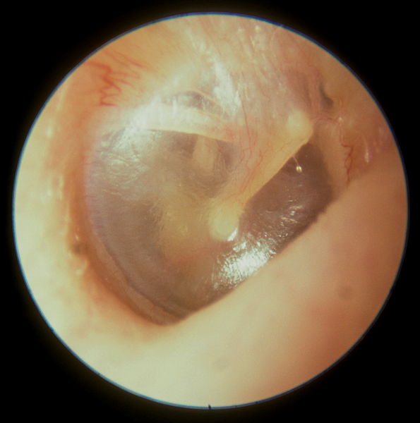
View of a normal tympanic membrane on an otoscopic exam:
Note the bony malleus attached to the tympanic membrane.

Image: “Normal tympanic membrane” by Michael Hawke, MD. License: CC BY 4.0

Tuning fork examination

  • 512 Hz tuning fork
  • Rinne test for CHL:
    • Procedure: The tuning fork is struck and placed behind the ear over the mastoid bone Bone Bone is a compact type of hardened connective tissue composed of bone cells, membranes, an extracellular mineralized matrix, and central bone marrow. The 2 primary types of bone are compact and spongy. Bones: Structure and Types; once no longer heard, the fork is moved 2.5 cm from the external ear.
    • Normal: air conduction > bone Bone Bone is a compact type of hardened connective tissue composed of bone cells, membranes, an extracellular mineralized matrix, and central bone marrow. The 2 primary types of bone are compact and spongy. Bones: Structure and Types conduction ( vibration Vibration A continuing periodic change in displacement with respect to a fixed reference. Neurological Examination is still heard near the external ear) 
    • CHL: bone Bone Bone is a compact type of hardened connective tissue composed of bone cells, membranes, an extracellular mineralized matrix, and central bone marrow. The 2 primary types of bone are compact and spongy. Bones: Structure and Types conduction > air conduction ( vibration Vibration A continuing periodic change in displacement with respect to a fixed reference. Neurological Examination is no longer heard once the tuning fork is moved in front of the ear canal)
  • Weber test:
    • The tuning fork is struck and placed on the top of the head.
    • Normal: Sound is heard equally in both ears.
    • CHL: Sound is louder in the affected ear.
    • SNHL: Sound is quieter in the affected ear.
Rinne and weber tests

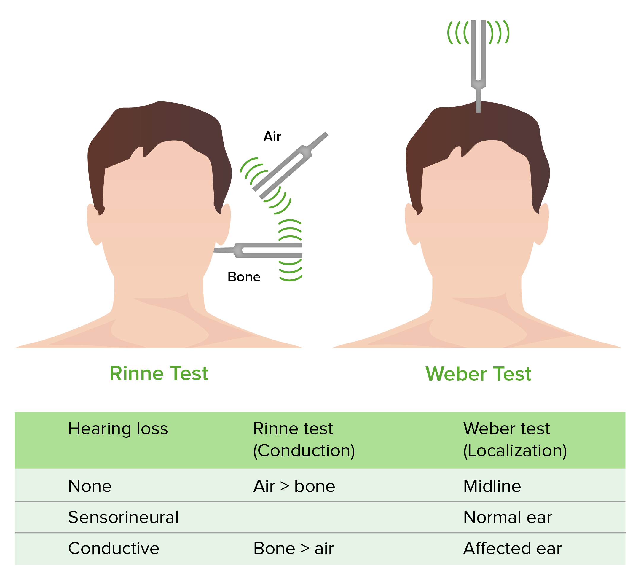
Weber and Rinne test

Image by Lecturio.

Diagnosis and Management

Diagnosis of hearing loss is based on physiologic and audiometric testing. Lab testing and imaging may also be needed to rule out another pathology.
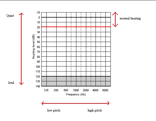
Physiologic testing

  • Auditory brainstem response (ABR) testing:
    • A “click” sound is used to evoke physiologic responses in the CN VIII and the auditory brainstem.
    • Recorded with surface electrodes Electrodes Electric conductors through which electric currents enter or leave a medium, whether it be an electrolytic solution, solid, molten mass, gas, or vacuum. Electrocardiogram (ECG)
    • Does not assess low frequency < 1500 Hz
  • Auditory steady-state response testing:
    • An auditory-evoked potential test (like ABR)
    • Tests several frequencies and gives specific information (unlike ABR)
  • Evoked otoacoustic emissions: assesses activity of the cochlear hair cells Hair cells Auditory sensory cells of organ of corti, usually placed in one row medially to the core of spongy bone (the modiolus). Inner hair cells are in fewer numbers than the outer auditory hair cells, and their stereocilia are approximately twice as thick as those of the outer hair cells. Auditory and Vestibular Pathways: Anatomy
  • Tympanometry: assesses mobility of the tympanic membrane Tympanic membrane An oval semitransparent membrane separating the external ear canal from the tympanic cavity. It contains three layers: the skin of the external ear canal; the core of radially and circularly arranged collagen fibers; and the mucosa of the middle ear. Ear: Anatomy and the middle ear Middle ear The space and structures directly internal to the tympanic membrane and external to the inner ear (labyrinth). Its major components include the auditory ossicles and the eustachian tube that connects the cavity of middle ear (tympanic cavity) to the upper part of the throat. Acute Otitis Media ossicles

Audiometry Audiometry The testing of the acuity of the sense of hearing to determine the thresholds of the loWest intensity levels at which an individual can hear a set of tones. The frequencies between 125 and 8000 hz are used to test air conduction thresholds and the frequencies between 250 and 4000 hz are used to test bone conduction thresholds. Ménière Disease

  • Behavioral testing for young children:
    • Indicated for children with delayed speech development
    • Behavioral observation audiometry Audiometry The testing of the acuity of the sense of hearing to determine the thresholds of the loWest intensity levels at which an individual can hear a set of tones. The frequencies between 125 and 8000 hz are used to test air conduction thresholds and the frequencies between 250 and 4000 hz are used to test bone conduction thresholds. Ménière Disease: birth to 6 months of age
    • Visual reinforcement audiometry Audiometry The testing of the acuity of the sense of hearing to determine the thresholds of the loWest intensity levels at which an individual can hear a set of tones. The frequencies between 125 and 8000 hz are used to test air conduction thresholds and the frequencies between 250 and 4000 hz are used to test bone conduction thresholds. Ménière Disease: 6 months to 2.5 years of age
  • Pure tone audiometry Audiometry The testing of the acuity of the sense of hearing to determine the thresholds of the loWest intensity levels at which an individual can hear a set of tones. The frequencies between 125 and 8000 hz are used to test air conduction thresholds and the frequencies between 250 and 4000 hz are used to test bone conduction thresholds. Ménière Disease:
    • Earphones are used to identify the threshold Threshold Minimum voltage necessary to generate an action potential (an all-or-none response) Skeletal Muscle Contraction heard for a defined set of frequencies.
    • Used in adults and children mature enough to cooperate
    • Indicated in any individual with unilateral or bilateral hearing loss 
    • Normal: within 15 dB of normal threshold Threshold Minimum voltage necessary to generate an action potential (an all-or-none response) Skeletal Muscle Contraction
    • High-frequency sounds are lost 1st with aging.
  • Hearing loss: Normal audible range is 0–180 dB (> 85 dB is damaging):
    • Severity:
      • Mild: 26–40 dB
      • Moderate: 41–55 dB
      • Moderately severe: 56–70 dB
      • Severe: 71–90 dB
      • Profound: 90 dB
    • Frequencies: Normal hearing is 20–20,000 Hz:
      • Low-frequency hearing loss: sounds < 500 Hz
      • Middle-frequency hearing loss: sounds 501–2,000 Hz
      • High-frequency hearing loss: sounds > 2,000 Hz 
    • Percentage of hearing impairment:
      • Determined by the pure tone average in dB at 500 Hz, 1,000 Hz, 2,000 Hz, and 3,000 Hz frequencies
      • 30% impairment = 70% residual hearing at 45 dB
      • 60% impairment = 40% residual hearing at 65 dB
      • 80% impairment = 20% residual hearing at 78 dB
      • 100% impairment = no residual hearing at 91 dB
  • Functional impairment:
    • May be misleading if based on pure tone average alone:
      • 30% impairment = 70% residual hearing at 45 dB
      • Conversational speech is approximately 50–60 dB → significant impact on the ability to function
    • A different rating scale Scale Dermatologic Examination is used for young children → mild impairments can cause significant difficulty with language development
Audiogram

Audiogram chart

Image: “Audiogram” by Audiology6. License: Public Domain

Additional testing

  • Lab tests:
    • Blood glucose Glucose A primary source of energy for living organisms. It is naturally occurring and is found in fruits and other parts of plants in its free state. It is used therapeutically in fluid and nutrient replacement. Lactose Intolerance: Diabetic vasculopathy can cause cochlear ischemia Ischemia A hypoperfusion of the blood through an organ or tissue caused by a pathologic constriction or obstruction of its blood vessels, or an absence of blood circulation. Ischemic Cell Damage.
    • CBC: check for anemia Anemia Anemia is a condition in which individuals have low Hb levels, which can arise from various causes. Anemia is accompanied by a reduced number of RBCs and may manifest with fatigue, shortness of breath, pallor, and weakness. Subtypes are classified by the size of RBCs, chronicity, and etiology. Anemia: Overview and Types and WBC count
    • Thyroid function tests Thyroid Function Tests Blood tests used to evaluate the functioning of the thyroid gland. Ion Channel Myopathy
    • Serologic test for syphilis Syphilis Syphilis is a bacterial infection caused by the spirochete Treponema pallidum pallidum (T. p. pallidum), which is usually spread through sexual contact. Syphilis has 4 clinical stages: primary, secondary, latent, and tertiary. Syphilis 
    • Tests for autoimmune disease (if suspected):
  • Imaging:
    • MRI: recommended in cases of asymmetric or unilateral SNHL
    • CT scan of the temporal bone Temporal bone Either of a pair of compound bones forming the lateral (left and right) surfaces and base of the skull which contains the organs of hearing. It is a large bone formed by the fusion of parts: the squamous (the flattened anterior-superior part), the tympanic (the curved anterior-inferior part), the mastoid (the irregular posterior portion), and the petrous (the part at the base of the skull). Jaw and Temporomandibular Joint: Anatomy:
      • Identifies abnormal bone Bone Bone is a compact type of hardened connective tissue composed of bone cells, membranes, an extracellular mineralized matrix, and central bone marrow. The 2 primary types of bone are compact and spongy. Bones: Structure and Types findings
      • Localizes effusions and abnormal soft tissue Soft Tissue Soft Tissue Abscess

Management

  • Medical management:
    • Give antibiotics if an infection is present.
    • Evacuation of obstruction: cerumen Cerumen The yellow or brown waxy secretions produced by vestigial apocrine sweat glands in the external ear canal. Otitis Externa or other foreign body Foreign Body Foreign Body Aspiration
    • Treatment of underlying conditions:
      • Autoimmune disease
      • Cerebrovascular disease
      • Meningitis Meningitis Meningitis is inflammation of the meninges, the protective membranes of the brain, and spinal cord. The causes of meningitis are varied, with the most common being bacterial or viral infection. The classic presentation of meningitis is a triad of fever, altered mental status, and nuchal rigidity. Meningitis
      • Multiple sclerosis Sclerosis A pathological process consisting of hardening or fibrosis of an anatomical structure, often a vessel or a nerve. Wilms Tumor
      • Syphilis Syphilis Syphilis is a bacterial infection caused by the spirochete Treponema pallidum pallidum (T. p. pallidum), which is usually spread through sexual contact. Syphilis has 4 clinical stages: primary, secondary, latent, and tertiary. Syphilis
  • Otolaryngology specialty referral:
    • Cochlear implant (surgical procedure):
      • Indicated for SNHL
      • > 6 months of age
    • Otologic emergency:
      • Sudden SNHL
      • Trauma
    • Surgical management depends on the etiology:
      • Repair of a perforated tympanic membrane Tympanic membrane An oval semitransparent membrane separating the external ear canal from the tympanic cavity. It contains three layers: the skin of the external ear canal; the core of radially and circularly arranged collagen fibers; and the mucosa of the middle ear. Ear: Anatomy
      • Stapedectomy for otosclerosis Otosclerosis Formation of spongy bone in the labyrinth capsule which can progress toward the stapes (stapedial fixation) or anteriorly toward the cochlea leading to conductive, sensorineural, or mixed hearing loss. Several genes are associated with familial otosclerosis with varied clinical signs. Vertigo 
      • Ossicular chain reconstruction 
      • Tumor Tumor Inflammation (e.g., acoustic neuroma Acoustic neuroma Acoustic neuroma, also referred to as vestibular schwannoma, is a benign tumor arising from Schwann cells of the vestibular component of the cranial nerve VIII. Acoustic neuroma forms within the internal auditory meatus and extends into the cerebellopontine angle. Acoustic Neuroma)

Clinical Relevance

Common conditions causing hearing loss: 

  • Otitis media: infection in the middle ear Middle ear The space and structures directly internal to the tympanic membrane and external to the inner ear (labyrinth). Its major components include the auditory ossicles and the eustachian tube that connects the cavity of middle ear (tympanic cavity) to the upper part of the throat. Acute Otitis Media characterized by mucosal inflammation Inflammation Inflammation is a complex set of responses to infection and injury involving leukocytes as the principal cellular mediators in the body’s defense against pathogenic organisms. Inflammation is also seen as a response to tissue injury in the process of wound healing. The 5 cardinal signs of inflammation are pain, heat, redness, swelling, and loss of function. Inflammation and retention of fluid. The most common pathogens are S. pneumoniae, H. influenzae H. influenzae A species of Haemophilus found on the mucous membranes of humans and a variety of animals. The species is further divided into biotypes I through VIII. Haemophilus, and Moraxella catarrhalis Moraxella catarrhalis Gram-negative aerobic cocci of low virulence that colonize the nasopharynx and occasionally cause meningitis; bacteremia; empyema; pericarditis; and pneumonia. Moraxella. Otitis media can present with fever Fever Fever is defined as a measured body temperature of at least 38°C (100.4°F). Fever is caused by circulating endogenous and/or exogenous pyrogens that increase levels of prostaglandin E2 in the hypothalamus. Fever is commonly associated with chills, rigors, sweating, and flushing of the skin. Fever, otalgia Otalgia Acute Otitis Media, and hearing loss. Diagnosis is made by history and physical exam. Management is with oral antibiotics. Procedures may be needed for recurring infections Infections Invasion of the host organism by microorganisms or their toxins or by parasites that can cause pathological conditions or diseases. Chronic Granulomatous Disease
  • Otitis externa Otitis externa Otitis externa (also known as external otitis or swimmer’s ear) is an infection of the external auditory canal that is most often caused by acute bacterial infection and is frequently associated with hot, humid weather and water exposure. Patients commonly present with ear pain, pruritus, discharge, and hearing loss. Otitis Externa (swimmers ear): infection of the external auditory canal External Auditory Canal Otitis Externa. Otitis external is most often caused by acute bacterial infection and is frequently associated with hot, humid weather and water exposure. Individuals commonly present with ear pain Ear Pain Acute Otitis Media, pruritus Pruritus An intense itching sensation that produces the urge to rub or scratch the skin to obtain relief. Atopic Dermatitis (Eczema), discharge, and hearing loss. Diagnosis is made by history and physical exam. Management is with topical antibiotics, pain Pain An unpleasant sensation induced by noxious stimuli which are detected by nerve endings of nociceptive neurons. Pain: Types and Pathways relief, and protection of the ear.
  • Meningitis Meningitis Meningitis is inflammation of the meninges, the protective membranes of the brain, and spinal cord. The causes of meningitis are varied, with the most common being bacterial or viral infection. The classic presentation of meningitis is a triad of fever, altered mental status, and nuchal rigidity. Meningitis: inflammation Inflammation Inflammation is a complex set of responses to infection and injury involving leukocytes as the principal cellular mediators in the body’s defense against pathogenic organisms. Inflammation is also seen as a response to tissue injury in the process of wound healing. The 5 cardinal signs of inflammation are pain, heat, redness, swelling, and loss of function. Inflammation of the meninges Meninges The brain and the spinal cord are enveloped by 3 overlapping layers of connective tissue called the meninges. The layers are, from the most external layer to the most internal layer, the dura mater, arachnoid mater, and pia mater. Between these layers are 3 potential spaces called the epidural, subdural, and subarachnoid spaces. Meninges: Anatomy (the protective membranes of the brain Brain The part of central nervous system that is contained within the skull (cranium). Arising from the neural tube, the embryonic brain is comprised of three major parts including prosencephalon (the forebrain); mesencephalon (the midbrain); and rhombencephalon (the hindbrain). The developed brain consists of cerebrum; cerebellum; and other structures in the brain stem. Nervous System: Anatomy, Structure, and Classification and spinal cord Spinal cord The spinal cord is the major conduction pathway connecting the brain to the body; it is part of the CNS. In cross section, the spinal cord is divided into an H-shaped area of gray matter (consisting of synapsing neuronal cell bodies) and a surrounding area of white matter (consisting of ascending and descending tracts of myelinated axons). Spinal Cord: Anatomy). The classic presentation of meningitis Meningitis Meningitis is inflammation of the meninges, the protective membranes of the brain, and spinal cord. The causes of meningitis are varied, with the most common being bacterial or viral infection. The classic presentation of meningitis is a triad of fever, altered mental status, and nuchal rigidity. Meningitis is a triad of fever Fever Fever is defined as a measured body temperature of at least 38°C (100.4°F). Fever is caused by circulating endogenous and/or exogenous pyrogens that increase levels of prostaglandin E2 in the hypothalamus. Fever is commonly associated with chills, rigors, sweating, and flushing of the skin. Fever, altered mental status Altered Mental Status Sepsis in Children, and nuchal rigidity Nuchal Rigidity Meningitis. Meningitis Meningitis Meningitis is inflammation of the meninges, the protective membranes of the brain, and spinal cord. The causes of meningitis are varied, with the most common being bacterial or viral infection. The classic presentation of meningitis is a triad of fever, altered mental status, and nuchal rigidity. Meningitis can cause SNHL due to the spread of infection to the cochlea Cochlea The part of the inner ear (labyrinth) that is concerned with hearing. It forms the anterior part of the labyrinth, as a snail-like structure that is situated almost horizontally anterior to the vestibular labyrinth. Ear: Anatomy. Diagnosis of meningitis Meningitis Meningitis is inflammation of the meninges, the protective membranes of the brain, and spinal cord. The causes of meningitis are varied, with the most common being bacterial or viral infection. The classic presentation of meningitis is a triad of fever, altered mental status, and nuchal rigidity. Meningitis is made clinically with full neurologic examination and CSF analysis CSF analysis Meningitis. Management includes immediate broad-spectrum Broad-Spectrum Fluoroquinolones antibiotics and supportive care. 
  • Multiple sclerosis Sclerosis A pathological process consisting of hardening or fibrosis of an anatomical structure, often a vessel or a nerve. Wilms Tumor: a chronic inflammatory autoimmune disease leading to demyelination Demyelination Multiple Sclerosis of the CNS. The clinical presentation varies widely depending on the site of the lesions. Hearing may be compromised with significant involvement of the CN VIII. The diagnosis is made by clinical and CSF exams and MRI imaging. Management involves corticosteroids Corticosteroids Chorioretinitis for acute exacerbations and disease-modifying agents to slow the progression of the disease.
  • Syphilis Syphilis Syphilis is a bacterial infection caused by the spirochete Treponema pallidum pallidum (T. p. pallidum), which is usually spread through sexual contact. Syphilis has 4 clinical stages: primary, secondary, latent, and tertiary. Syphilis: a bacterial infection caused by Treponema pallidum Treponema pallidum The causative agent of venereal and non-venereal syphilis as well as yaws. Treponema, a spirochete Spirochete Treponema is a gram-negative, microaerophilic spirochete. Owing to its very thin structure, it is not easily seen on Gram stain, but can be visualized using dark-field microscopy. This spirochete contains endoflagella, which allow for a characteristic corkscrew movement. Treponema, typically spread through sexual contact. Varying clinical presentations may range from a painless genital ulcer ( primary syphilis Primary Syphilis Syphilis), to a rash Rash Rocky Mountain Spotted Fever ( secondary syphilis Secondary Syphilis Syphilis), to severe neurological deterioration ( tertiary syphilis Tertiary Syphilis Syphilis). Individuals can experience hearing loss and vertigo Vertigo Vertigo is defined as the perceived sensation of rotational motion while remaining still. A very common complaint in primary care and the ER, vertigo is more frequently experienced by women and its prevalence increases with age. Vertigo is classified into peripheral or central based on its etiology. Vertigo due to ear involvement in secondary syphilis Secondary Syphilis Syphilis. Diagnosis is through lab testing. Treatment is with penicillin Penicillin Rheumatic Fever and the duration depends on the stage of the disease. 
  • Meniere disease: a disorder of the inner ear Inner ear The essential part of the hearing organ consists of two labyrinthine compartments: the bony labyrinthine and the membranous labyrinth. Ear: Anatomy characterized by hearing loss, fluctuating aural symptoms (e.g., tinnitus Tinnitus A nonspecific symptom of hearing disorder characterized by the sensation of buzzing, ringing, clicking, pulsations, and other noises in the ear. Objective tinnitus refers to noises generated from within the ear or adjacent structures that can be heard by other individuals. The term subjective tinnitus is used when the sound is audible only to the affected individual. Tinnitus may occur as a manifestation of cochlear diseases; vestibulocochlear nerve diseases; intracranial hypertension; craniocerebral trauma; and other conditions. Cranial Nerve Palsies), and spontaneous episodes of vertigo Vertigo Vertigo is defined as the perceived sensation of rotational motion while remaining still. A very common complaint in primary care and the ER, vertigo is more frequently experienced by women and its prevalence increases with age. Vertigo is classified into peripheral or central based on its etiology. Vertigo. Diagnosis is made with a thorough history, physical exam, and full otologic exam. An audiogram is a key component of the evaluation. Management includes diet and lifestyle modification, vasodilators Vasodilators Drugs used to cause dilation of the blood vessels. Thromboangiitis Obliterans (Buerger’s Disease), diuretics Diuretics Agents that promote the excretion of urine through their effects on kidney function. Heart Failure and Angina Medication, antihistamines Antihistamines Antihistamines are drugs that target histamine receptors, particularly H1 and H2 receptors. H1 antagonists are competitive and reversible inhibitors of H1 receptors. First-generation antihistamines cross the blood-brain barrier and can cause sedation. Antihistamines, benzodiazepines Benzodiazepines Benzodiazepines work on the gamma-aminobutyric acid type A (GABAA) receptor to produce inhibitory effects on the CNS. Benzodiazepines do not mimic GABA, the main inhibitory neurotransmitter in humans, but instead potentiate GABA activity. Benzodiazepines, antiemetics Antiemetics Antiemetics are medications used to treat and/or prevent nausea and vomiting. These drugs act on different target receptors. The main classes include benzodiazepines, corticosteroids, atypical antipsychotics, cannabinoids, and antagonists of the following receptors: serotonin, dopamine, and muscarinic and neurokinin receptors. Antiemetics, glucocorticoids Glucocorticoids Glucocorticoids are a class within the corticosteroid family. Glucocorticoids are chemically and functionally similar to endogenous cortisol. There are a wide array of indications, which primarily benefit from the antiinflammatory and immunosuppressive effects of this class of drugs. Glucocorticoids, surgical intervention, and/or hearing aids AIDS Chronic HIV infection and depletion of CD4 cells eventually results in acquired immunodeficiency syndrome (AIDS), which can be diagnosed by the presence of certain opportunistic diseases called AIDS-defining conditions. These conditions include a wide spectrum of bacterial, viral, fungal, and parasitic infections as well as several malignancies and generalized conditions. HIV Infection and AIDS.

References

  1. Shapiro, S.B., Noij, K.S., Naples, J.G., & Samy, R.N. (2021). Hearing Loss and Tinnitus. Medical Clinics of North America, 105(5), 799–811. https://doi.org/http://dx.doi.org/10.1016/j.mcna.2021.05.003 
  2. Molina, F.J. (2012). Chapter 18. Hearing Loss. In M. C. Henderson, L. M. Tierney, & G. W. Smetana (Eds.), The Patient History: An Evidence-Based Approach to Differential Diagnosis, 2nd ed. The McGraw-Hill Companies. http://accessmedicine.mhmedical.com/content.aspx?aid=56852049 
  3. Berkowitz, A.L. (2016). The auditory and vestibular pathways and approach to hearing loss and dizziness/vertigo: Cranial nerve 8. In Clinical Neurology and Neuroanatomy: A Localization-Based Approach. McGraw-Hill Education. http://accessmedicine.mhmedical.com/content.aspx?aid=1160204039 
  4. Krogmann, R.J., & Al Khalili, Y. (2021). Cochlear Implants. StatPearls. Retrieved September 23, 2021, from https://www.ncbi.nlm.nih.gov/books/NBK544280/
  5. Weber, P.C. (2020). Etiology of hearing loss. UpToDate. Retrieved September 23, 2021, from https://www.uptodate.com/contents/etiology-of-hearing-loss-in-adults
  6. Weber, P.C. (2021). Evaluation of hearing loss in adults. UpToDate. Retrieved September 23, 2021, from https://www.uptodate.com/contents/evaluation-of-hearing-loss-in-adults
  7. Smith, R.J.H. & Gooi, A. (2021). Hearing loss in children: Etiology. UpToDate. Retrieved September 23, 2021, from https://www.uptodate.com/contents/hearing-loss-in-children-etiology

Create your free account or log in to continue reading!

Sign up now and get free access to Lecturio with concept pages, medical videos, and questions for your medical education.

User Reviews

Details