Achieve Mastery of Medical Concepts

Study for medical school and boards with Lecturio

Alport Syndrome

Alport syndrome, also called hereditary nephritis, is a genetic disorder caused by a mutation Mutation Genetic mutations are errors in DNA that can cause protein misfolding and dysfunction. There are various types of mutations, including chromosomal, point, frameshift, and expansion mutations. Types of Mutations in the genes Genes A category of nucleic acid sequences that function as units of heredity and which code for the basic instructions for the development, reproduction, and maintenance of organisms. DNA Types and Structure encoding for the alpha chains of type IV collagen Collagen A polypeptide substance comprising about one third of the total protein in mammalian organisms. It is the main constituent of skin; connective tissue; and the organic substance of bones (bone and bones) and teeth (tooth). Connective Tissue: Histology, resulting in the production of abnormal type IV collagen Collagen A polypeptide substance comprising about one third of the total protein in mammalian organisms. It is the main constituent of skin; connective tissue; and the organic substance of bones (bone and bones) and teeth (tooth). Connective Tissue: Histology strands. Patients Patients Individuals participating in the health care system for the purpose of receiving therapeutic, diagnostic, or preventive procedures. Clinician–Patient Relationship present with glomerulonephritis, hypertension Hypertension Hypertension, or high blood pressure, is a common disease that manifests as elevated systemic arterial pressures. Hypertension is most often asymptomatic and is found incidentally as part of a routine physical examination or during triage for an unrelated medical encounter. Hypertension, edema Edema Edema is a condition in which excess serous fluid accumulates in the body cavity or interstitial space of connective tissues. Edema is a symptom observed in several medical conditions. It can be categorized into 2 types, namely, peripheral (in the extremities) and internal (in an organ or body cavity). Edema, hematuria Hematuria Presence of blood in the urine. Renal Cell Carcinoma, and proteinuria Proteinuria The presence of proteins in the urine, an indicator of kidney diseases. Nephrotic Syndrome in Children, as well as with ocular ( cataract Cataract Partial or complete opacity on or in the lens or capsule of one or both eyes, impairing vision or causing blindness. The many kinds of cataract are classified by their morphology (size, shape, location) or etiology (cause and time of occurrence). Neurofibromatosis Type 2, retinopathy) and auditory ( sensorineural hearing loss Sensorineural hearing loss Hearing loss resulting from damage to the cochlea and the sensorineural elements which lie internally beyond the oval and round windows. These elements include the auditory nerve and its connections in the brainstem. Hearing Loss) findings. Diagnosis is established with urinalysis Urinalysis Examination of urine by chemical, physical, or microscopic means. Routine urinalysis usually includes performing chemical screening tests, determining specific gravity, observing any unusual color or odor, screening for bacteriuria, and examining the sediment microscopically. Urinary Tract Infections (UTIs) in Children, urine microscopy, and a renal function panel. A renal biopsy Renal Biopsy Antineutrophil Cytoplasmic Antibody (ANCA)-Associated Vasculitis showing characteristic glomerular basement membrane Basement membrane A darkly stained mat-like extracellular matrix (ecm) that separates cell layers, such as epithelium from endothelium or a layer of connective tissue. The ecm layer that supports an overlying epithelium or endothelium is called basal lamina. Basement membrane (bm) can be formed by the fusion of either two adjacent basal laminae or a basal lamina with an adjacent reticular lamina of connective tissue. Bm, composed mainly of type IV collagen; glycoprotein laminin; and proteoglycan, provides barriers as well as channels between interacting cell layers. Thin Basement Membrane Nephropathy (TBMN) splitting Splitting Defense Mechanisms may be used to confirm the diagnosis. Treatment for Alport syndrome is focused on limiting disease progression with angiotensin receptor Receptor Receptors are proteins located either on the surface of or within a cell that can bind to signaling molecules known as ligands (e.g., hormones) and cause some type of response within the cell. Receptors blockers and angiotensin-converting enzyme inhibitors Angiotensin-converting enzyme inhibitors A class of drugs whose main indications are the treatment of hypertension and heart failure. They exert their hemodynamic effect mainly by inhibiting the renin-angiotensin system. They also modulate sympathetic nervous system activity and increase prostaglandin synthesis. They cause mainly vasodilation and mild natriuresis without affecting heart rate and contractility. Renin-Angiotensin-Aldosterone System Inhibitors. Hearing aids AIDS Chronic HIV infection and depletion of CD4 cells eventually results in acquired immunodeficiency syndrome (AIDS), which can be diagnosed by the presence of certain opportunistic diseases called AIDS-defining conditions. These conditions include a wide spectrum of bacterial, viral, fungal, and parasitic infections as well as several malignancies and generalized conditions. HIV Infection and AIDS are used for hearing loss Hearing loss Hearing loss, also known as hearing impairment, is any degree of impairment in the ability to apprehend sound as determined by audiometry to be below normal hearing thresholds. Clinical presentation may occur at birth or as a gradual loss of hearing with age, including a short-term or sudden loss at any point. Hearing Loss associated with Alport syndrome.

Last updated: Aug 18, 2022

Editorial responsibility: Stanley Oiseth, Lindsay Jones, Evelin Maza

Epidemiology and Etiology

Epidemiology

  • Most common type of hereditary nephritis
  • Affects 1 in 50,000 newborns
  • Men more commonly symptomatic than women ( X-linked X-linked Genetic diseases that are linked to gene mutations on the X chromosome in humans or the X chromosome in other species. Included here are animal models of human X-linked diseases. Common Variable Immunodeficiency (CVID) inheritance)
  • 80% of males affected with X-linked X-linked Genetic diseases that are linked to gene mutations on the X chromosome in humans or the X chromosome in other species. Included here are animal models of human X-linked diseases. Common Variable Immunodeficiency (CVID) Alport syndrome will develop some degree of hearing loss Hearing loss Hearing loss, also known as hearing impairment, is any degree of impairment in the ability to apprehend sound as determined by audiometry to be below normal hearing thresholds. Clinical presentation may occur at birth or as a gradual loss of hearing with age, including a short-term or sudden loss at any point. Hearing Loss by adolescence.

Etiology

  • Caused by mutation Mutation Genetic mutations are errors in DNA that can cause protein misfolding and dysfunction. There are various types of mutations, including chromosomal, point, frameshift, and expansion mutations. Types of Mutations in:  
    • COL4A3 
    • COL4A4 
    • COL4A5
  • X-linked X-linked Genetic diseases that are linked to gene mutations on the X chromosome in humans or the X chromosome in other species. Included here are animal models of human X-linked diseases. Common Variable Immunodeficiency (CVID) in 80% of cases
  • May be autosomal recessive Autosomal recessive Autosomal inheritance, both dominant and recessive, refers to the transmission of genes from the 22 autosomal chromosomes. Autosomal recessive diseases are only expressed when 2 copies of the recessive allele are inherited. Autosomal Recessive and Autosomal Dominant Inheritance or autosomal dominant Autosomal dominant Autosomal inheritance, both dominant and recessive, refers to the transmission of genes from the 22 autosomal chromosomes. Autosomal dominant diseases are expressed when only 1 copy of the dominant allele is inherited. Autosomal Recessive and Autosomal Dominant Inheritance:
    • Autosomal recessive Autosomal recessive Autosomal inheritance, both dominant and recessive, refers to the transmission of genes from the 22 autosomal chromosomes. Autosomal recessive diseases are only expressed when 2 copies of the recessive allele are inherited. Autosomal Recessive and Autosomal Dominant Inheritance: a mutation Mutation Genetic mutations are errors in DNA that can cause protein misfolding and dysfunction. There are various types of mutations, including chromosomal, point, frameshift, and expansion mutations. Types of Mutations in both COL4A3 and COL4A4
    • Autosomal dominant Autosomal dominant Autosomal inheritance, both dominant and recessive, refers to the transmission of genes from the 22 autosomal chromosomes. Autosomal dominant diseases are expressed when only 1 copy of the dominant allele is inherited. Autosomal Recessive and Autosomal Dominant Inheritance
      • Heterozygous mutation Mutation Genetic mutations are errors in DNA that can cause protein misfolding and dysfunction. There are various types of mutations, including chromosomal, point, frameshift, and expansion mutations. Types of Mutations
      • Compared to X-linked X-linked Genetic diseases that are linked to gene mutations on the X chromosome in humans or the X chromosome in other species. Included here are animal models of human X-linked diseases. Common Variable Immunodeficiency (CVID) inheritance, slower progression to end-stage renal disease (ESRD) 
      • Compared to X-linked X-linked Genetic diseases that are linked to gene mutations on the X chromosome in humans or the X chromosome in other species. Included here are animal models of human X-linked diseases. Common Variable Immunodeficiency (CVID) inheritance, less likely to have extrarenal manifestations

Related videos

Pathophysiology

Alport syndrome is a genetic disorder arising from a mutation Mutation Genetic mutations are errors in DNA that can cause protein misfolding and dysfunction. There are various types of mutations, including chromosomal, point, frameshift, and expansion mutations. Types of Mutations in genes Genes A category of nucleic acid sequences that function as units of heredity and which code for the basic instructions for the development, reproduction, and maintenance of organisms. DNA Types and Structure encoding for the alpha chains of type IV collagen Collagen A polypeptide substance comprising about one third of the total protein in mammalian organisms. It is the main constituent of skin; connective tissue; and the organic substance of bones (bone and bones) and teeth (tooth). Connective Tissue: Histology, which is primarily located in the lens Lens A transparent, biconvex structure of the eye, enclosed in a capsule and situated behind the iris and in front of the vitreous humor (vitreous body). It is slightly overlapped at its margin by the ciliary processes. Adaptation by the ciliary body is crucial for ocular accommodation. Eye: Anatomy, cochlea Cochlea The part of the inner ear (labyrinth) that is concerned with hearing. It forms the anterior part of the labyrinth, as a snail-like structure that is situated almost horizontally anterior to the vestibular labyrinth. Ear: Anatomy, and kidneys Kidneys The kidneys are a pair of bean-shaped organs located retroperitoneally against the posterior wall of the abdomen on either side of the spine. As part of the urinary tract, the kidneys are responsible for blood filtration and excretion of water-soluble waste in the urine. Kidneys: Anatomy (glomerulus).

  • Alport syndrome = production of impaired strands + deposition of type IV collagen Collagen A polypeptide substance comprising about one third of the total protein in mammalian organisms. It is the main constituent of skin; connective tissue; and the organic substance of bones (bone and bones) and teeth (tooth). Connective Tissue: Histology in basement membrane Basement membrane A darkly stained mat-like extracellular matrix (ecm) that separates cell layers, such as epithelium from endothelium or a layer of connective tissue. The ecm layer that supports an overlying epithelium or endothelium is called basal lamina. Basement membrane (bm) can be formed by the fusion of either two adjacent basal laminae or a basal lamina with an adjacent reticular lamina of connective tissue. Bm, composed mainly of type IV collagen; glycoprotein laminin; and proteoglycan, provides barriers as well as channels between interacting cell layers. Thin Basement Membrane Nephropathy (TBMN) 
  • Glomerular basement membrane Basement membrane A darkly stained mat-like extracellular matrix (ecm) that separates cell layers, such as epithelium from endothelium or a layer of connective tissue. The ecm layer that supports an overlying epithelium or endothelium is called basal lamina. Basement membrane (bm) can be formed by the fusion of either two adjacent basal laminae or a basal lamina with an adjacent reticular lamina of connective tissue. Bm, composed mainly of type IV collagen; glycoprotein laminin; and proteoglycan, provides barriers as well as channels between interacting cell layers. Thin Basement Membrane Nephropathy (TBMN) (GBM) in Alport syndrome is prone to proteolytic injury → adhesion Adhesion The process whereby platelets adhere to something other than platelets, e.g., collagen; basement membrane; microfibrils; or other ‘foreign’ surfaces. Coagulation Studies kinase activation in podocytes and endothelin receptors Receptors Receptors are proteins located either on the surface of or within a cell that can bind to signaling molecules known as ligands (e.g., hormones) and cause some type of response within the cell. Receptors
  • Pathologic results of kidney biopsy Biopsy Removal and pathologic examination of specimens from the living body. Ewing Sarcoma:
    • Light microscopy:
      • Normal on early light microscopy
      • With progression, nonspecific findings: focal and segmental glomerulosclerosis, interstitial fibrosis Fibrosis Any pathological condition where fibrous connective tissue invades any organ, usually as a consequence of inflammation or other injury. Bronchiolitis Obliterans, tubular atrophy Atrophy Decrease in the size of a cell, tissue, organ, or multiple organs, associated with a variety of pathological conditions such as abnormal cellular changes, ischemia, malnutrition, or hormonal changes. Cellular Adaptation, and presence of lymphocytes Lymphocytes Lymphocytes are heterogeneous WBCs involved in immune response. Lymphocytes develop from the bone marrow, starting from hematopoietic stem cells (HSCs) and progressing to common lymphoid progenitors (CLPs). B and T lymphocytes and natural killer (NK) cells arise from the lineage. Lymphocytes: Histology and plasma Plasma The residual portion of blood that is left after removal of blood cells by centrifugation without prior blood coagulation. Transfusion Products cells
    • Immunofluorescence: negative
    • Electron microscopy: 
Alport syndrome kidney biopsy

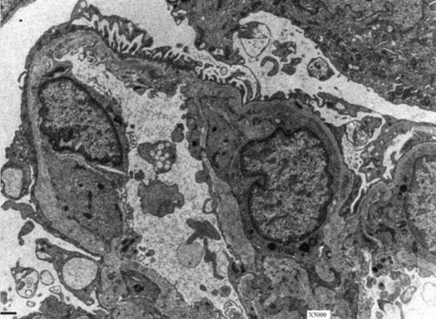
Glomerular basement membrane (GBM) in a patient with Alport syndrome:
Electron microscopy of a kidney shows the characteristic basket-weave appearance of the GBM in a patient with Alport syndrome.

Image: “Electron micrograph from kidney biopsy” by University of North Carolina School of Medicine, Chapel Hill, NC. License: CC BY 2.5

Clinical Presentation

Alport syndrome most often presents with renal, ocular, and auditory findings.

Renal presentation (glomerulonephritis)

  • Symptoms:
    • Gross hematuria Hematuria Presence of blood in the urine. Renal Cell Carcinoma following infection (commonly upper respiratory infections Infections Invasion of the host organism by microorganisms or their toxins or by parasites that can cause pathological conditions or diseases. Chronic Granulomatous Disease)
    • Edema Edema Edema is a condition in which excess serous fluid accumulates in the body cavity or interstitial space of connective tissues. Edema is a symptom observed in several medical conditions. It can be categorized into 2 types, namely, peripheral (in the extremities) and internal (in an organ or body cavity). Edema
    • Hypertension Hypertension Hypertension, or high blood pressure, is a common disease that manifests as elevated systemic arterial pressures. Hypertension is most often asymptomatic and is found incidentally as part of a routine physical examination or during triage for an unrelated medical encounter. Hypertension
  • Lab findings:
    • Proteinuria Proteinuria The presence of proteins in the urine, an indicator of kidney diseases. Nephrotic Syndrome in Children
    • Progressive decline in renal function → eventual ESRD (usually presents at 16–35 years of age)

Ocular findings

  • Cone-shaped lens Lens A transparent, biconvex structure of the eye, enclosed in a capsule and situated behind the iris and in front of the vitreous humor (vitreous body). It is slightly overlapped at its margin by the ciliary processes. Adaptation by the ciliary body is crucial for ocular accommodation. Eye: Anatomy (anterior lenticonus) leads to:
    • Decreased visual acuity Visual Acuity Clarity or sharpness of ocular vision or the ability of the eye to see fine details. Visual acuity depends on the functions of retina, neuronal transmission, and the interpretative ability of the brain. Normal visual acuity is expressed as 20/20 indicating that one can see at 20 feet what should normally be seen at that distance. Visual acuity can also be influenced by brightness, color, and contrast. Ophthalmic Exam 
    • Abnormal refraction Refraction Refractive Errors
  • Subcapsular cataract Cataract Partial or complete opacity on or in the lens or capsule of one or both eyes, impairing vision or causing blindness. The many kinds of cataract are classified by their morphology (size, shape, location) or etiology (cause and time of occurrence). Neurofibromatosis Type 2
  • Retinopathy: pigmentary retinal changes, with yellow or white flecks (e.g., dot-and-fleck retinopathy)
  • Corneal erosion Erosion Partial-thickness loss of the epidermis Generalized and Localized Rashes

Auditory findings

Sensorineural hearing loss Sensorineural hearing loss Hearing loss resulting from damage to the cochlea and the sensorineural elements which lie internally beyond the oval and round windows. These elements include the auditory nerve and its connections in the brainstem. Hearing Loss:

  • Begins with loss of high-frequency sounds
  • Apparent late in childhood or early puberty Puberty Puberty is a complex series of physical, psychosocial, and cognitive transitions usually experienced by adolescents (11-19 years of age). Puberty is marked by a growth in stature and the development of secondary sexual characteristics, achievement of fertility, and changes in most body systems. Puberty, prior to onset of ESRD
  • Secondary to type IV collagen Collagen A polypeptide substance comprising about one third of the total protein in mammalian organisms. It is the main constituent of skin; connective tissue; and the organic substance of bones (bone and bones) and teeth (tooth). Connective Tissue: Histology abnormalities in inner ear Inner ear The essential part of the hearing organ consists of two labyrinthine compartments: the bony labyrinthine and the membranous labyrinth. Ear: Anatomy
Anterior lenticonus in the eye of an individual with alport syndrome

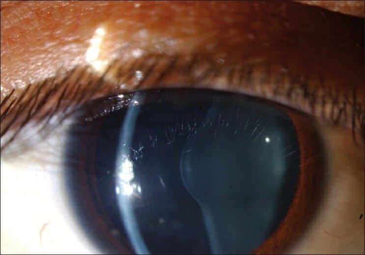
Anterior lenticonus in the eye of an individual with Alport syndrome

Image: “Figure 1 Anterior lenticonus in the right eye in a patient with Alport syndrome” by Ammar M Al-Mahmood, Samar A Al-Swailem, Abdulrahman Al-Khalaf, Ghada Y Al-Binali. License: CC BY 2.0

Diagnosis

A complete history, including family history Family History Adult Health Maintenance, and physical exam are required to make a diagnosis. 

  • Urinalysis Urinalysis Examination of urine by chemical, physical, or microscopic means. Routine urinalysis usually includes performing chemical screening tests, determining specific gravity, observing any unusual color or odor, screening for bacteriuria, and examining the sediment microscopically. Urinary Tract Infections (UTIs) in Children:
  • Urine microscopy:
  • Renal function panel: to assess presence and degree of renal failure Renal failure Conditions in which the kidneys perform below the normal level in the ability to remove wastes, concentrate urine, and maintain electrolyte balance; blood pressure; and calcium metabolism. Renal insufficiency can be classified by the degree of kidney damage (as measured by the level of proteinuria) and reduction in glomerular filtration rate. Crush Syndrome 
  • Renal biopsy Renal Biopsy Antineutrophil Cytoplasmic Antibody (ANCA)-Associated Vasculitis:
    • Indicated in patients Patients Individuals participating in the health care system for the purpose of receiving therapeutic, diagnostic, or preventive procedures. Clinician–Patient Relationship with:
      • Abnormal urinalysis Urinalysis Examination of urine by chemical, physical, or microscopic means. Routine urinalysis usually includes performing chemical screening tests, determining specific gravity, observing any unusual color or odor, screening for bacteriuria, and examining the sediment microscopically. Urinary Tract Infections (UTIs) in Children
      • Acanthocytes Acanthocytes Erythrocytes with protoplasmic projections giving the cell a thorny appearance. Kidney Function Tests on urine microscopy
      • Abnormal renal function panel
    • Light microscopy:
      • GBM splitting Splitting Defense Mechanisms
      • Hypercellular and inflamed glomeruli 
      • If light microscopy is normal, perform immunostaining for type IV collagen Collagen A polypeptide substance comprising about one third of the total protein in mammalian organisms. It is the main constituent of skin; connective tissue; and the organic substance of bones (bone and bones) and teeth (tooth). Connective Tissue: Histology of alpha-3, -4, and -5 chains.
  • Skin biopsy Skin Biopsy Secondary Skin Lesions:
    • Less invasive than renal biopsy Renal Biopsy Antineutrophil Cytoplasmic Antibody (ANCA)-Associated Vasculitis
    • Can also be used to establish diagnosis 
    • Usually used in children with suspected X-linked X-linked Genetic diseases that are linked to gene mutations on the X chromosome in humans or the X chromosome in other species. Included here are animal models of human X-linked diseases. Common Variable Immunodeficiency (CVID) Alport syndrome
    • Monoclonal antibody against alpha-5 chain of type IV collagen Collagen A polypeptide substance comprising about one third of the total protein in mammalian organisms. It is the main constituent of skin; connective tissue; and the organic substance of bones (bone and bones) and teeth (tooth). Connective Tissue: Histology, usually expressed in epidermis Epidermis The external, nonvascular layer of the skin. It is made up, from within outward, of five layers of epithelium: (1) basal layer (stratum basale epidermidis); (2) spinous layer (stratum spinosum epidermidis); (3) granular layer (stratum granulosum epidermidis); (4) clear layer (stratum lucidum epidermidis); and (5) horny layer (stratum corneum epidermidis). Skin: Structure and Functions
  • Genetic testing Genetic Testing Detection of a mutation; genotype; karyotype; or specific alleles associated with genetic traits, heritable diseases, or predisposition to a disease, or that may lead to the disease in descendants. It includes prenatal genetic testing. Myotonic Dystrophies
    • Used to confirm diagnosis
    • Used to identify inheritance pattern

Management and Prognosis

Management

Treatment is focused on limiting the progression of kidney disease and proteinuria Proteinuria The presence of proteins in the urine, an indicator of kidney diseases. Nephrotic Syndrome in Children. No definitive treatment is currently available.

  • Glomerulonephritis management:
    • Angiotensin-converting enzyme inhibitors Angiotensin-converting enzyme inhibitors A class of drugs whose main indications are the treatment of hypertension and heart failure. They exert their hemodynamic effect mainly by inhibiting the renin-angiotensin system. They also modulate sympathetic nervous system activity and increase prostaglandin synthesis. They cause mainly vasodilation and mild natriuresis without affecting heart rate and contractility. Renin-Angiotensin-Aldosterone System Inhibitors ( ACEis ACEIs A class of drugs whose main indications are the treatment of hypertension and heart failure. They exert their hemodynamic effect mainly by inhibiting the renin-angiotensin system. They also modulate sympathetic nervous system activity and increase prostaglandin synthesis. They cause mainly vasodilation and mild natriuresis without affecting heart rate and contractility. Heart Failure and Angina Medication)
    • Angiotensin receptor Receptor Receptors are proteins located either on the surface of or within a cell that can bind to signaling molecules known as ligands (e.g., hormones) and cause some type of response within the cell. Receptors blockers ( ARBs ARBs Agents that antagonize angiotensin receptors. Many drugs in this class specifically target the angiotensin type 1 receptor. Heart Failure and Angina Medication): 
    • Diuretics Diuretics Agents that promote the excretion of urine through their effects on kidney function. Heart Failure and Angina Medication may be used, depending on degree of proteinuria Proteinuria The presence of proteins in the urine, an indicator of kidney diseases. Nephrotic Syndrome in Children.
  • Dialysis Dialysis Renal replacement therapy refers to dialysis and/or kidney transplantation. Dialysis is a procedure by which toxins and excess water are removed from the circulation. Hemodialysis and peritoneal dialysis (PD) are the two types of dialysis, and their primary difference is the location of the filtration process (external to the body in hemodialysis versus inside the body for PD). Peritoneal Dialysis and Hemodialysis and kidney transplantation Kidney Transplantation The transference of a kidney from one human or animal to another. Organ Transplantation (once ESRD has developed):
    • Transplantation is curative for renal symptoms.
    • 3% risk of Alport post-transplantation nephritis or anti-GBM antibody disease developing de novo in 1st year after transplantation; high rate of retransplantation
  • Ocular management:  
    • Anterior lenticonus: Clear lens Lens A transparent, biconvex structure of the eye, enclosed in a capsule and situated behind the iris and in front of the vitreous humor (vitreous body). It is slightly overlapped at its margin by the ciliary processes. Adaptation by the ciliary body is crucial for ocular accommodation. Eye: Anatomy phacoemulsification Phacoemulsification A procedure for removal of the crystalline lens in cataract surgery in which an anterior capsulectomy is performed by means of a needle inserted through a small incision at the temporal limbus, allowing the lens contents to fall through the dilated pupil into the anterior chamber where they are broken up by the use of ultrasound and aspirated out of the eye through the incision. Cataracts in Adults with intraocular lens Lens A transparent, biconvex structure of the eye, enclosed in a capsule and situated behind the iris and in front of the vitreous humor (vitreous body). It is slightly overlapped at its margin by the ciliary processes. Adaptation by the ciliary body is crucial for ocular accommodation. Eye: Anatomy implantation Implantation Endometrial implantation of embryo, mammalian at the blastocyst stage. Fertilization and First Week may be considered.
  • Auditory management: 
    • Hearing aids AIDS Chronic HIV infection and depletion of CD4 cells eventually results in acquired immunodeficiency syndrome (AIDS), which can be diagnosed by the presence of certain opportunistic diseases called AIDS-defining conditions. These conditions include a wide spectrum of bacterial, viral, fungal, and parasitic infections as well as several malignancies and generalized conditions. HIV Infection and AIDS beneficial 
    • Hearing loss Hearing loss Hearing loss, also known as hearing impairment, is any degree of impairment in the ability to apprehend sound as determined by audiometry to be below normal hearing thresholds. Clinical presentation may occur at birth or as a gradual loss of hearing with age, including a short-term or sudden loss at any point. Hearing Loss not impacted by kidney transplantation Kidney Transplantation The transference of a kidney from one human or animal to another. Organ Transplantation

Prognosis Prognosis A prediction of the probable outcome of a disease based on a individual’s condition and the usual course of the disease as seen in similar situations. Non-Hodgkin Lymphomas

  • X-linked X-linked Genetic diseases that are linked to gene mutations on the X chromosome in humans or the X chromosome in other species. Included here are animal models of human X-linked diseases. Common Variable Immunodeficiency (CVID) male form:
    • 50% require dialysis Dialysis Renal replacement therapy refers to dialysis and/or kidney transplantation. Dialysis is a procedure by which toxins and excess water are removed from the circulation. Hemodialysis and peritoneal dialysis (PD) are the two types of dialysis, and their primary difference is the location of the filtration process (external to the body in hemodialysis versus inside the body for PD). Peritoneal Dialysis and Hemodialysis or kidney transplantation Kidney Transplantation The transference of a kidney from one human or animal to another. Organ Transplantation by age 30.
    • 90% develop ESRD by age 40.
  • X-linked X-linked Genetic diseases that are linked to gene mutations on the X chromosome in humans or the X chromosome in other species. Included here are animal models of human X-linked diseases. Common Variable Immunodeficiency (CVID) female form: 
    • 12% develop ESRD by age 40.
    • 30%–40% develop ESRD by age 6.
  • Autosomal recessive Autosomal recessive Autosomal inheritance, both dominant and recessive, refers to the transmission of genes from the 22 autosomal chromosomes. Autosomal recessive diseases are only expressed when 2 copies of the recessive allele are inherited. Autosomal Recessive and Autosomal Dominant Inheritance form: kidney failure by age 20
  • Autosomal dominant Autosomal dominant Autosomal inheritance, both dominant and recessive, refers to the transmission of genes from the 22 autosomal chromosomes. Autosomal dominant diseases are expressed when only 1 copy of the dominant allele is inherited. Autosomal Recessive and Autosomal Dominant Inheritance form: delay of ESRD until middle age

Differential Diagnosis

  • IgA IgA Represents 15-20% of the human serum immunoglobulins, mostly as the 4-chain polymer in humans or dimer in other mammals. Secretory iga is the main immunoglobulin in secretions. Immunoglobulins: Types and Functions nephropathy ( Berger’s disease Berger’s disease IgA nephropathy (Berger’s disease) is a renal disease characterized by IgA deposition in the mesangium. It is the most common cause of primary glomerulonephritis in most developed countries. Patients frequently present in the second and third decades of life and, historically, with a preceding upper respiratory or GI infection. IgA Nephropathy): occurs when IgA IgA Represents 15-20% of the human serum immunoglobulins, mostly as the 4-chain polymer in humans or dimer in other mammals. Secretory iga is the main immunoglobulin in secretions. Immunoglobulins: Types and Functions builds up in kidneys Kidneys The kidneys are a pair of bean-shaped organs located retroperitoneally against the posterior wall of the abdomen on either side of the spine. As part of the urinary tract, the kidneys are responsible for blood filtration and excretion of water-soluble waste in the urine. Kidneys: Anatomy: In most developed countries, IgA IgA Represents 15-20% of the human serum immunoglobulins, mostly as the 4-chain polymer in humans or dimer in other mammals. Secretory iga is the main immunoglobulin in secretions. Immunoglobulins: Types and Functions nephropathy is the most common cause of primary glomerulonephritis. Autoimmune disease is often triggered by an upper respiratory infection Upper respiratory infection Rhinitis, but it can also be triggered by a gastrointestinal infection or exercise. The disease presents most commonly in the 2nd–3rd decades of life. 
  • Thin GBM disease: characterized by thinning of GBM; cause of isolated microscopic hematuria Hematuria Presence of blood in the urine. Renal Cell Carcinoma: In patients Patients Individuals participating in the health care system for the purpose of receiving therapeutic, diagnostic, or preventive procedures. Clinician–Patient Relationship with thin GBM disease, blood pressure and protein excretion are normal. The characteristic manifestation is persistent or intermittent asymptomatic microscopic hematuria Hematuria Presence of blood in the urine. Renal Cell Carcinoma
  • Acute poststreptococcal glomerulonephritis Poststreptococcal Glomerulonephritis Post-streptococcal glomerulonephritis (PSGN) is a type of nephritis that is caused by a prior infection with group A beta-hemolytic Streptococcus (GAS). The clinical presentation of PSGN can range from asymptomatic, microscopic hematuria to full-blown acute nephritic syndrome, which is characterized by red-to-brown urine, proteinuria, edema, and acute kidney injury. Postinfectious Glomerulonephritis: immune response seen 2–4 weeks after group A streptococcal infection of pharynx Pharynx The pharynx is a component of the digestive system that lies posterior to the nasal cavity, oral cavity, and larynx. The pharynx can be divided into the oropharynx, nasopharynx, and laryngopharynx. Pharyngeal muscles play an integral role in vital processes such as breathing, swallowing, and speaking. Pharynx: Anatomy or skin Skin The skin, also referred to as the integumentary system, is the largest organ of the body. The skin is primarily composed of the epidermis (outer layer) and dermis (deep layer). The epidermis is primarily composed of keratinocytes that undergo rapid turnover, while the dermis contains dense layers of connective tissue. Skin: Structure and Functions: Acute poststreptococcal glomerulonephritis Poststreptococcal Glomerulonephritis Post-streptococcal glomerulonephritis (PSGN) is a type of nephritis that is caused by a prior infection with group A beta-hemolytic Streptococcus (GAS). The clinical presentation of PSGN can range from asymptomatic, microscopic hematuria to full-blown acute nephritic syndrome, which is characterized by red-to-brown urine, proteinuria, edema, and acute kidney injury. Postinfectious Glomerulonephritis is characterized by the sudden appearance of edema Edema Edema is a condition in which excess serous fluid accumulates in the body cavity or interstitial space of connective tissues. Edema is a symptom observed in several medical conditions. It can be categorized into 2 types, namely, peripheral (in the extremities) and internal (in an organ or body cavity). Edema, hematuria Hematuria Presence of blood in the urine. Renal Cell Carcinoma, hypertension Hypertension Hypertension, or high blood pressure, is a common disease that manifests as elevated systemic arterial pressures. Hypertension is most often asymptomatic and is found incidentally as part of a routine physical examination or during triage for an unrelated medical encounter. Hypertension, and proteinuria Proteinuria The presence of proteins in the urine, an indicator of kidney diseases. Nephrotic Syndrome in Children
  • Medullary cystic Cystic Fibrocystic Change disease: also known as autosomal dominant Autosomal dominant Autosomal inheritance, both dominant and recessive, refers to the transmission of genes from the 22 autosomal chromosomes. Autosomal dominant diseases are expressed when only 1 copy of the dominant allele is inherited. Autosomal Recessive and Autosomal Dominant Inheritance tubulointerstitial kidney disease: Medullary cystic Cystic Fibrocystic Change disease is an inherited disorder in which multiple, small, fluid-filled sacs ( cysts Cysts Any fluid-filled closed cavity or sac that is lined by an epithelium. Cysts can be of normal, abnormal, non-neoplastic, or neoplastic tissues. Fibrocystic Change) fill the medulla of the kidney. Scarring Scarring Inflammation or tubulointerstitial fibrosis Fibrosis Any pathological condition where fibrous connective tissue invades any organ, usually as a consequence of inflammation or other injury. Bronchiolitis Obliterans also occurs in tubules of the kidney. 
  • Multicystic renal dysplasia Renal Dysplasia Congenital Renal Abnormalities: congenital Congenital Chorioretinitis cystic Cystic Fibrocystic Change renal disease: Multicystic renal dysplasia Renal Dysplasia Congenital Renal Abnormalities often presents as an abdominal mass Mass Three-dimensional lesion that occupies a space within the breast Imaging of the Breast in infants. Patients Patients Individuals participating in the health care system for the purpose of receiving therapeutic, diagnostic, or preventive procedures. Clinician–Patient Relationship present with 1 or 2 large, nonfunctional kidneys Kidneys The kidneys are a pair of bean-shaped organs located retroperitoneally against the posterior wall of the abdomen on either side of the spine. As part of the urinary tract, the kidneys are responsible for blood filtration and excretion of water-soluble waste in the urine. Kidneys: Anatomy that are distended with fluid-filled sacs or cysts Cysts Any fluid-filled closed cavity or sac that is lined by an epithelium. Cysts can be of normal, abnormal, non-neoplastic, or neoplastic tissues. Fibrocystic Change.
  • Polycystic kidney disease: multisystem, progressive disorder that can be inherited in autosomal dominant Autosomal dominant Autosomal inheritance, both dominant and recessive, refers to the transmission of genes from the 22 autosomal chromosomes. Autosomal dominant diseases are expressed when only 1 copy of the dominant allele is inherited. Autosomal Recessive and Autosomal Dominant Inheritance or recessive form: Autosomal dominant polycystic kidney disease Autosomal dominant polycystic kidney disease Polycystic kidney disease (PKD) is an inherited genetic disorder leading to the development of numerous fluid-filled cysts in the kidneys. The 2 main types of PKD are autosomal dominant polycystic kidney disease (ADPKD), which is often diagnosed in adulthood, and autosomal recessive polycystic kidney disease (ARPKD), which is often diagnosed antenatally or shortly after birth. Autosomal dominant polycystic kidney disease (ADPKD) is more common and presents with large, multicystic kidneys Kidneys The kidneys are a pair of bean-shaped organs located retroperitoneally against the posterior wall of the abdomen on either side of the spine. As part of the urinary tract, the kidneys are responsible for blood filtration and excretion of water-soluble waste in the urine. Kidneys: Anatomy with involvement of pancreas Pancreas The pancreas lies mostly posterior to the stomach and extends across the posterior abdominal wall from the duodenum on the right to the spleen on the left. This organ has both exocrine and endocrine tissue. Pancreas: Anatomy, spleen Spleen The spleen is the largest lymphoid organ in the body, located in the LUQ of the abdomen, superior to the left kidney and posterior to the stomach at the level of the 9th-11th ribs just below the diaphragm. The spleen is highly vascular and acts as an important blood filter, cleansing the blood of pathogens and damaged erythrocytes. Spleen: Anatomy, and liver Liver The liver is the largest gland in the human body. The liver is found in the superior right quadrant of the abdomen and weighs approximately 1.5 kilograms. Its main functions are detoxification, metabolism, nutrient storage (e.g., iron and vitamins), synthesis of coagulation factors, formation of bile, filtration, and storage of blood. Liver: Anatomy.

References

  1. Crockett, D. K., Pont-Kingdon, G., Gedge, F., Sumner, K., Seamons, R., Lyon, E. (2010). The Alport syndrome COL4A5 variant database. Human Mutation 31:E1652-7.
  2. Delimont, D., Dufek, B. M., Meehan, D. T., Zallocchi ,M., Gratton, M. A., Phillips, G., Cosgrove, D. (2014). Laminin α2-mediated focal adhesion kinase activation triggers Alport glomerular pathogenesis. PLoS One 9(6):e99083.
  3. Dufek, B., Meehan, D. T., Delimont, D., Cheung, L., Gratton, M. A., Phillips, G., Song, W., Liu, S., Cosgrove, D. (2016). Endothelin A receptor activation on mesangial cells initiates Alport glomerular disease. Kidney International 90:300-10.
  4. Giani, M., Castorina, P., Bresin, E., Giachino, D., De Marchi, M., Mari, F., Bruttini, M., Renieri, A., Ariani, F. (2014). Unbiased next-generation sequencing analysis confirms the existence of autosomal dominant Alport syndrome in a relevant fraction of cases. Clinical Genetics 86:252-7.

Create your free account or log in to continue reading!

Sign up now and get free access to Lecturio with concept pages, medical videos, and questions for your medical education.

User Reviews

Details