The ear is a sensory Sensory Neurons which conduct nerve impulses to the central nervous system. Nervous System: Histology organ responsible for the sense of hearing and balance. Anatomically, the ear can be divided into 3 parts: the outer ear, the middle ear Middle ear The space and structures directly internal to the tympanic membrane and external to the inner ear (labyrinth). Its major components include the auditory ossicles and the eustachian tube that connects the cavity of middle ear (tympanic cavity) to the upper part of the throat. Acute Otitis Media, and the inner ear. The outer ear consists of the auricle and ear canal. The middle ear Middle ear The space and structures directly internal to the tympanic membrane and external to the inner ear (labyrinth). Its major components include the auditory ossicles and the eustachian tube that connects the cavity of middle ear (tympanic cavity) to the upper part of the throat. Acute Otitis Media houses the tympanic structures and ossicles, which are responsible for the detection and initial transmission of sound. Finally, the inner ear contains the bony labyrinth, along with other structures essential for spatial orientation Spatial orientation Change in position or alignment in response to an external stimulus. Psychiatric Assessment, hearing, and balance.
Last updated: Dec 15, 2025
Development of the ear begins at 3 weeks of gestation and is usually complete by 20 weeks of gestation.

Transverse section of the embryo featuring the otic pit becoming the otic vesicle, which migrates into the mesenchyme
Image by Lecturio.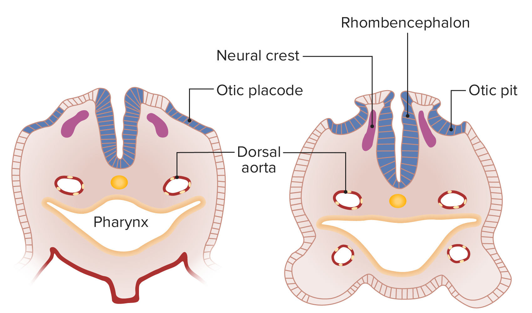
Transverse section of the embryo, featuring the otic placode originating from the ectoderm and invaginating to become the otic pit
Image by Lecturio.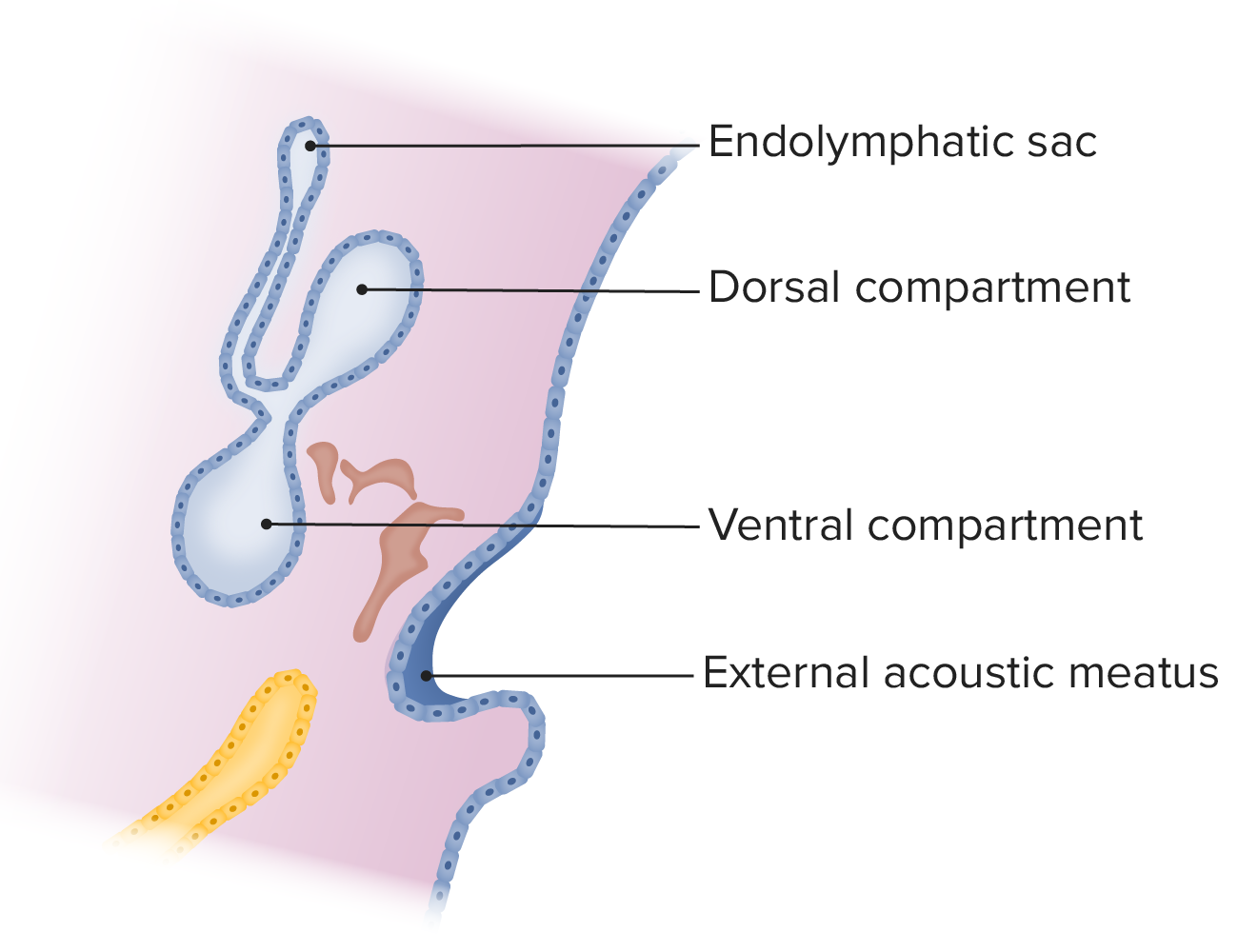
Diagram displaying the development of the otic vesicle into 2 compartments: dorsal and ventral:
Note that the dorsal compartment has an endolymphatic sac that extends toward the brain. The ossicle condensations are featured in brown, while the auditory tube is shown in yellow.
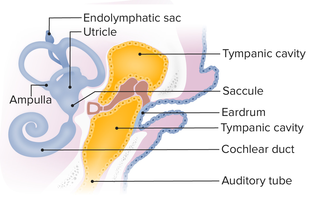
Diagram displaying the auditory tube, which extends from the 1st pharyngeal pouch as it surrounds the ossicles, creating the tympanic cavity
Image by Lecturio.
The 6 auricular hillocks in the fetus bulge outward from the pharyngeal arches and shift to form the characteristic shape of the external ear.
Image by Lecturio.The auricle is composed of musculocutaneous tissue and elastic Elastic Connective Tissue: Histology cartilage Cartilage Cartilage is a type of connective tissue derived from embryonic mesenchyme that is responsible for structural support, resilience, and the smoothness of physical actions. Perichondrium (connective tissue membrane surrounding cartilage) compensates for the absence of vasculature in cartilage by providing nutrition and support. Cartilage: Histology covered by skin Skin The skin, also referred to as the integumentary system, is the largest organ of the body. The skin is primarily composed of the epidermis (outer layer) and dermis (deep layer). The epidermis is primarily composed of keratinocytes that undergo rapid turnover, while the dermis contains dense layers of connective tissue. Skin: Structure and Functions. This structure is responsible for collecting and redirecting sound waves into the external auditory meatus.
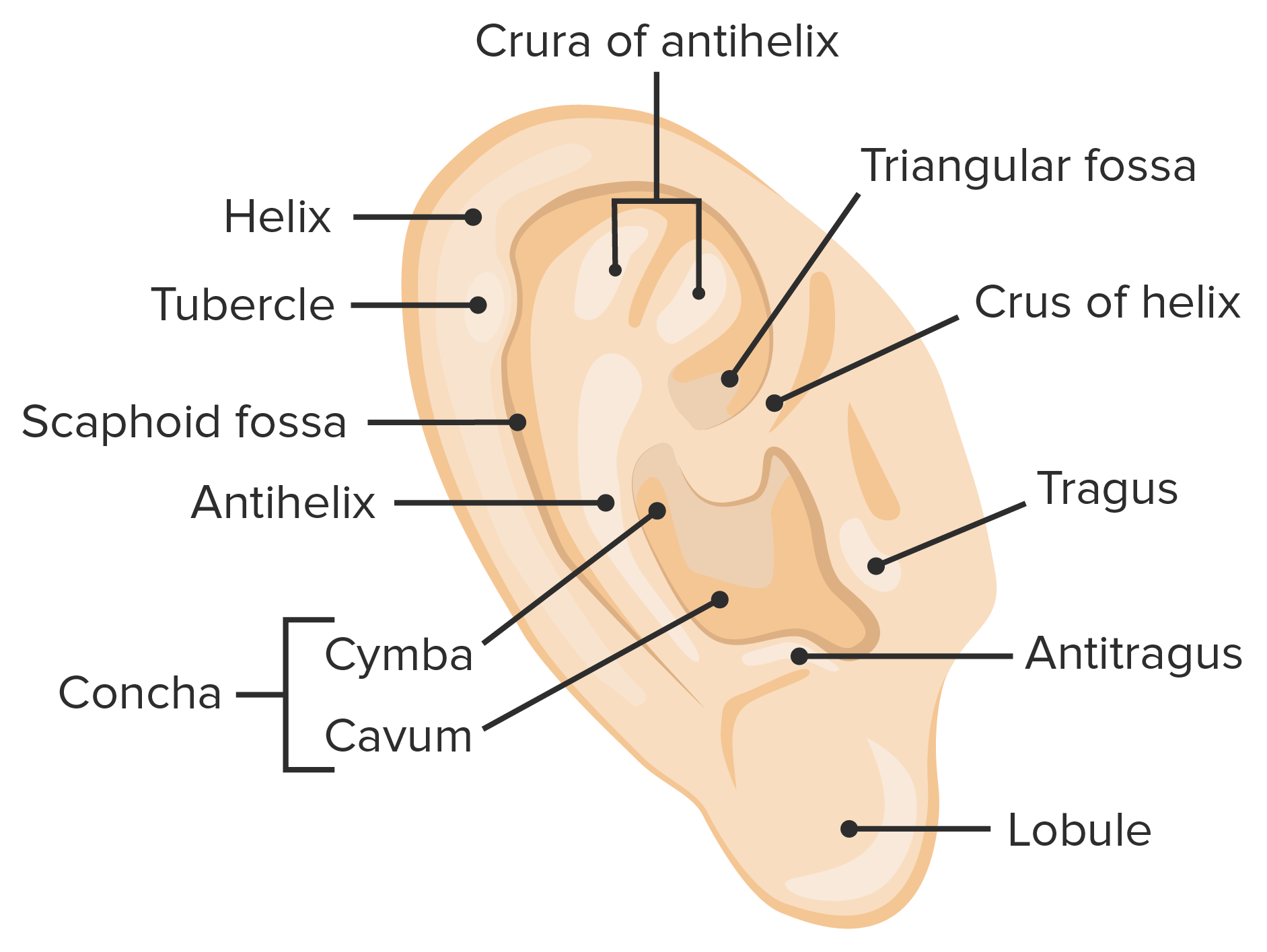
The outer ear, featuring the anatomical landmarks of the auricle
Image by Lecturio.The external auditory meatus is typically 1 in. (2.5 cm) in length and is S-shaped.
Transmits sound waves from the air to the ossicles → mechanical vibrations
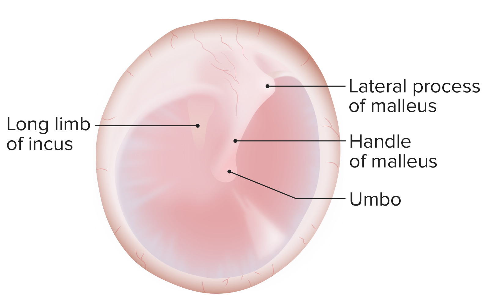
Tympanic membrane:
Pars flaccida (superior to insertion manubrium) and pars tensa (remainder of tympanic membrane)
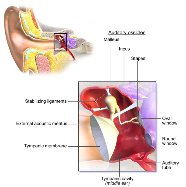
The middle ear, featuring the auditory ossicles connecting the tympanic membrane to the oval window
Image: “Anatomy of the middle ear” by BruceBlaus. License: CC BY 3.0Middle ear Middle ear The space and structures directly internal to the tympanic membrane and external to the inner ear (labyrinth). Its major components include the auditory ossicles and the eustachian tube that connects the cavity of middle ear (tympanic cavity) to the upper part of the throat. Acute Otitis Media muscles are responsible for movement of the tympanic membrane.
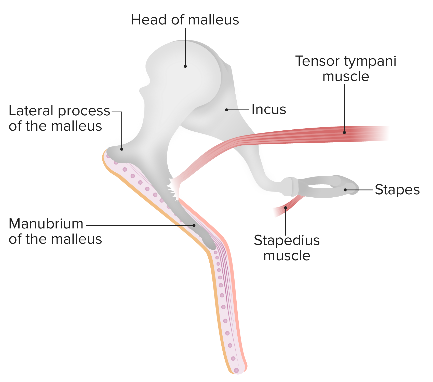
Muscles and ossicles of the middle ear:
The malleus connects to the tympanic membrane while the incus and stapes are completely within the middle ear. The stapedius muscle prevents excessive oscillation of the ossicles.
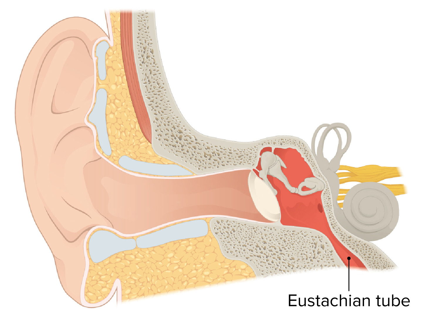
The Eustachian tube connects the middle ear with the pharynx:
This connection allows for the equalization of air pressure between the middle ear and the outside world.
The inner ear is located within the petrous part of the temporal bone Temporal bone Either of a pair of compound bones forming the lateral (left and right) surfaces and base of the skull which contains the organs of hearing. It is a large bone formed by the fusion of parts: the squamous (the flattened anterior-superior part), the tympanic (the curved anterior-inferior part), the mastoid (the irregular posterior portion), and the petrous (the part at the base of the skull). Jaw and Temporomandibular Joint: Anatomy and contains the organs of hearing and equilibrium Equilibrium Occurs when tumor cells survive the initial elimination attempt These cells are not able to progress, being maintained in a state of dormancy by the adaptive immune system. In this phase, tumor immunogenicity is edited, where T cells keep selectively attacking highly immunogenic tumor cells.This attack leaves other cells with less immunogenicity to potentially develop resistance to the immune response. Cancer Immunotherapy.
The bony labyrinth is filled with perilymph and is separated from the middle ear Middle ear The space and structures directly internal to the tympanic membrane and external to the inner ear (labyrinth). Its major components include the auditory ossicles and the eustachian tube that connects the cavity of middle ear (tympanic cavity) to the upper part of the throat. Acute Otitis Media by the medial wall of the tympanic cavity.
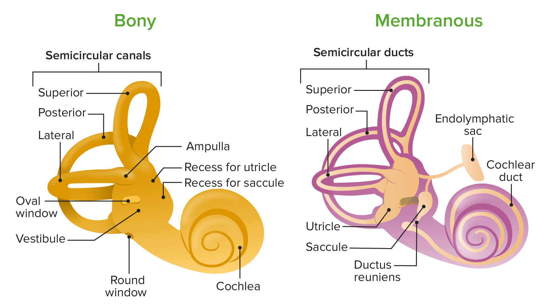
Diagram of the bony and membranous labyrinths, featuring their anatomical landmarks:
Note that the cochlear duct is found within the membranous labyrinth and the utricle and saccule are found within the vestibule.
The membranous labyrinth is filled with endolymph Endolymph The lymph fluid found in the membranous labyrinth of the ear. Vertigo and is composed of the vestibular and cochlear labyrinths.
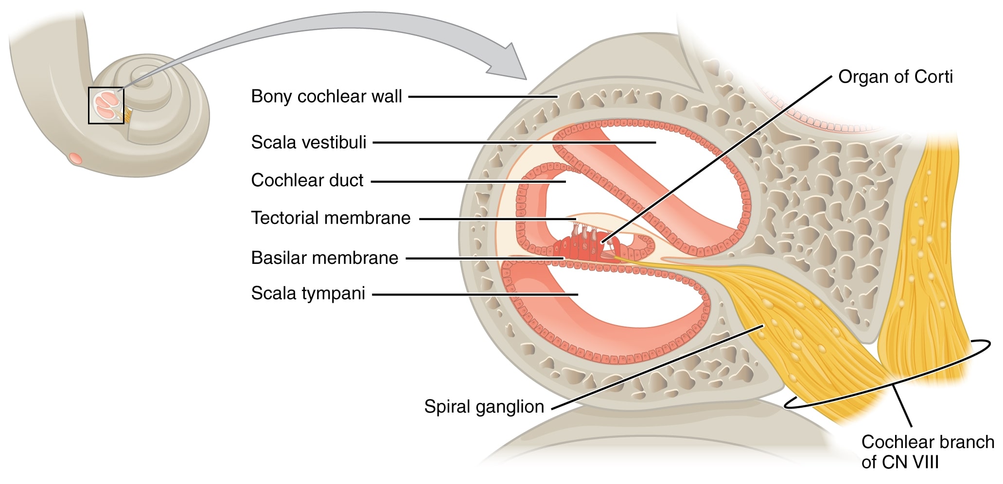
The organ of Corti is located within the scala media:
The organ of Corti detects sound in the form of vibration and transmits the information through the cochlear nerve.
CN: cranial nerve
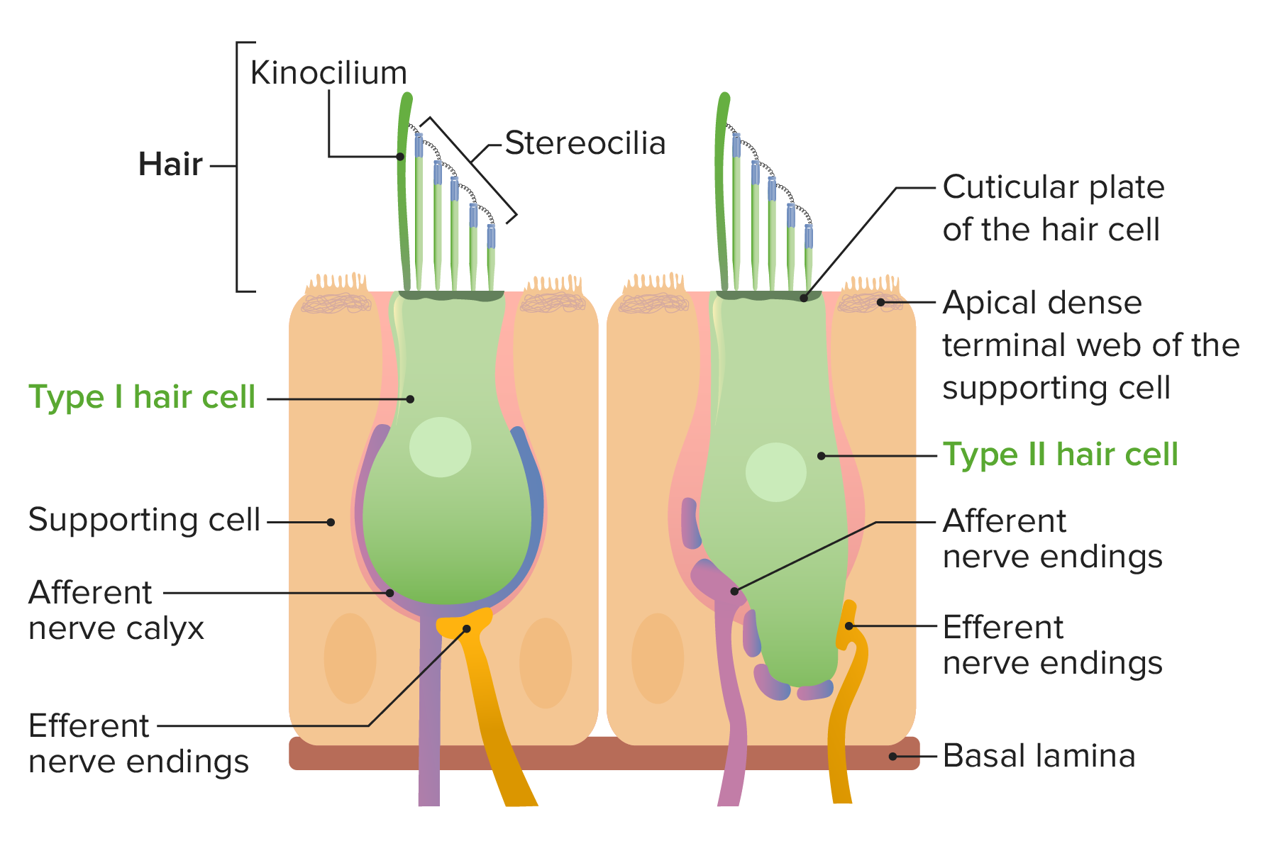
Types I and II hair cells:
Hair cells determine the movement and position of the head in space.
| Structure | Blood supply | Lymphatics | Innervation |
|---|---|---|---|
| Auricle |
|
Superficial parotid, mastoid, and cervical lymph nodes Lymph Nodes They are oval or bean shaped bodies (1 – 30 mm in diameter) located along the lymphatic system. Lymphatic Drainage System: Anatomy |
|
| Tympanic membrane |
|
Periauricular lymph nodes Lymph Nodes They are oval or bean shaped bodies (1 – 30 mm in diameter) located along the lymphatic system. Lymphatic Drainage System: Anatomy |
|
| Tympanic cavity |
|
Retroauricular cervical lymph nodes Lymph Nodes They are oval or bean shaped bodies (1 – 30 mm in diameter) located along the lymphatic system. Lymphatic Drainage System: Anatomy |
|
| Bony labyrinth |
|
Parotid, mastoid, and superficial cervical lymph nodes Lymph Nodes They are oval or bean shaped bodies (1 – 30 mm in diameter) located along the lymphatic system. Lymphatic Drainage System: Anatomy |
|
| Membranous labyrinth | Labyrinthine artery (branch of the basilar artery) |