The auditory and vestibular pathways are anatomically related but discrete pathways that permit conscious perceptionPerceptionThe process by which the nature and meaning of sensory stimuli are recognized and interpreted.Psychiatric Assessment of and reaction to sound and spatial orientationSpatial orientationChange in position or alignment in response to an external stimulus.Psychiatric Assessment. Stimulation of specialized hair cells in the cochleaCochleaThe part of the inner ear (labyrinth) that is concerned with hearing. It forms the anterior part of the labyrinth, as a snail-like structure that is situated almost horizontally anterior to the vestibular labyrinth.Ear: Anatomy and vestibular apparatus excite and send signals through partitions of the vestibulocochlear nerveVestibulocochlear nerveThe 8th cranial nerve. The vestibulocochlear nerve has a cochlear part (cochlear nerve) which is concerned with hearing and a vestibular part (vestibular nerve) which mediates the sense of balance and head position. The fibers of the cochlear nerve originate from neurons of the spiral ganglion and project to the cochlear nuclei (cochlear nucleus). The fibers of the vestibular nerve arise from neurons of scarpa's ganglion and project to the vestibular nuclei.The 12 Cranial Nerves: Overview and Functions (CN VIII) to the brainstem, where they synapseSynapseThe junction between 2 neurons is called a synapse. The synapse allows a neuron to pass an electrical or chemical signal to another neuron or target effector cell. Synapses and Neurotransmission on various targets, send and receive other projections, and ultimately contribute to spatial orientationSpatial orientationChange in position or alignment in response to an external stimulus.Psychiatric Assessment and perceptionPerceptionThe process by which the nature and meaning of sensory stimuli are recognized and interpreted.Psychiatric Assessment of sound.
The auditory pathway of the brainBrainThe part of central nervous system that is contained within the skull (cranium). Arising from the neural tube, the embryonic brain is comprised of three major parts including prosencephalon (the forebrain); mesencephalon (the midbrain); and rhombencephalon (the hindbrain). The developed brain consists of cerebrum; cerebellum; and other structures in the brain stem.Nervous System: Anatomy, Structure, and Classification begins with the external auditory canalExternal Auditory CanalOtitis Externa and includes the middle/inner earInner earThe essential part of the hearing organ consists of two labyrinthine compartments: the bony labyrinthine and the membranous labyrinth.Ear: Anatomy and eventually the brainstem nuclei before sending final signals to the primary auditory cortexPrimary auditory cortexThe region of the cerebral cortex that receives the auditory radiation from the medial geniculate body.Cerebral Cortex: Anatomy in the temporal lobeTemporal lobeLower lateral part of the cerebral hemisphere responsible for auditory, olfactory, and semantic processing. It is located inferior to the lateral fissure and anterior to the occipital lobe.Cerebral Cortex: Anatomy.
Peripheral components
Outer earOuter earThe outer part of the hearing system of the body. It includes the shell-like ear auricle which collects sound, and the external ear canal, the tympanic membrane, and the external ear cartilages.Ear: Anatomy:
Aurical (or pinna), auditory canal, and tympanic membraneTympanic membraneAn oval semitransparent membrane separating the external ear canal from the tympanic cavity. It contains three layers: the skin of the external ear canal; the core of radially and circularly arranged collagen fibers; and the mucosa of the middle ear.Ear: Anatomy
Directs sound waves to the tympanic membraneTympanic membraneAn oval semitransparent membrane separating the external ear canal from the tympanic cavity. It contains three layers: the skin of the external ear canal; the core of radially and circularly arranged collagen fibers; and the mucosa of the middle ear.Ear: Anatomy
Middle earMiddle earThe space and structures directly internal to the tympanic membrane and external to the inner ear (labyrinth). Its major components include the auditory ossicles and the eustachian tube that connects the cavity of middle ear (tympanic cavity) to the upper part of the throat.Acute Otitis Media:
Begins at the tympanic membraneTympanic membraneAn oval semitransparent membrane separating the external ear canal from the tympanic cavity. It contains three layers: the skin of the external ear canal; the core of radially and circularly arranged collagen fibers; and the mucosa of the middle ear.Ear: Anatomy
Airspace with 3 ossicles (malleus, incus, stapes)
Conducts and concentrates sound
The stapes directs sound vibrations into the vestibuleVestibuleAn oval, bony chamber of the inner ear, part of the bony labyrinth. It is continuous with bony cochlea anteriorly, and semicircular canals posteriorly. The vestibule contains two communicating sacs (utricle and saccule) of the balancing apparatus. The oval window on its lateral wall is occupied by the base of the stapes of the middle ear.Ear: Anatomy of the inner earInner earThe essential part of the hearing organ consists of two labyrinthine compartments: the bony labyrinthine and the membranous labyrinth.Ear: Anatomy through the oval window
Inner earInner earThe essential part of the hearing organ consists of two labyrinthine compartments: the bony labyrinthine and the membranous labyrinth.Ear: Anatomy:
Vibrations from the stapes are transferred to the perilymph in the scala vestibuli in the cochleaCochleaThe part of the inner ear (labyrinth) that is concerned with hearing. It forms the anterior part of the labyrinth, as a snail-like structure that is situated almost horizontally anterior to the vestibular labyrinth.Ear: Anatomy →
The vibrating perilymph pushes down on the vestibular membrane (which separates the scala vestibuli from the scala media) →
Causes vibrationVibrationA continuing periodic change in displacement with respect to a fixed reference.Neurological Examination of endolymphEndolymphThe lymph fluid found in the membranous labyrinth of the ear.Vertigo in the scala media →
Causes vibrationVibrationA continuing periodic change in displacement with respect to a fixed reference.Neurological Examination of the basilar membrane (separating the scala media from the scala tympani) →
As the basilar membrane vibrates up and down, the hair cells (which have mechanoreceptors) resting on top of the membrane move up and down.
The stereocilia on top of the hair cells are anchored to the tectorial membrane.
As the hair cells move, the stereocilia are bent back and forth by the tectorial membrane.
Bending of the stereocilia of the inner hair cells opens a mechanically gated ion channel, allowing in a quick burst of K+ from the surrounding endolymphEndolymphThe lymph fluid found in the membranous labyrinth of the ear.Vertigo →
Causes the hair cell to depolarize, effectively converting the sound vibrations into auditory nerve signals:
Loud sounds produce vigorous vibrations →
Activate more hair cells over a broader area of the basilar membrane
The signal is then passed to the cochlear branch of cranial nerve VIII (CN VIII), also known as the auditory nerve.
Tonotopy of basilar membrane: low frequency heard at apex and high frequency heard at base
Signals received from the apex (distal) are interpreted as low pitch.
Signals received from the base (proximal) are interpreted as high pitch.
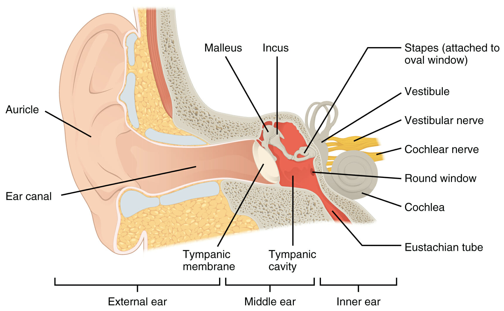
Anatomy of the ear
Image: “The external ear” by Phil Schatz. License: CC BY 4.0
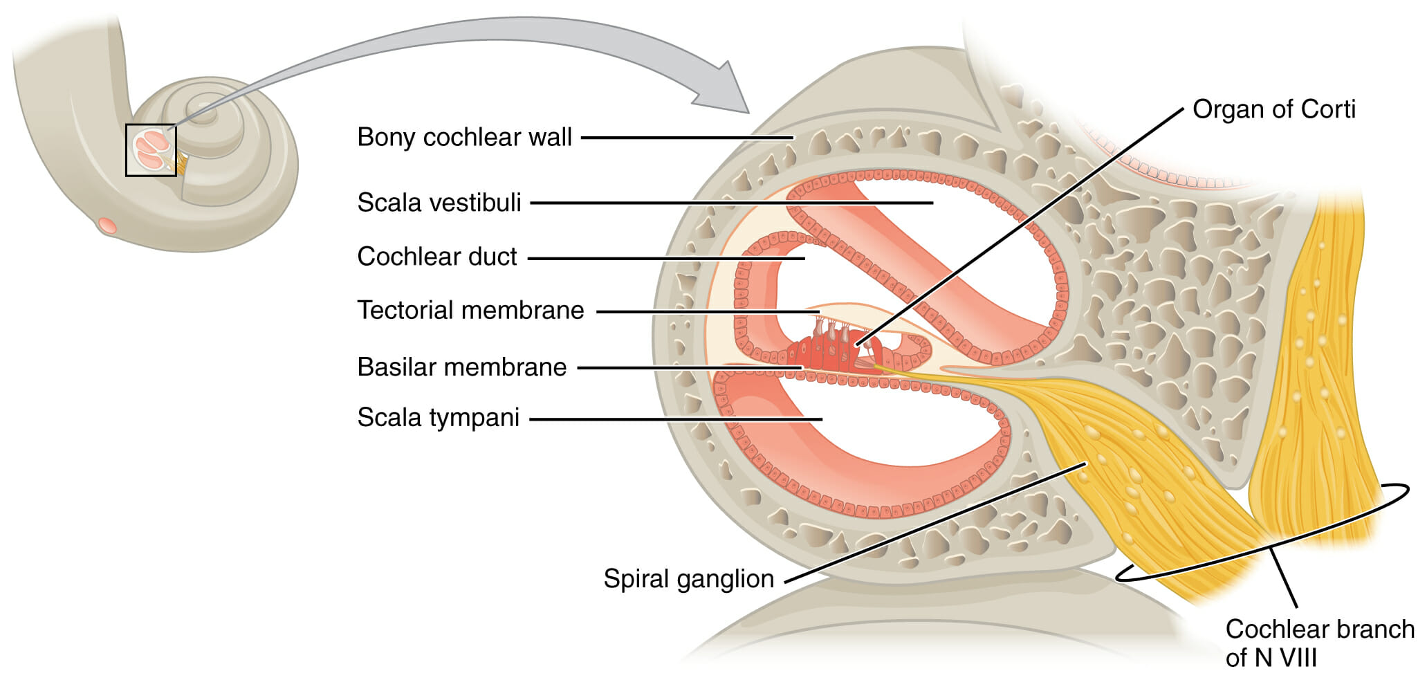
The organ of Corti is located within the scala media of the cochlea. The organ of Corti detects sound in the form of endolymph vibrations and converts them into nerve impulses, which are transmitted through the cochlear branch of cranial nerve (CN) VIII, the vestibulocochlear nerve.
Image by Lecturio
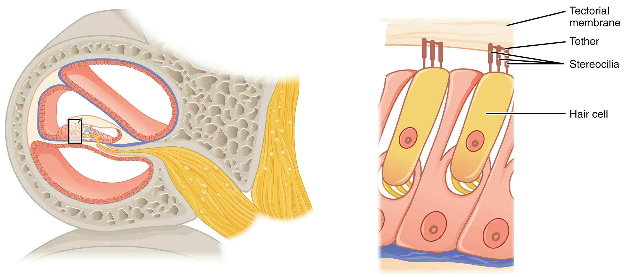
The organ of Corti contains hair cells with stereocilia in direct contact with the tectorial membrane. Vibrations in endolymph move the tectorial membrane, which moves the stereocilia and causes the hair cells (which are mechanoreceptors) to transmit nerve signals through the cochlear branch of cranial nerve VIII.
Image by Lecturio
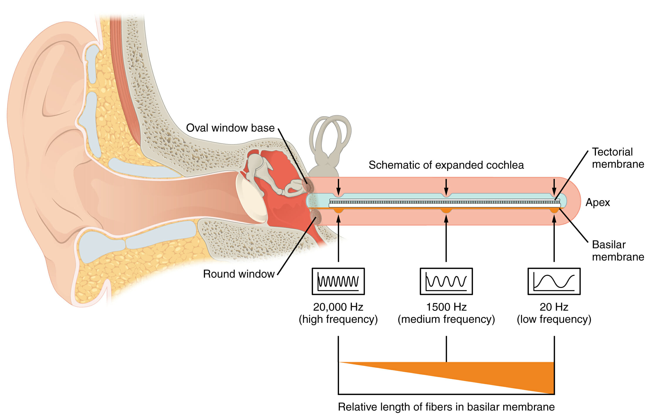
Sound waves cause vibrations in the perilymph of the scala vestibuli (the top of the pink portion of the expanded cochlea), causing vibrations in the basilar membrane. The organ of Corti sits atop the basilar membrane. The stereocilia of the hair cells are anchored in the tectorial membrane. As the basilar membrane vibrates, the stereocilia are bent back and forth by the tectorial membrane. The bending opens mechanically-gated ion channels, causing depolarization of the hair cells. Hair cells are sensitive to different frequencies along the length of the basilar membrane, which allows the brain to differentiate variations in pitch.
Image: “Frequency Coding in The Cochlea” by Phil Schatz. License: CC BY 4.0
Central components
AfferentAfferentNeurons which conduct nerve impulses to the central nervous system.Nervous System: Histology fibers in CN VIII convey information from the organ of Corti → auditory nuclei in the brainstem. The signal passes through brainBrainThe part of central nervous system that is contained within the skull (cranium). Arising from the neural tube, the embryonic brain is comprised of three major parts including prosencephalon (the forebrain); mesencephalon (the midbrain); and rhombencephalon (the hindbrain). The developed brain consists of cerebrum; cerebellum; and other structures in the brain stem.Nervous System: Anatomy, Structure, and Classification nuclei in the following order:
Cochlear nuclei: located at the dorsolateral side of the brainstem at the pontomedullary junction
Superior olivary nuclei: located in the ponsPonsThe front part of the hindbrain (rhombencephalon) that lies between the medulla and the midbrain (mesencephalon) ventral to the cerebellum. It is composed of two parts, the dorsal and the ventral. The pons serves as a relay station for neural pathways between the cerebellum to the cerebrum.Brain Stem: Anatomy
Sends signals back to the ear via CN V and CN VII efferentEfferentNeurons which send impulses peripherally to activate muscles or secretory cells.Nervous System: Histology fibers, which control the middle earMiddle earThe space and structures directly internal to the tympanic membrane and external to the inner ear (labyrinth). Its major components include the auditory ossicles and the eustachian tube that connects the cavity of middle ear (tympanic cavity) to the upper part of the throat.Acute Otitis Media muscles and cochlear tuning
Compares signals from the right and left to identify the direction of the sound
Lateral lemniscal nuclei: tract of axonsAxonsNerve fibers that are capable of rapidly conducting impulses away from the neuron cell body.Nervous System: Histology in the brainstem carrying auditory information to the inferior colliculusInferior colliculusThe posterior pair of the quadrigeminal bodies which contain centers for auditory function.Brain Stem: Anatomy of the midbrainMidbrainThe middle of the three primitive cerebral vesicles of the embryonic brain. Without further subdivision, midbrain develops into a short, constricted portion connecting the pons and the diencephalon. Midbrain contains two major parts, the dorsal tectum mesencephali and the ventral tegmentum mesencephali, housing components of auditory, visual, and other sensorimotor systems.Brain Stem: Anatomy
Inferior colliculusInferior colliculusThe posterior pair of the quadrigeminal bodies which contain centers for auditory function.Brain Stem: Anatomy: located in the midbrainMidbrainThe middle of the three primitive cerebral vesicles of the embryonic brain. Without further subdivision, midbrain develops into a short, constricted portion connecting the pons and the diencephalon. Midbrain contains two major parts, the dorsal tectum mesencephali and the ventral tegmentum mesencephali, housing components of auditory, visual, and other sensorimotor systems.Brain Stem: Anatomy
Processes fluctuations in pitch (important in understanding speech)
Helps locate the origin of the sound in space
Involved in the startle response (rapidly turning the head in response to sudden noise)
Medial geniculate body of the thalamusThalamusThe thalamus is a large, ovoid structure in the dorsal part of the diencephalon that is located between the cerebral cortex and midbrain. It consists of several interconnected nuclei of grey matter separated by the laminae of white matter. The thalamus is the main conductor of information that passes between the cerebral cortex and the periphery, spinal cord, or brain stem.Thalamus: Anatomy: part of thalamic relay system
Auditory cortex: located in the anterior and posterior transverse temporal areas
Conscious perceptionPerceptionThe process by which the nature and meaning of sensory stimuli are recognized and interpreted.Psychiatric Assessment of sound
Information processing
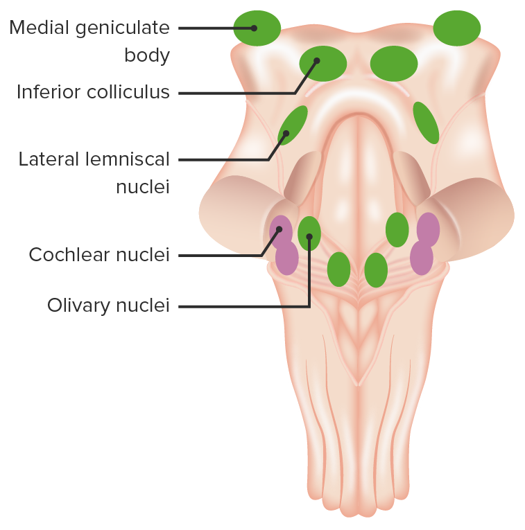
This image illustrates the nuclei involved in auditory sensation. Each of these plays an important role in the conduction and processing of auditory information from CN VIII up to the cortex.
Image by Lecturio.
Circuitry of pathway
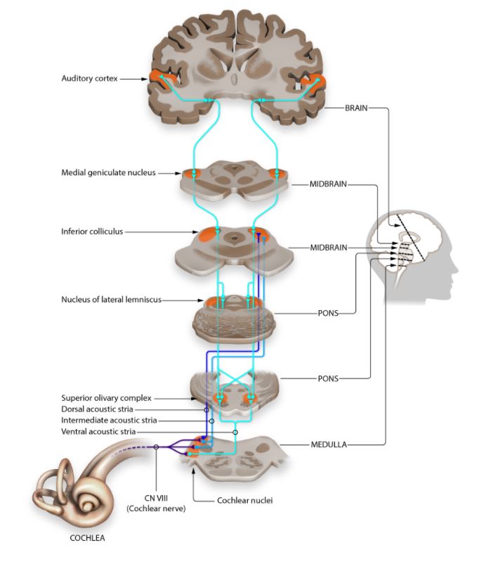
Outer earOuter earThe outer part of the hearing system of the body. It includes the shell-like ear auricle which collects sound, and the external ear canal, the tympanic membrane, and the external ear cartilages.Ear: Anatomy → inner earInner earThe essential part of the hearing organ consists of two labyrinthine compartments: the bony labyrinthine and the membranous labyrinth.Ear: Anatomy → depolarizationDepolarizationMembrane Potential of hair cells in the cochleaCochleaThe part of the inner ear (labyrinth) that is concerned with hearing. It forms the anterior part of the labyrinth, as a snail-like structure that is situated almost horizontally anterior to the vestibular labyrinth.Ear: Anatomy → both the ipsilateral and contralateral superior olivary nucleusNucleusWithin a eukaryotic cell, a membrane-limited body which contains chromosomes and one or more nucleoli (cell nucleolus). The nuclear membrane consists of a double unit-type membrane which is perforated by a number of pores; the outermost membrane is continuous with the endoplasmic reticulum. A cell may contain more than one nucleus.The Cell: Organelles → lateral lemniscus → inferior colliculusInferior colliculusThe posterior pair of the quadrigeminal bodies which contain centers for auditory function.Brain Stem: Anatomy → medial geniculate bodiesGeniculate BodiesPart of the diencephalon inferior to the caudal end of the dorsal thalamus. Includes the lateral geniculate body which relays visual impulses from the optic tract to the calcarine cortex, and the medial geniculate body which relays auditory impulses from the lateral lemniscus to the auditory cortex.Thalamus: Anatomy of the thalamusThalamusThe thalamus is a large, ovoid structure in the dorsal part of the diencephalon that is located between the cerebral cortex and midbrain. It consists of several interconnected nuclei of grey matter separated by the laminae of white matter. The thalamus is the main conductor of information that passes between the cerebral cortex and the periphery, spinal cord, or brain stem.Thalamus: Anatomy → auditory cortex of temporal lobeTemporal lobeLower lateral part of the cerebral hemisphere responsible for auditory, olfactory, and semantic processing. It is located inferior to the lateral fissure and anterior to the occipital lobe.Cerebral Cortex: Anatomy
Image depicting the pathway of sound from the cochlea to the level of the auditory cortex with multilevel axial slices through the brainstem
Image: “Auditory Pathway” by Jonathan E. Peelle. License: CC BY 4.0
The vestibular pathway of the brainBrainThe part of central nervous system that is contained within the skull (cranium). Arising from the neural tube, the embryonic brain is comprised of three major parts including prosencephalon (the forebrain); mesencephalon (the midbrain); and rhombencephalon (the hindbrain). The developed brain consists of cerebrum; cerebellum; and other structures in the brain stem.Nervous System: Anatomy, Structure, and Classification begins with the utricle and saccule, with additional input from the semicircular canals. Information eventually reaches the brainstem nuclei before sending final signals to the thalamusThalamusThe thalamus is a large, ovoid structure in the dorsal part of the diencephalon that is located between the cerebral cortex and midbrain. It consists of several interconnected nuclei of grey matter separated by the laminae of white matter. The thalamus is the main conductor of information that passes between the cerebral cortex and the periphery, spinal cord, or brain stem.Thalamus: Anatomy and cerebellumCerebellumThe cerebellum, Latin for “little brain,” is located in the posterior cranial fossa, dorsal to the pons and midbrain, and its principal role is in the coordination of movements. The cerebellum consists of 3 lobes on either side of its 2 hemispheres and is connected in the middle by the vermis. Cerebellum: Anatomy.
Functions of the vestibular pathway
The primary function of the vestibular system is to help the body maintain equilibriumEquilibriumOccurs when tumor cells survive the initial elimination attempt These cells are not able to progress, being maintained in a state of dormancy by the adaptive immune system. In this phase, tumor immunogenicity is edited, where T cells keep selectively attacking highly immunogenic tumor cells.This attack leaves other cells with less immunogenicity to potentially develop resistance to the immune response.Cancer Immunotherapyas it relates to balance and coordinationCoordinationCerebellar Disorders.
Types of equilibriumEquilibriumOccurs when tumor cells survive the initial elimination attempt These cells are not able to progress, being maintained in a state of dormancy by the adaptive immune system. In this phase, tumor immunogenicity is edited, where T cells keep selectively attacking highly immunogenic tumor cells.This attack leaves other cells with less immunogenicity to potentially develop resistance to the immune response.Cancer Immunotherapy:
Static equilibriumEquilibriumOccurs when tumor cells survive the initial elimination attempt These cells are not able to progress, being maintained in a state of dormancy by the adaptive immune system. In this phase, tumor immunogenicity is edited, where T cells keep selectively attacking highly immunogenic tumor cells.This attack leaves other cells with less immunogenicity to potentially develop resistance to the immune response.Cancer Immunotherapy: perceptionPerceptionThe process by which the nature and meaning of sensory stimuli are recognized and interpreted.Psychiatric Assessment of head orientationOrientationAwareness of oneself in relation to time, place and person.Psychiatric Assessment while stationary
Dynamic equilibriumEquilibriumOccurs when tumor cells survive the initial elimination attempt These cells are not able to progress, being maintained in a state of dormancy by the adaptive immune system. In this phase, tumor immunogenicity is edited, where T cells keep selectively attacking highly immunogenic tumor cells.This attack leaves other cells with less immunogenicity to potentially develop resistance to the immune response.Cancer Immunotherapy: perceptionPerceptionThe process by which the nature and meaning of sensory stimuli are recognized and interpreted.Psychiatric Assessment of motion and acceleration:
Linear acceleration
Angular acceleration
The vestibular system monitors the orientationOrientationAwareness of oneself in relation to time, place and person.Psychiatric Assessment of the body with respect to gravity.
Stimulates the vestibulospinal tracts to elicit compensatory movements
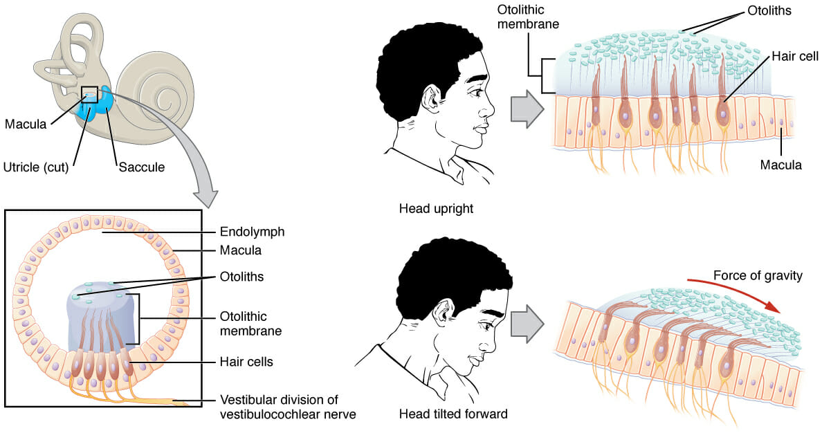
Head position relative to gravity (static equilibriumEquilibriumOccurs when tumor cells survive the initial elimination attempt These cells are not able to progress, being maintained in a state of dormancy by the adaptive immune system. In this phase, tumor immunogenicity is edited, where T cells keep selectively attacking highly immunogenic tumor cells.This attack leaves other cells with less immunogenicity to potentially develop resistance to the immune response.Cancer Immunotherapy and linear acceleration) is sensed by the 2 otolith organsOtolith OrgansVertigo, the utricle and saccule:
Utricle: oriented horizontally
Saccule: oriented vertically
Angular acceleration of the head is sensed by the 3 semicircular canals.
Physiology of the vestibular apparatus
Hair cells in the utricle, saccule, and semicircular canals are displaced based on their position relative to gravity, leading to depolarizationDepolarizationMembrane Potential and stimulation of the vestibular portion of CN VIII.
MaculaMaculaAn oval area in the retina, 3 to 5 mm in diameter, usually located temporal to the posterior pole of the eye and slightly below the level of the optic disk. It is characterized by the presence of a yellow pigment diffusely permeating the inner layers, contains the fovea centralis in its center, and provides the best phototropic visual acuity. It is devoid of retinal blood vessels, except in its periphery, and receives nourishment from the choriocapillaris of the choroid.Eye: Anatomy:
Groups of hair cells and supporting cells in the saccule and utricle:
MaculaMaculaAn oval area in the retina, 3 to 5 mm in diameter, usually located temporal to the posterior pole of the eye and slightly below the level of the optic disk. It is characterized by the presence of a yellow pigment diffusely permeating the inner layers, contains the fovea centralis in its center, and provides the best phototropic visual acuity. It is devoid of retinal blood vessels, except in its periphery, and receives nourishment from the choriocapillaris of the choroid.Eye: Anatomy sacculi: lies almost vertically on the saccule wall
MaculaMaculaAn oval area in the retina, 3 to 5 mm in diameter, usually located temporal to the posterior pole of the eye and slightly below the level of the optic disk. It is characterized by the presence of a yellow pigment diffusely permeating the inner layers, contains the fovea centralis in its center, and provides the best phototropic visual acuity. It is devoid of retinal blood vessels, except in its periphery, and receives nourishment from the choriocapillaris of the choroid.Eye: Anatomy utriculi: lies almost horizontally on the utricle floor
Otolithic membrane:
A gelatinous membrane that sits atop the maculaMaculaAn oval area in the retina, 3 to 5 mm in diameter, usually located temporal to the posterior pole of the eye and slightly below the level of the optic disk. It is characterized by the presence of a yellow pigment diffusely permeating the inner layers, contains the fovea centralis in its center, and provides the best phototropic visual acuity. It is devoid of retinal blood vessels, except in its periphery, and receives nourishment from the choriocapillaris of the choroid.Eye: Anatomy
Stereocilia of hair cells are embedded within.
Contains calcium carbonateCalcium carbonateCarbonic acid calcium salt. An odorless, tasteless powder or crystal that occurs in nature. It is used therapeutically as a phosphate buffer in hemodialysis patients and as a calcium supplement.Hypocalcemia deposits called otoliths, which make the otolithic membrane top-heavy and generate inertia
Detects linear acceleration
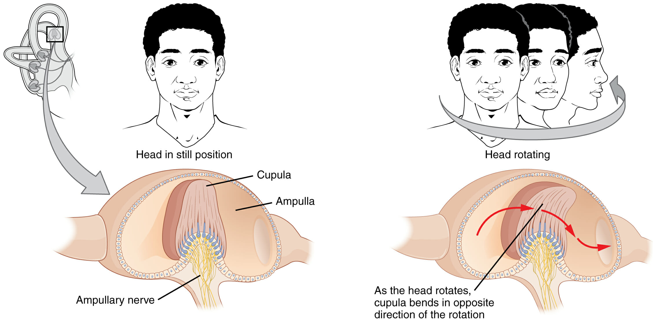
Crista ampullaris:
Groups of hair cells and supporting cells in the ampulla of the semicircular canals
Cupula:
A gelatinous membrane overlying the hair cells
Stereocilia of the hair cells are embedded within.
Connected to the roof of the ampulla, loosely anchoring it
Detects angular acceleration
Both the maculaMaculaAn oval area in the retina, 3 to 5 mm in diameter, usually located temporal to the posterior pole of the eye and slightly below the level of the optic disk. It is characterized by the presence of a yellow pigment diffusely permeating the inner layers, contains the fovea centralis in its center, and provides the best phototropic visual acuity. It is devoid of retinal blood vessels, except in its periphery, and receives nourishment from the choriocapillaris of the choroid.Eye: Anatomy and crista ampullaris are surrounded by endolymphEndolymphThe lymph fluid found in the membranous labyrinth of the ear.Vertigo.
Process:
Head movement causes movement of the otolithic membrane and/or cupula relative to the underlying maculaMaculaAn oval area in the retina, 3 to 5 mm in diameter, usually located temporal to the posterior pole of the eye and slightly below the level of the optic disk. It is characterized by the presence of a yellow pigment diffusely permeating the inner layers, contains the fovea centralis in its center, and provides the best phototropic visual acuity. It is devoid of retinal blood vessels, except in its periphery, and receives nourishment from the choriocapillaris of the choroid.Eye: Anatomy/crista ampullaris (due to gravity and inertia) →
Impulses are transmitted via the vestibular branch of CN VIII
Different head orientations and movements cause varying stimulation of the maculaMaculaAn oval area in the retina, 3 to 5 mm in diameter, usually located temporal to the posterior pole of the eye and slightly below the level of the optic disk. It is characterized by the presence of a yellow pigment diffusely permeating the inner layers, contains the fovea centralis in its center, and provides the best phototropic visual acuity. It is devoid of retinal blood vessels, except in its periphery, and receives nourishment from the choriocapillaris of the choroid.Eye: Anatomy and crista ampullaris on the right and left sides, which are interpreted by the brainBrainThe part of central nervous system that is contained within the skull (cranium). Arising from the neural tube, the embryonic brain is comprised of three major parts including prosencephalon (the forebrain); mesencephalon (the midbrain); and rhombencephalon (the hindbrain). The developed brain consists of cerebrum; cerebellum; and other structures in the brain stem.Nervous System: Anatomy, Structure, and Classification.
Linear acceleration:
Linear acceleration is detected primarily by the macula in the utricle and saccule. Stereocilia on the hair cells are embedded in a gelatinous otolithic membrane. Movement causes a shift in the otolithic membrane due to the inertia of the gelatinous mass. The shift causes the stereocilia to bend, opening mechanically-gated ion channels, which triggers depolarization of the hair cells.
Image: “Maculae and Equilibrium” by Phil Schatz. License: CC BY 4.0
Angular acceleration:
Angular acceleration is detected primarily by the crista ampullaris in the ampulla of the semicircular canals. Stereocilia on the hair cells are embedded in a gelatinous mass called the cupula. Head movement causes a shift in the cupula due to the inertia of the gelatinous mass. The shift causes the stereocilia to bend, opening mechanically gated ion channels, which triggers depolarization of the hair cells.
Image: “Equilibrium and Semicircular Canals” by Phil Schatz. License: CC BY 4.0.
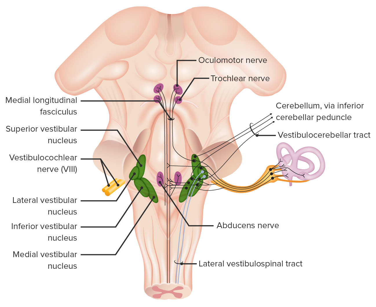
This image demonstrates the intricate pathways of the vestibular system. Note the flow of information, which starts with depolarization of hair cells in the inner ear sends movement and positional information to the vestibular ganglia, which transmits this information to the vestibular nuclei (also receives input from the cerebellum). From here, the vestibular nuclei send information to CN III, IV, & VI to dictate eye movement. The vestibular nuclei also sends information to the thalamus, cerebellum, and medial and lateral vestibulospinal tracts, allowing for perception, integration, and adjustments of body position within space.
Image by Lecturio.
Neural pathway within the brainBrainThe part of central nervous system that is contained within the skull (cranium). Arising from the neural tube, the embryonic brain is comprised of three major parts including prosencephalon (the forebrain); mesencephalon (the midbrain); and rhombencephalon (the hindbrain). The developed brain consists of cerebrum; cerebellum; and other structures in the brain stem.Nervous System: Anatomy, Structure, and Classification
The base of the hair cells synapses with sensorySensoryNeurons which conduct nerve impulses to the central nervous system.Nervous System: Histology fibers in the vestibular branch of CN VIII. Cranial nerve VIII enters the brainstem at the pontomedullary junction (at the cerebellopontine angleCerebellopontine angleJunction between the cerebellum and the pons.Acoustic Neuroma) and then sends fibers to the vestibular nucleusNucleusWithin a eukaryotic cell, a membrane-limited body which contains chromosomes and one or more nucleoli (cell nucleolus). The nuclear membrane consists of a double unit-type membrane which is perforated by a number of pores; the outermost membrane is continuous with the endoplasmic reticulum. A cell may contain more than one nucleus.The Cell: Organelles and flocculonodular lobeFlocculonodular lobeCerebellum: Anatomy in the cerebellumCerebellumThe cerebellum, Latin for “little brain,” is located in the posterior cranial fossa, dorsal to the pons and midbrain, and its principal role is in the coordination of movements. The cerebellum consists of 3 lobes on either side of its 2 hemispheres and is connected in the middle by the vermis. Cerebellum: Anatomy:
Vestibular nucleusNucleusWithin a eukaryotic cell, a membrane-limited body which contains chromosomes and one or more nucleoli (cell nucleolus). The nuclear membrane consists of a double unit-type membrane which is perforated by a number of pores; the outermost membrane is continuous with the endoplasmic reticulum. A cell may contain more than one nucleus.The Cell: Organelles:
Includes superior, lateral, inferior, and medial portions
Receives the majority of afferents from CN VIII
Sends fibers to:
The flocculonodular lobeFlocculonodular lobeCerebellum: Anatomy of the cerebellumCerebellumThe cerebellum, Latin for “little brain,” is located in the posterior cranial fossa, dorsal to the pons and midbrain, and its principal role is in the coordination of movements. The cerebellum consists of 3 lobes on either side of its 2 hemispheres and is connected in the middle by the vermis. Cerebellum: Anatomy, which helps coordinate:
Nuclei for CN XI that control movement of the head and neckNeckThe part of a human or animal body connecting the head to the rest of the body.Peritonsillar Abscess
Have axonsAxonsNerve fibers that are capable of rapidly conducting impulses away from the neuron cell body.Nervous System: Histology that terminate at all spinal levels
Innervate axialAxialComputed Tomography (CT) (e.g., intercostal, neckNeckThe part of a human or animal body connecting the head to the rest of the body.Peritonsillar Abscess, and back) and extensor muscles important for maintaining balance and equilibriumEquilibriumOccurs when tumor cells survive the initial elimination attempt These cells are not able to progress, being maintained in a state of dormancy by the adaptive immune system. In this phase, tumor immunogenicity is edited, where T cells keep selectively attacking highly immunogenic tumor cells.This attack leaves other cells with less immunogenicity to potentially develop resistance to the immune response.Cancer Immunotherapy
ThalamusThalamusThe thalamus is a large, ovoid structure in the dorsal part of the diencephalon that is located between the cerebral cortex and midbrain. It consists of several interconnected nuclei of grey matter separated by the laminae of white matter. The thalamus is the main conductor of information that passes between the cerebral cortex and the periphery, spinal cord, or brain stem.Thalamus: Anatomy: related to conscious perceptionPerceptionThe process by which the nature and meaning of sensory stimuli are recognized and interpreted.Psychiatric Assessment of equilibriumEquilibriumOccurs when tumor cells survive the initial elimination attempt These cells are not able to progress, being maintained in a state of dormancy by the adaptive immune system. In this phase, tumor immunogenicity is edited, where T cells keep selectively attacking highly immunogenic tumor cells.This attack leaves other cells with less immunogenicity to potentially develop resistance to the immune response.Cancer Immunotherapy
Almost all efferentEfferentNeurons which send impulses peripherally to activate muscles or secretory cells.Nervous System: Histology output from the vestibular nucleusNucleusWithin a eukaryotic cell, a membrane-limited body which contains chromosomes and one or more nucleoli (cell nucleolus). The nuclear membrane consists of a double unit-type membrane which is perforated by a number of pores; the outermost membrane is continuous with the endoplasmic reticulum. A cell may contain more than one nucleus.The Cell: Organelles is reflexive (i.e., very difficult to voluntarily inhibit).
Located in the cerebellumCerebellumThe cerebellum, Latin for “little brain,” is located in the posterior cranial fossa, dorsal to the pons and midbrain, and its principal role is in the coordination of movements. The cerebellum consists of 3 lobes on either side of its 2 hemispheres and is connected in the middle by the vermis. Cerebellum: Anatomy
Synapses with fibers leading to the:
Vestibular nucleusNucleusWithin a eukaryotic cell, a membrane-limited body which contains chromosomes and one or more nucleoli (cell nucleolus). The nuclear membrane consists of a double unit-type membrane which is perforated by a number of pores; the outermost membrane is continuous with the endoplasmic reticulum. A cell may contain more than one nucleus.The Cell: Organelles (helps coordinate smooth-tracking eye movements)
Fastigial nucleusNucleusWithin a eukaryotic cell, a membrane-limited body which contains chromosomes and one or more nucleoli (cell nucleolus). The nuclear membrane consists of a double unit-type membrane which is perforated by a number of pores; the outermost membrane is continuous with the endoplasmic reticulum. A cell may contain more than one nucleus.The Cell: Organelles (provides information regarding skeletal muscle and spinal cordSpinal cordThe spinal cord is the major conduction pathway connecting the brain to the body; it is part of the CNS. In cross section, the spinal cord is divided into an H-shaped area of gray matter (consisting of synapsing neuronal cell bodies) and a surrounding area of white matter (consisting of ascending and descending tracts of myelinated axons). Spinal Cord: Anatomy activity necessary to coordinate vestibular responses)
Vestibular pathway outputs
Table: Vestibular pathway outputs
Anatomical structure
Function
Cranial nerve nuclei
Control over eye movements
ThalamusThalamusThe thalamus is a large, ovoid structure in the dorsal part of the diencephalon that is located between the cerebral cortex and midbrain. It consists of several interconnected nuclei of grey matter separated by the laminae of white matter. The thalamus is the main conductor of information that passes between the cerebral cortex and the periphery, spinal cord, or brain stem.Thalamus: Anatomy
Conscious perceptionPerceptionThe process by which the nature and meaning of sensory stimuli are recognized and interpreted.Psychiatric Assessment of movement and gravity through connections to cortex
CerebellumCerebellumThe cerebellum, Latin for “little brain,” is located in the posterior cranial fossa, dorsal to the pons and midbrain, and its principal role is in the coordination of movements. The cerebellum consists of 3 lobes on either side of its 2 hemispheres and is connected in the middle by the vermis. Cerebellum: Anatomy (flocculonodular lobeFlocculonodular lobeCerebellum: Anatomy)
Acoustic neuromaAcoustic neuromaAcoustic neuroma, also referred to as vestibular schwannoma, is a benign tumor arising from Schwann cells of the vestibular component of the cranial nerve VIII. Acoustic neuroma forms within the internal auditory meatus and extends into the cerebellopontine angle. Acoustic Neuroma: an acoustic neuromaAcoustic neuromaAcoustic neuroma, also referred to as vestibular schwannoma, is a benign tumor arising from Schwann cells of the vestibular component of the cranial nerve VIII. Acoustic neuroma forms within the internal auditory meatus and extends into the cerebellopontine angle. Acoustic Neuroma is a benignBenignFibroadenomatumorTumorInflammation of Schwann cells that involves the cranial nervesCranial nervesThere are 12 pairs of cranial nerves (CNs), which run from the brain to various parts of the head, neck, and trunk. The CNs can be sensory or motor or both. The CNs are named and numbered in Roman numerals according to their location, from the front to the back of the brain.The 12 Cranial Nerves: Overview and Functions within the craniumCraniumThe skull (cranium) is the skeletal structure of the head supporting the face and forming a protective cavity for the brain. The skull consists of 22 bones divided into the viscerocranium (facial skeleton) and the neurocranium.Skull: Anatomy. Acoustic neuromas most frequently affect the vestibular branch of CN VIII. Acoustic neuromas often present with hearing lossHearing lossHearing loss, also known as hearing impairment, is any degree of impairment in the ability to apprehend sound as determined by audiometry to be below normal hearing thresholds. Clinical presentation may occur at birth or as a gradual loss of hearing with age, including a short-term or sudden loss at any point. Hearing Loss and tinnitusTinnitusA nonspecific symptom of hearing disorder characterized by the sensation of buzzing, ringing, clicking, pulsations, and other noises in the ear. Objective tinnitus refers to noises generated from within the ear or adjacent structures that can be heard by other individuals. The term subjective tinnitus is used when the sound is audible only to the affected individual. Tinnitus may occur as a manifestation of cochlear diseases; vestibulocochlear nerve diseases; intracranial hypertension; craniocerebral trauma; and other conditions.Cranial Nerve Palsies. Treatment is with surgical removal.
Hearing lossHearing lossHearing loss, also known as hearing impairment, is any degree of impairment in the ability to apprehend sound as determined by audiometry to be below normal hearing thresholds. Clinical presentation may occur at birth or as a gradual loss of hearing with age, including a short-term or sudden loss at any point. Hearing Loss: hearing impairments are classified into conductive hearing lossConductive hearing lossHearing loss due to interference with the mechanical reception or amplification of sound to the cochlea. The interference is in the outer or middle ear involving the ear canal; tympanic membrane; or ear ossicles.Hearing Loss and sensorineural hearing lossSensorineural hearing lossHearing loss resulting from damage to the cochlea and the sensorineural elements which lie internally beyond the oval and round windows. These elements include the auditory nerve and its connections in the brainstem.Hearing Loss. Conductive hearing lossConductive hearing lossHearing loss due to interference with the mechanical reception or amplification of sound to the cochlea. The interference is in the outer or middle ear involving the ear canal; tympanic membrane; or ear ossicles.Hearing Loss comes about when there is a problem transferring sound waves anywhere along the pathway from the outer earOuter earThe outer part of the hearing system of the body. It includes the shell-like ear auricle which collects sound, and the external ear canal, the tympanic membrane, and the external ear cartilages.Ear: Anatomy, the tympanic membraneTympanic membraneAn oval semitransparent membrane separating the external ear canal from the tympanic cavity. It contains three layers: the skin of the external ear canal; the core of radially and circularly arranged collagen fibers; and the mucosa of the middle ear.Ear: Anatomy, or middle earMiddle earThe space and structures directly internal to the tympanic membrane and external to the inner ear (labyrinth). Its major components include the auditory ossicles and the eustachian tube that connects the cavity of middle ear (tympanic cavity) to the upper part of the throat.Acute Otitis Media. In cases of sensorineural hearing lossSensorineural hearing lossHearing loss resulting from damage to the cochlea and the sensorineural elements which lie internally beyond the oval and round windows. These elements include the auditory nerve and its connections in the brainstem.Hearing Loss, there is an errorErrorRefers to any act of commission (doing something wrong) or omission (failing to do something right) that exposes patients to potentially hazardous situations.Disclosure of Information in the transmission of auditory stimuli from the cochleaCochleaThe part of the inner ear (labyrinth) that is concerned with hearing. It forms the anterior part of the labyrinth, as a snail-like structure that is situated almost horizontally anterior to the vestibular labyrinth.Ear: Anatomy to the auditory nuclei.
VertigoVertigoVertigo is defined as the perceived sensation of rotational motion while remaining still. A very common complaint in primary care and the ER, vertigo is more frequently experienced by women and its prevalence increases with age. Vertigo is classified into peripheral or central based on its etiology. Vertigo: the sensation of movement between oneself and the surroundings when no movement is actually occurring. VertigoVertigoVertigo is defined as the perceived sensation of rotational motion while remaining still. A very common complaint in primary care and the ER, vertigo is more frequently experienced by women and its prevalence increases with age. Vertigo is classified into peripheral or central based on its etiology. Vertigo is not limited to a feeling of rotationRotationMotion of an object in which either one or more points on a line are fixed. It is also the motion of a particle about a fixed point.X-rays (spinning); other forms include upward lifting, swaying, rocking, and unsystematic movement. VertigoVertigoVertigo is defined as the perceived sensation of rotational motion while remaining still. A very common complaint in primary care and the ER, vertigo is more frequently experienced by women and its prevalence increases with age. Vertigo is classified into peripheral or central based on its etiology. Vertigo most often occurs due to problems within the semicircular canals.
Molina, FJ. (2012). Chapter 18: Hearing loss. In Henderson, MC, Tierney, LM, & Smetana, GW. (Eds.), The Patient History: An Evidence-Based Approach to Differential Diagnosis, 2e. The McGraw-Hill Companies. http://accessmedicine.mhmedical.com/content.aspx?aid=56852049
Berkowitz, AL. (2016). The auditory and vestibular pathways and approach to hearing loss and dizziness/vertigo: Cranial nerve 8. In Clinical Neurology and Neuroanatomy: A Localization-Based Approach. McGraw-Hill Education. http://accessmedicine.mhmedical.com/content.aspx?aid=1160204039