Achieve Mastery of Medical Concepts

Study for medical school and boards with Lecturio

Autosomal dominant polycystic kidney disease (ADPKD)

Polycystic kidney disease (PKD) is an inherited genetic disorder leading to the development of numerous fluid-filled cysts Cysts Any fluid-filled closed cavity or sac that is lined by an epithelium. Cysts can be of normal, abnormal, non-neoplastic, or neoplastic tissues. Fibrocystic Change in the kidneys Kidneys The kidneys are a pair of bean-shaped organs located retroperitoneally against the posterior wall of the abdomen on either side of the spine. As part of the urinary tract, the kidneys are responsible for blood filtration and excretion of water-soluble waste in the urine. Kidneys: Anatomy. The 2 main types of PKD are autosomal dominant Autosomal dominant Autosomal inheritance, both dominant and recessive, refers to the transmission of genes from the 22 autosomal chromosomes. Autosomal dominant diseases are expressed when only 1 copy of the dominant allele is inherited. Autosomal Recessive and Autosomal Dominant Inheritance polycystic kidney disease (ADPKD), which is often diagnosed in adulthood, and autosomal recessive polycystic kidney disease Autosomal Recessive Polycystic Kidney Disease Polycystic kidney disease (PKD) is an inherited genetic disorder leading to the development of numerous fluid-filled cysts in the kidneys. Autosomal recessive polycystic kidney disease is primarily characterized by cystic dilatations of the renal collecting ducts and intrahepatic bile duct dilatation with hepatic fibrosis. Autosomal Recessive Polycystic Kidney Disease (ARPKD) ( ARPKD ARPKD Polycystic kidney disease (PKD) is an inherited genetic disorder leading to the development of numerous fluid-filled cysts in the kidneys. Autosomal recessive polycystic kidney disease is primarily characterized by cystic dilatations of the renal collecting ducts and intrahepatic bile duct dilatation with hepatic fibrosis. Autosomal Recessive Polycystic Kidney Disease (ARPKD)), which is often diagnosed antenatally or shortly after birth. ADPKD patients Patients Individuals participating in the health care system for the purpose of receiving therapeutic, diagnostic, or preventive procedures. Clinician–Patient Relationship commonly present with hypertension Hypertension Hypertension, or high blood pressure, is a common disease that manifests as elevated systemic arterial pressures. Hypertension is most often asymptomatic and is found incidentally as part of a routine physical examination or during triage for an unrelated medical encounter. Hypertension, hematuria Hematuria Presence of blood in the urine. Renal Cell Carcinoma, and flank pain Flank pain Pain emanating from below the ribs and above the ilium. Renal Cell Carcinoma. Extrarenal manifestations include intracerebral aneurysm Aneurysm An aneurysm is a bulging, weakened area of a blood vessel that causes an abnormal widening of its diameter > 1.5 times the size of the native vessel. Aneurysms occur more often in arteries than in veins and are at risk of dissection and rupture, which can be life-threatening. Thoracic Aortic Aneurysms, hepatic and pancreatic cysts Cysts Any fluid-filled closed cavity or sac that is lined by an epithelium. Cysts can be of normal, abnormal, non-neoplastic, or neoplastic tissues. Fibrocystic Change, and cardiac valvular abnormalities. Diagnosis is by history, physical exam, and ultrasonography. Management requires a multidisciplinary approach and many patients Patients Individuals participating in the health care system for the purpose of receiving therapeutic, diagnostic, or preventive procedures. Clinician–Patient Relationship require renal replacement therapy. The end goal is to slow the progression of renal disease by controlling hypertension Hypertension Hypertension, or high blood pressure, is a common disease that manifests as elevated systemic arterial pressures. Hypertension is most often asymptomatic and is found incidentally as part of a routine physical examination or during triage for an unrelated medical encounter. Hypertension, proteinuria Proteinuria The presence of proteins in the urine, an indicator of kidney diseases. Nephrotic Syndrome in Children, and symptoms. ADPKD prognosis Prognosis A prediction of the probable outcome of a disease based on a individual's condition and the usual course of the disease as seen in similar situations. Non-Hodgkin Lymphomas is dependent on a variety of factors. A cerebral aneurysm Cerebral aneurysm Brain aneurysms, also known as intracranial or cerebral aneurysms, are dilations of the arteries along points of weakness in the brain. The majority of the aneurysms are berry (saccular) in nature and located within the anterior circulation of the circle of Willis. Brain Aneurysms is a complication associated with a particularly poor prognosis Prognosis A prediction of the probable outcome of a disease based on a individual's condition and the usual course of the disease as seen in similar situations. Non-Hodgkin Lymphomas.

Last updated: Feb 17, 2023

Editorial responsibility: Stanley Oiseth, Lindsay Jones, Evelin Maza

Epidemiology and Etiology

Epidemiology

Polycystic kidney disease (PKD) affects about 500,000 people in the United States. One of the 2 main types of PKD is autosomal dominant Autosomal dominant Autosomal inheritance, both dominant and recessive, refers to the transmission of genes from the 22 autosomal chromosomes. Autosomal dominant diseases are expressed when only 1 copy of the dominant allele is inherited. Autosomal Recessive and Autosomal Dominant Inheritance polycystic kidney disease (ADPKD).

  • Most commonly inherited kidney disease
  • Prevalence Prevalence The total number of cases of a given disease in a specified population at a designated time. It is differentiated from incidence, which refers to the number of new cases in the population at a given time. Measures of Disease Frequency: 1 in 1,000 live births
  • Cause of CKD CKD Chronic kidney disease (CKD) is kidney impairment that lasts for ≥ 3 months, implying that it is irreversible. Hypertension and diabetes are the most common causes; however, there are a multitude of other etiologies. In the early to moderate stages, CKD is usually asymptomatic and is primarily diagnosed by laboratory abnormalities. Chronic Kidney Disease in 5% of annual dialysis Dialysis Renal replacement therapy refers to dialysis and/or kidney transplantation. Dialysis is a procedure by which toxins and excess water are removed from the circulation. Hemodialysis and peritoneal dialysis (PD) are the two types of dialysis, and their primary difference is the location of the filtration process (external to the body in hemodialysis versus inside the body for PD). Peritoneal Dialysis and Hemodialysis initiation
  • Decline in renal function often noted by the 4th decade of life

Etiology

  • Autosomal dominant Autosomal dominant Autosomal inheritance, both dominant and recessive, refers to the transmission of genes from the 22 autosomal chromosomes. Autosomal dominant diseases are expressed when only 1 copy of the dominant allele is inherited. Autosomal Recessive and Autosomal Dominant Inheritance (positive family history Family History Adult Health Maintenance)
  • Either a mutation Mutation Genetic mutations are errors in DNA that can cause protein misfolding and dysfunction. There are various types of mutations, including chromosomal, point, frameshift, and expansion mutations. Types of Mutations of:
    • PKD1 (encodes polycystin-1) on chromosome Chromosome In a prokaryotic cell or in the nucleus of a eukaryotic cell, a structure consisting of or containing DNA which carries the genetic information essential to the cell. Basic Terms of Genetics 16 OR
    • PKD2 (encodes polycystin-2) on chromosome Chromosome In a prokaryotic cell or in the nucleus of a eukaryotic cell, a structure consisting of or containing DNA which carries the genetic information essential to the cell. Basic Terms of Genetics 4

Clinical Presentation

The age of symptom onset is variable Variable Variables represent information about something that can change. The design of the measurement scales, or of the methods for obtaining information, will determine the data gathered and the characteristics of that data. As a result, a variable can be qualitative or quantitative, and may be further classified into subgroups. Types of Variables, but typically in adulthood. Patients Patients Individuals participating in the health care system for the purpose of receiving therapeutic, diagnostic, or preventive procedures. Clinician–Patient Relationship with PKD1 mutation Mutation Genetic mutations are errors in DNA that can cause protein misfolding and dysfunction. There are various types of mutations, including chromosomal, point, frameshift, and expansion mutations. Types of Mutations present with symptoms earlier than patients Patients Individuals participating in the health care system for the purpose of receiving therapeutic, diagnostic, or preventive procedures. Clinician–Patient Relationship with PKD2.

Renal manifestations

  • Hypertension Hypertension Hypertension, or high blood pressure, is a common disease that manifests as elevated systemic arterial pressures. Hypertension is most often asymptomatic and is found incidentally as part of a routine physical examination or during triage for an unrelated medical encounter. Hypertension
  • Hematuria Hematuria Presence of blood in the urine. Renal Cell Carcinoma
  • Proteinuria Proteinuria The presence of proteins in the urine, an indicator of kidney diseases. Nephrotic Syndrome in Children
  • Kidney function impairment
  • Flank or abdominal pain Abdominal Pain Acute Abdomen
  • Enlarged, palpable kidneys Kidneys The kidneys are a pair of bean-shaped organs located retroperitoneally against the posterior wall of the abdomen on either side of the spine. As part of the urinary tract, the kidneys are responsible for blood filtration and excretion of water-soluble waste in the urine. Kidneys: Anatomy on physical exam ( patients Patients Individuals participating in the health care system for the purpose of receiving therapeutic, diagnostic, or preventive procedures. Clinician–Patient Relationship with PKD1 mutation Mutation Genetic mutations are errors in DNA that can cause protein misfolding and dysfunction. There are various types of mutations, including chromosomal, point, frameshift, and expansion mutations. Types of Mutations have larger kidneys Kidneys The kidneys are a pair of bean-shaped organs located retroperitoneally against the posterior wall of the abdomen on either side of the spine. As part of the urinary tract, the kidneys are responsible for blood filtration and excretion of water-soluble waste in the urine. Kidneys: Anatomy and more cysts Cysts Any fluid-filled closed cavity or sac that is lined by an epithelium. Cysts can be of normal, abnormal, non-neoplastic, or neoplastic tissues. Fibrocystic Change than patients Patients Individuals participating in the health care system for the purpose of receiving therapeutic, diagnostic, or preventive procedures. Clinician–Patient Relationship with PKD2)
  • Recurrent urinary tract Urinary tract The urinary tract is located in the abdomen and pelvis and consists of the kidneys, ureters, urinary bladder, and urethra. The structures permit the excretion of urine from the body. Urine flows from the kidneys through the ureters to the urinary bladder and out through the urethra. Urinary Tract: Anatomy infections Infections Invasion of the host organism by microorganisms or their toxins or by parasites that can cause pathological conditions or diseases. Chronic Granulomatous Disease (UTIs)
  • Nephrolithiasis Nephrolithiasis Nephrolithiasis is the formation of a stone, or calculus, anywhere along the urinary tract caused by precipitations of solutes in the urine. The most common type of kidney stone is the calcium oxalate stone, but other types include calcium phosphate, struvite (ammonium magnesium phosphate), uric acid, and cystine stones. Nephrolithiasis 

Extrarenal manifestations

  • Cerebral aneurysm Cerebral aneurysm Brain aneurysms, also known as intracranial or cerebral aneurysms, are dilations of the arteries along points of weakness in the brain. The majority of the aneurysms are berry (saccular) in nature and located within the anterior circulation of the circle of Willis. Brain Aneurysms:
    • 4 times higher risk than the general population
    • Can rupture with subarachnoid hemorrhage Subarachnoid Hemorrhage Subarachnoid hemorrhage (SAH) is a type of cerebrovascular accident (stroke) resulting from intracranial hemorrhage into the subarachnoid space between the arachnoid and the pia mater layers of the meninges surrounding the brain. Most SAHs originate from a saccular aneurysm in the circle of Willis but may also occur as a result of trauma, uncontrolled hypertension, vasculitis, anticoagulant use, or stimulant use. Subarachnoid Hemorrhage or intracerebral hemorrhage Intracerebral Hemorrhage Intracerebral hemorrhage (ICH) refers to a spontaneous or traumatic bleed into the brain parenchyma and is the 2nd-most common cause of cerebrovascular accidents (CVAs), commonly known as stroke, after ischemic CVAs. Intracerebral Hemorrhage ( ICH ICH Intracerebral hemorrhage (ICH) refers to a spontaneous or traumatic bleed into the brain parenchyma and is the 2nd-most common cause of cerebrovascular accidents (CVAs), commonly known as stroke, after ischemic cvas. Intracerebral Hemorrhage)
    • The most serious risk factor associated with ADPKD
  • Benign Benign Fibroadenoma, asymptomatic hepatic cysts Cysts Any fluid-filled closed cavity or sac that is lined by an epithelium. Cysts can be of normal, abnormal, non-neoplastic, or neoplastic tissues. Fibrocystic Change:
    • Higher prevalence Prevalence The total number of cases of a given disease in a specified population at a designated time. It is differentiated from incidence, which refers to the number of new cases in the population at a given time. Measures of Disease Frequency as age increases
    • Can be complicated by cyst infection or hemorrhage
  • Benign Benign Fibroadenoma, asymptomatic pancreatic cysts Cysts Any fluid-filled closed cavity or sac that is lined by an epithelium. Cysts can be of normal, abnormal, non-neoplastic, or neoplastic tissues. Fibrocystic Change
  • Seminal vesicle Vesicle Primary Skin Lesions cysts Cysts Any fluid-filled closed cavity or sac that is lined by an epithelium. Cysts can be of normal, abnormal, non-neoplastic, or neoplastic tissues. Fibrocystic Change
  • Cardiac valvular abnormalities:
  • Colonic diverticula
  • Abdominal wall Abdominal wall The outer margins of the abdomen, extending from the osteocartilaginous thoracic cage to the pelvis. Though its major part is muscular, the abdominal wall consists of at least seven layers: the skin, subcutaneous fat, deep fascia; abdominal muscles, transversalis fascia, extraperitoneal fat, and the parietal peritoneum. Surgical Anatomy of the Abdomen and inguinal hernias Inguinal Hernias An abdominal hernia with an external bulge in the groin region. It can be classified by the location of herniation. Indirect inguinal hernias occur through the internal inguinal ring. Direct inguinal hernias occur through defects in the abdominal wall (transversalis fascia) in Hesselbach’s triangle. The former type is commonly seen in children and young adults; the latter in adults. Inguinal Canal: Anatomy and Hernias

Diagnosis

Diagnosis is made with the combination of positive family history Family History Adult Health Maintenance and the presence of multiple, bilateral renal cysts Renal Cysts Imaging of the Urinary System.

Physical exam

  • Assess for palpable kidneys Kidneys The kidneys are a pair of bean-shaped organs located retroperitoneally against the posterior wall of the abdomen on either side of the spine. As part of the urinary tract, the kidneys are responsible for blood filtration and excretion of water-soluble waste in the urine. Kidneys: Anatomy.
  • Assess for syndromic features and extrarenal manifestations.

Imaging studies

  • Ultrasonography of abdomen to detect renal cysts Renal Cysts Imaging of the Urinary System (1st line)
  • CT or MRI:
    • If ultrasound findings inconclusive
    • Can detect cysts Cysts Any fluid-filled closed cavity or sac that is lined by an epithelium. Cysts can be of normal, abnormal, non-neoplastic, or neoplastic tissues. Fibrocystic Change of smaller size
    • Determines total kidney volume (helps assess the risk of kidney disease progression)
Ultrasound of autosomal dominant polycystic kidney disease

Ultrasound of autosomal dominant polycystic kidney disease (ADPKD):
sagittal gray-scale ultrasound scan of both kidneys shows numerous cystic, space-occupying lesions with marked internal echogenic debris and thick septa, representing the numerous infected cysts

Image: “Value of ultrasound-guided irrigation and drainage of refractory pyocysts in ADPKD” by Saedi D, Varedi P, Varedi P, Mahmoodi S, Gashti HN, Darabi M. License: CC BY 2.0

Genetic testing Genetic Testing Detection of a mutation; genotype; karyotype; or specific alleles associated with genetic traits, heritable diseases, or predisposition to a disease, or that may lead to the disease in descendants. It includes prenatal genetic testing. Myotonic Dystrophies

  • If imaging tests uncertain
  • If atypical features:
    • Early, severe ADPKD
    • Renal failure Renal failure Conditions in which the kidneys perform below the normal level in the ability to remove wastes, concentrate urine, and maintain electrolyte balance; blood pressure; and calcium metabolism. Renal insufficiency can be classified by the degree of kidney damage (as measured by the level of proteinuria) and reduction in glomerular filtration rate. Crush Syndrome without significantly enlarged kidneys Kidneys The kidneys are a pair of bean-shaped organs located retroperitoneally against the posterior wall of the abdomen on either side of the spine. As part of the urinary tract, the kidneys are responsible for blood filtration and excretion of water-soluble waste in the urine. Kidneys: Anatomy
    • The disease is significantly discordant with family members.
    • Significant asymmetry Asymmetry Examination of the Upper Limbs of disease between each kidney
    • ADPKD without a family history Family History Adult Health Maintenance
  • If information is needed for reproductive counseling

Management and Prognosis

Management approach

Goals:

  • To slow renal disease progression
  • To control blood pressure and lower cardiovascular risk

Treatment measures:

  • ACE inhibitors ACE inhibitors Truncus Arteriosus and ARBs ARBs Agents that antagonize angiotensin receptors. Many drugs in this class specifically target the angiotensin type 1 receptor. Heart Failure and Angina Medication for blood pressure control
  • Restrict dietary sodium Sodium A member of the alkali group of metals. It has the atomic symbol na, atomic number 11, and atomic weight 23. Hyponatremia.
  • Increase fluid intake.
  • Manage complications (e.g., UTIs, nephrolithiasis Nephrolithiasis Nephrolithiasis is the formation of a stone, or calculus, anywhere along the urinary tract caused by precipitations of solutes in the urine. The most common type of kidney stone is the calcium oxalate stone, but other types include calcium phosphate, struvite (ammonium magnesium phosphate), uric acid, and cystine stones. Nephrolithiasis).
  • Tolvaptan Tolvaptan A benzazepine derivative and selective vasopressin v2 receptor antagonist that is used to treat euvolemic and hypervolemic hyponatremia. It is also used in the treatment of rapidly progressing autosomal dominant polycystic kidney disease to slow the rate of cyst development and renal insufficiency. Syndrome of Inappropriate Antidiuretic Hormone Secretion (SIADH) for high-risk patients Patients Individuals participating in the health care system for the purpose of receiving therapeutic, diagnostic, or preventive procedures. Clinician–Patient Relationship:
    • Blocks the positive effect of vasopressin on cyst production
    • Can decrease cyst growth 

For end-stage renal disease (ESRD):

Screening Screening Preoperative Care

  • Benefits of genetic counseling Genetic Counseling An educational process that provides information and advice to individuals or families about a genetic condition that may affect them. The purpose is to help individuals make informed decisions about marriage, reproduction, and other health management issues based on information about the genetic disease, the available diagnostic tests, and management programs. Psychosocial support is usually offered. Myotonic Dystrophies and screening Screening Preoperative Care for patients Patients Individuals participating in the health care system for the purpose of receiving therapeutic, diagnostic, or preventive procedures. Clinician–Patient Relationship and family:
    • Information on diagnosis and prevention/treatment of complications
    • Family planning (risks of passing condition to offspring)
    • Possible kidney transplant donor among relatives
  • Screening Screening Preoperative Care for intracerebral aneurysms with presymptomatic imaging ( MRA MRA Imaging of the Heart and Great Vessels or CT angiography Angiography Radiography of blood vessels after injection of a contrast medium. Cardiac Surgery):
    • Family history Family History Adult Health Maintenance of intracranial aneurysm Intracranial aneurysm Abnormal outpouching in the wall of intracranial blood vessels. Most common are the saccular (berry) aneurysms located at branch points in circle of willis at the base of the brain. Vessel rupture results in subarachnoid hemorrhage or intracranial hemorrhages. Giant aneurysms (>2. 5 cm in diameter) may compress adjacent structures, including the oculomotor nerve. Brain Aneurysms
    • Plans to have major surgery
    • High-risk occupations (ruptured aneurysm Aneurysm An aneurysm is a bulging, weakened area of a blood vessel that causes an abnormal widening of its diameter > 1.5 times the size of the native vessel. Aneurysms occur more often in arteries than in veins and are at risk of dissection and rupture, which can be life-threatening. Thoracic Aortic Aneurysms could put other lives at risk, e.g., pilot)
    • Requires anticoagulation Anticoagulation Pulmonary Hypertension Drugs (increased risk of bleeding)

Prognosis Prognosis A prediction of the probable outcome of a disease based on a individual’s condition and the usual course of the disease as seen in similar situations. Non-Hodgkin Lymphomas

  • Varies from person to person
  • Generally a slow rate of disease progression
  • About ½ of patients Patients Individuals participating in the health care system for the purpose of receiving therapeutic, diagnostic, or preventive procedures. Clinician–Patient Relationship ≥ 75 years of age require dialysis Dialysis Renal replacement therapy refers to dialysis and/or kidney transplantation. Dialysis is a procedure by which toxins and excess water are removed from the circulation. Hemodialysis and peritoneal dialysis (PD) are the two types of dialysis, and their primary difference is the location of the filtration process (external to the body in hemodialysis versus inside the body for PD). Peritoneal Dialysis and Hemodialysis and/or kidney transplantation Kidney Transplantation The transference of a kidney from one human or animal to another. Organ Transplantation.
  • Most patients Patients Individuals participating in the health care system for the purpose of receiving therapeutic, diagnostic, or preventive procedures. Clinician–Patient Relationship die from cardiac causes.
  • Particularly poor prognosis Prognosis A prediction of the probable outcome of a disease based on a individual’s condition and the usual course of the disease as seen in similar situations. Non-Hodgkin Lymphomas with a ruptured cerebral aneurysm Cerebral aneurysm Brain aneurysms, also known as intracranial or cerebral aneurysms, are dilations of the arteries along points of weakness in the brain. The majority of the aneurysms are berry (saccular) in nature and located within the anterior circulation of the circle of Willis. Brain Aneurysms
Gross pathology from nephrectomy in a patient with autosomal dominant polycystic kidney disease

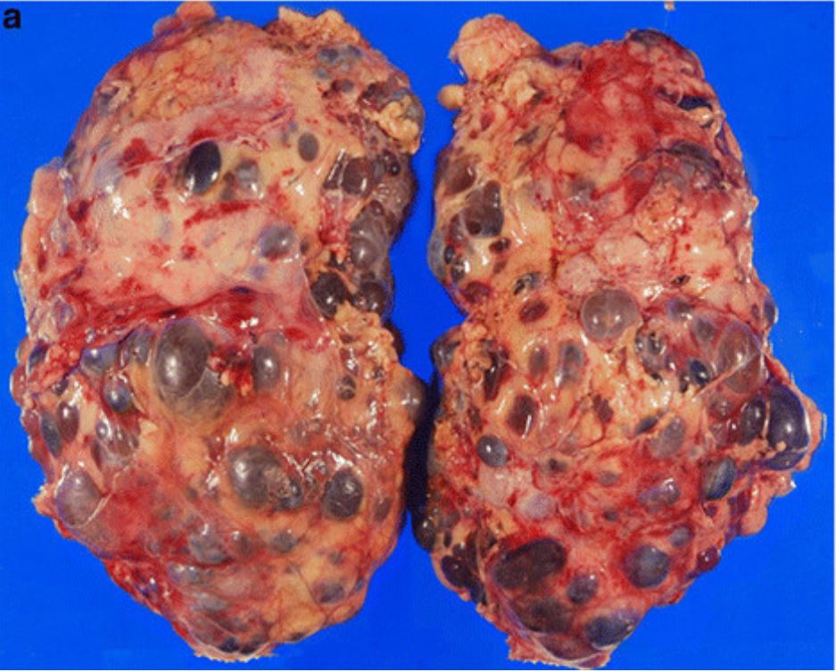
Gross pathology from nephrectomy in a patient with autosomal dominant polycystic kidney disease (ADPKD)

Image: “Incidental renal cell carcinoma presenting in a renal transplant recipient with autosomal dominant polycystic kidney disease” by Misumi T, Ide K, Onoe T, Banshodani M, Tazawa H, Teraoka Y, Hotta R, Yamashita M, Tashiro H, Ohdan H. License: CC BY 2.0, cropped by Lecturio.

Comparison

The 2 main types of polycystic kidney disease are ADPKD and autosomal recessive polycystic kidney disease Autosomal Recessive Polycystic Kidney Disease Polycystic kidney disease (PKD) is an inherited genetic disorder leading to the development of numerous fluid-filled cysts in the kidneys. Autosomal recessive polycystic kidney disease is primarily characterized by cystic dilatations of the renal collecting ducts and intrahepatic bile duct dilatation with hepatic fibrosis. Autosomal Recessive Polycystic Kidney Disease (ARPKD) ( ARPKD ARPKD Polycystic kidney disease (PKD) is an inherited genetic disorder leading to the development of numerous fluid-filled cysts in the kidneys. Autosomal recessive polycystic kidney disease is primarily characterized by cystic dilatations of the renal collecting ducts and intrahepatic bile duct dilatation with hepatic fibrosis. Autosomal Recessive Polycystic Kidney Disease (ARPKD)).

Table: ADPKD vs ARPKD ARPKD Polycystic kidney disease (PKD) is an inherited genetic disorder leading to the development of numerous fluid-filled cysts in the kidneys. Autosomal recessive polycystic kidney disease is primarily characterized by cystic dilatations of the renal collecting ducts and intrahepatic bile duct dilatation with hepatic fibrosis. Autosomal Recessive Polycystic Kidney Disease (ARPKD)
ADPKD ARPKD ARPKD Polycystic kidney disease (PKD) is an inherited genetic disorder leading to the development of numerous fluid-filled cysts in the kidneys. Autosomal recessive polycystic kidney disease is primarily characterized by cystic dilatations of the renal collecting ducts and intrahepatic bile duct dilatation with hepatic fibrosis. Autosomal Recessive Polycystic Kidney Disease (ARPKD)
Inheritance Autosomal dominant Autosomal dominant Autosomal inheritance, both dominant and recessive, refers to the transmission of genes from the 22 autosomal chromosomes. Autosomal dominant diseases are expressed when only 1 copy of the dominant allele is inherited. Autosomal Recessive and Autosomal Dominant Inheritance Autosomal recessive Autosomal recessive Autosomal inheritance, both dominant and recessive, refers to the transmission of genes from the 22 autosomal chromosomes. Autosomal recessive diseases are only expressed when 2 copies of the recessive allele are inherited. Autosomal Recessive and Autosomal Dominant Inheritance
Genes Genes A category of nucleic acid sequences that function as units of heredity and which code for the basic instructions for the development, reproduction, and maintenance of organisms. DNA Types and Structure involved PKD1, PKD2 PKHD1 PKHD1 Autosomal Recessive Polycystic Kidney Disease (ARPKD)
Associated proteins Proteins Linear polypeptides that are synthesized on ribosomes and may be further modified, crosslinked, cleaved, or assembled into complex proteins with several subunits. The specific sequence of amino acids determines the shape the polypeptide will take, during protein folding, and the function of the protein. Energy Homeostasis Polycystin-1, polycystin-2 Fibrocystin
Age of presentation Adulthood Antenatal, neonatal, infant
Clinical features
  • Impaired renal function
  • Enlarged kidneys Kidneys The kidneys are a pair of bean-shaped organs located retroperitoneally against the posterior wall of the abdomen on either side of the spine. As part of the urinary tract, the kidneys are responsible for blood filtration and excretion of water-soluble waste in the urine. Kidneys: Anatomy
  • Cerebral aneurysm Cerebral aneurysm Brain aneurysms, also known as intracranial or cerebral aneurysms, are dilations of the arteries along points of weakness in the brain. The majority of the aneurysms are berry (saccular) in nature and located within the anterior circulation of the circle of Willis. Brain Aneurysms
  • Hepatic and pancreatic cysts Cysts Any fluid-filled closed cavity or sac that is lined by an epithelium. Cysts can be of normal, abnormal, non-neoplastic, or neoplastic tissues. Fibrocystic Change
  • Colon Colon The large intestines constitute the last portion of the digestive system. The large intestine consists of the cecum, appendix, colon (with ascending, transverse, descending, and sigmoid segments), rectum, and anal canal. The primary function of the colon is to remove water and compact the stool prior to expulsion from the body via the rectum and anal canal. Colon, Cecum, and Appendix: Anatomy diverticulosis Diverticulosis A pathological condition characterized by the presence of a number of colonic diverticula in the colon. Its pathogenesis is multifactorial, including colon aging, motor dysfunction, increases in intraluminal pressure, and lack of dietary fibers. Diverticular Disease
  • Abdominal hernias
Gross and pathologic morphology
  • Large kidneys Kidneys The kidneys are a pair of bean-shaped organs located retroperitoneally against the posterior wall of the abdomen on either side of the spine. As part of the urinary tract, the kidneys are responsible for blood filtration and excretion of water-soluble waste in the urine. Kidneys: Anatomy, surface full of cysts Cysts Any fluid-filled closed cavity or sac that is lined by an epithelium. Cysts can be of normal, abnormal, non-neoplastic, or neoplastic tissues. Fibrocystic Change, distorted renal architecture
  • Round cysts Cysts Any fluid-filled closed cavity or sac that is lined by an epithelium. Cysts can be of normal, abnormal, non-neoplastic, or neoplastic tissues. Fibrocystic Change of varying size (microcysts and macrocysts)
  • Functioning nephrons between cysts Cysts Any fluid-filled closed cavity or sac that is lined by an epithelium. Cysts can be of normal, abnormal, non-neoplastic, or neoplastic tissues. Fibrocystic Change
  • Large kidneys Kidneys The kidneys are a pair of bean-shaped organs located retroperitoneally against the posterior wall of the abdomen on either side of the spine. As part of the urinary tract, the kidneys are responsible for blood filtration and excretion of water-soluble waste in the urine. Kidneys: Anatomy, smooth surface, preserved general shape of kidneys Kidneys The kidneys are a pair of bean-shaped organs located retroperitoneally against the posterior wall of the abdomen on either side of the spine. As part of the urinary tract, the kidneys are responsible for blood filtration and excretion of water-soluble waste in the urine. Kidneys: Anatomy
  • Microcysts (usually < 2 mm MM Multiple myeloma (MM) is a malignant condition of plasma cells (activated B lymphocytes) primarily seen in the elderly. Monoclonal proliferation of plasma cells results in cytokine-driven osteoclastic activity and excessive secretion of IgG antibodies. Multiple Myeloma), which are tubular (affecting the collecting ducts and tubules), radiate from the medulla to the cortex.
  • Dilation of collecting ducts
Ultrasound findings Multiple cysts Cysts Any fluid-filled closed cavity or sac that is lined by an epithelium. Cysts can be of normal, abnormal, non-neoplastic, or neoplastic tissues. Fibrocystic Change (based on age):
  • < 40 years of age: ≥ 3
  • 40–59 years of age: ≥ 2 each kidney
  • ≥ 60 years of age: ≥ 4 each kidney
  • Enlarged, echogenic kidneys Kidneys The kidneys are a pair of bean-shaped organs located retroperitoneally against the posterior wall of the abdomen on either side of the spine. As part of the urinary tract, the kidneys are responsible for blood filtration and excretion of water-soluble waste in the urine. Kidneys: Anatomy
  • Poor corticomedullary differentiation
  • Multiple tiny cysts Cysts Any fluid-filled closed cavity or sac that is lined by an epithelium. Cysts can be of normal, abnormal, non-neoplastic, or neoplastic tissues. Fibrocystic Change
ADPKD: autosomal dominant polycystic kidney disease
ARPKD: autosomal recessive polycystic kidney disease

Differential Diagnosis

  • Multiple benign Benign Fibroadenoma simple cysts Cysts Any fluid-filled closed cavity or sac that is lined by an epithelium. Cysts can be of normal, abnormal, non-neoplastic, or neoplastic tissues. Fibrocystic Change: Renal cysts Renal Cysts Imaging of the Urinary System are a common, asymptomatic, incidental finding in adults. The cysts Cysts Any fluid-filled closed cavity or sac that is lined by an epithelium. Cysts can be of normal, abnormal, non-neoplastic, or neoplastic tissues. Fibrocystic Change increase in number with age. Differentiation from mild ADPKD may be difficult. Patients Patients Individuals participating in the health care system for the purpose of receiving therapeutic, diagnostic, or preventive procedures. Clinician–Patient Relationship without ADPKD generally do not have a family history Family History Adult Health Maintenance, gross hematuria Hematuria Presence of blood in the urine. Renal Cell Carcinoma, flank pain Flank pain Pain emanating from below the ribs and above the ilium. Renal Cell Carcinoma, or renal insufficiency.
  • Localized renal cystic Cystic Fibrocystic Change disease: a localized, benign Benign Fibroadenoma condition confused with PKD. Unlike PKD, the condition is a localized cystic Cystic Fibrocystic Change disease and neither bilateral nor progressive. Patients Patients Individuals participating in the health care system for the purpose of receiving therapeutic, diagnostic, or preventive procedures. Clinician–Patient Relationship have no family history Family History Adult Health Maintenance of ADPKD. 
  • Acquired renal cystic Cystic Fibrocystic Change disease: chronic kidney disease Chronic Kidney Disease Chronic kidney disease (CKD) is kidney impairment that lasts for ≥ 3 months, implying that it is irreversible. Hypertension and diabetes are the most common causes; however, there are a multitude of other etiologies. In the early to moderate stages, CKD is usually asymptomatic and is primarily diagnosed by laboratory abnormalities. Chronic Kidney Disease is frequently associated with the development of multiple small cysts Cysts Any fluid-filled closed cavity or sac that is lined by an epithelium. Cysts can be of normal, abnormal, non-neoplastic, or neoplastic tissues. Fibrocystic Change bilaterally. The cysts Cysts Any fluid-filled closed cavity or sac that is lined by an epithelium. Cysts can be of normal, abnormal, non-neoplastic, or neoplastic tissues. Fibrocystic Change are often small (< 0.5 cm) and patients Patients Individuals participating in the health care system for the purpose of receiving therapeutic, diagnostic, or preventive procedures. Clinician–Patient Relationship have no family history Family History Adult Health Maintenance. The kidneys Kidneys The kidneys are a pair of bean-shaped organs located retroperitoneally against the posterior wall of the abdomen on either side of the spine. As part of the urinary tract, the kidneys are responsible for blood filtration and excretion of water-soluble waste in the urine. Kidneys: Anatomy have a smooth contour and are small to normal in size. Associated extrarenal manifestations are not noted.
  • Medullary sponge kidney: malformation of the collecting ducts (tubular dilatation) limited to the medullary pyramids. The condition may mimic PKD on urographic studies. Patients Patients Individuals participating in the health care system for the purpose of receiving therapeutic, diagnostic, or preventive procedures. Clinician–Patient Relationship can present with kidney stones Kidney stones Nephrolithiasis is the formation of a stone, or calculus, anywhere along the urinary tract caused by precipitations of solutes in the urine. The most common type of kidney stone is the calcium oxalate stone, but other types include calcium phosphate, struvite (ammonium magnesium phosphate), uric acid, and cystine stones. Nephrolithiasis, hematuria Hematuria Presence of blood in the urine. Renal Cell Carcinoma, and UTIs. Unlike PKD, the renal cortex is spared, which can be seen on CT or MRI.
  • Autosomal dominant Autosomal dominant Autosomal inheritance, both dominant and recessive, refers to the transmission of genes from the 22 autosomal chromosomes. Autosomal dominant diseases are expressed when only 1 copy of the dominant allele is inherited. Autosomal Recessive and Autosomal Dominant Inheritance tuberous sclerosis Sclerosis A pathological process consisting of hardening or fibrosis of an anatomical structure, often a vessel or a nerve. Wilms Tumor complex: multiple, bilateral kidney cysts Cysts Any fluid-filled closed cavity or sac that is lined by an epithelium. Cysts can be of normal, abnormal, non-neoplastic, or neoplastic tissues. Fibrocystic Change. Other findings include renal angiomyolipomas, facial angiofibromas, hypomelanotic macules, and retinal nodular hamartomas. Diagnosis is based on clinical findings and genetic testing Genetic Testing Detection of a mutation; genotype; karyotype; or specific alleles associated with genetic traits, heritable diseases, or predisposition to a disease, or that may lead to the disease in descendants. It includes prenatal genetic testing. Myotonic Dystrophies (TSC1 or TSC2 pathogenic mutation Mutation Genetic mutations are errors in DNA that can cause protein misfolding and dysfunction. There are various types of mutations, including chromosomal, point, frameshift, and expansion mutations. Types of Mutations).
  • Autosomal dominant Autosomal dominant Autosomal inheritance, both dominant and recessive, refers to the transmission of genes from the 22 autosomal chromosomes. Autosomal dominant diseases are expressed when only 1 copy of the dominant allele is inherited. Autosomal Recessive and Autosomal Dominant Inheritance tubulointerstitial kidney disease: a rare genetic disorder characterized by a gradual decline in kidney function from the teenage years. The onset of ESRD is variable Variable Variables represent information about something that can change. The design of the measurement scales, or of the methods for obtaining information, will determine the data gathered and the characteristics of that data. As a result, a variable can be qualitative or quantitative, and may be further classified into subgroups. Types of Variables. Presentation includes early-onset gout Gout Gout is a heterogeneous metabolic disease associated with elevated serum uric acid levels (> 6.8 mg/dL) and abnormal deposits of monosodium urate in tissues. The condition is often familial and is initially characterized by painful, recurring, and usually monoarticular acute arthritis, or “gout flare,” followed later by chronic deforming arthritis. Gout, hyperuricemia Hyperuricemia Excessive uric acid or urate in blood as defined by its solubility in plasma at 37 degrees c; greater than 0. 42 mmol per liter (7. 0 mg/dl) in men or 0. 36 mmol per liter (6. 0 mg/dl) in women. Gout, and, in some, renal cysts Renal Cysts Imaging of the Urinary System. Unlike PKD, cysts Cysts Any fluid-filled closed cavity or sac that is lined by an epithelium. Cysts can be of normal, abnormal, non-neoplastic, or neoplastic tissues. Fibrocystic Change are at the corticomedullary junction and the kidneys Kidneys The kidneys are a pair of bean-shaped organs located retroperitoneally against the posterior wall of the abdomen on either side of the spine. As part of the urinary tract, the kidneys are responsible for blood filtration and excretion of water-soluble waste in the urine. Kidneys: Anatomy are small to normal in size.

References

  1. Bennett W.M., Torres V.E. (2020). Autosomal dominant polycystic kidney disease (ADPKD): Extrarenal manifestations. UpToDate. Retrieved Feb 25, 2021, from https://www.uptodate.com/contents/autosomal-dominant-polycystic-kidney-disease-adpkd-extrarenal-manifestations
  2. Chapman AB, Rahbari-Oskoui FF, Bennett WM. (2020). Autosomal dominant polycystic kidney disease (ADPKD): Renal manifestations. UpToDate. Retrieved Feb 25, 2021, from https://www.uptodate.com/contents/autosomal-dominant-polycystic-kidney-disease-adpkd-renal-manifestations
  3. Chapman A.B., Rahbari-Oskoui, F.F., Bennett, W.M. (2020). Autosomal dominant polycystic kidney disease (ADPKD): Treatment. UpToDate. Retrieved Feb 25, 2021, from https://www.uptodate.com/contents/autosomal-dominant-polycystic-kidney-disease-adpkd-treatment
  4. Genetic and Rare Disease Information Center. (2020). Autosomal dominant polycystic kidney disease. Retrieved February 25, 2021, from https://rarediseases.info.nih.gov/diseases/10413/autosomal-dominant-polycystic-kidney-disease 
  5. Guler, S., Cimen, S., Hurton, S., et al. (2015). Diagnosis and Treatment Modalities of Symptomatic Polycystic Kidney Disease. In Li X (Ed.), Polycystic Kidney Disease [Internet]. Brisbane (AU): Codon Publications. https://www.ncbi.nlm.nih.gov/books/NBK373387/
  6. Halvorson, C.R., Bremmer, M.S., Jacobs, S.C. (2010). Polycystic kidney disease: inheritance, pathophysiology, prognosis, and treatment. Int. J. Nephrol. Renovasc. Dis. 3:69–83.
  7. Kruskal, J.B., Richie, J.P. (2021). Simple and complex renal cysts in adults. UpToDate. Retrieved Feb 25, 2021, from https://www.uptodate.com/contents/simple-and-complex-renal-cysts-in-adults
  8. Merck Manual. (2019). Autosomal Dominant Polycystic Kidney Disease (ADPKD). https://rarediseases.info.nih.gov/diseases/10413/autosomal-dominant-polycystic-kidney-disease
  9. National Institute of Diabetes and Digestive and Kidney Diseases. (2017). What is Polycystic Kidney Disease? https://www.niddk.nih.gov/health-information/kidney-disease/polycystic-kidney-disease/what-is-pkd#symptoms
  10. Patil A., Sweeney W.E. Jr., Avner E.D., et al. (2015). Childhood Polycystic Kidney Disease. Polycystic Kidney Disease. https://www.ncbi.nlm.nih.gov/books/NBK373381/
  11. Torres V.E., Bennett W.M. (2021). Autosomal dominant polycystic kidney disease (ADPKD) in adults: Epidemiology, clinical presentation, and diagnosis. UpToDate. Retrieved Feb 25, 2021, from https://www.uptodate.com/contents/autosomal-dominant-polycystic-kidney-disease-adpkd-in-adults-epidemiology-clinical-presentation-and-diagnosis
  12. Zhou J, & Pollak M.R. (2018). Polycystic kidney disease and other inherited disorders of tubule growth and development. In Jameson LJ, et al. (Ed.), Harrison’s Principles of Internal Medicine (20th ed.) https://accessmedicine-mhmedical-com.aucmed.idm.oclc.org/content.aspx?bookid=2129&sectionid=192281464

Create your free account or log in to continue reading!

Sign up now and get free access to Lecturio with concept pages, medical videos, and questions for your medical education.

User Reviews

Details