Achieve Mastery of Medical Concepts

Study for medical school and boards with Lecturio

Autosomal Recessive Polycystic Kidney Disease (ARPKD)

Polycystic kidney disease (PKD) is an inherited genetic disorder leading to the development of numerous fluid-filled cysts Cysts Any fluid-filled closed cavity or sac that is lined by an epithelium. Cysts can be of normal, abnormal, non-neoplastic, or neoplastic tissues. Fibrocystic Change in the kidneys Kidneys The kidneys are a pair of bean-shaped organs located retroperitoneally against the posterior wall of the abdomen on either side of the spine. As part of the urinary tract, the kidneys are responsible for blood filtration and excretion of water-soluble waste in the urine. Kidneys: Anatomy. The 2 main types of PKD are autosomal recessive Autosomal recessive Autosomal inheritance, both dominant and recessive, refers to the transmission of genes from the 22 autosomal chromosomes. Autosomal recessive diseases are only expressed when 2 copies of the recessive allele are inherited. Autosomal Recessive and Autosomal Dominant Inheritance polycystic kidney disease (ARPKD), which is often diagnosed antenatally or shortly after birth, and autosomal dominant polycystic kidney disease Autosomal dominant polycystic kidney disease Polycystic kidney disease (PKD) is an inherited genetic disorder leading to the development of numerous fluid-filled cysts in the kidneys. The 2 main types of PKD are autosomal dominant polycystic kidney disease (ADPKD), which is often diagnosed in adulthood, and autosomal recessive polycystic kidney disease (ARPKD), which is often diagnosed antenatally or shortly after birth. Autosomal dominant polycystic kidney disease (ADPKD) ( ADPKD ADPKD Polycystic kidney disease (PKD) is an inherited genetic disorder leading to the development of numerous fluid-filled cysts in the kidneys. The 2 main types of PKD are autosomal dominant polycystic kidney disease (ADPKD), which is often diagnosed in adulthood, and autosomal recessive polycystic kidney disease (ARPKD), which is often diagnosed antenatally or shortly after birth. Autosomal dominant polycystic kidney disease (ADPKD)), which is often diagnosed in adulthood. Autosomal recessive Autosomal recessive Autosomal inheritance, both dominant and recessive, refers to the transmission of genes from the 22 autosomal chromosomes. Autosomal recessive diseases are only expressed when 2 copies of the recessive allele are inherited. Autosomal Recessive and Autosomal Dominant Inheritance polycystic kidney disease is primarily characterized by cystic Cystic Fibrocystic Change dilatations of the renal collecting ducts and intrahepatic bile Bile An emulsifying agent produced in the liver and secreted into the duodenum. Its composition includes bile acids and salts; cholesterol; and electrolytes. It aids digestion of fats in the duodenum. Gallbladder and Biliary Tract: Anatomy duct dilatation with hepatic fibrosis Fibrosis Any pathological condition where fibrous connective tissue invades any organ, usually as a consequence of inflammation or other injury. Bronchiolitis Obliterans. Diagnosis is through physical exam and ultrasonography. Management requires a multidisciplinary approach to slow the progression of renal disease by controlling hypertension Hypertension Hypertension, or high blood pressure, is a common disease that manifests as elevated systemic arterial pressures. Hypertension is most often asymptomatic and is found incidentally as part of a routine physical examination or during triage for an unrelated medical encounter. Hypertension, proteinuria Proteinuria The presence of proteins in the urine, an indicator of kidney diseases. Nephrotic Syndrome in Children, and symptoms. The prognosis Prognosis A prediction of the probable outcome of a disease based on a individual's condition and the usual course of the disease as seen in similar situations. Non-Hodgkin Lymphomas of ARPKD depends on the age at presentation, along with the degree of hepatic and renal involvement. Patients Patients Individuals participating in the health care system for the purpose of receiving therapeutic, diagnostic, or preventive procedures. Clinician–Patient Relationship progressing to end-stage renal disease (ESRD) will need renal replacement therapy.

Last updated: Nov 17, 2022

Editorial responsibility: Stanley Oiseth, Lindsay Jones, Evelin Maza

Epidemiology and Etiology

Epidemiology

Polycystic kidney disease (PKD) affects about 500,000 people in the United States. Autosomal recessive Autosomal recessive Autosomal inheritance, both dominant and recessive, refers to the transmission of genes from the 22 autosomal chromosomes. Autosomal recessive diseases are only expressed when 2 copies of the recessive allele are inherited. Autosomal Recessive and Autosomal Dominant Inheritance polycystic kidney disease (ARPKD) (formerly known as infantile PKD), is 1 of the 2 main types of PKD.

  • Prevalence Prevalence The total number of cases of a given disease in a specified population at a designated time. It is differentiated from incidence, which refers to the number of new cases in the population at a given time. Measures of Disease Frequency: 1 per 20,000 live births
  • ⅓ present before 1 year old
  • ⅓ present between 1–20 years old
  • ⅓ present after 20 years old

Etiology

  • ARPKD: autosomal recessive Autosomal recessive Autosomal inheritance, both dominant and recessive, refers to the transmission of genes from the 22 autosomal chromosomes. Autosomal recessive diseases are only expressed when 2 copies of the recessive allele are inherited. Autosomal Recessive and Autosomal Dominant Inheritance
  • Either a mutation Mutation Genetic mutations are errors in DNA that can cause protein misfolding and dysfunction. There are various types of mutations, including chromosomal, point, frameshift, and expansion mutations. Types of Mutations of:
    • PKHD1 on chromosome Chromosome In a prokaryotic cell or in the nucleus of a eukaryotic cell, a structure consisting of or containing DNA which carries the genetic information essential to the cell. Basic Terms of Genetics 6p21 OR:
      • Encodes fibrocystin (large integral membrane protein) found in the cortical and medullary collecting ducts of the kidney and hepatic bile Bile An emulsifying agent produced in the liver and secreted into the duodenum. Its composition includes bile acids and salts; cholesterol; and electrolytes. It aids digestion of fats in the duodenum. Gallbladder and Biliary Tract: Anatomy duct cells
      • Defect in fibrocystin → dysfunction of renal cilia
    • DZIP1L (less common):
      • Located on the 2nd locus Locus Specific regions that are mapped within a genome. Genetic loci are usually identified with a shorthand notation that indicates the chromosome number and the position of a specific band along the P or Q arm of the chromosome where they are found. For example the locus 6p21 is found within band 21 of the P-arm of chromosome 6. Many well known genetic loci are also known by common names that are associated with a genetic function or hereditary disease. Basic Terms of Genetics for ARPKD
      • Presentation includes enlarged hyperechogenic kidneys Kidneys The kidneys are a pair of bean-shaped organs located retroperitoneally against the posterior wall of the abdomen on either side of the spine. As part of the urinary tract, the kidneys are responsible for blood filtration and excretion of water-soluble waste in the urine. Kidneys: Anatomy and arterial hypertension Hypertension Hypertension, or high blood pressure, is a common disease that manifests as elevated systemic arterial pressures. Hypertension is most often asymptomatic and is found incidentally as part of a routine physical examination or during triage for an unrelated medical encounter. Hypertension

Clinical Presentation

General manifestations

Manifestations vary by age and affect the following:

  • Kidneys Kidneys The kidneys are a pair of bean-shaped organs located retroperitoneally against the posterior wall of the abdomen on either side of the spine. As part of the urinary tract, the kidneys are responsible for blood filtration and excretion of water-soluble waste in the urine. Kidneys: Anatomy:
    • Cystic Cystic Fibrocystic Change dilations of the collecting ducts
    • Microcysts < 2 mm MM Multiple myeloma (MM) is a malignant condition of plasma cells (activated B lymphocytes) primarily seen in the elderly. Monoclonal proliferation of plasma cells results in cytokine-driven osteoclastic activity and excessive secretion of IgG antibodies. Multiple Myeloma in size
    • Impaired renal function
  • Hepatobiliary tract: intrahepatic bile Bile An emulsifying agent produced in the liver and secreted into the duodenum. Its composition includes bile acids and salts; cholesterol; and electrolytes. It aids digestion of fats in the duodenum. Gallbladder and Biliary Tract: Anatomy duct dilatation and hepatic fibrosis Fibrosis Any pathological condition where fibrous connective tissue invades any organ, usually as a consequence of inflammation or other injury. Bronchiolitis Obliterans

Antenatal and neonatal

  • Can be detected antenatally (by 24-weeks gestational age Gestational age The age of the conceptus, beginning from the time of fertilization. In clinical obstetrics, the gestational age is often estimated as the time from the last day of the last menstruation which is about 2 weeks before ovulation and fertilization. Pregnancy: Diagnosis, Physiology, and Care):
  • Potter sequence, which is due to severe oligohydramnios Oligohydramnios Oligohydramnios refers to amniotic fluid volume less than expected for the current gestational age. Oligohydramnios is diagnosed by ultrasound and defined as an amniotic fluid index (AFI) of ‰¤ 5 cm or a single deep pocket (SDP) of < 2 cm in the 2nd or 3rd trimester. Oligohydramnios and the compressive effect of enlarged kidneys Enlarged kidneys Autosomal dominant polycystic kidney disease (ADPKD):
  • Kidney manifestations:
    • Protruding abdomen due to bilaterally enlarged kidneys Enlarged kidneys Autosomal dominant polycystic kidney disease (ADPKD)
    • Chronic renal failure Renal failure Conditions in which the kidneys perform below the normal level in the ability to remove wastes, concentrate urine, and maintain electrolyte balance; blood pressure; and calcium metabolism. Renal insufficiency can be classified by the degree of kidney damage (as measured by the level of proteinuria) and reduction in glomerular filtration rate. Crush Syndrome or even end-stage renal disease (ESRD)
    • Hypertension Hypertension Hypertension, or high blood pressure, is a common disease that manifests as elevated systemic arterial pressures. Hypertension is most often asymptomatic and is found incidentally as part of a routine physical examination or during triage for an unrelated medical encounter. Hypertension
    • Hyponatremia Hyponatremia Hyponatremia is defined as a decreased serum sodium (sNa+) concentration less than 135 mmol/L. Serum sodium is the greatest contributor to plasma osmolality, which is very tightly controlled via antidiuretic hormone (ADH) release from the hypothalamus and by the thirst mechanism. Hyponatremia
  • Respiratory distress: noted in 50% of patients Patients Individuals participating in the health care system for the purpose of receiving therapeutic, diagnostic, or preventive procedures. Clinician–Patient Relationship due to pulmonary insufficiency and hypoplasia Hypoplasia Hypoplastic Left Heart Syndrome (HLHS) 
Arpkd

Autosomal recessive polycystic kidney disease (ARPKD):
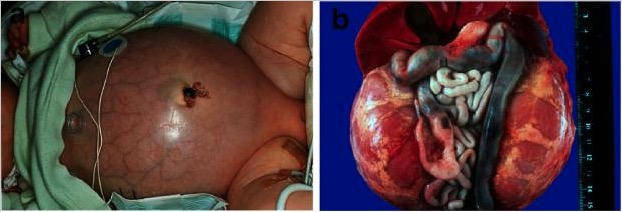
a) Baby with a distended abdomen due to voluminous kidneys, leading to respiratory problems and early demise
b) Abdominal situs of a perinatally demised ARPKD patient with symmetrically enlarged kidneys in their reniform configuration

Image: “Fig. 2” by Carsten Bergmann. License: CC BY 2.0

Infancy and childhood

In the 1st 3 years, survivors of the neonatal period experience a temporary improvement of renal function, followed by a decline.

  • Kidney manifestations:
    • Protruding abdomen due to bilaterally enlarged kidneys Enlarged kidneys Autosomal dominant polycystic kidney disease (ADPKD)
    • Polyuria Polyuria Urination of a large volume of urine with an increase in urinary frequency, commonly seen in diabetes. Renal Potassium Regulation and polydipsia Polydipsia Excessive thirst manifested by excessive fluid intake. It is characteristic of many diseases such as diabetes mellitus; diabetes insipidus; and nephrogenic diabetes insipidus. The condition may be psychogenic in origin. Diabetes Insipidus (usually the 1st signs of renal insufficiency)
    • Hypertension Hypertension Hypertension, or high blood pressure, is a common disease that manifests as elevated systemic arterial pressures. Hypertension is most often asymptomatic and is found incidentally as part of a routine physical examination or during triage for an unrelated medical encounter. Hypertension
    • Metabolic acidosis Acidosis A pathologic condition of acid accumulation or depletion of base in the body. The two main types are respiratory acidosis and metabolic acidosis, due to metabolic acid build up. Respiratory Acidosis
    • Recurrent urinary tract Urinary tract The urinary tract is located in the abdomen and pelvis and consists of the kidneys, ureters, urinary bladder, and urethra. The structures permit the excretion of urine from the body. Urine flows from the kidneys through the ureters to the urinary bladder and out through the urethra. Urinary Tract: Anatomy infections Infections Invasion of the host organism by microorganisms or their toxins or by parasites that can cause pathological conditions or diseases. Chronic Granulomatous Disease (UTIs)
    • Urinary abnormalities such as:
      • Proteinuria Proteinuria The presence of proteins in the urine, an indicator of kidney diseases. Nephrotic Syndrome in Children
      • Glucosuria Glucosuria Diabetes Mellitus
      • Hyperphosphaturia
      • Increased magnesium Magnesium A metallic element that has the atomic symbol mg, atomic number 12, and atomic weight 24. 31. It is important for the activity of many enzymes, especially those involved in oxidative phosphorylation. Electrolytes excretion in urine
    • Progression to CKD CKD Chronic kidney disease (CKD) is kidney impairment that lasts for ≥ 3 months, implying that it is irreversible. Hypertension and diabetes are the most common causes; however, there are a multitude of other etiologies. In the early to moderate stages, CKD is usually asymptomatic and is primarily diagnosed by laboratory abnormalities. Chronic Kidney Disease:
      • Cyst formation and fibrosis Fibrosis Any pathological condition where fibrous connective tissue invades any organ, usually as a consequence of inflammation or other injury. Bronchiolitis Obliterans
      • Decreased renal function
  • Liver Liver The liver is the largest gland in the human body. The liver is found in the superior right quadrant of the abdomen and weighs approximately 1.5 kilograms. Its main functions are detoxification, metabolism, nutrient storage (e.g., iron and vitamins), synthesis of coagulation factors, formation of bile, filtration, and storage of blood. Liver: Anatomy manifestations:
    • May be a prominent feature of ARPKD in patients Patients Individuals participating in the health care system for the purpose of receiving therapeutic, diagnostic, or preventive procedures. Clinician–Patient Relationship presenting at an older age
    • Can present with hepatomegaly
    • Congenital Congenital Chorioretinitis hepatic fibrosis Fibrosis Any pathological condition where fibrous connective tissue invades any organ, usually as a consequence of inflammation or other injury. Bronchiolitis Obliterans
    • Liver failure Liver failure Severe inability of the liver to perform its normal metabolic functions, as evidenced by severe jaundice and abnormal serum levels of ammonia; bilirubin; alkaline phosphatase; aspartate aminotransferase; lactate dehydrogenases; and albumin/globulin ratio. Autoimmune Hepatitis
    • Portal hypertension Portal hypertension Portal hypertension is increased pressure in the portal venous system. This increased pressure can lead to splanchnic vasodilation, collateral blood flow through portosystemic anastomoses, and increased hydrostatic pressure. There are a number of etiologies, including cirrhosis, right-sided congestive heart failure, schistosomiasis, portal vein thrombosis, hepatitis, and Budd-Chiari syndrome. Portal Hypertension
  • Other findings: impaired growth and neurologic development, left ventricular hypertrophy Ventricular Hypertrophy Tetralogy of Fallot

Diagnosis

Physical exam

  • Assess for Potter syndrome.
  • Assess for palpable kidneys Kidneys The kidneys are a pair of bean-shaped organs located retroperitoneally against the posterior wall of the abdomen on either side of the spine. As part of the urinary tract, the kidneys are responsible for blood filtration and excretion of water-soluble waste in the urine. Kidneys: Anatomy
  • Abdominal tenderness to palpation Palpation Application of fingers with light pressure to the surface of the body to determine consistency of parts beneath in physical diagnosis; includes palpation for determining the outlines of organs. Dermatologic Examination 
  • Assess for pulmonary or liver Liver The liver is the largest gland in the human body. The liver is found in the superior right quadrant of the abdomen and weighs approximately 1.5 kilograms. Its main functions are detoxification, metabolism, nutrient storage (e.g., iron and vitamins), synthesis of coagulation factors, formation of bile, filtration, and storage of blood. Liver: Anatomy manifestations of the disease.

Imaging studies

  • Ultrasonography of the abdomen:
    • Kidneys Kidneys The kidneys are a pair of bean-shaped organs located retroperitoneally against the posterior wall of the abdomen on either side of the spine. As part of the urinary tract, the kidneys are responsible for blood filtration and excretion of water-soluble waste in the urine. Kidneys: Anatomy:
      • Enlarged, hyperechogenic kidneys Kidneys The kidneys are a pair of bean-shaped organs located retroperitoneally against the posterior wall of the abdomen on either side of the spine. As part of the urinary tract, the kidneys are responsible for blood filtration and excretion of water-soluble waste in the urine. Kidneys: Anatomy bilaterally
      • Poor corticomedullary differentiation
      • Numerous tiny cysts Cysts Any fluid-filled closed cavity or sac that is lined by an epithelium. Cysts can be of normal, abnormal, non-neoplastic, or neoplastic tissues. Fibrocystic Change and ductal dilatation
    • Liver Liver The liver is the largest gland in the human body. The liver is found in the superior right quadrant of the abdomen and weighs approximately 1.5 kilograms. Its main functions are detoxification, metabolism, nutrient storage (e.g., iron and vitamins), synthesis of coagulation factors, formation of bile, filtration, and storage of blood. Liver: Anatomy:
      • Hepatomegaly 
      • Hyperechogenic cysts Cysts Any fluid-filled closed cavity or sac that is lined by an epithelium. Cysts can be of normal, abnormal, non-neoplastic, or neoplastic tissues. Fibrocystic Change
      • Dilatation of the main bile Bile An emulsifying agent produced in the liver and secreted into the duodenum. Its composition includes bile acids and salts; cholesterol; and electrolytes. It aids digestion of fats in the duodenum. Gallbladder and Biliary Tract: Anatomy ducts and the peripheral intrahepatic ducts
  • MRI or CT for further characterization of the disease, or if inconclusive ultrasound findings:
    • MRI: renal enlargement with microcysts in a hyperintense Hyperintense Magnetic Resonance Imaging (MRI) radial pattern in the cortex and medulla
    • CT: not the initial choice due to radiation Radiation Emission or propagation of acoustic waves (sound), electromagnetic energy waves (such as light; radio waves; gamma rays; or x-rays), or a stream of subatomic particles (such as electrons; neutrons; protons; or alpha particles). Osteosarcoma
Ultrasonography of the right kidney demonstrating autosomal recessive polycystic kidney disease

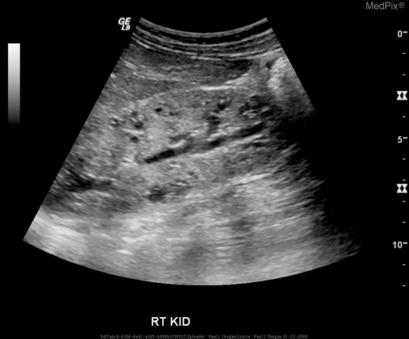
Ultrasonography of the right kidney demonstrating autosomal recessive polycystic kidney disease (ARPKD):
diffusely increased echogenicity, loss of corticomedullary differentiation, and multiple microcysts within the renal parenchyma

Image: “Autosomal Recessive Polycystic Kidney Disease” by Shogan PJS. License: CC BY 4.0

Genetic testing Genetic Testing Detection of a mutation; genotype; karyotype; or specific alleles associated with genetic traits, heritable diseases, or predisposition to a disease, or that may lead to the disease in descendants. It includes prenatal genetic testing. Myotonic Dystrophies

  • Diagnosis is inconclusive after imaging.
  • Prenatal diagnosis
  • Genetic counseling Genetic Counseling An educational process that provides information and advice to individuals or families about a genetic condition that may affect them. The purpose is to help individuals make informed decisions about marriage, reproduction, and other health management issues based on information about the genetic disease, the available diagnostic tests, and management programs. Psychosocial support is usually offered. Myotonic Dystrophies (determine familial carriers Carriers The Cell: Cell Membrane of the mutated PKHD1 gene Gene A category of nucleic acid sequences that function as units of heredity and which code for the basic instructions for the development, reproduction, and maintenance of organisms. Basic Terms of Genetics)

Management and Prognosis

Management approach

Antenatal management (if detected early):

  • Ultrasound monitoring every 2–3 weeks (check renal size and amniotic fluid Amniotic fluid A clear, yellowish liquid that envelopes the fetus inside the sac of amnion. In the first trimester, it is likely a transudate of maternal or fetal plasma. In the second trimester, amniotic fluid derives primarily from fetal lung and kidney. Cells or substances in this fluid can be removed for prenatal diagnostic tests (amniocentesis). Placenta, Umbilical Cord, and Amniotic Cavity volume)
  • Neonatal intensive care planning

Neonatal management:

  • Supportive respiratory care (if respiratory distress due to pulmonary hypoplasia Hypoplasia Hypoplastic Left Heart Syndrome (HLHS))
  • Mechanical ventilation Ventilation The total volume of gas inspired or expired per unit of time, usually measured in liters per minute. Ventilation: Mechanics of Breathing
  • Monitor/manage renal function and serum electrolytes Electrolytes Electrolytes are mineral salts that dissolve in water and dissociate into charged particles called ions, which can be either be positively (cations) or negatively (anions) charged. Electrolytes are distributed in the extracellular and intracellular compartments in different concentrations. Electrolytes are essential for various basic life-sustaining functions. Electrolytes
  • ACE inhibitor for hypertension Hypertension Hypertension, or high blood pressure, is a common disease that manifests as elevated systemic arterial pressures. Hypertension is most often asymptomatic and is found incidentally as part of a routine physical examination or during triage for an unrelated medical encounter. Hypertension
  • Fluid restriction for hyponatremia Hyponatremia Hyponatremia is defined as a decreased serum sodium (sNa+) concentration less than 135 mmol/L. Serum sodium is the greatest contributor to plasma osmolality, which is very tightly controlled via antidiuretic hormone (ADH) release from the hypothalamus and by the thirst mechanism. Hyponatremia
  • Supplemental nasogastric feedings for feeding difficulties
  • Dialysis Dialysis Renal replacement therapy refers to dialysis and/or kidney transplantation. Dialysis is a procedure by which toxins and excess water are removed from the circulation. Hemodialysis and peritoneal dialysis (PD) are the two types of dialysis, and their primary difference is the location of the filtration process (external to the body in hemodialysis versus inside the body for PD). Peritoneal Dialysis and Hemodialysis if ESRD
  • Nephrectomy Nephrectomy Excision of kidney. Renal Cell Carcinoma if absolutely needed for survival

Infancy and childhood management:

  • Monitor kidney function.
  • Monitor liver Liver The liver is the largest gland in the human body. The liver is found in the superior right quadrant of the abdomen and weighs approximately 1.5 kilograms. Its main functions are detoxification, metabolism, nutrient storage (e.g., iron and vitamins), synthesis of coagulation factors, formation of bile, filtration, and storage of blood. Liver: Anatomy status:
    • Check for signs of portal hypertension Portal hypertension Portal hypertension is increased pressure in the portal venous system. This increased pressure can lead to splanchnic vasodilation, collateral blood flow through portosystemic anastomoses, and increased hydrostatic pressure. There are a number of etiologies, including cirrhosis, right-sided congestive heart failure, schistosomiasis, portal vein thrombosis, hepatitis, and Budd-Chiari syndrome. Portal Hypertension.
    • Yearly abdominal ultrasound
  • May require supplemental feedings for feeding intolerance and growth
  • ACE inhibitor or ARB for blood pressure control
  • Manage complications (e.g., UTI UTI Urinary tract infections (UTIs) represent a wide spectrum of diseases, from self-limiting simple cystitis to severe pyelonephritis that can result in sepsis and death. Urinary tract infections are most commonly caused by Escherichia coli, but may also be caused by other bacteria and fungi. Urinary tract infections (UTIs))
  • Kidney transplantation Kidney Transplantation The transference of a kidney from one human or animal to another. Organ Transplantation for ESRD
  • May need a liver Liver The liver is the largest gland in the human body. The liver is found in the superior right quadrant of the abdomen and weighs approximately 1.5 kilograms. Its main functions are detoxification, metabolism, nutrient storage (e.g., iron and vitamins), synthesis of coagulation factors, formation of bile, filtration, and storage of blood. Liver: Anatomy transplant

Prognosis Prognosis A prediction of the probable outcome of a disease based on a individual’s condition and the usual course of the disease as seen in similar situations. Non-Hodgkin Lymphomas

  • Affected largely by the degree of renal and hepatic involvement (usually dependent on the age of presentation)
  • Worst prognosis Prognosis A prediction of the probable outcome of a disease based on a individual’s condition and the usual course of the disease as seen in similar situations. Non-Hodgkin Lymphomas ( mortality Mortality All deaths reported in a given population. Measures of Health Status rate of 30%): presentation as a neonate Neonate An infant during the first 28 days after birth. Physical Examination of the Newborn with severe renal disease and pulmonary insufficiency
  • Survival past the 1st month: > 80% chance of survival past 15 years old
Autosomal recessive polycystic kidney disease

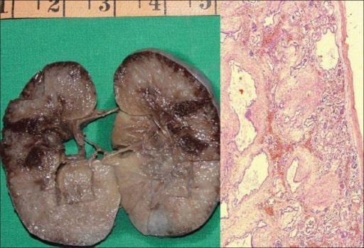
Autosomal recessive polycystic kidney disease (ARPKD) in a fetus:
On the left, a gross specimen is shown of a right kidney measuring 10 × 7 × 4 cm. The cut surface is spongy with poor corticomedullary differentiation. There are multiple, tiny cysts with some at right angles to the cortical surface.
On the right, a photomicrograph is showing numerous cysts lined by a single layer of low cuboidal epithelial cells with thick, peritubular mesenchyme. The glomeruli are normal (H&E, 40x).

Image: “Autosomal recessive polycystic kidney” by Department of Urology, Kasturba Medical College, Manipal, India. License: CC BY 2.0

Comparison

The 2 main types of PKD are autosomal dominant polycystic kidney disease Autosomal dominant polycystic kidney disease Polycystic kidney disease (PKD) is an inherited genetic disorder leading to the development of numerous fluid-filled cysts in the kidneys. The 2 main types of PKD are autosomal dominant polycystic kidney disease (ADPKD), which is often diagnosed in adulthood, and autosomal recessive polycystic kidney disease (ARPKD), which is often diagnosed antenatally or shortly after birth. Autosomal dominant polycystic kidney disease (ADPKD) ( ADPKD ADPKD Polycystic kidney disease (PKD) is an inherited genetic disorder leading to the development of numerous fluid-filled cysts in the kidneys. The 2 main types of PKD are autosomal dominant polycystic kidney disease (ADPKD), which is often diagnosed in adulthood, and autosomal recessive polycystic kidney disease (ARPKD), which is often diagnosed antenatally or shortly after birth. Autosomal dominant polycystic kidney disease (ADPKD)) and ARPKD.

Table: ADPKD ADPKD Polycystic kidney disease (PKD) is an inherited genetic disorder leading to the development of numerous fluid-filled cysts in the kidneys. The 2 main types of PKD are autosomal dominant polycystic kidney disease (ADPKD), which is often diagnosed in adulthood, and autosomal recessive polycystic kidney disease (ARPKD), which is often diagnosed antenatally or shortly after birth. Autosomal dominant polycystic kidney disease (ADPKD) vs. ARPKD
ADPKD ADPKD Polycystic kidney disease (PKD) is an inherited genetic disorder leading to the development of numerous fluid-filled cysts in the kidneys. The 2 main types of PKD are autosomal dominant polycystic kidney disease (ADPKD), which is often diagnosed in adulthood, and autosomal recessive polycystic kidney disease (ARPKD), which is often diagnosed antenatally or shortly after birth. Autosomal dominant polycystic kidney disease (ADPKD) ARPKD
Inheritance Autosomal dominant Autosomal dominant Autosomal inheritance, both dominant and recessive, refers to the transmission of genes from the 22 autosomal chromosomes. Autosomal dominant diseases are expressed when only 1 copy of the dominant allele is inherited. Autosomal Recessive and Autosomal Dominant Inheritance Autosomal recessive Autosomal recessive Autosomal inheritance, both dominant and recessive, refers to the transmission of genes from the 22 autosomal chromosomes. Autosomal recessive diseases are only expressed when 2 copies of the recessive allele are inherited. Autosomal Recessive and Autosomal Dominant Inheritance
Genes Genes A category of nucleic acid sequences that function as units of heredity and which code for the basic instructions for the development, reproduction, and maintenance of organisms. DNA Types and Structure involved PKD1, PKD2 PKD1, PKD2 Autosomal dominant polycystic kidney disease (ADPKD) PKHD1
Associated proteins Proteins Linear polypeptides that are synthesized on ribosomes and may be further modified, crosslinked, cleaved, or assembled into complex proteins with several subunits. The specific sequence of amino acids determines the shape the polypeptide will take, during protein folding, and the function of the protein. Energy Homeostasis Polycystin-1, polycystin-2 Fibrocystin
Age of presentation Adulthood Antenatal, neonatal, infant
Clinical features
  • Impaired renal function
  • Enlarged kidneys Enlarged kidneys Autosomal dominant polycystic kidney disease (ADPKD)
  • Cerebral aneurysm Cerebral aneurysm Brain aneurysms, also known as intracranial or cerebral aneurysms, are dilations of the arteries along points of weakness in the brain. The majority of the aneurysms are berry (saccular) in nature and located within the anterior circulation of the circle of Willis. Brain Aneurysms
  • Hepatic and pancreatic cysts Cysts Any fluid-filled closed cavity or sac that is lined by an epithelium. Cysts can be of normal, abnormal, non-neoplastic, or neoplastic tissues. Fibrocystic Change
  • Colon Colon The large intestines constitute the last portion of the digestive system. The large intestine consists of the cecum, appendix, colon (with ascending, transverse, descending, and sigmoid segments), rectum, and anal canal. The primary function of the colon is to remove water and compact the stool prior to expulsion from the body via the rectum and anal canal. Colon, Cecum, and Appendix: Anatomy diverticulosis Diverticulosis A pathological condition characterized by the presence of a number of colonic diverticula in the colon. Its pathogenesis is multifactorial, including colon aging, motor dysfunction, increases in intraluminal pressure, and lack of dietary fibers. Diverticular Disease
  • Abdominal hernias
Gross and pathologic morphology
  • Large kidneys Kidneys The kidneys are a pair of bean-shaped organs located retroperitoneally against the posterior wall of the abdomen on either side of the spine. As part of the urinary tract, the kidneys are responsible for blood filtration and excretion of water-soluble waste in the urine. Kidneys: Anatomy, surface full of cysts Cysts Any fluid-filled closed cavity or sac that is lined by an epithelium. Cysts can be of normal, abnormal, non-neoplastic, or neoplastic tissues. Fibrocystic Change, distorted renal architecture
  • Round cysts Cysts Any fluid-filled closed cavity or sac that is lined by an epithelium. Cysts can be of normal, abnormal, non-neoplastic, or neoplastic tissues. Fibrocystic Change of varying size (microcysts and macrocysts)
  • Functioning nephrons between cysts Cysts Any fluid-filled closed cavity or sac that is lined by an epithelium. Cysts can be of normal, abnormal, non-neoplastic, or neoplastic tissues. Fibrocystic Change
  • Large kidneys Kidneys The kidneys are a pair of bean-shaped organs located retroperitoneally against the posterior wall of the abdomen on either side of the spine. As part of the urinary tract, the kidneys are responsible for blood filtration and excretion of water-soluble waste in the urine. Kidneys: Anatomy, smooth surface, preserved general shape of kidneys Kidneys The kidneys are a pair of bean-shaped organs located retroperitoneally against the posterior wall of the abdomen on either side of the spine. As part of the urinary tract, the kidneys are responsible for blood filtration and excretion of water-soluble waste in the urine. Kidneys: Anatomy
  • Microcysts (usually < 2 mm MM Multiple myeloma (MM) is a malignant condition of plasma cells (activated B lymphocytes) primarily seen in the elderly. Monoclonal proliferation of plasma cells results in cytokine-driven osteoclastic activity and excessive secretion of IgG antibodies. Multiple Myeloma), which are tubular (affecting the collecting ducts and tubules), radiating from the medulla to the cortex
  • Dilation of collecting ducts
Ultrasound findings Multiple cysts Cysts Any fluid-filled closed cavity or sac that is lined by an epithelium. Cysts can be of normal, abnormal, non-neoplastic, or neoplastic tissues. Fibrocystic Change (based on age):
  • < 40 years of age: ≥ 3
  • 40–59 years of age: ≥ 2 each kidney
  • 60 years and above: ≥ 4 each kidney
  • Enlarged, echogenic kidneys Kidneys The kidneys are a pair of bean-shaped organs located retroperitoneally against the posterior wall of the abdomen on either side of the spine. As part of the urinary tract, the kidneys are responsible for blood filtration and excretion of water-soluble waste in the urine. Kidneys: Anatomy
  • Poor corticomedullary differentiation
  • Multiple tiny cysts Cysts Any fluid-filled closed cavity or sac that is lined by an epithelium. Cysts can be of normal, abnormal, non-neoplastic, or neoplastic tissues. Fibrocystic Change
ADPKD: autosomal dominant polycystic kidney disease
ARPKD: autosomal recessive polycystic kidney disease

Differential Diagnosis

  • Multiple benign Benign Fibroadenoma simple cysts Cysts Any fluid-filled closed cavity or sac that is lined by an epithelium. Cysts can be of normal, abnormal, non-neoplastic, or neoplastic tissues. Fibrocystic Change: asymptomatic renal cysts Renal Cysts Imaging of the Urinary System found in children. The cysts Cysts Any fluid-filled closed cavity or sac that is lined by an epithelium. Cysts can be of normal, abnormal, non-neoplastic, or neoplastic tissues. Fibrocystic Change increase in number with age. Patients Patients Individuals participating in the health care system for the purpose of receiving therapeutic, diagnostic, or preventive procedures. Clinician–Patient Relationship without PKD generally do not have gross hematuria Hematuria Presence of blood in the urine. Renal Cell Carcinoma, flank pain Flank pain Pain emanating from below the ribs and above the ilium. Renal Cell Carcinoma, or renal insufficiency. Conservative management is recommended.
  • Acquired renal cystic Cystic Fibrocystic Change disease: chronic kidney disease Chronic Kidney Disease Chronic kidney disease (CKD) is kidney impairment that lasts for ≥ 3 months, implying that it is irreversible. Hypertension and diabetes are the most common causes; however, there are a multitude of other etiologies. In the early to moderate stages, CKD is usually asymptomatic and is primarily diagnosed by laboratory abnormalities. Chronic Kidney Disease is frequently associated with the development of multiple small cysts Cysts Any fluid-filled closed cavity or sac that is lined by an epithelium. Cysts can be of normal, abnormal, non-neoplastic, or neoplastic tissues. Fibrocystic Change bilaterally. The cysts Cysts Any fluid-filled closed cavity or sac that is lined by an epithelium. Cysts can be of normal, abnormal, non-neoplastic, or neoplastic tissues. Fibrocystic Change are often small (< 0.5 cm) and patients Patients Individuals participating in the health care system for the purpose of receiving therapeutic, diagnostic, or preventive procedures. Clinician–Patient Relationship have no family history Family History Adult Health Maintenance. The kidneys Kidneys The kidneys are a pair of bean-shaped organs located retroperitoneally against the posterior wall of the abdomen on either side of the spine. As part of the urinary tract, the kidneys are responsible for blood filtration and excretion of water-soluble waste in the urine. Kidneys: Anatomy have a smooth contour and are small to normal in size. Associated extrarenal manifestations are not noted.
  • Renal cysts Renal Cysts Imaging of the Urinary System and diabetes Diabetes Diabetes mellitus (DM) is a metabolic disease characterized by hyperglycemia and dysfunction of the regulation of glucose metabolism by insulin. Type 1 DM is diagnosed mostly in children and young adults as the result of autoimmune destruction of β cells in the pancreas and the resulting lack of insulin. Type 2 DM has a significant association with obesity and is characterized by insulin resistance. Diabetes Mellitus syndrome: associated with TCF2 gene mutation Gene Mutation Myotonic Dystrophies; characterized by maturity-onset diabetes Diabetes Diabetes mellitus (DM) is a metabolic disease characterized by hyperglycemia and dysfunction of the regulation of glucose metabolism by insulin. Type 1 DM is diagnosed mostly in children and young adults as the result of autoimmune destruction of β cells in the pancreas and the resulting lack of insulin. Type 2 DM has a significant association with obesity and is characterized by insulin resistance. Diabetes Mellitus of the young and renal cysts Renal Cysts Imaging of the Urinary System. Other findings include abnormal liver Liver The liver is the largest gland in the human body. The liver is found in the superior right quadrant of the abdomen and weighs approximately 1.5 kilograms. Its main functions are detoxification, metabolism, nutrient storage (e.g., iron and vitamins), synthesis of coagulation factors, formation of bile, filtration, and storage of blood. Liver: Anatomy enzymes Enzymes Enzymes are complex protein biocatalysts that accelerate chemical reactions without being consumed by them. Due to the body’s constant metabolic needs, the absence of enzymes would make life unsustainable, as reactions would occur too slowly without these molecules. Basics of Enzymes, elevated uric acid Uric acid An oxidation product, via xanthine oxidase, of oxypurines such as xanthine and hypoxanthine. It is the final oxidation product of purine catabolism in humans and primates, whereas in most other mammals urate oxidase further oxidizes it to allantoin. Nephrolithiasis, and pancreatic malformations. Renal function varies from mild reduction to ESRD.
  • Nephronophthisis: autosomal-recessive disorder associated with dysfunction of the primary cilia, basal bodies, and centrosomes. The kidney appears echogenic with loss of corticomedullary differentiation on ultrasound. Renal size, however, is usually normal or slightly small. Hepatic fibrosis Fibrosis Any pathological condition where fibrous connective tissue invades any organ, usually as a consequence of inflammation or other injury. Bronchiolitis Obliterans is also found.
  • Autosomal dominant Autosomal dominant Autosomal inheritance, both dominant and recessive, refers to the transmission of genes from the 22 autosomal chromosomes. Autosomal dominant diseases are expressed when only 1 copy of the dominant allele is inherited. Autosomal Recessive and Autosomal Dominant Inheritance tuberous sclerosis Sclerosis A pathological process consisting of hardening or fibrosis of an anatomical structure, often a vessel or a nerve. Wilms Tumor complex: multiple, bilateral kidney cysts Cysts Any fluid-filled closed cavity or sac that is lined by an epithelium. Cysts can be of normal, abnormal, non-neoplastic, or neoplastic tissues. Fibrocystic Change. Other findings include renal angiomyolipomas, facial angiofibromas, hypomelanotic macules, and retinal nodular hamartomas. Diagnosis is based on clinical findings and genetic testing Genetic Testing Detection of a mutation; genotype; karyotype; or specific alleles associated with genetic traits, heritable diseases, or predisposition to a disease, or that may lead to the disease in descendants. It includes prenatal genetic testing. Myotonic Dystrophies (TSC1 or TSC2 pathogenic mutation Mutation Genetic mutations are errors in DNA that can cause protein misfolding and dysfunction. There are various types of mutations, including chromosomal, point, frameshift, and expansion mutations. Types of Mutations).
  • Multicystic dysplastic kidney Multicystic dysplastic kidney A nongenetic defect due to malformation of the kidney which appears as a bunch of grapes with multiple renal cysts but lacking the normal renal bean shape, and the collection drainage system. This condition can be detected in-utero with ultrasonography. Hydronephrosis: a severe form of cystic Cystic Fibrocystic Change renal disease; the kidneys Kidneys The kidneys are a pair of bean-shaped organs located retroperitoneally against the posterior wall of the abdomen on either side of the spine. As part of the urinary tract, the kidneys are responsible for blood filtration and excretion of water-soluble waste in the urine. Kidneys: Anatomy fill with cysts Cysts Any fluid-filled closed cavity or sac that is lined by an epithelium. Cysts can be of normal, abnormal, non-neoplastic, or neoplastic tissues. Fibrocystic Change without renal tissue. Most cases are unilateral (boys more affected). Diagnosis is made with antenatal ultrasound. The majority of affected kidneys Kidneys The kidneys are a pair of bean-shaped organs located retroperitoneally against the posterior wall of the abdomen on either side of the spine. As part of the urinary tract, the kidneys are responsible for blood filtration and excretion of water-soluble waste in the urine. Kidneys: Anatomy undergo involution, therefore observation and long-term follow-up are recommended.

References

  1. Guler, S., Cimen, S., Hurton, S., et al. (2015). Diagnosis and Treatment Modalities of Symptomatic Polycystic Kidney Disease. In Li X (Ed.), Polycystic Kidney Disease [Internet]. https://www.ncbi.nlm.nih.gov/books/NBK373387/
  2. Halvorson, C.R., Bremmer, M.S., Jacobs, S.C. (2010). Polycystic kidney disease: inheritance, pathophysiology, prognosis, and treatment. Int. J. Nephrol. Renovasc. Dis. 3:69–83.
  3. Kruskal, J.B., Richie, J.P. (2021). Simple and complex renal cysts in adults. UpToDate. Retrieved Feb 25, 2021, from https://www.uptodate.com/contents/simple-and-complex-renal-cysts-in-adults
  4. National Institute of Diabetes and Digestive and Kidney Diseases. (2017). Autosomal Recessive Polycystic Kidney Disease. Retrieved February 25, 2021, from https://www.niddk.nih.gov/health-information/kidney-disease/polycystic-kidney-disease/autosomal-recessive-pkd#treat 
  5. National Institute of Diabetes and Digestive and Kidney Diseases. (2017). What is Polycystic Kidney Disease? Retrieved February 25, 2021, from https://www.niddk.nih.gov/health-information/kidney-disease/polycystic-kidney-disease/what-is-pkd#symptoms
  6. National Organization for Rare Disorders. (2020). Autosomal Recessive Polycystic Kidney Disease. Retrieved February 25, 2021, from https://rarediseases.org/rare-diseases/autosomal-recessive-polycystic-kidney-disease/#symptoms 
  7. Niaudet P. (2020). Autosomal recessive polycystic kidney disease in children. UpToDate. Retrieved Feb 25, 2021, from https://www.uptodate.com/contents/autosomal-recessive-polycystic-kidney-disease-in-children
  8. Niaudet, P. (2020). Renal cystic diseases in children. UpToDate. Retrieved 16 Apr 2021 from https://www.uptodate.com/contents/renal-cystic-diseases-in-children
  9. Patil A., Sweeney W.E. Jr., Avner E.D., et al. (2015). Childhood Polycystic Kidney Disease. Polycystic Kidney Disease. https://www.ncbi.nlm.nih.gov/books/NBK373381/
  10. Srinath A., Shneider B.L. (2012). Congenital hepatic fibrosis and autosomal recessive polycystic kidney disease. J Pediatr Gastroenterol Nutr. 54, 580–7.
  11. Zhou J, & Pollak M.R. (2018). Polycystic kidney disease and other inherited disorders of tubule growth and development. Jameson JL, et al. (Ed.), Harrison’s Principles of Internal Medicine (20th ed.) https://accessmedicine-mhmedical-com.aucmed.idm.oclc.org/content.aspx?bookid=2129&sectionid=192281464

Create your free account or log in to continue reading!

Sign up now and get free access to Lecturio with concept pages, medical videos, and questions for your medical education.

User Reviews

Details