

Osteosarcoma is a primary malignant tumor of the bone characterized by the production of osteoid or immature bone by the tumor cells. The disease is most common in children and young adults and most frequently affects the metaphyses of long bones around the knee, although it can involve any bone. Osteosarcoma can present with pain Pain An unpleasant sensation induced by noxious stimuli which are detected by nerve endings of nociceptive neurons. Pain: Types and Pathways, swelling Swelling Inflammation, and palpable mass Mass Three-dimensional lesion that occupies a space within the breast Imaging of the Breast, and sometimes with a pathologic fracture Pathologic Fracture Chondrosarcoma. Diagnosis is established with imaging studies and biopsy Biopsy Removal and pathologic examination of specimens from the living body. Ewing Sarcoma. Treatment involves systemic chemotherapy and surgical resection. Long-term survival can be expected with appropriate treatment in the absence of macrometastatic disease.
Last updated: Sep 29, 2026
Osteosarcoma, also called osteogenic sarcoma, is a highly malignant mesenchymal bone Bone Bone is a compact type of hardened connective tissue composed of bone cells, membranes, an extracellular mineralized matrix, and central bone marrow. The 2 primary types of bone are compact and spongy. Bones: Structure and Types tumor Tumor Inflammation characterized by the direct production of osteoid Osteoid Bones: Development and Ossification (immature bone Bone Bone is a compact type of hardened connective tissue composed of bone cells, membranes, an extracellular mineralized matrix, and central bone marrow. The 2 primary types of bone are compact and spongy. Bones: Structure and Types) by the tumor Tumor Inflammation cells.
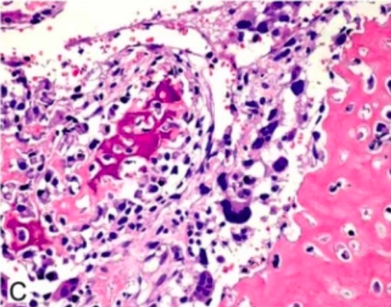
Hematoxylin and eosin stain of osteoblastic osteosarcoma with irregular immature bone deposition by anaplastic tumor cells
Image: “Osteoblastic osteosarcoma” by Department of Orthopaedic Surgery, Graduate School of Medical Sciences, Kyushu University, 3-1-1, Higashi-ku, Maidashi, Fukuoka, 812-8582, Japan. License: CC BY 2.5, cropped by Lecturio.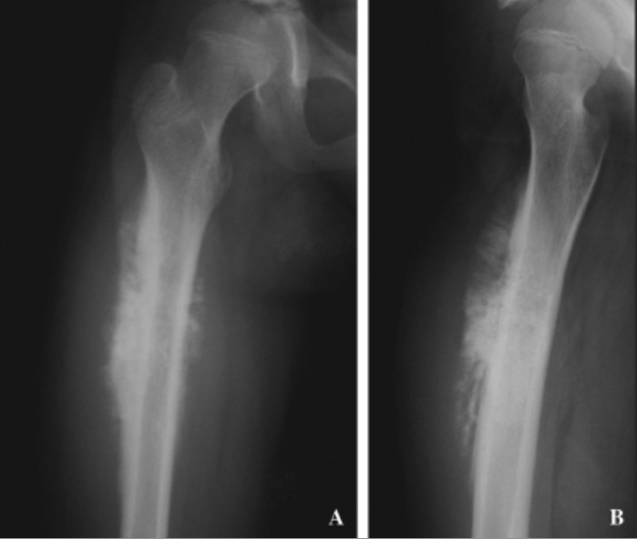
Osteosarcoma:
Plain X-rays of the proximal femur reveal a mixed sclerotic and lytic intramedullary lesion with the periosteal perpendicular spiculation with extensive periosteal reaction and Codman’s triangle reaction.
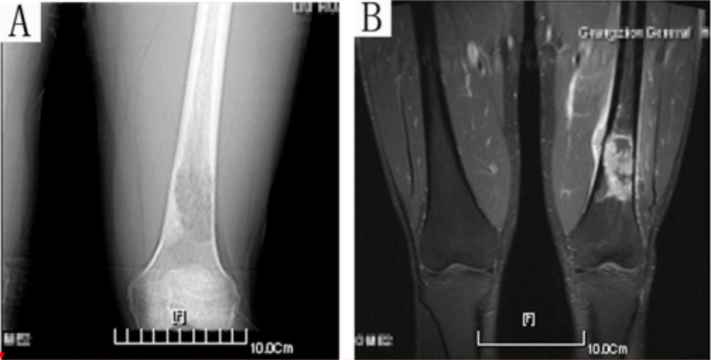
A 21-year-old patient with osteosarcoma of the distal femur:
A: anteroposterior CT scan image
B: MRI image
Both CT and MRI images reveal an osteosarcoma in the left distal femur.