Achieve Mastery of Medical Concepts

Study for medical school and boards with Lecturio

Inguinal Canal: Anatomy and Hernias

The inguinal region Inguinal region Anterior Abdominal Wall: Anatomy, or the groin Groin The external junctural region between the lower part of the abdomen and the thigh. Male Genitourinary Examination, is located in the RLQ and LLQ of the anterior abdominal wall Abdominal wall The outer margins of the abdomen, extending from the osteocartilaginous thoracic cage to the pelvis. Though its major part is muscular, the abdominal wall consists of at least seven layers: the skin, subcutaneous fat, deep fascia; abdominal muscles, transversalis fascia, extraperitoneal fat, and the parietal peritoneum. Surgical Anatomy of the Abdomen, bordered by the thigh Thigh The thigh is the region of the lower limb found between the hip and the knee joint. There is a single bone in the thigh called the femur, which is surrounded by large muscles grouped into 3 fascial compartments. Thigh: Anatomy inferiorly, the pubis medially, and the iliac crest superolaterally. The inguinal canal is a tubular structure that runs in a straight line from the anterior superior iliac spine Anterior Superior Iliac Spine Chronic Apophyseal Injury to the pubic tubercle. The canal contains the spermatic cord Spermatic Cord Either of a pair of tubular structures formed by ductus deferens; arteries; veins; lymphatic vessels; and nerves. The spermatic cord extends from the deep inguinal ring through the inguinal canal to the testis in the scrotum. Testicles: Anatomy in men and the round ligament Round ligament A fibromuscular band that attaches to the uterus and then passes along the broad ligament, out through the inguinal ring, and into the labium majus. Uterus, Cervix, and Fallopian Tubes: Anatomy in women. An inguinal hernia Hernia Protrusion of tissue, structure, or part of an organ through the bone, muscular tissue, or the membrane by which it is normally contained. Hernia may involve tissues such as the abdominal wall or the respiratory diaphragm. Hernias may be internal, external, congenital, or acquired. Abdominal Hernias occurs when tissue or an organ (such as a portion of the intestine) protrudes through in the abdominal wall Abdominal wall The outer margins of the abdomen, extending from the osteocartilaginous thoracic cage to the pelvis. Though its major part is muscular, the abdominal wall consists of at least seven layers: the skin, subcutaneous fat, deep fascia; abdominal muscles, transversalis fascia, extraperitoneal fat, and the parietal peritoneum. Surgical Anatomy of the Abdomen and into the inguinal canal. Inguinal hernias are the most common type of hernia Hernia Protrusion of tissue, structure, or part of an organ through the bone, muscular tissue, or the membrane by which it is normally contained. Hernia may involve tissues such as the abdominal wall or the respiratory diaphragm. Hernias may be internal, external, congenital, or acquired. Abdominal Hernias, and may be classified as indirect (tissue protrudes through the deep inguinal ring) or direct (tissue protrudes through the posterior wall of the inguinal canal). A hernia Hernia Protrusion of tissue, structure, or part of an organ through the bone, muscular tissue, or the membrane by which it is normally contained. Hernia may involve tissues such as the abdominal wall or the respiratory diaphragm. Hernias may be internal, external, congenital, or acquired. Abdominal Hernias may cause pain Pain An unpleasant sensation induced by noxious stimuli which are detected by nerve endings of nociceptive neurons. Pain: Types and Pathways or discomfort, and there is a risk of bowel obstruction Bowel obstruction Any impairment, arrest, or reversal of the normal flow of intestinal contents toward the anal canal. Ascaris/Ascariasis due to bowel incarceration with possible strangulation and infarction. Surgery is indicated for inguinal hernias which are high-risk or cause significant pain Pain An unpleasant sensation induced by noxious stimuli which are detected by nerve endings of nociceptive neurons. Pain: Types and Pathways.

Last updated: Jan 17, 2024

Editorial responsibility: Stanley Oiseth, Lindsay Jones, Evelin Maza

Overview

  • The inguinal region Inguinal region Anterior Abdominal Wall: Anatomy, or the groin Groin The external junctural region between the lower part of the abdomen and the thigh. Male Genitourinary Examination, is located in the RLQ and LLQ of the anterior abdominal wall Abdominal wall The outer margins of the abdomen, extending from the osteocartilaginous thoracic cage to the pelvis. Though its major part is muscular, the abdominal wall consists of at least seven layers: the skin, subcutaneous fat, deep fascia; abdominal muscles, transversalis fascia, extraperitoneal fat, and the parietal peritoneum. Surgical Anatomy of the Abdomen.
  • Boundaries:
    • Thigh Thigh The thigh is the region of the lower limb found between the hip and the knee joint. There is a single bone in the thigh called the femur, which is surrounded by large muscles grouped into 3 fascial compartments. Thigh: Anatomy inferiorly
    • Pubic tubercle medially
    • Anterior superior iliac spine Anterior Superior Iliac Spine Chronic Apophyseal Injury laterally
  • The inguinal canal runs in a straight line from the anterior superior iliac spine Anterior Superior Iliac Spine Chronic Apophyseal Injury to the pubic tubercle.
  • Contents of inguinal canal:
    • Men:
      • Spermatic cord Spermatic Cord Either of a pair of tubular structures formed by ductus deferens; arteries; veins; lymphatic vessels; and nerves. The spermatic cord extends from the deep inguinal ring through the inguinal canal to the testis in the scrotum. Testicles: Anatomy (with genitofemoral nerve)
      • Ilioinguinal nerve
    • Women:
  • Hesselbach triangle:
    • A triangle of the abdominal wall Abdominal wall The outer margins of the abdomen, extending from the osteocartilaginous thoracic cage to the pelvis. Though its major part is muscular, the abdominal wall consists of at least seven layers: the skin, subcutaneous fat, deep fascia; abdominal muscles, transversalis fascia, extraperitoneal fat, and the parietal peritoneum. Surgical Anatomy of the Abdomen
    • Located medial to the epigastric vessels
    • Site of direct inguinal hernias

Embryologic Development

Formation of the inguinal canal

  • Independent of testicular descent, in the 12th week of gestation, the anterior abdominal wall Abdominal wall The outer margins of the abdomen, extending from the osteocartilaginous thoracic cage to the pelvis. Though its major part is muscular, the abdominal wall consists of at least seven layers: the skin, subcutaneous fat, deep fascia; abdominal muscles, transversalis fascia, extraperitoneal fat, and the parietal peritoneum. Surgical Anatomy of the Abdomen musculature and fascia Fascia Layers of connective tissue of variable thickness. The superficial fascia is found immediately below the skin; the deep fascia invests muscles, nerves, and other organs. Cellulitis form an evagination on each side of the midline known as the processus vaginalis.
  • The processus vaginalis, in combination with the muscle and fascia Fascia Layers of connective tissue of variable thickness. The superficial fascia is found immediately below the skin; the deep fascia invests muscles, nerves, and other organs. Cellulitis of the anterior abdominal wall Abdominal wall The outer margins of the abdomen, extending from the osteocartilaginous thoracic cage to the pelvis. Though its major part is muscular, the abdominal wall consists of at least seven layers: the skin, subcutaneous fat, deep fascia; abdominal muscles, transversalis fascia, extraperitoneal fat, and the parietal peritoneum. Surgical Anatomy of the Abdomen, forms the inguinal canal.
  • In women, the ovum descends into the pelvis Pelvis The pelvis consists of the bony pelvic girdle, the muscular and ligamentous pelvic floor, and the pelvic cavity, which contains viscera, vessels, and multiple nerves and muscles. The pelvic girdle, composed of 2 “hip” bones and the sacrum, is a ring-like bony structure of the axial skeleton that links the vertebral column with the lower extremities. Pelvis: Anatomy, and the round ligament Round ligament A fibromuscular band that attaches to the uterus and then passes along the broad ligament, out through the inguinal ring, and into the labium majus. Uterus, Cervix, and Fallopian Tubes: Anatomy of the uterus Uterus The uterus, cervix, and fallopian tubes are part of the internal female reproductive system. The uterus has a thick wall made of smooth muscle (the myometrium) and an inner mucosal layer (the endometrium). The most inferior portion of the uterus is the cervix, which connects the uterine cavity to the vagina. Uterus, Cervix, and Fallopian Tubes: Anatomy travels through the inguinal canal to the labia majora Labia majora Vagina, Vulva, and Pelvic Floor: Anatomy.

Male inguinal canal development

  • The testes Testes Gonadal Hormones originally reside in the posterior aspect of the abdominal cavity.
  • Between 28 and 33 weeks’ gestation, the testes Testes Gonadal Hormones pass through the inguinal region Inguinal region Anterior Abdominal Wall: Anatomy and into the scrotum Scrotum A cutaneous pouch of skin containing the testicles and spermatic cords. Testicles: Anatomy.
  • Descent of the testes Testes Gonadal Hormones creates a weakness in the abdominal wall Abdominal wall The outer margins of the abdomen, extending from the osteocartilaginous thoracic cage to the pelvis. Though its major part is muscular, the abdominal wall consists of at least seven layers: the skin, subcutaneous fat, deep fascia; abdominal muscles, transversalis fascia, extraperitoneal fat, and the parietal peritoneum. Surgical Anatomy of the Abdomen in the region of the inguinal canal, making men particularly susceptible to hernia Hernia Protrusion of tissue, structure, or part of an organ through the bone, muscular tissue, or the membrane by which it is normally contained. Hernia may involve tissues such as the abdominal wall or the respiratory diaphragm. Hernias may be internal, external, congenital, or acquired. Abdominal Hernias formation.
  • The connection between the processus vaginalis and the peritoneum Peritoneum The peritoneum is a serous membrane lining the abdominopelvic cavity. This lining is formed by connective tissue and originates from the mesoderm. The membrane lines both the abdominal walls (as parietal peritoneum) and all of the visceral organs (as visceral peritoneum). Peritoneum: Anatomy will obliterate at birth, but a serous sac will remain around the testes Testes Gonadal Hormones, known as the tunica vaginalis Tunica vaginalis Testicles: Anatomy testis.
  • Abdominal wall Abdominal wall The outer margins of the abdomen, extending from the osteocartilaginous thoracic cage to the pelvis. Though its major part is muscular, the abdominal wall consists of at least seven layers: the skin, subcutaneous fat, deep fascia; abdominal muscles, transversalis fascia, extraperitoneal fat, and the parietal peritoneum. Surgical Anatomy of the Abdomen contributions:
    • Transversalis fascia Fascia Layers of connective tissue of variable thickness. The superficial fascia is found immediately below the skin; the deep fascia invests muscles, nerves, and other organs. Cellulitis forms the internal spermatic fascia Fascia Layers of connective tissue of variable thickness. The superficial fascia is found immediately below the skin; the deep fascia invests muscles, nerves, and other organs. Cellulitis.
    • Internal oblique Internal oblique Muscles of the anterolateral abdominal wall consisting of the external oblique and the internal oblique muscles. The external abdominal oblique muscle fibers extend from lower thoracic ribs to the linea alba and the iliac crest. The internal abdominal oblique extend superomedially beneath the external oblique muscles. Anterior Abdominal Wall: Anatomy muscle forms the cremasteric fascia Fascia Layers of connective tissue of variable thickness. The superficial fascia is found immediately below the skin; the deep fascia invests muscles, nerves, and other organs. Cellulitis and muscle.
    • External oblique External oblique Muscles of the anterolateral abdominal wall consisting of the external oblique and the internal oblique muscles. The external abdominal oblique muscle fibers extend from lower thoracic ribs to the linea alba and the iliac crest. The internal abdominal oblique extend superomedially beneath the external oblique muscles. Anterior Abdominal Wall: Anatomy muscle forms the external spermatic fascia Fascia Layers of connective tissue of variable thickness. The superficial fascia is found immediately below the skin; the deep fascia invests muscles, nerves, and other organs. Cellulitis.
Pathway of testicular descent

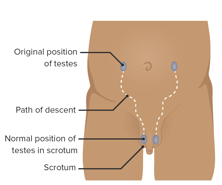
Pathway of testicular descent from the posterior abdominal wall into the scrotum: The testicles pass through the inguinal canal.

Image by Lecturio.

Inguinal Canal Anatomy

Anterior abdominal wall Abdominal wall The outer margins of the abdomen, extending from the osteocartilaginous thoracic cage to the pelvis. Though its major part is muscular, the abdominal wall consists of at least seven layers: the skin, subcutaneous fat, deep fascia; abdominal muscles, transversalis fascia, extraperitoneal fat, and the parietal peritoneum. Surgical Anatomy of the Abdomen

  • The anterior abdominal wall Abdominal wall The outer margins of the abdomen, extending from the osteocartilaginous thoracic cage to the pelvis. Though its major part is muscular, the abdominal wall consists of at least seven layers: the skin, subcutaneous fat, deep fascia; abdominal muscles, transversalis fascia, extraperitoneal fat, and the parietal peritoneum. Surgical Anatomy of the Abdomen is composed of several muscles:
    • Rectus abdominis Rectus Abdominis A long flat muscle that extends along the whole length of both sides of the abdomen. It flexes the vertebral column, particularly the lumbar portion; it also tenses the anterior abdominal wall and assists in compressing the abdominal contents. It is frequently the site of hematomas. In reconstructive surgery it is often used for the creation of myocutaneous flaps. Anterior Abdominal Wall: Anatomy
    • External oblique External oblique Muscles of the anterolateral abdominal wall consisting of the external oblique and the internal oblique muscles. The external abdominal oblique muscle fibers extend from lower thoracic ribs to the linea alba and the iliac crest. The internal abdominal oblique extend superomedially beneath the external oblique muscles. Anterior Abdominal Wall: Anatomy
    • Internal oblique Internal oblique Muscles of the anterolateral abdominal wall consisting of the external oblique and the internal oblique muscles. The external abdominal oblique muscle fibers extend from lower thoracic ribs to the linea alba and the iliac crest. The internal abdominal oblique extend superomedially beneath the external oblique muscles. Anterior Abdominal Wall: Anatomy
    • Transversus abdominis Transversus abdominis Anterior Abdominal Wall: Anatomy
  • The inner transversalis fascia Fascia Layers of connective tissue of variable thickness. The superficial fascia is found immediately below the skin; the deep fascia invests muscles, nerves, and other organs. Cellulitis continues into the inguinal canal, forming the internal spermatic fascia Fascia Layers of connective tissue of variable thickness. The superficial fascia is found immediately below the skin; the deep fascia invests muscles, nerves, and other organs. Cellulitis.

Course of the inguinal canal

  • Approximately 4 cm long from anterior superior iliac spine Anterior Superior Iliac Spine Chronic Apophyseal Injury to pubic tubercle (superolaterally to inferomedially)
  • Deep inguinal ring: an evagination of the transversalis fascia Fascia Layers of connective tissue of variable thickness. The superficial fascia is found immediately below the skin; the deep fascia invests muscles, nerves, and other organs. Cellulitis (surrounding the spermatic cord Spermatic Cord Either of a pair of tubular structures formed by ductus deferens; arteries; veins; lymphatic vessels; and nerves. The spermatic cord extends from the deep inguinal ring through the inguinal canal to the testis in the scrotum. Testicles: Anatomy as the internal spermatic fascia Fascia Layers of connective tissue of variable thickness. The superficial fascia is found immediately below the skin; the deep fascia invests muscles, nerves, and other organs. Cellulitis)
  • Superficial inguinal ring: a fissure Fissure A crack or split that extends into the dermis Generalized and Localized Rashes in the aponeurosis of the external oblique External oblique Muscles of the anterolateral abdominal wall consisting of the external oblique and the internal oblique muscles. The external abdominal oblique muscle fibers extend from lower thoracic ribs to the linea alba and the iliac crest. The internal abdominal oblique extend superomedially beneath the external oblique muscles. Anterior Abdominal Wall: Anatomy muscle
The layers of the anterior abdominal wall

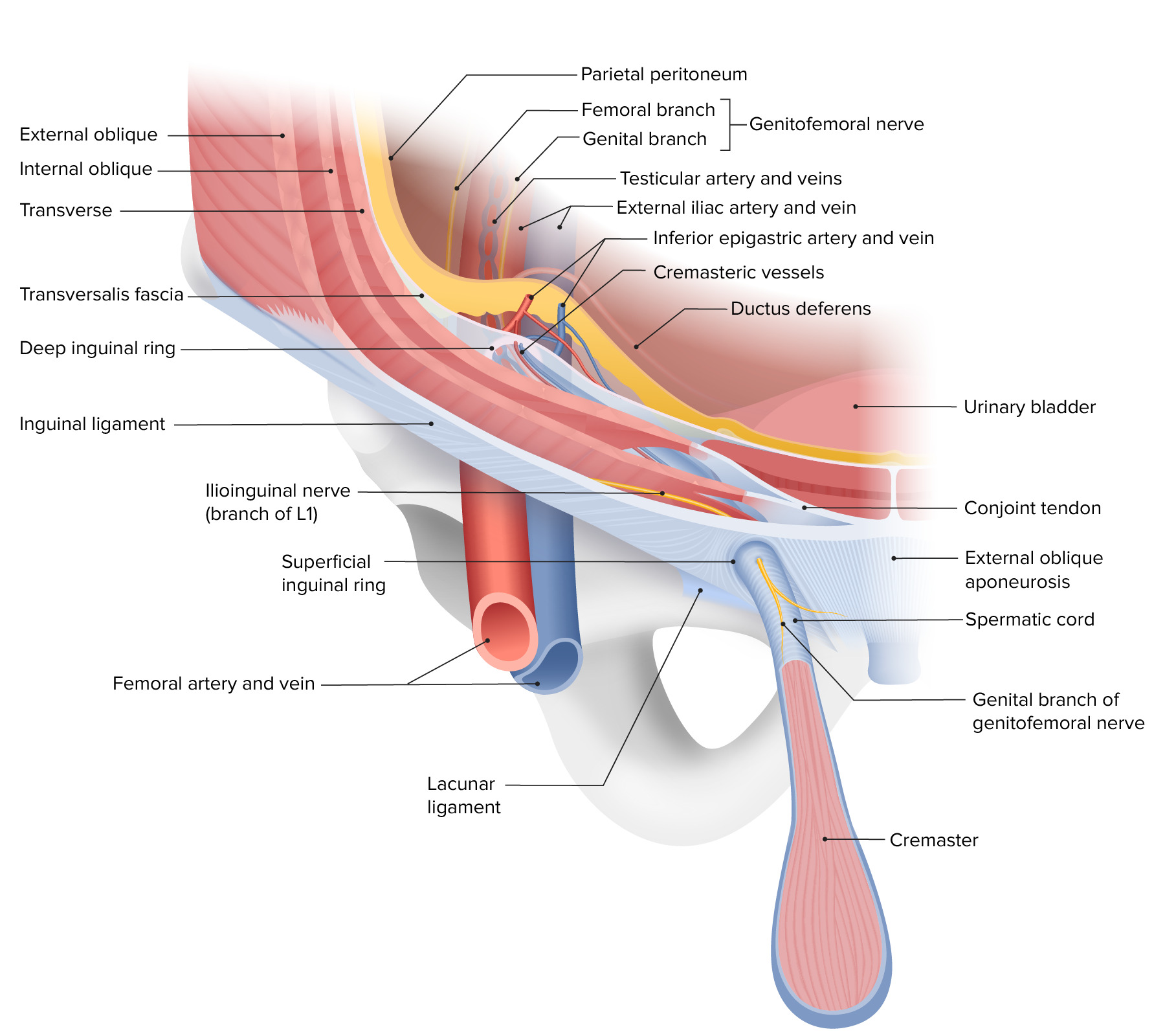
The layers of the anterior abdominal wall, depicting the course of the inguinal canal and the composition of the deep and superficial inguinal rings

Image by Lecturio. License: CC BY-NC-SA 4.0

Boundaries of the inguinal canal

The boundaries of the inguinal canal vary throughout its course.

  • At the level of the deep ring:
    • Anterior wall:
      • Internal oblique Internal oblique Muscles of the anterolateral abdominal wall consisting of the external oblique and the internal oblique muscles. The external abdominal oblique muscle fibers extend from lower thoracic ribs to the linea alba and the iliac crest. The internal abdominal oblique extend superomedially beneath the external oblique muscles. Anterior Abdominal Wall: Anatomy muscle
      • External oblique External oblique Muscles of the anterolateral abdominal wall consisting of the external oblique and the internal oblique muscles. The external abdominal oblique muscle fibers extend from lower thoracic ribs to the linea alba and the iliac crest. The internal abdominal oblique extend superomedially beneath the external oblique muscles. Anterior Abdominal Wall: Anatomy muscle
    • Posterior wall: transversalis fascia Fascia Layers of connective tissue of variable thickness. The superficial fascia is found immediately below the skin; the deep fascia invests muscles, nerves, and other organs. Cellulitis
    • Roof: transversalis fascia Fascia Layers of connective tissue of variable thickness. The superficial fascia is found immediately below the skin; the deep fascia invests muscles, nerves, and other organs. Cellulitis
    • Floor: inguinal ligament Inguinal Ligament Femoral Region and Hernias: Anatomy
  • At the middle of the inguinal canal:
    • Anterior wall: external oblique External oblique Muscles of the anterolateral abdominal wall consisting of the external oblique and the internal oblique muscles. The external abdominal oblique muscle fibers extend from lower thoracic ribs to the linea alba and the iliac crest. The internal abdominal oblique extend superomedially beneath the external oblique muscles. Anterior Abdominal Wall: Anatomy aponeurosis
    • Posterior wall: transversalis fascia Fascia Layers of connective tissue of variable thickness. The superficial fascia is found immediately below the skin; the deep fascia invests muscles, nerves, and other organs. Cellulitis
    • Roof: arching fibers of the internal oblique Internal oblique Muscles of the anterolateral abdominal wall consisting of the external oblique and the internal oblique muscles. The external abdominal oblique muscle fibers extend from lower thoracic ribs to the linea alba and the iliac crest. The internal abdominal oblique extend superomedially beneath the external oblique muscles. Anterior Abdominal Wall: Anatomy muscle and transversus abdominis Transversus abdominis Anterior Abdominal Wall: Anatomy
    • Floor: inguinal ligament Inguinal Ligament Femoral Region and Hernias: Anatomy
  • At the level of the superficial ring:
    • Anterior wall: external oblique External oblique Muscles of the anterolateral abdominal wall consisting of the external oblique and the internal oblique muscles. The external abdominal oblique muscle fibers extend from lower thoracic ribs to the linea alba and the iliac crest. The internal abdominal oblique extend superomedially beneath the external oblique muscles. Anterior Abdominal Wall: Anatomy aponeurosis
    • Posterior wall: conjoint tendon
    • Roof: medial crus of external oblique External oblique Muscles of the anterolateral abdominal wall consisting of the external oblique and the internal oblique muscles. The external abdominal oblique muscle fibers extend from lower thoracic ribs to the linea alba and the iliac crest. The internal abdominal oblique extend superomedially beneath the external oblique muscles. Anterior Abdominal Wall: Anatomy muscle
    • Floor: lacunar ligament
Boundaries and contents of the male inguinal canal

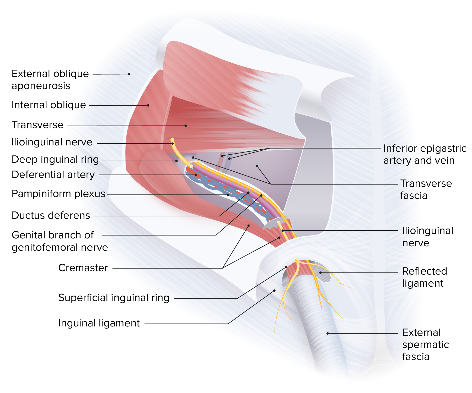
Boundaries and contents of the male inguinal canal:
Note that the ilioinguinal nerve runs along the inguinal canal externally from the spermatic cord.

Image by Lecturio. License: CC BY-NC-SA 4.0

Borders of the Hesselbach triangle

  • Refers to a triangle of the anterior abdominal wall Abdominal wall The outer margins of the abdomen, extending from the osteocartilaginous thoracic cage to the pelvis. Though its major part is muscular, the abdominal wall consists of at least seven layers: the skin, subcutaneous fat, deep fascia; abdominal muscles, transversalis fascia, extraperitoneal fat, and the parietal peritoneum. Surgical Anatomy of the Abdomen
  • Boundaries:
    • Medial: lateral margin of the rectus abdominis Rectus Abdominis A long flat muscle that extends along the whole length of both sides of the abdomen. It flexes the vertebral column, particularly the lumbar portion; it also tenses the anterior abdominal wall and assists in compressing the abdominal contents. It is frequently the site of hematomas. In reconstructive surgery it is often used for the creation of myocutaneous flaps. Anterior Abdominal Wall: Anatomy (linea semilunaris)
    • Superolateral: inferior epigastric vessels
    • Inferior: inguinal and pectineal ligament
  • Direct hernias occur within the triangle, and indirect hernias occur lateral to the triangle.

Epidemiology and Etiology of Inguinal Hernias

Epidemiology

  • Most common type of hernia Hernia Protrusion of tissue, structure, or part of an organ through the bone, muscular tissue, or the membrane by which it is normally contained. Hernia may involve tissues such as the abdominal wall or the respiratory diaphragm. Hernias may be internal, external, congenital, or acquired. Abdominal Hernias (> 75% of cases)
  • Indirect inguinal hernias > direct inguinal hernias
  • Ages of peak incidence Incidence The number of new cases of a given disease during a given period in a specified population. It also is used for the rate at which new events occur in a defined population. It is differentiated from prevalence, which refers to all cases in the population at a given time. Measures of Disease Frequency:
    • 0‒5 years of age
    • 75‒80 years of age
  • Lifetime risk of approximately 25% for men and < 5% for women

Risk factors

  • History of hernia Hernia Protrusion of tissue, structure, or part of an organ through the bone, muscular tissue, or the membrane by which it is normally contained. Hernia may involve tissues such as the abdominal wall or the respiratory diaphragm. Hernias may be internal, external, congenital, or acquired. Abdominal Hernias or previous hernia Hernia Protrusion of tissue, structure, or part of an organ through the bone, muscular tissue, or the membrane by which it is normally contained. Hernia may involve tissues such as the abdominal wall or the respiratory diaphragm. Hernias may be internal, external, congenital, or acquired. Abdominal Hernias repair
  • Male sex Sex The totality of characteristics of reproductive structure, functions, phenotype, and genotype, differentiating the male from the female organism. Gender Dysphoria
  • Older age
  • Chronic obstructive pulmonary disease Pulmonary disease Diseases involving the respiratory system. Blastomyces/Blastomycosis ( COPD COPD Chronic obstructive pulmonary disease (COPD) is a lung disease characterized by progressive, largely irreversible airflow obstruction. The condition usually presents in middle-aged or elderly persons with a history of cigarette smoking. Signs and symptoms include prolonged expiration, wheezing, diminished breath sounds, progressive dyspnea, and chronic cough. Chronic Obstructive Pulmonary Disease (COPD))
  • Chronic constipation Constipation Constipation is common and may be due to a variety of causes. Constipation is generally defined as bowel movement frequency < 3 times per week. Patients who are constipated often strain to pass hard stools. The condition is classified as primary (also known as idiopathic or functional constipation) or secondary, and as acute or chronic. Constipation
  • Weight lifting 
  • Obesity Obesity Obesity is a condition associated with excess body weight, specifically with the deposition of excessive adipose tissue. Obesity is considered a global epidemic. Major influences come from the western diet and sedentary lifestyles, but the exact mechanisms likely include a mixture of genetic and environmental factors. Obesity
  • Smoking Smoking Willful or deliberate act of inhaling and exhaling smoke from burning substances or agents held by hand. Interstitial Lung Diseases

Etiology

  • Congenital Congenital Chorioretinitis:
    • Due to abnormal development
    • Failed closure of the processus vaginalis
  • Acquired:
    • Develop later in life due to progressive weakness of previously normal tissues
    • Conditions with increased intraabdominal pressure, such as strenuous physical activity, coughing, or obesity Obesity Obesity is a condition associated with excess body weight, specifically with the deposition of excessive adipose tissue. Obesity is considered a global epidemic. Major influences come from the western diet and sedentary lifestyles, but the exact mechanisms likely include a mixture of genetic and environmental factors. Obesity
    • Sometimes iatrogenic Iatrogenic Any adverse condition in a patient occurring as the result of treatment by a physician, surgeon, or other health professional, especially infections acquired by a patient during the course of treatment. Anterior Cord Syndrome due to injury or abdominal surgery
  • All direct hernias are acquired, while indirect ones may be congenital Congenital Chorioretinitis or acquired.

Classification and Clinical Presentation

Classification

  • Indirect hernias:
    • Lateral to the inferior epigastric blood vessels and the Hesselbach triangle 
    • Contents pass through the deep inguinal ring, traverse the entire trajectory of the inguinal canal, and exit the canal through the superficial inguinal ring.
    • Contents are encased by the coverings of the spermatic cord Spermatic Cord Either of a pair of tubular structures formed by ductus deferens; arteries; veins; lymphatic vessels; and nerves. The spermatic cord extends from the deep inguinal ring through the inguinal canal to the testis in the scrotum. Testicles: Anatomy.
  • Direct hernias:
    • Medial to the inferior epigastric blood vessels and within the Hesselbach triangle
    • Contents protrude directly through the posterior wall of the inguinal canal and through the superficial inguinal ring, encased only by the external spermatic fascia Fascia Layers of connective tissue of variable thickness. The superficial fascia is found immediately below the skin; the deep fascia invests muscles, nerves, and other organs. Cellulitis.
  • Pantaloon hernia Hernia Protrusion of tissue, structure, or part of an organ through the bone, muscular tissue, or the membrane by which it is normally contained. Hernia may involve tissues such as the abdominal wall or the respiratory diaphragm. Hernias may be internal, external, congenital, or acquired. Abdominal Hernias: inguinal hernia Hernia Protrusion of tissue, structure, or part of an organ through the bone, muscular tissue, or the membrane by which it is normally contained. Hernia may involve tissues such as the abdominal wall or the respiratory diaphragm. Hernias may be internal, external, congenital, or acquired. Abdominal Hernias with both direct and indirect components
  • Amyand hernia Hernia Protrusion of tissue, structure, or part of an organ through the bone, muscular tissue, or the membrane by which it is normally contained. Hernia may involve tissues such as the abdominal wall or the respiratory diaphragm. Hernias may be internal, external, congenital, or acquired. Abdominal Hernias: The appendix Appendix A worm-like blind tube extension from the cecum. Colon, Cecum, and Appendix: Anatomy is found within the hernia Hernia Protrusion of tissue, structure, or part of an organ through the bone, muscular tissue, or the membrane by which it is normally contained. Hernia may involve tissues such as the abdominal wall or the respiratory diaphragm. Hernias may be internal, external, congenital, or acquired. Abdominal Hernias sac.
Anatomy of the inguinal region and hernia

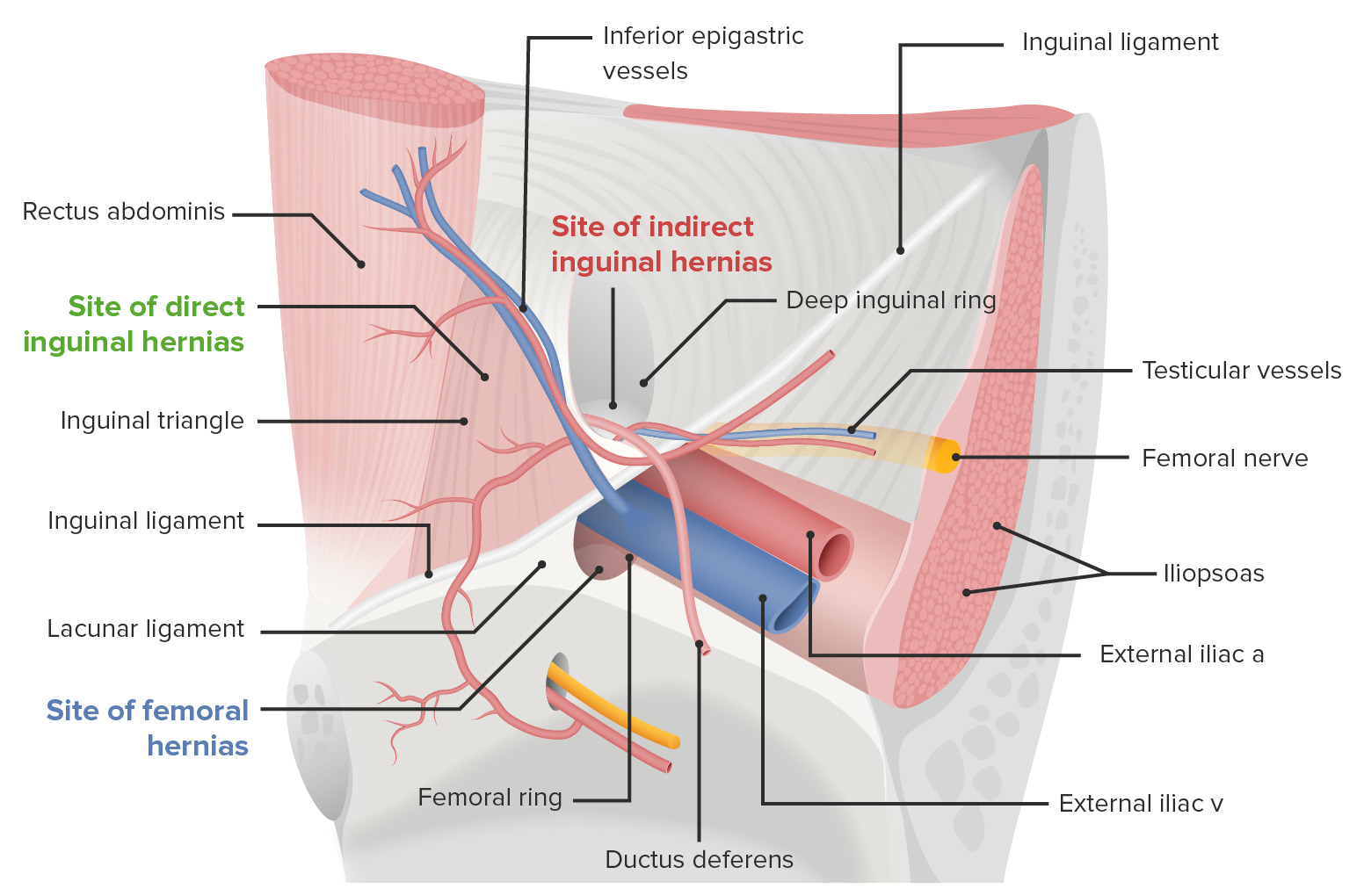
View of the left inguinal canal: Indirect inguinal hernias arise lateral to the epigastric vessels, while direct inguinal hernias arise medial to the epigastric vessels.

Image by Lecturio.

Clinical presentation

  • Mild inguinal pain Pain An unpleasant sensation induced by noxious stimuli which are detected by nerve endings of nociceptive neurons. Pain: Types and Pathways or discomfort
  • Visible bulge in the groin Groin The external junctural region between the lower part of the abdomen and the thigh. Male Genitourinary Examination area, which increases upon standing or during physical activities that increase intraabdominal pressure (coughing, sneezing Sneezing The sudden, forceful, involuntary expulsion of air from the nose and mouth caused by irritation to the mucous membranes of the upper respiratory tract. Rhinovirus, weight lifting) and reduces upon lying down.
  • May be associated with a communicating hydrocele Hydrocele Accumulation of serous fluid between the layers of membrane (tunica vaginalis) covering the testis in the scrotum. Varicocele, Hydrocele, and Spermatocele
  • Erythema Erythema Redness of the skin produced by congestion of the capillaries. This condition may result from a variety of disease processes. Chalazion may be noted over the hernia Hernia Protrusion of tissue, structure, or part of an organ through the bone, muscular tissue, or the membrane by which it is normally contained. Hernia may involve tissues such as the abdominal wall or the respiratory diaphragm. Hernias may be internal, external, congenital, or acquired. Abdominal Hernias if there is strangulation and tissue death.

Complications

  • Incarceration:
    • Inability of the contents of the hernia Hernia Protrusion of tissue, structure, or part of an organ through the bone, muscular tissue, or the membrane by which it is normally contained. Hernia may involve tissues such as the abdominal wall or the respiratory diaphragm. Hernias may be internal, external, congenital, or acquired. Abdominal Hernias to return to their original cavity
    • Presents with severe pain Pain An unpleasant sensation induced by noxious stimuli which are detected by nerve endings of nociceptive neurons. Pain: Types and Pathways and a non-reducible bulge
    • If the intestines are incarcerated, symptoms of intestinal obstruction Intestinal obstruction Any impairment, arrest, or reversal of the normal flow of intestinal contents toward the anal canal. Ascaris/Ascariasis develop.
  • Strangulation:
    • Contents of the hernia Hernia Protrusion of tissue, structure, or part of an organ through the bone, muscular tissue, or the membrane by which it is normally contained. Hernia may involve tissues such as the abdominal wall or the respiratory diaphragm. Hernias may be internal, external, congenital, or acquired. Abdominal Hernias must first become incarcerated.
    • Blood supply to the incarcerated organs is compromised, which causes ischemia Ischemia A hypoperfusion of the blood through an organ or tissue caused by a pathologic constriction or obstruction of its blood vessels, or an absence of blood circulation. Ischemic Cell Damage and resultant tissue death.

Diagnosis and Management

Diagnosis

  • Medical history and physical exam
  • Palpation Palpation Application of fingers with light pressure to the surface of the body to determine consistency of parts beneath in physical diagnosis; includes palpation for determining the outlines of organs. Dermatologic Examination of the inguinal canal:
    • With the patient standing, palpate from the scrotal skin Skin The skin, also referred to as the integumentary system, is the largest organ of the body. The skin is primarily composed of the epidermis (outer layer) and dermis (deep layer). The epidermis is primarily composed of keratinocytes that undergo rapid turnover, while the dermis contains dense layers of connective tissue. Skin: Structure and Functions toward the superficial inguinal ring.
    • Ask the patient to cough ( Valsalva maneuver Valsalva maneuver Forced expiratory effort against a closed glottis. Rectal Prolapse).
    • Bulging can be felt at the fingertip.
  • Imaging:
    • Used for confirmation in uncertain cases and occasionally surgical planning
    • Ultrasound :
      • Best initial imaging study in patients Patients Individuals participating in the health care system for the purpose of receiving therapeutic, diagnostic, or preventive procedures. Clinician–Patient Relationship without physical evidence of a hernia Hernia Protrusion of tissue, structure, or part of an organ through the bone, muscular tissue, or the membrane by which it is normally contained. Hernia may involve tissues such as the abdominal wall or the respiratory diaphragm. Hernias may be internal, external, congenital, or acquired. Abdominal Hernias 
      • The diagnostic finding is an increased diameter of the inguinal canal (normally < 13 mm at the deep inguinal ring).
    • CT scans: particularly useful to distinguish between different subtypes of inguinal hernias
    • MRI:
      • Best imaging modality to differentiate between inguinal and femoral hernias with a sensitivity and specificity Sensitivity and Specificity Binary classification measures to assess test results. Sensitivity or recall rate is the proportion of true positives. Specificity is the probability of correctly determining the absence of a condition. Epidemiological Values of Diagnostic Tests greater than 95%
      • Due to the cost and limited availability of MRIs, CT scans are still more frequently used.

Management

  • Surgical hernia Hernia Protrusion of tissue, structure, or part of an organ through the bone, muscular tissue, or the membrane by which it is normally contained. Hernia may involve tissues such as the abdominal wall or the respiratory diaphragm. Hernias may be internal, external, congenital, or acquired. Abdominal Hernias repair:
    • Complicated hernias
    • Uncomplicated hernias with moderate symptoms 
    • Selectively for uncomplicated hernias with mild symptomatology Symptomatology Scarlet Fever or high risk of incarceration
    • Specifics of surgical repair techniques can be found in abdominal hernias.
  • Surgical techniques:
    • Reinforcing the posterior wall of the inguinal canal with synthetic mesh
    • Reduction in the diameters of the inguinal rings

References

  1. Sadler, T. W. (2012). Chapter 16: Urogenital system. In Langman’s Medical Embryology. 12th ed., pp. 232–259. Philadelphia: Wolters Kluwer Health/Lippincott Williams & Wilkins.
  2. Malangoni, M. A., & Rosen, M. J. (2012). Hernias. In Mattox, K. L., et al. (Eds.), Sabiston Textbook of Surgery: The Biological Basis of Modern Surgical Practice, 19th ed., pp. 1114‒1140. Elsevier.
  3. Brooks, D. V., & Hawn, M. (2019). Classification, clinical features, and diagnosis of inguinal and femoral hernias in adults. In Chen, W. (Ed.). UpToDate. Retrieved June 19, 2021, from https://www.uptodate.com/contents/classification-clinical-features-and-diagnosis-of-inguinal-and-femoral-hernias-in-adults

Create your free account or log in to continue reading!

Sign up now and get free access to Lecturio with concept pages, medical videos, and questions for your medical education.

User Reviews

Details