Achieve Mastery of Medical Concepts

Study for medical school and boards with Lecturio

Meningioma

Meningiomas are slow-growing tumors that arise from the meninges Meninges The brain and the spinal cord are enveloped by 3 overlapping layers of connective tissue called the meninges. The layers are, from the most external layer to the most internal layer, the dura mater, arachnoid mater, and pia mater. Between these layers are 3 potential spaces called the epidural, subdural, and subarachnoid spaces. Meninges: Anatomy of the brain Brain The part of central nervous system that is contained within the skull (cranium). Arising from the neural tube, the embryonic brain is comprised of three major parts including prosencephalon (the forebrain); mesencephalon (the midbrain); and rhombencephalon (the hindbrain). The developed brain consists of cerebrum; cerebellum; and other structures in the brain stem. Nervous System: Anatomy, Structure, and Classification and spinal cord Spinal cord The spinal cord is the major conduction pathway connecting the brain to the body; it is part of the CNS. In cross section, the spinal cord is divided into an H-shaped area of gray matter (consisting of synapsing neuronal cell bodies) and a surrounding area of white matter (consisting of ascending and descending tracts of myelinated axons). Spinal Cord: Anatomy. The vast majority are benign Benign Fibroadenoma. These tumors commonly occur in individuals with a history of high doses of skull Skull The skull (cranium) is the skeletal structure of the head supporting the face and forming a protective cavity for the brain. The skull consists of 22 bones divided into the viscerocranium (facial skeleton) and the neurocranium. Skull: Anatomy radiation Radiation Emission or propagation of acoustic waves (sound), electromagnetic energy waves (such as light; radio waves; gamma rays; or x-rays), or a stream of subatomic particles (such as electrons; neutrons; protons; or alpha particles). Osteosarcoma, head trauma Head trauma Head trauma occurs when external forces are directed to the skull and brain structures, resulting in damage to the skull, brain, and intracranial structures. Head injuries can be classified as open (penetrating) or closed (blunt), and primary (from the initial trauma) or secondary (indirect brain injury), and range from mild to severe and life-threatening. Head Trauma, and neurofibromatosis type 2 Type 2 Spinal Muscular Atrophy. Clinical symptoms of meningioma depend on the location and progression of the tumor Tumor Inflammation, with a large proportion of meningiomas being asymptomatic. When symptoms do develop, they most commonly include headache Headache The symptom of pain in the cranial region. It may be an isolated benign occurrence or manifestation of a wide variety of headache disorders. Brain Abscess, seizures Seizures A seizure is abnormal electrical activity of the neurons in the cerebral cortex that can manifest in numerous ways depending on the region of the brain affected. Seizures consist of a sudden imbalance that occurs between the excitatory and inhibitory signals in cortical neurons, creating a net excitation. The 2 major classes of seizures are focal and generalized. Seizures, visual disturbances, and other focal neurologic deficits Neurologic Deficits High-Risk Headaches. Diagnosis is made based on neuroimaging Neuroimaging Non-invasive methods of visualizing the central nervous system, especially the brain, by various imaging modalities. Febrile Infant (MRI is the gold standard) and biopsy Biopsy Removal and pathologic examination of specimens from the living body. Ewing Sarcoma. Management involves surveillance Surveillance Developmental Milestones and Normal Growth for asymptomatic patients Patients Individuals participating in the health care system for the purpose of receiving therapeutic, diagnostic, or preventive procedures. Clinician–Patient Relationship and, for symptomatic patients Patients Individuals participating in the health care system for the purpose of receiving therapeutic, diagnostic, or preventive procedures. Clinician–Patient Relationship, surgical resection. Radiation Radiation Emission or propagation of acoustic waves (sound), electromagnetic energy waves (such as light; radio waves; gamma rays; or x-rays), or a stream of subatomic particles (such as electrons; neutrons; protons; or alpha particles). Osteosarcoma is also used in some cases.

Last updated: Oct 24, 2022

Editorial responsibility: Stanley Oiseth, Lindsay Jones, Evelin Maza

Overview

Definition

A meningioma is a slow-growing tumor Tumor Inflammation that arises from the meninges Meninges The brain and the spinal cord are enveloped by 3 overlapping layers of connective tissue called the meninges. The layers are, from the most external layer to the most internal layer, the dura mater, arachnoid mater, and pia mater. Between these layers are 3 potential spaces called the epidural, subdural, and subarachnoid spaces. Meninges: Anatomy of the brain Brain The part of central nervous system that is contained within the skull (cranium). Arising from the neural tube, the embryonic brain is comprised of three major parts including prosencephalon (the forebrain); mesencephalon (the midbrain); and rhombencephalon (the hindbrain). The developed brain consists of cerebrum; cerebellum; and other structures in the brain stem. Nervous System: Anatomy, Structure, and Classification and spinal cord Spinal cord The spinal cord is the major conduction pathway connecting the brain to the body; it is part of the CNS. In cross section, the spinal cord is divided into an H-shaped area of gray matter (consisting of synapsing neuronal cell bodies) and a surrounding area of white matter (consisting of ascending and descending tracts of myelinated axons). Spinal Cord: Anatomy.

  • Meninges Meninges The brain and the spinal cord are enveloped by 3 overlapping layers of connective tissue called the meninges. The layers are, from the most external layer to the most internal layer, the dura mater, arachnoid mater, and pia mater. Between these layers are 3 potential spaces called the epidural, subdural, and subarachnoid spaces. Meninges: Anatomy are the fibrous Fibrous Fibrocystic Change layers that encase the brain Brain The part of central nervous system that is contained within the skull (cranium). Arising from the neural tube, the embryonic brain is comprised of three major parts including prosencephalon (the forebrain); mesencephalon (the midbrain); and rhombencephalon (the hindbrain). The developed brain consists of cerebrum; cerebellum; and other structures in the brain stem. Nervous System: Anatomy, Structure, and Classification and spinal cord Spinal cord The spinal cord is the major conduction pathway connecting the brain to the body; it is part of the CNS. In cross section, the spinal cord is divided into an H-shaped area of gray matter (consisting of synapsing neuronal cell bodies) and a surrounding area of white matter (consisting of ascending and descending tracts of myelinated axons). Spinal Cord: Anatomy, and consist of 3 layers:
    • Dura mater Dura mater The outermost of the three meninges, a fibrous membrane of connective tissue that covers the brain and the spinal cord. Meninges: Anatomy: the outer layer, which forms a tough collagenous sheath
    • Arachnoid mater Arachnoid mater A delicate membrane enveloping the brain and spinal cord. It lies between the pia mater and the dura mater. It is separated from the pia mater by the subarachnoid cavity which is filled with cerebrospinal fluid. Meninges: Anatomy: the middle layer, which consists of simple squamous epithelium Simple squamous epithelium Surface Epithelium: Histology with a loose mesh of collagen Collagen A polypeptide substance comprising about one third of the total protein in mammalian organisms. It is the main constituent of skin; connective tissue; and the organic substance of bones (bone and bones) and teeth (tooth). Connective Tissue: Histology and elastic Elastic Connective Tissue: Histology fibers
    • Pia mater Pia mater The innermost layer of the three meninges covering the brain and spinal cord. It is the fine vascular membrane that lies under the arachnoid and the dura mater. Meninges: Anatomy: the thin inner layer, which is in contact with the brain Brain The part of central nervous system that is contained within the skull (cranium). Arising from the neural tube, the embryonic brain is comprised of three major parts including prosencephalon (the forebrain); mesencephalon (the midbrain); and rhombencephalon (the hindbrain). The developed brain consists of cerebrum; cerebellum; and other structures in the brain stem. Nervous System: Anatomy, Structure, and Classification and spinal cord Spinal cord The spinal cord is the major conduction pathway connecting the brain to the body; it is part of the CNS. In cross section, the spinal cord is divided into an H-shaped area of gray matter (consisting of synapsing neuronal cell bodies) and a surrounding area of white matter (consisting of ascending and descending tracts of myelinated axons). Spinal Cord: Anatomy
  • Meningiomas arise from the arachnoid layer.
Table: Classification of nervous system Nervous system The nervous system is a small and complex system that consists of an intricate network of neural cells (or neurons) and even more glial cells (for support and insulation). It is divided according to its anatomical components as well as its functional characteristics. The brain and spinal cord are referred to as the central nervous system, and the branches of nerves from these structures are referred to as the peripheral nervous system. Nervous System: Anatomy, Structure, and Classification tumors
Categories Specific tumors
Neuroepithelial tumors in the CNS
  • Astrocytomas, including glioblastoma multiforme Glioblastoma multiforme Glioblastoma multiforme is a high-grade astrocytoma, an aggressive brain tumor arising from astrocytes, with an unknown cause and a poorly understood link to risk factors. There are two main types: primary, a more aggressive form seen more commonly in older patients, and secondary, developing from lower-grade astrocytomas and seen more commonly in younger patients. Glioblastoma Multiforme
  • Oligodendrogliomas
  • Ependymoma Ependymoma Ependymomas are glial cell tumors arising from CSF-producing ependymal cells lining the ventricular system. Ependymomas most commonly occur within the posterior fossa in contact with the 4th ventricle, or within the intramedullary spinal cord. Ependymoma and choroid-plexus tumors
  • Medulloblastomas (embryonal tumors)
Meningeal tumors
  • Meningiomas
  • Hemangioblastomas Hemangioblastomas A benign tumor of the nervous system that may occur sporadically or in association with von Hippel-Lindau disease. It accounts for approximately 2% of intracranial tumors, arising most frequently in the cerebellar hemispheres and vermis. Histologically, the tumors are composed of multiple capillary and sinusoidal channels lined with endothelial cells and clusters of lipid-laden pseudoxanthoma cells. Usually solitary, these tumors can be multiple and may also occur in the brain stem, spinal cord, retina, and supratentorial compartment. Cerebellar hemangioblastomas usually present in the third decade with intracranial hypertension, and ataxia. Von Hippel-Lindau Disease
Sellar region tumors
  • Craniopharyngioma Craniopharyngioma Craniopharyngiomas are rare squamous epithelial tumors with a solid and/or cystic structure that arise from the remnants of Rathke’s pouch along the pituitary stalk, in the suprasellar region. Craniopharyngiomas are histologically benign but tend to invade surrounding structures; thus, they should be treated as low-grade malignancies. Craniopharyngioma
  • Pituitary Pituitary A small, unpaired gland situated in the sella turcica. It is connected to the hypothalamus by a short stalk which is called the infundibulum. Hormones: Overview and Types adenoma
  • Pinealoma/pinealoblastoma
Other primary CNS tumors Primary CNS lymphoma Lymphoma A general term for various neoplastic diseases of the lymphoid tissue. Imaging of the Mediastinum
Metastasis Metastasis The transfer of a neoplasm from one organ or part of the body to another remote from the primary site. Grading, Staging, and Metastasis to the brain Brain The part of central nervous system that is contained within the skull (cranium). Arising from the neural tube, the embryonic brain is comprised of three major parts including prosencephalon (the forebrain); mesencephalon (the midbrain); and rhombencephalon (the hindbrain). The developed brain consists of cerebrum; cerebellum; and other structures in the brain stem. Nervous System: Anatomy, Structure, and Classification (5x more common than primary brain Brain The part of central nervous system that is contained within the skull (cranium). Arising from the neural tube, the embryonic brain is comprised of three major parts including prosencephalon (the forebrain); mesencephalon (the midbrain); and rhombencephalon (the hindbrain). The developed brain consists of cerebrum; cerebellum; and other structures in the brain stem. Nervous System: Anatomy, Structure, and Classification tumors) Most commonly arising from:
  • Lung, breast, and renal cell carcinomas
  • Melanoma Melanoma Melanoma is a malignant tumor arising from melanocytes, the melanin-producing cells of the epidermis. These tumors are most common in fair-skinned individuals with a history of excessive sun exposure and sunburns. Melanoma
Peripheral tumors
  • Schwannomas, including acoustic neuroma Acoustic neuroma Acoustic neuroma, also referred to as vestibular schwannoma, is a benign tumor arising from Schwann cells of the vestibular component of the cranial nerve VIII. Acoustic neuroma forms within the internal auditory meatus and extends into the cerebellopontine angle. Acoustic Neuroma
  • Neuroblastoma Neuroblastoma Neuroblastoma is a malignancy that arises from the neural crest cell derivatives along the sympathetic chain (neuroblasts) and is most commonly located in the adrenal medulla. The tumor often presents in childhood with a flank mass that crosses the midline. Neuroblastoma

Epidemiology

  • Incidence Incidence The number of new cases of a given disease during a given period in a specified population. It also is used for the rate at which new events occur in a defined population. It is differentiated from prevalence, which refers to all cases in the population at a given time. Measures of Disease Frequency:
    • Meningiomas are the most common type of primary CNS tumors. 
    • Account for about ⅓ of all primary brain Brain The part of central nervous system that is contained within the skull (cranium). Arising from the neural tube, the embryonic brain is comprised of three major parts including prosencephalon (the forebrain); mesencephalon (the midbrain); and rhombencephalon (the hindbrain). The developed brain consists of cerebrum; cerebellum; and other structures in the brain stem. Nervous System: Anatomy, Structure, and Classification and spinal tumors
    • Cranial meningiomas: 90%
    • Spinal meningiomas: 10%
  • The median age of diagnosis is 65 years: 
    • Incidence Incidence The number of new cases of a given disease during a given period in a specified population. It also is used for the rate at which new events occur in a defined population. It is differentiated from prevalence, which refers to all cases in the population at a given time. Measures of Disease Frequency progressively increases with age. 
    • Rare in children except when there is an association with neurofibromatosis type 2 Type 2 Spinal Muscular Atrophy
  • Gender Gender Gender Dysphoria: more common in women
    • Women-to-men ratio:
      • 2–3:1 for cranial meningiomas
      • 9:1 for spinal meningiomas
    • Female predominance is highest between 35 and 54 years.
  • Incidence Incidence The number of new cases of a given disease during a given period in a specified population. It also is used for the rate at which new events occur in a defined population. It is differentiated from prevalence, which refers to all cases in the population at a given time. Measures of Disease Frequency by WHO grade:
    • Grade 1: 80%–85%
    • Grade 2: 15%–18%
    • Grade 3: 1%–3% 

Classification

Meningiomas are classified morphologically by WHO into 3 grades:

  • Grade 1: benign Benign Fibroadenoma tumors of the meninges Meninges The brain and the spinal cord are enveloped by 3 overlapping layers of connective tissue called the meninges. The layers are, from the most external layer to the most internal layer, the dura mater, arachnoid mater, and pia mater. Between these layers are 3 potential spaces called the epidural, subdural, and subarachnoid spaces. Meninges: Anatomy, which include many subtypes
  • Grade 2: tumors showing some brain Brain The part of central nervous system that is contained within the skull (cranium). Arising from the neural tube, the embryonic brain is comprised of three major parts including prosencephalon (the forebrain); mesencephalon (the midbrain); and rhombencephalon (the hindbrain). The developed brain consists of cerebrum; cerebellum; and other structures in the brain stem. Nervous System: Anatomy, Structure, and Classification invasion and more mitotic activity
    • Atypical meningioma
    • Clear-cell meningioma
    • Choroid Choroid The thin, highly vascular membrane covering most of the posterior of the eye between the retina and sclera. Eye: Anatomy meningioma
  • Grade 3: more aggressive malignant tumors 
    • Papillary meningioma
    • Anaplastic meningioma
    • Rhabdoid meningioma

Risk factors

The causes of meningioma have not been established. However, certain factors at high risk of causing meningiomas have been identified:

  • Radiation Radiation Emission or propagation of acoustic waves (sound), electromagnetic energy waves (such as light; radio waves; gamma rays; or x-rays), or a stream of subatomic particles (such as electrons; neutrons; protons; or alpha particles). Osteosarcoma exposure: ionizing radiation Radiation Emission or propagation of acoustic waves (sound), electromagnetic energy waves (such as light; radio waves; gamma rays; or x-rays), or a stream of subatomic particles (such as electrons; neutrons; protons; or alpha particles). Osteosarcoma to the skull Skull The skull (cranium) is the skeletal structure of the head supporting the face and forming a protective cavity for the brain. The skull consists of 22 bones divided into the viscerocranium (facial skeleton) and the neurocranium. Skull: Anatomy
  • Head trauma Head trauma Head trauma occurs when external forces are directed to the skull and brain structures, resulting in damage to the skull, brain, and intracranial structures. Head injuries can be classified as open (penetrating) or closed (blunt), and primary (from the initial trauma) or secondary (indirect brain injury), and range from mild to severe and life-threatening. Head Trauma
  • Female hormones Hormones Hormones are messenger molecules that are synthesized in one part of the body and move through the bloodstream to exert specific regulatory effects on another part of the body. Hormones play critical roles in coordinating cellular activities throughout the body in response to the constant changes in both the internal and external environments. Hormones: Overview and Types: high incidence Incidence The number of new cases of a given disease during a given period in a specified population. It also is used for the rate at which new events occur in a defined population. It is differentiated from prevalence, which refers to all cases in the population at a given time. Measures of Disease Frequency in postpubertal women and during peak reproductive years
  • Breast cancer Breast cancer Breast cancer is a disease characterized by malignant transformation of the epithelial cells of the breast. Breast cancer is the most common form of cancer and 2nd most common cause of cancer-related death among women. Breast Cancer: increased risk of occurrence
  • Inherited nervous system Nervous system The nervous system is a small and complex system that consists of an intricate network of neural cells (or neurons) and even more glial cells (for support and insulation). It is divided according to its anatomical components as well as its functional characteristics. The brain and spinal cord are referred to as the central nervous system, and the branches of nerves from these structures are referred to as the peripheral nervous system. Nervous System: Anatomy, Structure, and Classification disorders:
  • Obesity Obesity Obesity is a condition associated with excess body weight, specifically with the deposition of excessive adipose tissue. Obesity is considered a global epidemic. Major influences come from the western diet and sedentary lifestyles, but the exact mechanisms likely include a mixture of genetic and environmental factors. Obesity

Pathophysiology

Sites of meningioma

  • Parasagittal and falcine
  • Cerebral convexity
  • Sphenoid wing
  • Olfactory groove
  • Cavernous sinus 
  • Suprasellar
  • Foramen magnum
  • Posterior fossa
  • Intraventricular
  • Intraorbital
  • Spinal
Meningioma locations

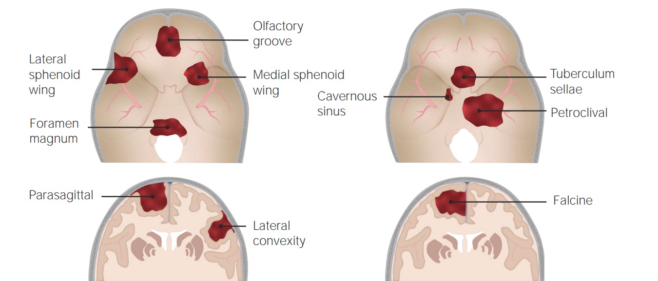
Common sites of occurrence of meningiomas

Image by Lecturio. License: CC BY-NC-SA 4.0

Pathophysiology

Meningiomas commonly arise from the arachnoid cells near the vicinity of the venous sinuses Venous sinuses Veins: Histology. As the tumor Tumor Inflammation enlarges, it can cause symptoms due to:

Clinical Presentation

Some patients Patients Individuals participating in the health care system for the purpose of receiving therapeutic, diagnostic, or preventive procedures. Clinician–Patient Relationship are asymptomatic. For symptomatic patients Patients Individuals participating in the health care system for the purpose of receiving therapeutic, diagnostic, or preventive procedures. Clinician–Patient Relationship, the clinical presentation of meningiomas depends on the location of the tumor Tumor Inflammation.

  • Headaches
  • Seizures Seizures A seizure is abnormal electrical activity of the neurons in the cerebral cortex that can manifest in numerous ways depending on the region of the brain affected. Seizures consist of a sudden imbalance that occurs between the excitatory and inhibitory signals in cortical neurons, creating a net excitation. The 2 major classes of seizures are focal and generalized. Seizures
  • Altered mental status Altered Mental Status Sepsis in Children
  • Focal neurologic deficits Neurologic Deficits High-Risk Headaches:
    • Subfrontal: apathy Apathy Lack of emotion or emotional expression; a disorder of motivation that persists over time. Wernicke Encephalopathy and Korsakoff Syndrome, urinary incontinence Urinary incontinence Urinary incontinence (UI) is involuntary loss of bladder control or unintentional voiding, which represents a hygienic or social problem to the patient. Urinary incontinence is a symptom, a sign, and a disorder. The 5 types of UI include stress, urge, mixed, overflow, and functional. Urinary Incontinence
    • Cavernous sinus: decreased vision Vision Ophthalmic Exam, diplopia Diplopia A visual symptom in which a single object is perceived by the visual cortex as two objects rather than one. Disorders associated with this condition include refractive errors; strabismus; oculomotor nerve diseases; trochlear nerve diseases; abducens nerve diseases; and diseases of the brain stem and occipital lobe. Myasthenia Gravis, facial numbness
    • Occipital lobe Occipital lobe Posterior portion of the cerebral hemispheres responsible for processing visual sensory information. It is located posterior to the parieto-occipital sulcus and extends to the preoccipital notch. Cerebral Cortex: Anatomy: Contralateral hemianopia Hemianopia Transient Ischemic Attack (TIA)
    • Posterior fossa: obstructive hydrocephalus Obstructive Hydrocephalus Hydrocephalus in Children papilledema Papilledema Swelling of the optic disk, usually in association with increased intracranial pressure, characterized by hyperemia, blurring of the disk margins, microhemorrhages, blind spot enlargement, and engorgement of retinal veins. Chronic papilledema may cause optic atrophy and visual loss. Idiopathic Intracranial Hypertension and morning headaches
    • Spinal cord Spinal cord The spinal cord is the major conduction pathway connecting the brain to the body; it is part of the CNS. In cross section, the spinal cord is divided into an H-shaped area of gray matter (consisting of synapsing neuronal cell bodies) and a surrounding area of white matter (consisting of ascending and descending tracts of myelinated axons). Spinal Cord: Anatomy: localized pain Pain An unpleasant sensation induced by noxious stimuli which are detected by nerve endings of nociceptive neurons. Pain: Types and Pathways, hemispinal cord defects
    • Olfactory groove: anosmia Anosmia Complete or severe loss of the subjective sense of smell. Loss of smell may be caused by many factors such as a cold, allergy, olfactory nerve diseases, viral respiratory tract infections (e.g., COVID-19), aging and various neurological disorders (e.g., Alzheimer disease). Cranial Nerve Palsies, papilledema Papilledema Swelling of the optic disk, usually in association with increased intracranial pressure, characterized by hyperemia, blurring of the disk margins, microhemorrhages, blind spot enlargement, and engorgement of retinal veins. Chronic papilledema may cause optic atrophy and visual loss. Idiopathic Intracranial Hypertension, optic atrophy Atrophy Decrease in the size of a cell, tissue, organ, or multiple organs, associated with a variety of pathological conditions such as abnormal cellular changes, ischemia, malnutrition, or hormonal changes. Cellular Adaptation 
    • Sphenoid: seizures Seizures A seizure is abnormal electrical activity of the neurons in the cerebral cortex that can manifest in numerous ways depending on the region of the brain affected. Seizures consist of a sudden imbalance that occurs between the excitatory and inhibitory signals in cortical neurons, creating a net excitation. The 2 major classes of seizures are focal and generalized. Seizures, cranial nerve palsies Cranial Nerve Palsies Cranial nerve palsy is a congenital or acquired dysfunction of 1 or more cranial nerves that will, in turn, lead to focal neurologic abnormalities in movement or autonomic dysfunction of its territory. Head/neck trauma, mass effect, infectious processes, and ischemia/infarction are among the many etiologies for these dysfunctions. Diagnosis is initially clinical and supported by diagnostic aids. Management includes both symptomatic measures and interventions aimed at correcting the underlying cause. Cranial Nerve Palsies 
    • Tentorial: headaches, seizures Seizures A seizure is abnormal electrical activity of the neurons in the cerebral cortex that can manifest in numerous ways depending on the region of the brain affected. Seizures consist of a sudden imbalance that occurs between the excitatory and inhibitory signals in cortical neurons, creating a net excitation. The 2 major classes of seizures are focal and generalized. Seizures, difficulty walking

Diagnosis

Laboratory studies are of little use in the diagnosis of meningiomas; imaging is a more helpful diagnostic tool. Biopsy Biopsy Removal and pathologic examination of specimens from the living body. Ewing Sarcoma is required to confirm the diagnosis.
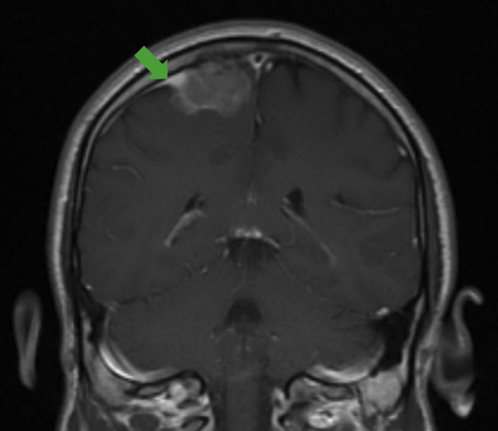
Imaging

Dural tail sign

Dural tail sign:
MRI showing the classic dural tail sign (green arrow), which can be seen in imaging of a meningioma.

Image by Roy Strowd, MD. License: CC BY-NC-SA 4.0

Biopsy Biopsy Removal and pathologic examination of specimens from the living body. Ewing Sarcoma

Biopsy Biopsy Removal and pathologic examination of specimens from the living body. Ewing Sarcoma provides a definitive diagnosis. Histologic findings include:

  • Lobulated architecture with meningothelial whorls
  • Psammoma bodies Psammoma Bodies Cellular Accumulations: round calcifications
  • Syncytial cells
  • Eosinophilic cytoplasm
  • Uniform nuclei
  • Grade 2 tumors:
    • ↑ Mitotic activity ( ≥ 4 mitotic figures per high-powered field)
    • Demonstrate 3 or more of the following features:
      • ↑ Cellularity
      • Small cells with ↑ nuclear-to-cytoplasmic ratio
      • Prominent nucleoli
      • Sheet-like growth 
      • Areas of necrosis Necrosis The death of cells in an organ or tissue due to disease, injury or failure of the blood supply. Ischemic Cell Damage
  • Grade 3 tumors:
    • Mitotic activity ( ≥ 20 mitotic figures per high-powered field)
    • Infiltration into underlying brain Brain The part of central nervous system that is contained within the skull (cranium). Arising from the neural tube, the embryonic brain is comprised of three major parts including prosencephalon (the forebrain); mesencephalon (the midbrain); and rhombencephalon (the hindbrain). The developed brain consists of cerebrum; cerebellum; and other structures in the brain stem. Nervous System: Anatomy, Structure, and Classification
    • Atypical cells
    • Multiple areas of necrosis Necrosis The death of cells in an organ or tissue due to disease, injury or failure of the blood supply. Ischemic Cell Damage
Meningioma histology

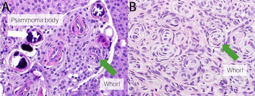
Meningioma histology:
Psammoma bodies (white arrow) and whorls (green arrows) are seen.

Image by Roy Strowd, MD. License: CC BY-NC-SA 4.0

Management

Observation

The majority of meningiomas can be observed, at least initially.

  • Observation is appropriate in patients Patients Individuals participating in the health care system for the purpose of receiving therapeutic, diagnostic, or preventive procedures. Clinician–Patient Relationship who:
    • Are asymptomatic (with meningiomas discovered incidentally on imaging)
    • Have small tumors (generally ≤ 2 cm) without evidence of invasion
  • Reassess in 3–6 months → if stable, can space out followup imaging 
  • Indications for treatment: 
    • Significant enlargement of a known tumor Tumor Inflammation
    • If the patient becomes symptomatic

Surgical intervention and postsurgical prognosis Prognosis A prediction of the probable outcome of a disease based on a individual’s condition and the usual course of the disease as seen in similar situations. Non-Hodgkin Lymphomas

  • Indications:
    • Large or expanding tumors
    • Infiltrating tumors
    • Associated with surrounding edema Edema Edema is a condition in which excess serous fluid accumulates in the body cavity or interstitial space of connective tissues. Edema is a symptom observed in several medical conditions. It can be categorized into 2 types, namely, peripheral (in the extremities) and internal (in an organ or body cavity). Edema
  • Used in uncomplicated and easily accessible meningiomas
  • The surgical approach to meningioma is based on: 
    • Size and location of the meningioma
    • Rate of growth of the tumor Tumor Inflammation
    • Risks of surgery to the patient based on age, health, and comorbidities Comorbidities The presence of co-existing or additional diseases with reference to an initial diagnosis or with reference to the index condition that is the subject of study. Comorbidity may affect the ability of affected individuals to function and also their survival; it may be used as a prognostic indicator for length of hospital stay, cost factors, and outcome or survival. St. Louis Encephalitis Virus
  • Surgical resection of meningiomas is considered to be a permanent cure.
  • Complete resection of the tumor Tumor Inflammation and its dural attachment is important to prevent recurrence.
  • Prognosis Prognosis A prediction of the probable outcome of a disease based on a individual’s condition and the usual course of the disease as seen in similar situations. Non-Hodgkin Lymphomas based on the extent of surgical resection, as graded according to the Simpson grading Grading Methods which attempt to express in replicable terms the level of cell differentiation in neoplasms as increasing anaplasia correlates with the aggressiveness of the neoplasm. Grading, Staging, and Metastasis scale Scale Dermatologic Examination.
    • Lower grades (I–III): more complete removal of the tumor Tumor Inflammation with lower recurrence risks
    • Higher grades (IV–V): less complete resection with higher recurrence risks
Table: Surgery resection grading Grading Methods which attempt to express in replicable terms the level of cell differentiation in neoplasms as increasing anaplasia correlates with the aggressiveness of the neoplasm. Grading, Staging, and Metastasis: Simpson grading Grading Methods which attempt to express in replicable terms the level of cell differentiation in neoplasms as increasing anaplasia correlates with the aggressiveness of the neoplasm. Grading, Staging, and Metastasis scale Scale Dermatologic Examination (recurrence rate based on the extent of resection)
Grade Extent of resection Recurrence risk (10-year interval)
I Macroscopically complete removal of tumor Tumor Inflammation, involved bones, venous sinuses Venous sinuses Veins: Histology, and dural tail 9%
II Macroscopically complete removal of tumor Tumor Inflammation, coagulation of dural tail 19%
III Macroscopically complete removal of tumor Tumor Inflammation, resection of dural tail 29%
IV Partial tumor Tumor Inflammation removal, no resection of dural tail 44%
V Simple decompression, no resection of dural tail 100%

Radiation Radiation Emission or propagation of acoustic waves (sound), electromagnetic energy waves (such as light; radio waves; gamma rays; or x-rays), or a stream of subatomic particles (such as electrons; neutrons; protons; or alpha particles). Osteosarcoma

  • Indications:
    • When the tumor Tumor Inflammation is inoperable
    • In case of incomplete resection
    • Higher-grade tumors (grades II and III)
  • Goals: 
Table: Radiation Radiation Emission or propagation of acoustic waves (sound), electromagnetic energy waves (such as light; radio waves; gamma rays; or x-rays), or a stream of subatomic particles (such as electrons; neutrons; protons; or alpha particles). Osteosarcoma therapy for meningiomas: Requirement of radiation Radiation Emission or propagation of acoustic waves (sound), electromagnetic energy waves (such as light; radio waves; gamma rays; or x-rays), or a stream of subatomic particles (such as electrons; neutrons; protons; or alpha particles). Osteosarcoma therapy based on WHO grade and type of surgery undergone
Grade Type of surgery Requirement of radiation Radiation Emission or propagation of acoustic waves (sound), electromagnetic energy waves (such as light; radio waves; gamma rays; or x-rays), or a stream of subatomic particles (such as electrons; neutrons; protons; or alpha particles). Osteosarcoma therapy
WHO grade I Gross total resection Observe
Subtotal resection Observe versus radiation Radiation Emission or propagation of acoustic waves (sound), electromagnetic energy waves (such as light; radio waves; gamma rays; or x-rays), or a stream of subatomic particles (such as electrons; neutrons; protons; or alpha particles). Osteosarcoma therapy
No surgery Radiation Radiation Emission or propagation of acoustic waves (sound), electromagnetic energy waves (such as light; radio waves; gamma rays; or x-rays), or a stream of subatomic particles (such as electrons; neutrons; protons; or alpha particles). Osteosarcoma therapy
WHO grade II Gross total resection Observe versus radiation Radiation Emission or propagation of acoustic waves (sound), electromagnetic energy waves (such as light; radio waves; gamma rays; or x-rays), or a stream of subatomic particles (such as electrons; neutrons; protons; or alpha particles). Osteosarcoma therapy
Subtotal resection Radiation Radiation Emission or propagation of acoustic waves (sound), electromagnetic energy waves (such as light; radio waves; gamma rays; or x-rays), or a stream of subatomic particles (such as electrons; neutrons; protons; or alpha particles). Osteosarcoma therapy
WHO grade III Any surgery Radiation Radiation Emission or propagation of acoustic waves (sound), electromagnetic energy waves (such as light; radio waves; gamma rays; or x-rays), or a stream of subatomic particles (such as electrons; neutrons; protons; or alpha particles). Osteosarcoma therapy

Differential Diagnosis

  • Hemangioblastoma Hemangioblastoma Hemangioblastomas are vascular neoplasms of the CNS. Hemangioblastomas are rare and are often associated with von Hippel-Lindau disease (VHL). The most common presentation is a headache and, depending on the size and location of the tumor, patients may present with sensory deficits and motor weakness. Hemangioblastoma: rare vascular tumors of the CNS often associated with von Hippel-Lindau disease von Hippel-Lindau disease Von Hippel-Lindau (VHL) disease is an autosomal dominant genetic condition resulting from a deletion or mutation in the VHL gene. Individuals diagnosed with VHL disease have tumors and cysts in various parts of their bodies and may present with hemangioblastomas, renal cell carcinoma (RCC), pheochromocytoma, endolymphatic sac tumors of the middle ear, pancreatic tumors, and papillary cystadenomas of the epididymis or the broad ligament. Von Hippel-Lindau Disease, which usually affects the brain Brain The part of central nervous system that is contained within the skull (cranium). Arising from the neural tube, the embryonic brain is comprised of three major parts including prosencephalon (the forebrain); mesencephalon (the midbrain); and rhombencephalon (the hindbrain). The developed brain consists of cerebrum; cerebellum; and other structures in the brain stem. Nervous System: Anatomy, Structure, and Classification but can also involve the spinal cord Spinal cord The spinal cord is the major conduction pathway connecting the brain to the body; it is part of the CNS. In cross section, the spinal cord is divided into an H-shaped area of gray matter (consisting of synapsing neuronal cell bodies) and a surrounding area of white matter (consisting of ascending and descending tracts of myelinated axons). Spinal Cord: Anatomy or retina Retina The ten-layered nervous tissue membrane of the eye. It is continuous with the optic nerve and receives images of external objects and transmits visual impulses to the brain. Its outer surface is in contact with the choroid and the inner surface with the vitreous body. The outermost layer is pigmented, whereas the inner nine layers are transparent. Eye: Anatomy. Presenting symptoms include headache Headache The symptom of pain in the cranial region. It may be an isolated benign occurrence or manifestation of a wide variety of headache disorders. Brain Abscess and neurologic deficits Neurologic Deficits High-Risk Headaches based on the location of the tumor Tumor Inflammation. Diagnosis is based on imaging and biopsy Biopsy Removal and pathologic examination of specimens from the living body. Ewing Sarcoma. Treatment includes surgical excision of the tumor Tumor Inflammation and, potentially, radiation Radiation Emission or propagation of acoustic waves (sound), electromagnetic energy waves (such as light; radio waves; gamma rays; or x-rays), or a stream of subatomic particles (such as electrons; neutrons; protons; or alpha particles). Osteosarcoma therapy.
  • Glioblastoma multiforme Glioblastoma multiforme Glioblastoma multiforme is a high-grade astrocytoma, an aggressive brain tumor arising from astrocytes, with an unknown cause and a poorly understood link to risk factors. There are two main types: primary, a more aggressive form seen more commonly in older patients, and secondary, developing from lower-grade astrocytomas and seen more commonly in younger patients. Glioblastoma Multiforme: a rapidly progressive WHO grade IV astrocytoma Grade IV Astrocytoma Astrocytoma that arises from astrocytes Astrocytes A class of large neuroglial (macroglial) cells in the central nervous system – the largest and most numerous neuroglial cells in the brain and spinal cord. Astrocytes (from ‘star’ cells) are irregularly shaped with many long processes, including those with ‘end feet’ which form the glial (limiting) membrane and directly and indirectly contribute to the blood-brain barrier. They regulate the extracellular ionic and chemical environment, and ‘reactive astrocytes’ (along with microglia) respond to injury. Nervous System: Histology ( glial cells Glial Cells The non-neuronal cells of the nervous system. They not only provide physical support, but also respond to injury, regulate the ionic and chemical composition of the extracellular milieu, participate in the blood-brain barrier and blood-retinal barrier, form the myelin insulation of nervous pathways, guide neuronal migration during development, and exchange metabolites with neurons. Neuroglia have high-affinity transmitter uptake systems, voltage-dependent and transmitter-gated ion channels, and can release transmitters, but their role in signaling (as in many other functions) is unclear. Nervous System: Histology in the brain Brain The part of central nervous system that is contained within the skull (cranium). Arising from the neural tube, the embryonic brain is comprised of three major parts including prosencephalon (the forebrain); mesencephalon (the midbrain); and rhombencephalon (the hindbrain). The developed brain consists of cerebrum; cerebellum; and other structures in the brain stem. Nervous System: Anatomy, Structure, and Classification) and clinically presents as headache Headache The symptom of pain in the cranial region. It may be an isolated benign occurrence or manifestation of a wide variety of headache disorders. Brain Abscess, nausea Nausea An unpleasant sensation in the stomach usually accompanied by the urge to vomit. Common causes are early pregnancy, sea and motion sickness, emotional stress, intense pain, food poisoning, and various enteroviruses. Antiemetics, drowsiness, blurred vision Blurred Vision Retinal Detachment, personality changes, and seizures Seizures A seizure is abnormal electrical activity of the neurons in the cerebral cortex that can manifest in numerous ways depending on the region of the brain affected. Seizures consist of a sudden imbalance that occurs between the excitatory and inhibitory signals in cortical neurons, creating a net excitation. The 2 major classes of seizures are focal and generalized. Seizures. Imaging, clinical presentation, and biopsy Biopsy Removal and pathologic examination of specimens from the living body. Ewing Sarcoma are the mainstays of diagnosis. Management includes radiotherapy, chemotherapy Chemotherapy Osteosarcoma, and surgical excision. Prognosis Prognosis A prediction of the probable outcome of a disease based on a individual’s condition and the usual course of the disease as seen in similar situations. Non-Hodgkin Lymphomas is poor.
  • Oligodendroglioma Oligodendroglioma Oligodendrogliomas are malignant CNS tumors arising from neural glial cell precursors. Oligodendrogliomas often arise in the frontal lobes of the brain and have a generally favorable prognosis when compared to other gliomas. Oligodendrogliomas are the 3rd most common CNS tumor. The most frequent presenting symptom is a seizure. Oligodendroglioma: a central nervous system Central nervous system The main information-processing organs of the nervous system, consisting of the brain, spinal cord, and meninges. Nervous System: Anatomy, Structure, and Classification tumor Tumor Inflammation arising from oligodendrocytes, which can present with focal neurological deficits Focal Neurological Deficits Brain Abscess, seizures Seizures A seizure is abnormal electrical activity of the neurons in the cerebral cortex that can manifest in numerous ways depending on the region of the brain affected. Seizures consist of a sudden imbalance that occurs between the excitatory and inhibitory signals in cortical neurons, creating a net excitation. The 2 major classes of seizures are focal and generalized. Seizures, and personality changes depending on the exact location. Diagnosis is made by MRI imaging and biopsy Biopsy Removal and pathologic examination of specimens from the living body. Ewing Sarcoma. Contact with the meninges Meninges The brain and the spinal cord are enveloped by 3 overlapping layers of connective tissue called the meninges. The layers are, from the most external layer to the most internal layer, the dura mater, arachnoid mater, and pia mater. Between these layers are 3 potential spaces called the epidural, subdural, and subarachnoid spaces. Meninges: Anatomy and a dural tail can help differentiate many meningiomas from oligodendrogliomas, which most commonly develop in the cerebral hemispheres, usually the frontal lobe Frontal lobe The part of the cerebral hemisphere anterior to the central sulcus, and anterior and superior to the lateral sulcus. Cerebral Cortex: Anatomy. Management involves surgical resection possibly accompanied by radiation Radiation Emission or propagation of acoustic waves (sound), electromagnetic energy waves (such as light; radio waves; gamma rays; or x-rays), or a stream of subatomic particles (such as electrons; neutrons; protons; or alpha particles). Osteosarcoma and/or chemotherapy Chemotherapy Osteosarcoma.
  • Craniopharyngioma Craniopharyngioma Craniopharyngiomas are rare squamous epithelial tumors with a solid and/or cystic structure that arise from the remnants of Rathke’s pouch along the pituitary stalk, in the suprasellar region. Craniopharyngiomas are histologically benign but tend to invade surrounding structures; thus, they should be treated as low-grade malignancies. Craniopharyngioma: a rare pituitary Pituitary A small, unpaired gland situated in the sella turcica. It is connected to the hypothalamus by a short stalk which is called the infundibulum. Hormones: Overview and Types tumor Tumor Inflammation derived from embryonic tissue. Clinically presents with headache Headache The symptom of pain in the cranial region. It may be an isolated benign occurrence or manifestation of a wide variety of headache disorders. Brain Abscess, hypersomnia Hypersomnia Disorders characterized by hypersomnolence during normal waking hours that may impair cognitive functioning. Subtypes include primary hypersomnia disorders (e.g., idiopathic hypersomnolence; narcolepsy; and kleine-levin syndrome) and secondary hypersomnia disorders where excessive somnolence can be attributed to a known cause (e.g., drug affect, mental disorders, and sleep apnea syndrome). Whipple’s Disease, polydipsia Polydipsia Excessive thirst manifested by excessive fluid intake. It is characteristic of many diseases such as diabetes mellitus; diabetes insipidus; and nephrogenic diabetes insipidus. The condition may be psychogenic in origin. Diabetes Insipidus, polyuria Polyuria Urination of a large volume of urine with an increase in urinary frequency, commonly seen in diabetes. Renal Potassium Regulation, loss of vision Vision Ophthalmic Exam, and vomiting Vomiting The forcible expulsion of the contents of the stomach through the mouth. Hypokalemia. Diagnosed by MRI and biopsy Biopsy Removal and pathologic examination of specimens from the living body. Ewing Sarcoma. Management includes surgical excision of the tumor Tumor Inflammation and radiotherapy if indicated. 
  • Metastatic tumor Tumor Inflammation: neoplastic cells that have spread to the brain Brain The part of central nervous system that is contained within the skull (cranium). Arising from the neural tube, the embryonic brain is comprised of three major parts including prosencephalon (the forebrain); mesencephalon (the midbrain); and rhombencephalon (the hindbrain). The developed brain consists of cerebrum; cerebellum; and other structures in the brain stem. Nervous System: Anatomy, Structure, and Classification from primary tumors elsewhere in the body. Metastatic neoplasms Neoplasms New abnormal growth of tissue. Malignant neoplasms show a greater degree of anaplasia and have the properties of invasion and metastasis, compared to benign neoplasms. Benign Bone Tumors are the most common neoplasms Neoplasms New abnormal growth of tissue. Malignant neoplasms show a greater degree of anaplasia and have the properties of invasion and metastasis, compared to benign neoplasms. Benign Bone Tumors in the brain Brain The part of central nervous system that is contained within the skull (cranium). Arising from the neural tube, the embryonic brain is comprised of three major parts including prosencephalon (the forebrain); mesencephalon (the midbrain); and rhombencephalon (the hindbrain). The developed brain consists of cerebrum; cerebellum; and other structures in the brain stem. Nervous System: Anatomy, Structure, and Classification. Neuroimaging Neuroimaging Non-invasive methods of visualizing the central nervous system, especially the brain, by various imaging modalities. Febrile Infant often shows multiple foci of the carcinoma, suggesting nonbrain origin. Clinical presentation depends on the primary tumor Tumor Inflammation and location and extent of brain Brain The part of central nervous system that is contained within the skull (cranium). Arising from the neural tube, the embryonic brain is comprised of three major parts including prosencephalon (the forebrain); mesencephalon (the midbrain); and rhombencephalon (the hindbrain). The developed brain consists of cerebrum; cerebellum; and other structures in the brain stem. Nervous System: Anatomy, Structure, and Classification metastasis Metastasis The transfer of a neoplasm from one organ or part of the body to another remote from the primary site. Grading, Staging, and Metastasis. Treatment is directed at the underlying neoplasm and may involve surgical resection, radiation Radiation Emission or propagation of acoustic waves (sound), electromagnetic energy waves (such as light; radio waves; gamma rays; or x-rays), or a stream of subatomic particles (such as electrons; neutrons; protons; or alpha particles). Osteosarcoma therapy, and chemotherapy Chemotherapy Osteosarcoma.
  • Migraine Migraine Migraine headache is a primary headache disorder and is among the most prevalent disorders in the world. Migraine is characterized by episodic, moderate to severe headaches that may be associated with increased sensitivity to light and sound, as well as nausea and/or vomiting. Migraine Headache: a headache Headache The symptom of pain in the cranial region. It may be an isolated benign occurrence or manifestation of a wide variety of headache disorders. Brain Abscess that is usually severe and often accompanied by nausea Nausea An unpleasant sensation in the stomach usually accompanied by the urge to vomit. Common causes are early pregnancy, sea and motion sickness, emotional stress, intense pain, food poisoning, and various enteroviruses. Antiemetics, photosensitivity Photosensitivity Tetracyclines, and sensitivity to sound. Diagnosis is based on family history Family History Adult Health Maintenance, clinical history, and presentation. Migraine Migraine Migraine headache is a primary headache disorder and is among the most prevalent disorders in the world. Migraine is characterized by episodic, moderate to severe headaches that may be associated with increased sensitivity to light and sound, as well as nausea and/or vomiting. Migraine Headache can be differentiated from meningiomas based on neuroimaging Neuroimaging Non-invasive methods of visualizing the central nervous system, especially the brain, by various imaging modalities. Febrile Infant findings. Management involves analgesics and preventive medicines such as alpha-blockers Alpha-blockers Drugs that bind to but do not activate alpha-adrenergic receptors thereby blocking the actions of endogenous or exogenous adrenergic agonists. Adrenergic alpha-antagonists are used in the treatment of hypertension, vasospasm, peripheral vascular disease, shock, and pheochromocytoma. Antiadrenergic Drugs.

References

  1. Mayo Clinic. (2020). Meningioma. https://www.mayoclinic.org/diseases-conditions/meningioma/symptoms-causes/syc-20355643
  2. The Johns Hopkins University. (2021). Meningioma. https://www.hopkinsmedicine.org/health/conditions-and-diseases/meningioma
  3. Haddad, G., Turkmani, A. (2018). Meningioma. Medscape. https://emedicine.medscape.com/article/1156552-overview
  4. Park, J.K. Epidemiology, pathology, clinical features, and diagnosis of meningioma. UpToDate. Retrieved May 19, 2021, from https://www.uptodate.com/contents/epidemiology-pathology-clinical-features-and-diagnosis-of-meningioma
  5. Park, J.K. Management of known or presumed benign (WHO grade 1) meningioma. UpToDate. Retrieved May 19, 2021, from https://www.uptodate.com/contents/management-of-known-or-presumed-benign-who-grade-i-meningioma
  6. Shih, H.A., Park, J.K. Management of atypical and malignant (WHO grade 2 and 3) meningioma. UpToDate. Retrieved May 19, 2021, from https://www.uptodate.com/contents/management-of-atypical-and-malignant-who-grade-ii-and-iii-meningioma

Create your free account or log in to continue reading!

Sign up now and get free access to Lecturio with concept pages, medical videos, and questions for your medical education.

User Reviews

Details