Achieve Mastery of Medical Concepts

Study for medical school and boards with Lecturio

Oligodendroglioma

Oligodendrogliomas are malignant CNS tumors arising from neural glial cell precursors. Oligodendrogliomas often arise in the frontal Frontal The bone that forms the frontal aspect of the skull. Its flat part forms the forehead, articulating inferiorly with the nasal bone and the cheek bone on each side of the face. Skull: Anatomy lobes of the brain Brain The part of central nervous system that is contained within the skull (cranium). Arising from the neural tube, the embryonic brain is comprised of three major parts including prosencephalon (the forebrain); mesencephalon (the midbrain); and rhombencephalon (the hindbrain). The developed brain consists of cerebrum; cerebellum; and other structures in the brain stem. Nervous System: Anatomy, Structure, and Classification and have a generally favorable prognosis Prognosis A prediction of the probable outcome of a disease based on a individual's condition and the usual course of the disease as seen in similar situations. Non-Hodgkin Lymphomas when compared to other gliomas. Oligodendrogliomas are the 3rd most common CNS tumor Tumor Inflammation. The most frequent presenting symptom is a seizure; other symptoms include headaches, visual loss, and focal neurologic deficits Neurologic Deficits High-Risk Headaches. Diagnosis is established by brain biopsy Brain biopsy JC Virus and BK Virus that demonstrates a classic “fried egg” appearance (round nuclei with clear cytoplasm). Oligodendrogliomas are slow-growing, but because they are life-limiting, they are managed with a combination of surgical resection, radiation Radiation Emission or propagation of acoustic waves (sound), electromagnetic energy waves (such as light; radio waves; gamma rays; or x-rays), or a stream of subatomic particles (such as electrons; neutrons; protons; or alpha particles). Osteosarcoma, and chemotherapy Chemotherapy Osteosarcoma.

Last updated: Nov 3, 2022

Editorial responsibility: Stanley Oiseth, Lindsay Jones, Evelin Maza

Overview

Definition

Oligodendrogliomas are malignant neuroepithelial tumors arising from neural glial cell precursors. These tumor Tumor Inflammation cells appear histologically similar to oligodendrocytes, but lack the myelinating ability of oligodendrocytes. Oligodendrogliomas are a type of glioma:

  • Gliomas arise from glial cells Glial Cells The non-neuronal cells of the nervous system. They not only provide physical support, but also respond to injury, regulate the ionic and chemical composition of the extracellular milieu, participate in the blood-brain barrier and blood-retinal barrier, form the myelin insulation of nervous pathways, guide neuronal migration during development, and exchange metabolites with neurons. Neuroglia have high-affinity transmitter uptake systems, voltage-dependent and transmitter-gated ion channels, and can release transmitters, but their role in signaling (as in many other functions) is unclear. Nervous System: Histology, which are supportive tissues within the brain Brain The part of central nervous system that is contained within the skull (cranium). Arising from the neural tube, the embryonic brain is comprised of three major parts including prosencephalon (the forebrain); mesencephalon (the midbrain); and rhombencephalon (the hindbrain). The developed brain consists of cerebrum; cerebellum; and other structures in the brain stem. Nervous System: Anatomy, Structure, and Classification and spinal cord Spinal cord The spinal cord is the major conduction pathway connecting the brain to the body; it is part of the CNS. In cross section, the spinal cord is divided into an H-shaped area of gray matter (consisting of synapsing neuronal cell bodies) and a surrounding area of white matter (consisting of ascending and descending tracts of myelinated axons). Spinal Cord: Anatomy.
  • Oligodendrocytes are glial cells Glial Cells The non-neuronal cells of the nervous system. They not only provide physical support, but also respond to injury, regulate the ionic and chemical composition of the extracellular milieu, participate in the blood-brain barrier and blood-retinal barrier, form the myelin insulation of nervous pathways, guide neuronal migration during development, and exchange metabolites with neurons. Neuroglia have high-affinity transmitter uptake systems, voltage-dependent and transmitter-gated ion channels, and can release transmitters, but their role in signaling (as in many other functions) is unclear. Nervous System: Histology in the CNS that produce myelin.
  • Astrocytomas and ependymomas are the other common glial cell tumors.

Classification of nervous system Nervous system The nervous system is a small and complex system that consists of an intricate network of neural cells (or neurons) and even more glial cells (for support and insulation). It is divided according to its anatomical components as well as its functional characteristics. The brain and spinal cord are referred to as the central nervous system, and the branches of nerves from these structures are referred to as the peripheral nervous system. Nervous System: Anatomy, Structure, and Classification tumors

Table: Classification of nervous system Nervous system The nervous system is a small and complex system that consists of an intricate network of neural cells (or neurons) and even more glial cells (for support and insulation). It is divided according to its anatomical components as well as its functional characteristics. The brain and spinal cord are referred to as the central nervous system, and the branches of nerves from these structures are referred to as the peripheral nervous system. Nervous System: Anatomy, Structure, and Classification tumors
Categories Specific tumors
Neuroepithelial tumors in the CNS
  • Astrocytomas, including glioblastoma multiforme Glioblastoma multiforme Glioblastoma multiforme is a high-grade astrocytoma, an aggressive brain tumor arising from astrocytes, with an unknown cause and a poorly understood link to risk factors. There are two main types: primary, a more aggressive form seen more commonly in older patients, and secondary, developing from lower-grade astrocytomas and seen more commonly in younger patients. Glioblastoma Multiforme
  • Oligodendroglioma
  • Ependymoma Ependymoma Ependymomas are glial cell tumors arising from CSF-producing ependymal cells lining the ventricular system. Ependymomas most commonly occur within the posterior fossa in contact with the 4th ventricle, or within the intramedullary spinal cord. Ependymoma and choroid-plexus tumors
  • Medulloblastomas (embryonal tumors)
Meningeal tumors
  • Meningiomas
  • Hemangioblastomas Hemangioblastomas A benign tumor of the nervous system that may occur sporadically or in association with von Hippel-Lindau disease. It accounts for approximately 2% of intracranial tumors, arising most frequently in the cerebellar hemispheres and vermis. Histologically, the tumors are composed of multiple capillary and sinusoidal channels lined with endothelial cells and clusters of lipid-laden pseudoxanthoma cells. Usually solitary, these tumors can be multiple and may also occur in the brain stem, spinal cord, retina, and supratentorial compartment. Cerebellar hemangioblastomas usually present in the third decade with intracranial hypertension, and ataxia. Von Hippel-Lindau Disease
Sellar region tumors
  • Craniopharyngioma Craniopharyngioma Craniopharyngiomas are rare squamous epithelial tumors with a solid and/or cystic structure that arise from the remnants of Rathke’s pouch along the pituitary stalk, in the suprasellar region. Craniopharyngiomas are histologically benign but tend to invade surrounding structures; thus, they should be treated as low-grade malignancies. Craniopharyngioma
  • Pituitary Pituitary A small, unpaired gland situated in the sella turcica. It is connected to the hypothalamus by a short stalk which is called the infundibulum. Hormones: Overview and Types adenoma
  • Pinealoma/pinealoblastoma
Primary CNS lymphoma Lymphoma A general term for various neoplastic diseases of the lymphoid tissue. Imaging of the Mediastinum Primary CNS lymphoma Lymphoma A general term for various neoplastic diseases of the lymphoid tissue. Imaging of the Mediastinum
Metastasis Metastasis The transfer of a neoplasm from one organ or part of the body to another remote from the primary site. Grading, Staging, and Metastasis to the brain Brain The part of central nervous system that is contained within the skull (cranium). Arising from the neural tube, the embryonic brain is comprised of three major parts including prosencephalon (the forebrain); mesencephalon (the midbrain); and rhombencephalon (the hindbrain). The developed brain consists of cerebrum; cerebellum; and other structures in the brain stem. Nervous System: Anatomy, Structure, and Classification (5× more common than primary brain Brain The part of central nervous system that is contained within the skull (cranium). Arising from the neural tube, the embryonic brain is comprised of three major parts including prosencephalon (the forebrain); mesencephalon (the midbrain); and rhombencephalon (the hindbrain). The developed brain consists of cerebrum; cerebellum; and other structures in the brain stem. Nervous System: Anatomy, Structure, and Classification tumors) Most commonly arising from:
  • Lung, breast, and renal cell carcinomas
  • Melanoma Melanoma Melanoma is a malignant tumor arising from melanocytes, the melanin-producing cells of the epidermis. These tumors are most common in fair-skinned individuals with a history of excessive sun exposure and sunburns. Melanoma
Peripheral tumors
  • Schwannomas, including acoustic neuroma Acoustic neuroma Acoustic neuroma, also referred to as vestibular schwannoma, is a benign tumor arising from Schwann cells of the vestibular component of the cranial nerve VIII. Acoustic neuroma forms within the internal auditory meatus and extends into the cerebellopontine angle. Acoustic Neuroma
  • Neuroblastoma Neuroblastoma Neuroblastoma is a malignancy that arises from the neural crest cell derivatives along the sympathetic chain (neuroblasts) and is most commonly located in the adrenal medulla. The tumor often presents in childhood with a flank mass that crosses the midline. Neuroblastoma

Epidemiology

  • Incidence Incidence The number of new cases of a given disease during a given period in a specified population. It also is used for the rate at which new events occur in a defined population. It is differentiated from prevalence, which refers to all cases in the population at a given time. Measures of Disease Frequency:
    • 3rd most common primary brain Brain The part of central nervous system that is contained within the skull (cranium). Arising from the neural tube, the embryonic brain is comprised of three major parts including prosencephalon (the forebrain); mesencephalon (the midbrain); and rhombencephalon (the hindbrain). The developed brain consists of cerebrum; cerebellum; and other structures in the brain stem. Nervous System: Anatomy, Structure, and Classification neoplasm (behind glioblastoma multiforme Glioblastoma multiforme Glioblastoma multiforme is a high-grade astrocytoma, an aggressive brain tumor arising from astrocytes, with an unknown cause and a poorly understood link to risk factors. There are two main types: primary, a more aggressive form seen more commonly in older patients, and secondary, developing from lower-grade astrocytomas and seen more commonly in younger patients. Glioblastoma Multiforme and low-grade diffuse astrocytoma Low-Grade Diffuse Astrocytoma Astrocytoma)
    • Accounts for approximately 5% of primary CNS tumors
    • 0.2 per 100,000 population
    • Approximately 1000 patients Patients Individuals participating in the health care system for the purpose of receiving therapeutic, diagnostic, or preventive procedures. Clinician–Patient Relationship diagnosed with oligodendrogliomas each year in the United States.
  • Median age at diagnosis: 25–45 years
  • Slightly more common in males than in females

Pathophysiology

Pathophysiology

  • Location: 
    • Usually arise in the cerebral hemispheres (cortical or subcortical)
    • Rarely can occur within the cerebellum Cerebellum The cerebellum, Latin for “little brain,” is located in the posterior cranial fossa, dorsal to the pons and midbrain, and its principal role is in the coordination of movements. The cerebellum consists of 3 lobes on either side of its 2 hemispheres and is connected in the middle by the vermis. Cerebellum: Anatomy, ventricles, or spinal cord Spinal cord The spinal cord is the major conduction pathway connecting the brain to the body; it is part of the CNS. In cross section, the spinal cord is divided into an H-shaped area of gray matter (consisting of synapsing neuronal cell bodies) and a surrounding area of white matter (consisting of ascending and descending tracts of myelinated axons). Spinal Cord: Anatomy
  • Regional effects on brain Brain The part of central nervous system that is contained within the skull (cranium). Arising from the neural tube, the embryonic brain is comprised of three major parts including prosencephalon (the forebrain); mesencephalon (the midbrain); and rhombencephalon (the hindbrain). The developed brain consists of cerebrum; cerebellum; and other structures in the brain stem. Nervous System: Anatomy, Structure, and Classification parenchyma include:
  • Intracranial pressure Intracranial Pressure Idiopathic Intracranial Hypertension ( ICP ICP Normal intracranial pressure (ICP) is defined as < 15 mm Hg, whereas pathologically increased ICP is any pressure ≥ 20 mm Hg. Increased ICP may result from several etiologies, including trauma, intracranial hemorrhage, mass lesions, cerebral edema, increased CSF production, and decreased CSF absorption. Increased Intracranial Pressure (ICP)) may be due to:
    • Direct mass Mass Three-dimensional lesion that occupies a space within the breast Imaging of the Breast effect
    • Edema Edema Edema is a condition in which excess serous fluid accumulates in the body cavity or interstitial space of connective tissues. Edema is a symptom observed in several medical conditions. It can be categorized into 2 types, namely, peripheral (in the extremities) and internal (in an organ or body cavity). Edema in surrounding brain Brain The part of central nervous system that is contained within the skull (cranium). Arising from the neural tube, the embryonic brain is comprised of three major parts including prosencephalon (the forebrain); mesencephalon (the midbrain); and rhombencephalon (the hindbrain). The developed brain consists of cerebrum; cerebellum; and other structures in the brain stem. Nervous System: Anatomy, Structure, and Classification tissue
    • ↑ Blood volume
    • ↑ CSF volume/ hydrocephalus Hydrocephalus Excessive accumulation of cerebrospinal fluid within the cranium which may be associated with dilation of cerebral ventricles, intracranial. Subarachnoid Hemorrhage
  • Disruption of normal parenchymal functions are due to:
    • Hypoxia Hypoxia Sub-optimal oxygen levels in the ambient air of living organisms. Ischemic Cell Damage
    • Competition for nutrients
    • Release Release Release of a virus from the host cell following virus assembly and maturation. Egress can occur by host cell lysis, exocytosis, or budding through the plasma membrane. Virology of metabolic end products:
      • Free radicals Free radicals Highly reactive molecules with an unsatisfied electron valence pair. Free radicals are produced in both normal and pathological processes. They are proven or suspected agents of tissue damage in a wide variety of circumstances including radiation, damage from environment chemicals, and aging. Natural and pharmacological prevention of free radical damage is being actively investigated. Ischemic Cell Damage
      • Altered electrolytes Electrolytes Electrolytes are mineral salts that dissolve in water and dissociate into charged particles called ions, which can be either be positively (cations) or negatively (anions) charged. Electrolytes are distributed in the extracellular and intracellular compartments in different concentrations. Electrolytes are essential for various basic life-sustaining functions. Electrolytes
      • Neurotransmitters
    • Release Release Release of a virus from the host cell following virus assembly and maturation. Egress can occur by host cell lysis, exocytosis, or budding through the plasma membrane. Virology and recruitment Recruitment Skeletal Muscle Contraction of cellular mediators (e.g., cytokines Cytokines Non-antibody proteins secreted by inflammatory leukocytes and some non-leukocytic cells, that act as intercellular mediators. They differ from classical hormones in that they are produced by a number of tissue or cell types rather than by specialized glands. They generally act locally in a paracrine or autocrine rather than endocrine manner. Adaptive Immune Response)

Pathogenesis

Oligodendrogliomas are histologically defined tumors that include both isocitrate dehydrogenase Isocitrate dehydrogenase An enzyme of the oxidoreductase class that catalyzes the conversion of isocitrate and NAD+ to yield 2-ketoglutarate, carbon dioxide, and nadh. It occurs in cell mitochondria. The enzyme requires mg2+, mn2+; it is activated by adp, citrate, and Ca2+, and inhibited by nadh, NADPH, and ATP. The reaction is the key rate-limiting step of the citric acid (tricarboxylic) cycle. Citric Acid Cycle (IDH) 1 and 2 mutations and codeletions of chromosomal arms 1p and 19q.

  • Mutations in IDH1 (found in the cytosol Cytosol A cell’s cytoskeleton is a network of intracellular protein fibers that provides structural support, anchors organelles, and aids intra- and extracellular movement. The Cell: Cytosol and Cytoskeleton) and/or IDH2 (found in the mitochondria Mitochondria Semiautonomous, self-reproducing organelles that occur in the cytoplasm of all cells of most, but not all, eukaryotes. Each mitochondrion is surrounded by a double limiting membrane. The inner membrane is highly invaginated, and its projections are called cristae. Mitochondria are the sites of the reactions of oxidative phosphorylation, which result in the formation of ATP. They contain distinctive ribosomes, transfer RNAs; amino Acyl tRNA synthetases; and elongation and termination factors. Mitochondria depend upon genes within the nucleus of the cells in which they reside for many essential messenger RNAs. Mitochondria are believed to have arisen from aerobic bacteria that established a symbiotic relationship with primitive protoeukaryotes. The Cell: Organelles):
    • IDH:
      • Catalyzes the reversible oxidative decarboxylation Decarboxylation The removal of a carboxyl group, usually in the form of carbon dioxide, from a chemical compound. Catabolism of Amino Acids of isocitrate → α-ketoglutarate (α-KG) in the TCA cycle TCA cycle The citric acid cycle, also known as the tricarboxylic acid (TCA) cycle or the krebs cycle, is a cyclic set of reactions that occurs in the mitochondrial matrix. The tca cycle is the continuation of any metabolic pathway that produces pyruvate, which is converted into its main substrate, acetyl-coa. Citric Acid Cycle 
      • Primary producer of NADPH NADPH Nicotinamide adenine dinucleotide phosphate. A coenzyme composed of ribosylnicotinamide 5′-phosphate (nmn) coupled by pyrophosphate linkage to the 5′-phosphate adenosine 2. Pentose Phosphate Pathway in most tissues, especially the brain Brain The part of central nervous system that is contained within the skull (cranium). Arising from the neural tube, the embryonic brain is comprised of three major parts including prosencephalon (the forebrain); mesencephalon (the midbrain); and rhombencephalon (the hindbrain). The developed brain consists of cerebrum; cerebellum; and other structures in the brain stem. Nervous System: Anatomy, Structure, and Classification
      • Also involved in mitigating oxidative damage
    • Mutations lead to production and buildup of 2-hydroxyglutarate 2-Hydroxyglutarate Astrocytoma ( 2-HG 2-HG Astrocytoma):
      • 2-HG 2-HG Astrocytoma inhibits enzymatic function of α-KG–dependent dioxygenases, which are involved in DNA DNA A deoxyribonucleotide polymer that is the primary genetic material of all cells. Eukaryotic and prokaryotic organisms normally contain DNA in a double-stranded state, yet several important biological processes transiently involve single-stranded regions. DNA, which consists of a polysugar-phosphate backbone possessing projections of purines (adenine and guanine) and pyrimidines (thymine and cytosine), forms a double helix that is held together by hydrogen bonds between these purines and pyrimidines (adenine to thymine and guanine to cytosine). DNA Types and Structure demethylation.
      • 2-HG 2-HG Astrocytoma causes epigenetic dysregulation → can lead to tumor Tumor Inflammation development
  • Codeletions of chromosomal arms 1p and 19q:
    • Leads to epigenetic changes and hypermethylation of the genome Genome The complete genetic complement contained in the DNA of a set of chromosomes in a human. The length of the human genome is about 3 billion base pairs. Basic Terms of Genetics
    • Typically occur because of an unbalanced translocation between chromosomes Chromosomes In a prokaryotic cell or in the nucleus of a eukaryotic cell, a structure consisting of or containing DNA which carries the genetic information essential to the cell. DNA Types and Structure 1 and 19
  • Other mutations:
    • Activating mutations in the telomerase Telomerase An essential ribonucleoprotein reverse transcriptase that adds telomeric DNA to the ends of eukaryotic chromosomes. DNA Replication reverse transcriptase Reverse transcriptase A reverse transcriptase encoded by the pol gene of HIV. It is a heterodimer of 66 kda and 51 kda subunits that are derived from a common precursor protein. The heterodimer also includes an RNAse h activity that plays an essential role the viral replication process. HIV Infection and AIDS (TERT) promoter:
      • TERT is a catalytic subunit in telomerase Telomerase An essential ribonucleoprotein reverse transcriptase that adds telomeric DNA to the ends of eukaryotic chromosomes. DNA Replication → activating mutations allow tumor Tumor Inflammation cells to become “immortal”
      • A primary diagnostic marker for glioblastoma multiforme Glioblastoma multiforme Glioblastoma multiforme is a high-grade astrocytoma, an aggressive brain tumor arising from astrocytes, with an unknown cause and a poorly understood link to risk factors. There are two main types: primary, a more aggressive form seen more commonly in older patients, and secondary, developing from lower-grade astrocytomas and seen more commonly in younger patients. Glioblastoma Multiforme, though can also be present in oligodendrogliomas
    • Methylation Methylation Addition of methyl groups. In histo-chemistry methylation is used to esterify carboxyl groups and remove sulfate groups by treating tissue sections with hot methanol in the presence of hydrochloric acid. . Glucocorticoids (i.e., silencing) of the O-6-methylguanine-DNA methyltransferase (MGMT) promoter: MGMT is an enzyme involved in DNA DNA A deoxyribonucleotide polymer that is the primary genetic material of all cells. Eukaryotic and prokaryotic organisms normally contain DNA in a double-stranded state, yet several important biological processes transiently involve single-stranded regions. DNA, which consists of a polysugar-phosphate backbone possessing projections of purines (adenine and guanine) and pyrimidines (thymine and cytosine), forms a double helix that is held together by hydrogen bonds between these purines and pyrimidines (adenine to thymine and guanine to cytosine). DNA Types and Structure repair.

Classification

Oligodendrogliomas are classified by the WHO according to their histology and the types of mutations they harbor. (Grade I tumors are considered benign Benign Fibroadenoma, so, unlike astrocytomas, there are no grade I oligodendrogliomas.)

  • WHO grade II: 
    • Oligodendroglioma, IDH-mutant and 1p/19q-codeleted
    • Oligodendroglioma, not otherwise specified
  • WHO grade III: 
    • Anaplastic oligodendroglioma, IDH-mutant and 1p/19q-codeleted
    • Anaplastic oligodendroglioma, not otherwise specified
  • Tumors not otherwise specified underwent genetic testing Genetic Testing Detection of a mutation; genotype; karyotype; or specific alleles associated with genetic traits, heritable diseases, or predisposition to a disease, or that may lead to the disease in descendants. It includes prenatal genetic testing. Myotonic Dystrophies that was inconclusive or genetic testing Genetic Testing Detection of a mutation; genotype; karyotype; or specific alleles associated with genetic traits, heritable diseases, or predisposition to a disease, or that may lead to the disease in descendants. It includes prenatal genetic testing. Myotonic Dystrophies was not done.
  • The diagnosis of oligoastrocytoma is no longer preferred, as tumors with mixed histology should be classified according to their molecular signature as either astrocytomas or oligodendrogliomas. If the 1p/19q codeletion is absent, the tumor Tumor Inflammation is typically classified as an astrocytoma Astrocytoma Astrocytomas are neuroepithelial tumors that arise from astrocytes, which are star-shaped glial cells (supporting tissues of the CNS). Astrocytomas are a type of glioma. There are 4 grades of astrocytomas. Astrocytoma.

Clinical Presentation

Most of the signs and symptoms depend on the exact location of the tumor Tumor Inflammation. Oligodendrogliomas are slow-growing and often asymptomatic for years prior to diagnosis. The most common presenting symptom is a seizure.

  • Seizures Seizures A seizure is abnormal electrical activity of the neurons in the cerebral cortex that can manifest in numerous ways depending on the region of the brain affected. Seizures consist of a sudden imbalance that occurs between the excitatory and inhibitory signals in cortical neurons, creating a net excitation. The 2 major classes of seizures are focal and generalized. Seizures: can be either partial or generalized
  • General symptoms: symptoms that can occur with tumors in any location
    • Headache Headache The symptom of pain in the cranial region. It may be an isolated benign occurrence or manifestation of a wide variety of headache disorders. Brain Abscess (usually in the morning)
    • Nausea Nausea An unpleasant sensation in the stomach usually accompanied by the urge to vomit. Common causes are early pregnancy, sea and motion sickness, emotional stress, intense pain, food poisoning, and various enteroviruses. Antiemetics and/or vomiting Vomiting The forcible expulsion of the contents of the stomach through the mouth. Hypokalemia 
    • Cognitive difficulties: 
      • Memory Memory Complex mental function having four distinct phases: (1) memorizing or learning, (2) retention, (3) recall, and (4) recognition. Clinically, it is usually subdivided into immediate, recent, and remote memory. Psychiatric Assessment problems
      • Mood or personality changes (mostly seen in frontal lobe Frontal lobe The part of the cerebral hemisphere anterior to the central sulcus, and anterior and superior to the lateral sulcus. Cerebral Cortex: Anatomy tumors)
    • Papilledema Papilledema Swelling of the optic disk, usually in association with increased intracranial pressure, characterized by hyperemia, blurring of the disk margins, microhemorrhages, blind spot enlargement, and engorgement of retinal veins. Chronic papilledema may cause optic atrophy and visual loss. Idiopathic Intracranial Hypertension
  • Focal neurologic deficits Neurologic Deficits High-Risk Headaches: depends on particular location of tumor Tumor Inflammation
    • Muscle weakness (damage to contralateral motor Motor Neurons which send impulses peripherally to activate muscles or secretory cells. Nervous System: Histology cortex)
    • Sensory Sensory Neurons which conduct nerve impulses to the central nervous system. Nervous System: Histology abnormalities (damage to contralateral somatosensory cortex Somatosensory cortex Area of the parietal lobe concerned with receiving sensations such as movement, pain, pressure, position, temperature, touch, and vibration. It lies posterior to the central sulcus. Cerebral Cortex: Anatomy)
    • Apraxia Apraxia A group of cognitive disorders characterized by the inability to perform previously learned skills that cannot be attributed to deficits of motor or sensory function. The two major subtypes of this condition are ideomotor and ideational apraxia, which refers to loss of the ability to mentally formulate the processes involved with performing an action. For example, dressing apraxia may result from an inability to mentally formulate the act of placing clothes on the body. Apraxias are generally associated with lesions of the dominant parietal lobe and supramarginal gyrus. Cranial Nerve Palsies 
    • Aphasia Aphasia A cognitive disorder marked by an impaired ability to comprehend or express language in its written or spoken form. This condition is caused by diseases which affect the language areas of the dominant hemisphere. Clinical features are used to classify the various subtypes of this condition. General categories include receptive, expressive, and mixed forms of aphasia. Ischemic Stroke
    • Visual field Visual Field The Visual Pathway and Related Disorders deficits 
    • Hemiparesis Hemiparesis The term hemiparesis refers to mild to moderate weakness involving one side of the body. Epidural Hemorrhage

Diagnosis

Imaging

Head MRI is the imaging method of choice when evaluating for brain Brain The part of central nervous system that is contained within the skull (cranium). Arising from the neural tube, the embryonic brain is comprised of three major parts including prosencephalon (the forebrain); mesencephalon (the midbrain); and rhombencephalon (the hindbrain). The developed brain consists of cerebrum; cerebellum; and other structures in the brain stem. Nervous System: Anatomy, Structure, and Classification tumors.
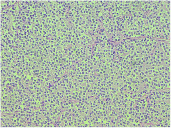
  • MRI of the brain Brain The part of central nervous system that is contained within the skull (cranium). Arising from the neural tube, the embryonic brain is comprised of three major parts including prosencephalon (the forebrain); mesencephalon (the midbrain); and rhombencephalon (the hindbrain). The developed brain consists of cerebrum; cerebellum; and other structures in the brain stem. Nervous System: Anatomy, Structure, and Classification:
  • Head CT:
    • Done when there are contraindications Contraindications A condition or factor associated with a recipient that makes the use of a drug, procedure, or physical agent improper or inadvisable. Contraindications may be absolute (life threatening) or relative (higher risk of complications in which benefits may outweigh risks). Noninvasive Ventilation to MRI or in the acute care setting 
    • Findings include: 
      • Hypodense lesions with surrounding edema Edema Edema is a condition in which excess serous fluid accumulates in the body cavity or interstitial space of connective tissues. Edema is a symptom observed in several medical conditions. It can be categorized into 2 types, namely, peripheral (in the extremities) and internal (in an organ or body cavity). Edema
      • Calcifications and hemorrhage can be seen.
      • Typically, only anaplastic lesions enhance.

Biopsy Biopsy Removal and pathologic examination of specimens from the living body. Ewing Sarcoma

  • Gross appearance: 
    • Well-circumscribed, gray, gelatinous mass Mass Three-dimensional lesion that occupies a space within the breast Imaging of the Breast 
    • Often with areas of cysts Cysts Any fluid-filled closed cavity or sac that is lined by an epithelium. Cysts can be of normal, abnormal, non-neoplastic, or neoplastic tissues. Fibrocystic Change, hemorrhages, and calcifications
  • Microscopic appearance: 
    • Composed of well-differentiated cells resembling oligodendrocytes
    • Sheets of fried-egg cells (large cells with clear cytoplasm and round hyperchromatic nucleus Nucleus Within a eukaryotic cell, a membrane-limited body which contains chromosomes and one or more nucleoli (cell nucleolus). The nuclear membrane consists of a double unit-type membrane which is perforated by a number of pores; the outermost membrane is continuous with the endoplasmic reticulum. A cell may contain more than one nucleus. The Cell: Organelles)
    • Chicken-wire capillaries Capillaries Capillaries are the primary structures in the circulatory system that allow the exchange of gas, nutrients, and other materials between the blood and the extracellular fluid (ECF). Capillaries are the smallest of the blood vessels. Because a capillary diameter is so small, only 1 RBC may pass through at a time. Capillaries: Histology: networks of thin blood vessels throughout the tumor Tumor Inflammation due to significant angiogenesis Angiogenesis Bartonella
    • Anaplastic tumors exhibit:
  • Molecular testing:
    • Presence of IDH1/2 mutations and 1p/19q codeletion:
      • Immunohistochemical staining for IDH1/2 mutations
      • FISH FISH A type of in situ hybridization in which target sequences are stained with fluorescent dye so their location and size can be determined using fluorescence microscopy. This staining is sufficiently distinct that the hybridization signal can be seen both in metaphase spreads and in interphase nuclei. Chromosome Testing studies for 1p/19q codeletions
    • Lack myelin basic protein (MBP) found in normal myelin-producing oligodendrocytes and Schwann cells
    • May stain positive for oligodendrocyte Oligodendrocyte A class of large neuroglial (macroglial) cells in the central nervous system. Oligodendroglia may be called interfascicular, perivascular, or perineuronal (not the same as satellite cells, perineuronal of ganglia) according to their location. They form the insulating myelin sheath of axons in the central nervous system. Nervous System: Histology transcription factor Transcription factor Generic term for proteins necessary for transcription Regulation of Transcription 2 (Olig2), a marker for oligodendrocyte Oligodendrocyte A class of large neuroglial (macroglial) cells in the central nervous system. Oligodendroglia may be called interfascicular, perivascular, or perineuronal (not the same as satellite cells, perineuronal of ganglia) according to their location. They form the insulating myelin sheath of axons in the central nervous system. Nervous System: Histology progenitor cells that is involved in cell differentiation
    • May stain positive for glial fibrillary acidic protein (GFAP), a common marker of astrocytes Astrocytes A class of large neuroglial (macroglial) cells in the central nervous system – the largest and most numerous neuroglial cells in the brain and spinal cord. Astrocytes (from ‘star’ cells) are irregularly shaped with many long processes, including those with ‘end feet’ which form the glial (limiting) membrane and directly and indirectly contribute to the blood-brain barrier. They regulate the extracellular ionic and chemical environment, and ‘reactive astrocytes’ (along with microglia) respond to injury. Nervous System: Histology
Oligodendroglioma

Oligodendroglioma:
Note the fried-egg cells with dark, round nuclei and chicken-wire capillaries throughout the tissue sample.

Image: “Morphologic spectrum of glial tumors: an increased trend towards oligodendroglial tumors in Pakistan” by Hashmi AA, Faridi N, Malik B, Edhi MM, Khurshid A, Khan M. License: CC BY 4.0

Management and Prognosis

Management

Oligodendrogliomas are infiltrative tumors that are commonly considered incurable owing to high risk of relapse Relapse Relapsing Fever/recurrence. Optimal management typically involves surgery, radiation Radiation Emission or propagation of acoustic waves (sound), electromagnetic energy waves (such as light; radio waves; gamma rays; or x-rays), or a stream of subatomic particles (such as electrons; neutrons; protons; or alpha particles). Osteosarcoma, and chemotherapy Chemotherapy Osteosarcoma.

  • Surgery: diagnostic and therapeutic roles
    • Confirms the diagnosis, which can guide adjuvant Adjuvant Substances that augment, stimulate, activate, potentiate, or modulate the immune response at either the cellular or humoral level. The classical agents (freund’s adjuvant, bcg, corynebacterium parvum, et al.) contain bacterial antigens. Some are endogenous (e.g., histamine, interferon, transfer factor, tuftsin, interleukin-1). Their mode of action is either non-specific, resulting in increased immune responsiveness to a wide variety of antigens, or antigen-specific, i.e., affecting a restricted type of immune response to a narrow group of antigens. The therapeutic efficacy of many biological response modifiers is related to their antigen-specific immunoadjuvanticity. Vaccination therapies
    • For best outcomes, the goal of surgery is a gross total resection; however, this is not always possible, depending on the region of the brain Brain The part of central nervous system that is contained within the skull (cranium). Arising from the neural tube, the embryonic brain is comprised of three major parts including prosencephalon (the forebrain); mesencephalon (the midbrain); and rhombencephalon (the hindbrain). The developed brain consists of cerebrum; cerebellum; and other structures in the brain stem. Nervous System: Anatomy, Structure, and Classification affected.
  • Radiation Radiation Emission or propagation of acoustic waves (sound), electromagnetic energy waves (such as light; radio waves; gamma rays; or x-rays), or a stream of subatomic particles (such as electrons; neutrons; protons; or alpha particles). Osteosarcoma therapy:
    • Typically follows surgery
    • Administered to a restricted area around the surgical site to maximize therapeutic benefit while minimizing side effects
    • Typically withheld from very young children owing to serious comorbidity in that age group
  • Chemotherapy Chemotherapy Osteosarcoma:
    • Typically given following radiation Radiation Emission or propagation of acoustic waves (sound), electromagnetic energy waves (such as light; radio waves; gamma rays; or x-rays), or a stream of subatomic particles (such as electrons; neutrons; protons; or alpha particles). Osteosarcoma
    • Improves overall survival when combined with adjuvant Adjuvant Substances that augment, stimulate, activate, potentiate, or modulate the immune response at either the cellular or humoral level. The classical agents (freund’s adjuvant, bcg, corynebacterium parvum, et al.) contain bacterial antigens. Some are endogenous (e.g., histamine, interferon, transfer factor, tuftsin, interleukin-1). Their mode of action is either non-specific, resulting in increased immune responsiveness to a wide variety of antigens, or antigen-specific, i.e., affecting a restricted type of immune response to a narrow group of antigens. The therapeutic efficacy of many biological response modifiers is related to their antigen-specific immunoadjuvanticity. Vaccination radiation Radiation Emission or propagation of acoustic waves (sound), electromagnetic energy waves (such as light; radio waves; gamma rays; or x-rays), or a stream of subatomic particles (such as electrons; neutrons; protons; or alpha particles). Osteosarcoma therapy
    • The standard regimen is PCV:
    • Can also consider temozolomide Temozolomide A dacarbazine derivative that is used as an alkylating antineoplastic agent for the treatment of malignant glioma and malignant melanoma. Alkylating Agents and Platinum as a 2nd-line agent in oligodendroglioma (it is 1st-line for astrocytomas)
  • Other therapies:
    • Anticonvulsant Anticonvulsant Anticonvulsant drugs are pharmacological agents used to achieve seizure control and/or prevent seizure episodes. Anticonvulsants encompass various drugs with different mechanisms of action including ion-channel (Na+ and Ca+2) blocking and GABA reuptake inhibition. First-Generation Anticonvulsant Drugs therapy in patients Patients Individuals participating in the health care system for the purpose of receiving therapeutic, diagnostic, or preventive procedures. Clinician–Patient Relationship with seizures Seizures A seizure is abnormal electrical activity of the neurons in the cerebral cortex that can manifest in numerous ways depending on the region of the brain affected. Seizures consist of a sudden imbalance that occurs between the excitatory and inhibitory signals in cortical neurons, creating a net excitation. The 2 major classes of seizures are focal and generalized. Seizures
    • Corticosteroids Corticosteroids Chorioretinitis for brain Brain The part of central nervous system that is contained within the skull (cranium). Arising from the neural tube, the embryonic brain is comprised of three major parts including prosencephalon (the forebrain); mesencephalon (the midbrain); and rhombencephalon (the hindbrain). The developed brain consists of cerebrum; cerebellum; and other structures in the brain stem. Nervous System: Anatomy, Structure, and Classification edema Edema Edema is a condition in which excess serous fluid accumulates in the body cavity or interstitial space of connective tissues. Edema is a symptom observed in several medical conditions. It can be categorized into 2 types, namely, peripheral (in the extremities) and internal (in an organ or body cavity). Edema

Prognosis Prognosis A prediction of the probable outcome of a disease based on a individual’s condition and the usual course of the disease as seen in similar situations. Non-Hodgkin Lymphomas

  • Average 5-year survival: 
    • Low-grade oligodendroglioma: 5–10 years 
    • Anaplastic oligodendroglioma: 3–4 years 
  • Factors affecting prognosis Prognosis A prediction of the probable outcome of a disease based on a individual’s condition and the usual course of the disease as seen in similar situations. Non-Hodgkin Lymphomas:
    • Age of the patient at diagnosis (younger patients Patients Individuals participating in the health care system for the purpose of receiving therapeutic, diagnostic, or preventive procedures. Clinician–Patient Relationship do better)
    • Location of the tumor Tumor Inflammation
    • Extent of surgical resection
    • Histologic grade of the tumor Tumor Inflammation
    • Postoperative performance status

Related videos

Differential Diagnosis

  • Astrocytoma Astrocytoma Astrocytomas are neuroepithelial tumors that arise from astrocytes, which are star-shaped glial cells (supporting tissues of the CNS). Astrocytomas are a type of glioma. There are 4 grades of astrocytomas. Astrocytoma: CNS tumor Tumor Inflammation arising from astrocytes Astrocytes A class of large neuroglial (macroglial) cells in the central nervous system – the largest and most numerous neuroglial cells in the brain and spinal cord. Astrocytes (from ‘star’ cells) are irregularly shaped with many long processes, including those with ‘end feet’ which form the glial (limiting) membrane and directly and indirectly contribute to the blood-brain barrier. They regulate the extracellular ionic and chemical environment, and ‘reactive astrocytes’ (along with microglia) respond to injury. Nervous System: Histology. Aggressive, grade IV astrocytomas are called glioblastoma multiforme Glioblastoma multiforme Glioblastoma multiforme is a high-grade astrocytoma, an aggressive brain tumor arising from astrocytes, with an unknown cause and a poorly understood link to risk factors. There are two main types: primary, a more aggressive form seen more commonly in older patients, and secondary, developing from lower-grade astrocytomas and seen more commonly in younger patients. Glioblastoma Multiforme, while grade II astrocytomas are called low-grade diffuse astrocytomas. Astrocytomas can present similarly to oligodendrogliomas and are diagnosed using MRI and biopsy Biopsy Removal and pathologic examination of specimens from the living body. Ewing Sarcoma. Mutations in IDH and codeletion of 1p/19q are required to diagnose oligodendroglioma; if only IDH mutations are present, this likely represents a low-grade astrocytoma Astrocytoma Astrocytomas are neuroepithelial tumors that arise from astrocytes, which are star-shaped glial cells (supporting tissues of the CNS). Astrocytomas are a type of glioma. There are 4 grades of astrocytomas. Astrocytoma. Like oligodendrogliomas, treatment for astrocytomas includes surgical excision, radiation Radiation Emission or propagation of acoustic waves (sound), electromagnetic energy waves (such as light; radio waves; gamma rays; or x-rays), or a stream of subatomic particles (such as electrons; neutrons; protons; or alpha particles). Osteosarcoma, and chemotherapy Chemotherapy Osteosarcoma.
  • Meningioma Meningioma Meningiomas are slow-growing tumors that arise from the meninges of the brain and spinal cord. The vast majority are benign. These tumors commonly occur in individuals with a history of high doses of skull radiation, head trauma, and neurofibromatosis 2. Meningioma: benign Benign Fibroadenoma tumor Tumor Inflammation arising from the meninges Meninges The brain and the spinal cord are enveloped by 3 overlapping layers of connective tissue called the meninges. The layers are, from the most external layer to the most internal layer, the dura mater, arachnoid mater, and pia mater. Between these layers are 3 potential spaces called the epidural, subdural, and subarachnoid spaces. Meninges: Anatomy of the brain Brain The part of central nervous system that is contained within the skull (cranium). Arising from the neural tube, the embryonic brain is comprised of three major parts including prosencephalon (the forebrain); mesencephalon (the midbrain); and rhombencephalon (the hindbrain). The developed brain consists of cerebrum; cerebellum; and other structures in the brain stem. Nervous System: Anatomy, Structure, and Classification or the spinal cord Spinal cord The spinal cord is the major conduction pathway connecting the brain to the body; it is part of the CNS. In cross section, the spinal cord is divided into an H-shaped area of gray matter (consisting of synapsing neuronal cell bodies) and a surrounding area of white matter (consisting of ascending and descending tracts of myelinated axons). Spinal Cord: Anatomy. Meningiomas present with headache Headache The symptom of pain in the cranial region. It may be an isolated benign occurrence or manifestation of a wide variety of headache disorders. Brain Abscess, vision Vision Ophthalmic Exam disturbances, and possible seizures Seizures A seizure is abnormal electrical activity of the neurons in the cerebral cortex that can manifest in numerous ways depending on the region of the brain affected. Seizures consist of a sudden imbalance that occurs between the excitatory and inhibitory signals in cortical neurons, creating a net excitation. The 2 major classes of seizures are focal and generalized. Seizures. Diagnosis is confirmed using MRI and biopsy Biopsy Removal and pathologic examination of specimens from the living body. Ewing Sarcoma, and meningiomas are treated with surgical excision. Unlike meningiomas, oligodendrogliomas are more commonly symptomatic and have no association with the dura. 
  • Metastatic tumor Tumor Inflammation: neoplastic cells that have spread to the brain Brain The part of central nervous system that is contained within the skull (cranium). Arising from the neural tube, the embryonic brain is comprised of three major parts including prosencephalon (the forebrain); mesencephalon (the midbrain); and rhombencephalon (the hindbrain). The developed brain consists of cerebrum; cerebellum; and other structures in the brain stem. Nervous System: Anatomy, Structure, and Classification from primary tumors elsewhere in the body. Metastatic neoplasms Neoplasms New abnormal growth of tissue. Malignant neoplasms show a greater degree of anaplasia and have the properties of invasion and metastasis, compared to benign neoplasms. Benign Bone Tumors are the most common neoplasms Neoplasms New abnormal growth of tissue. Malignant neoplasms show a greater degree of anaplasia and have the properties of invasion and metastasis, compared to benign neoplasms. Benign Bone Tumors in the brain Brain The part of central nervous system that is contained within the skull (cranium). Arising from the neural tube, the embryonic brain is comprised of three major parts including prosencephalon (the forebrain); mesencephalon (the midbrain); and rhombencephalon (the hindbrain). The developed brain consists of cerebrum; cerebellum; and other structures in the brain stem. Nervous System: Anatomy, Structure, and Classification. Neuroimaging Neuroimaging Non-invasive methods of visualizing the central nervous system, especially the brain, by various imaging modalities. Febrile Infant often shows multiple foci of the carcinoma, suggesting an origin outside the brain Brain The part of central nervous system that is contained within the skull (cranium). Arising from the neural tube, the embryonic brain is comprised of three major parts including prosencephalon (the forebrain); mesencephalon (the midbrain); and rhombencephalon (the hindbrain). The developed brain consists of cerebrum; cerebellum; and other structures in the brain stem. Nervous System: Anatomy, Structure, and Classification. Clinical presentation depends on the primary tumor Tumor Inflammation and location and extent of brain Brain The part of central nervous system that is contained within the skull (cranium). Arising from the neural tube, the embryonic brain is comprised of three major parts including prosencephalon (the forebrain); mesencephalon (the midbrain); and rhombencephalon (the hindbrain). The developed brain consists of cerebrum; cerebellum; and other structures in the brain stem. Nervous System: Anatomy, Structure, and Classification metastasis Metastasis The transfer of a neoplasm from one organ or part of the body to another remote from the primary site. Grading, Staging, and Metastasis. Treatment is directed at the underlying neoplasm and may involve surgical resection, radiation Radiation Emission or propagation of acoustic waves (sound), electromagnetic energy waves (such as light; radio waves; gamma rays; or x-rays), or a stream of subatomic particles (such as electrons; neutrons; protons; or alpha particles). Osteosarcoma therapy, and chemotherapy Chemotherapy Osteosarcoma.
  • Cerebral toxoplasmosis Toxoplasmosis Toxoplasmosis is an infectious disease caused by Toxoplasma gondii, an obligate intracellular protozoan parasite. Felines are the definitive host, but transmission to humans can occur through contact with cat feces or the consumption of contaminated foods. The clinical presentation and complications depend on the host’s immune status. Toxoplasma/Toxoplasmosis: infection with Toxoplasma Toxoplasma Toxoplasmosis is an infectious disease caused by Toxoplasma gondii, an obligate intracellular protozoan parasite. Felines are the definitive host, but transmission to humans can occur through contact with cat feces or the consumption of contaminated foods. The clinical presentation and complications depend on the host’s immune status. Toxoplasma/Toxoplasmosis gondii in the CNS in immunocompromised immunocompromised A human or animal whose immunologic mechanism is deficient because of an immunodeficiency disorder or other disease or as the result of the administration of immunosuppressive drugs or radiation. Gastroenteritis patients Patients Individuals participating in the health care system for the purpose of receiving therapeutic, diagnostic, or preventive procedures. Clinician–Patient Relationship (e.g., HIV HIV Anti-HIV Drugs patients Patients Individuals participating in the health care system for the purpose of receiving therapeutic, diagnostic, or preventive procedures. Clinician–Patient Relationship). This disease is usually symptomatic only in patients Patients Individuals participating in the health care system for the purpose of receiving therapeutic, diagnostic, or preventive procedures. Clinician–Patient Relationship with CD4 counts < 100 cells/µL, and it presents with fever Fever Fever is defined as a measured body temperature of at least 38°C (100.4°F). Fever is caused by circulating endogenous and/or exogenous pyrogens that increase levels of prostaglandin E2 in the hypothalamus. Fever is commonly associated with chills, rigors, sweating, and flushing of the skin. Fever, headache Headache The symptom of pain in the cranial region. It may be an isolated benign occurrence or manifestation of a wide variety of headache disorders. Brain Abscess, focal neurologic deficits Neurologic Deficits High-Risk Headaches, and/or seizures Seizures A seizure is abnormal electrical activity of the neurons in the cerebral cortex that can manifest in numerous ways depending on the region of the brain affected. Seizures consist of a sudden imbalance that occurs between the excitatory and inhibitory signals in cortical neurons, creating a net excitation. The 2 major classes of seizures are focal and generalized. Seizures. Cerebral toxoplasmosis Toxoplasmosis Toxoplasmosis is an infectious disease caused by Toxoplasma gondii, an obligate intracellular protozoan parasite. Felines are the definitive host, but transmission to humans can occur through contact with cat feces or the consumption of contaminated foods. The clinical presentation and complications depend on the host’s immune status. Toxoplasma/Toxoplasmosis is diagnosed with serologic studies and brain Brain The part of central nervous system that is contained within the skull (cranium). Arising from the neural tube, the embryonic brain is comprised of three major parts including prosencephalon (the forebrain); mesencephalon (the midbrain); and rhombencephalon (the hindbrain). The developed brain consists of cerebrum; cerebellum; and other structures in the brain stem. Nervous System: Anatomy, Structure, and Classification MRI showing ring-enhancing lesions (which is unlike oligodendrogliomas). Cerebral toxoplasmosis Toxoplasmosis Toxoplasmosis is an infectious disease caused by Toxoplasma gondii, an obligate intracellular protozoan parasite. Felines are the definitive host, but transmission to humans can occur through contact with cat feces or the consumption of contaminated foods. The clinical presentation and complications depend on the host’s immune status. Toxoplasma/Toxoplasmosis is treated with pyrimethamine Pyrimethamine One of the folic acid antagonists that is used as an antimalarial or with a sulfonamide to treat toxoplasmosis. Antimalarial Drugs, sulfadiazine Sulfadiazine One of the short-acting sulfonamides used in combination with pyrimethamine to treat toxoplasmosis in patients with acquired immunodeficiency syndrome and in newborns with congenital infections. Sulfonamides and Trimethoprim, and leucovorin, along with antiretroviral therapy Antiretroviral therapy Antiretroviral therapy (ART) targets the replication cycle of the human immunodeficiency virus (HIV) and is classified based on the viral enzyme or mechanism that is inhibited. The goal of therapy is to suppress viral replication to reach the outcome of undetected viral load. Anti-HIV Drugs

References

  1. Van den Bent, M. (2021). Clinical features, diagnosis, and pathology of IDH-mutant, 1p/19q-codeleted oligodendrogliomas. UpToDate. Retrieved May 21, 2021, from https://www.uptodate.com/contents/clinical-features-diagnosis-and-pathology-of-idh-mutant-1p-19q-codeleted-oligodendrogliomas 
  2. Van den Bent, M. (2021). Treatment and prognosis of IDH-mutant, 1p/19q-codeleted (grade II and III) oligodendrogliomas. UpToDate. Retrieved May 21, 2021, from https://www.uptodate.com/contents/treatment-and-prognosis-of-idh-mutant-1p-19q-codeleted-grade-ii-and-iii-oligodendrogliomas 
  3. Uddin, S. (2020). Oligodendroglioma. Medscape. Retrieved May 21, 2021, from https://emedicine.medscape.com/article/1156699-overview 
  4. Tork, C.A., Atkinson, C. (2020). Oligodendroglioma. StatPearls. Retrieved May 21, 2021, from https://www.ncbi.nlm.nih.gov/books/NBK559184/ 

Create your free account or log in to continue reading!

Sign up now and get free access to Lecturio with concept pages, medical videos, and questions for your medical education.

User Reviews

Details