Achieve Mastery of Medical Concepts

Study for medical school and boards with Lecturio

Ependymoma

Ependymomas are glial cell tumors arising from CSF-producing ependymal cells Ependymal Cells The macroglial cells of ependyma. They are characterized by bipolar cell body shape and processes that contact basal lamina around blood vessels and/or the pia mater and the cerebral ventricles. Muller cells of the retina are included based on similar microenvironmental contacts and morphology. Nervous System: Histology lining the ventricular system Ventricular System The ventricular system is an extension of the subarachnoid space into the brain consisting of a series of interconnecting spaces and channels. Four chambers are filled with cerebrospinal fluid (CSF): the paired lateral ventricles, the unpaired 3rd ventricle, and the unpaired 4th ventricle. Ventricular System: Anatomy. Ependymomas most commonly occur within the posterior fossa in contact with the 4th ventricle, or within the intramedullary spinal cord Spinal cord The spinal cord is the major conduction pathway connecting the brain to the body; it is part of the CNS. In cross section, the spinal cord is divided into an H-shaped area of gray matter (consisting of synapsing neuronal cell bodies) and a surrounding area of white matter (consisting of ascending and descending tracts of myelinated axons). Spinal Cord: Anatomy. The clinical presentation of ependymomas varies depending on the location of the tumor Tumor Inflammation. Magnetic resonance imaging is the imaging modality of choice, but histologic confirmation is required for diagnosis. The mainstay of treatment for intracranial ependymoma is surgical resection and adjuvant Adjuvant Substances that augment, stimulate, activate, potentiate, or modulate the immune response at either the cellular or humoral level. The classical agents (freund's adjuvant, bcg, corynebacterium parvum, et al.) contain bacterial antigens. Some are endogenous (e.g., histamine, interferon, transfer factor, tuftsin, interleukin-1). Their mode of action is either non-specific, resulting in increased immune responsiveness to a wide variety of antigens, or antigen-specific, i.e., affecting a restricted type of immune response to a narrow group of antigens. The therapeutic efficacy of many biological response modifiers is related to their antigen-specific immunoadjuvanticity. Vaccination radiation Radiation Emission or propagation of acoustic waves (sound), electromagnetic energy waves (such as light; radio waves; gamma rays; or x-rays), or a stream of subatomic particles (such as electrons; neutrons; protons; or alpha particles). Osteosarcoma therapy; young patients Patients Individuals participating in the health care system for the purpose of receiving therapeutic, diagnostic, or preventive procedures. Clinician–Patient Relationship receive chemotherapy Chemotherapy Osteosarcoma instead. Spinal cord Spinal cord The spinal cord is the major conduction pathway connecting the brain to the body; it is part of the CNS. In cross section, the spinal cord is divided into an H-shaped area of gray matter (consisting of synapsing neuronal cell bodies) and a surrounding area of white matter (consisting of ascending and descending tracts of myelinated axons). Spinal Cord: Anatomy ependymomas are treated with maximal surgical resection.

Last updated: Nov 17, 2022

Editorial responsibility: Stanley Oiseth, Lindsay Jones, Evelin Maza

Overview

Definition

Ependymomas are glial cell tumors in the CNS arising from ependymal cells Ependymal Cells The macroglial cells of ependyma. They are characterized by bipolar cell body shape and processes that contact basal lamina around blood vessels and/or the pia mater and the cerebral ventricles. Muller cells of the retina are included based on similar microenvironmental contacts and morphology. Nervous System: Histology.

  • Glial cells Glial Cells The non-neuronal cells of the nervous system. They not only provide physical support, but also respond to injury, regulate the ionic and chemical composition of the extracellular milieu, participate in the blood-brain barrier and blood-retinal barrier, form the myelin insulation of nervous pathways, guide neuronal migration during development, and exchange metabolites with neurons. Neuroglia have high-affinity transmitter uptake systems, voltage-dependent and transmitter-gated ion channels, and can release transmitters, but their role in signaling (as in many other functions) is unclear. Nervous System: Histology are supportive tissues within the brain Brain The part of central nervous system that is contained within the skull (cranium). Arising from the neural tube, the embryonic brain is comprised of three major parts including prosencephalon (the forebrain); mesencephalon (the midbrain); and rhombencephalon (the hindbrain). The developed brain consists of cerebrum; cerebellum; and other structures in the brain stem. Nervous System: Anatomy, Structure, and Classification and nervous system Nervous system The nervous system is a small and complex system that consists of an intricate network of neural cells (or neurons) and even more glial cells (for support and insulation). It is divided according to its anatomical components as well as its functional characteristics. The brain and spinal cord are referred to as the central nervous system, and the branches of nerves from these structures are referred to as the peripheral nervous system. Nervous System: Anatomy, Structure, and Classification.
  • Ependymal cells Ependymal Cells The macroglial cells of ependyma. They are characterized by bipolar cell body shape and processes that contact basal lamina around blood vessels and/or the pia mater and the cerebral ventricles. Muller cells of the retina are included based on similar microenvironmental contacts and morphology. Nervous System: Histology produce CSF.
  • May arise anywhere in the ventricular system Ventricular System The ventricular system is an extension of the subarachnoid space into the brain consisting of a series of interconnecting spaces and channels. Four chambers are filled with cerebrospinal fluid (CSF): the paired lateral ventricles, the unpaired 3rd ventricle, and the unpaired 4th ventricle. Ventricular System: Anatomy (intracranial ependymoma) or spinal canal Spinal Canal The cavity within the spinal column through which the spinal cord passes. Spinal Cord Injuries (spinal ependymoma), but most commonly:
    • Children: the posterior fossa at the floor of the 4th ventricle 
    • Adults: lumbosacral spinal cord Spinal cord The spinal cord is the major conduction pathway connecting the brain to the body; it is part of the CNS. In cross section, the spinal cord is divided into an H-shaped area of gray matter (consisting of synapsing neuronal cell bodies) and a surrounding area of white matter (consisting of ascending and descending tracts of myelinated axons). Spinal Cord: Anatomy or filum terminale Filum terminale Spinal Cord: Anatomy

Classification of nervous system Nervous system The nervous system is a small and complex system that consists of an intricate network of neural cells (or neurons) and even more glial cells (for support and insulation). It is divided according to its anatomical components as well as its functional characteristics. The brain and spinal cord are referred to as the central nervous system, and the branches of nerves from these structures are referred to as the peripheral nervous system. Nervous System: Anatomy, Structure, and Classification tumors

Table: Classification of nervous system Nervous system The nervous system is a small and complex system that consists of an intricate network of neural cells (or neurons) and even more glial cells (for support and insulation). It is divided according to its anatomical components as well as its functional characteristics. The brain and spinal cord are referred to as the central nervous system, and the branches of nerves from these structures are referred to as the peripheral nervous system. Nervous System: Anatomy, Structure, and Classification tumors
Categories Specific tumors
Neuroepithelial tumors in the CNS
  • Astrocytoma Astrocytoma Astrocytomas are neuroepithelial tumors that arise from astrocytes, which are star-shaped glial cells (supporting tissues of the CNS). Astrocytomas are a type of glioma. There are 4 grades of astrocytomas. Astrocytoma, including glioblastoma multiforme Glioblastoma multiforme Glioblastoma multiforme is a high-grade astrocytoma, an aggressive brain tumor arising from astrocytes, with an unknown cause and a poorly understood link to risk factors. There are two main types: primary, a more aggressive form seen more commonly in older patients, and secondary, developing from lower-grade astrocytomas and seen more commonly in younger patients. Glioblastoma Multiforme
  • Oligodendroglioma Oligodendroglioma Oligodendrogliomas are malignant CNS tumors arising from neural glial cell precursors. Oligodendrogliomas often arise in the frontal lobes of the brain and have a generally favorable prognosis when compared to other gliomas. Oligodendrogliomas are the 3rd most common CNS tumor. The most frequent presenting symptom is a seizure. Oligodendroglioma
  • Ependymoma and choroid-plexus tumor Tumor Inflammation
  • Medulloblastoma Medulloblastoma Medulloblastomas are malignant primitive neuroectodermal tumors that arise in the posterior fossa in children. Medulloblastomas are the most common malignant brain tumors in children. Patients with medulloblastoma present with symptoms of increased intracranial pressure and cerebellar signs, which both evolve and worsen over weeks to a few months. Medulloblastoma (embryonal tumor Tumor Inflammation)
Meningeal tumors
  • Meningioma Meningioma Meningiomas are slow-growing tumors that arise from the meninges of the brain and spinal cord. The vast majority are benign. These tumors commonly occur in individuals with a history of high doses of skull radiation, head trauma, and neurofibromatosis 2. Meningioma
  • Hemangioblastoma Hemangioblastoma Hemangioblastomas are vascular neoplasms of the CNS. Hemangioblastomas are rare and are often associated with von Hippel-Lindau disease (VHL). The most common presentation is a headache and, depending on the size and location of the tumor, patients may present with sensory deficits and motor weakness. Hemangioblastoma
Sellar-region tumors
  • Craniopharyngioma Craniopharyngioma Craniopharyngiomas are rare squamous epithelial tumors with a solid and/or cystic structure that arise from the remnants of Rathke’s pouch along the pituitary stalk, in the suprasellar region. Craniopharyngiomas are histologically benign but tend to invade surrounding structures; thus, they should be treated as low-grade malignancies. Craniopharyngioma
  • Pituitary Pituitary A small, unpaired gland situated in the sella turcica. It is connected to the hypothalamus by a short stalk which is called the infundibulum. Hormones: Overview and Types adenoma
  • Pinealoma/pinealoblastoma
Primary CNS lymphoma Lymphoma A general term for various neoplastic diseases of the lymphoid tissue. Imaging of the Mediastinum Primary CNS lymphoma Lymphoma A general term for various neoplastic diseases of the lymphoid tissue. Imaging of the Mediastinum
Metastasis Metastasis The transfer of a neoplasm from one organ or part of the body to another remote from the primary site. Grading, Staging, and Metastasis to the brain Brain The part of central nervous system that is contained within the skull (cranium). Arising from the neural tube, the embryonic brain is comprised of three major parts including prosencephalon (the forebrain); mesencephalon (the midbrain); and rhombencephalon (the hindbrain). The developed brain consists of cerebrum; cerebellum; and other structures in the brain stem. Nervous System: Anatomy, Structure, and Classification (5x more common than primary brain Brain The part of central nervous system that is contained within the skull (cranium). Arising from the neural tube, the embryonic brain is comprised of three major parts including prosencephalon (the forebrain); mesencephalon (the midbrain); and rhombencephalon (the hindbrain). The developed brain consists of cerebrum; cerebellum; and other structures in the brain stem. Nervous System: Anatomy, Structure, and Classification tumors) Most commonly arising from:
  • Lung, breast, and renal cell carcinomas
  • Melanoma Melanoma Melanoma is a malignant tumor arising from melanocytes, the melanin-producing cells of the epidermis. These tumors are most common in fair-skinned individuals with a history of excessive sun exposure and sunburns. Melanoma
Peripheral tumors
  • Schwannoma Schwannoma Schwannomas (also known as neurilemmomas) are benign nerve sheath tumors in the peripheral nervous system (PNS), arising from Schwann cells that encase the peripheral nerves. Schwannomas are the most common tumors in the PNS. Schwannoma, including acoustic neuroma Acoustic neuroma Acoustic neuroma, also referred to as vestibular schwannoma, is a benign tumor arising from Schwann cells of the vestibular component of the cranial nerve VIII. Acoustic neuroma forms within the internal auditory meatus and extends into the cerebellopontine angle. Acoustic Neuroma
  • Neuroblastoma Neuroblastoma Neuroblastoma is a malignancy that arises from the neural crest cell derivatives along the sympathetic chain (neuroblasts) and is most commonly located in the adrenal medulla. The tumor often presents in childhood with a flank mass that crosses the midline. Neuroblastoma

Epidemiology

Intracranial ependymoma:

  • Incidence Incidence The number of new cases of a given disease during a given period in a specified population. It also is used for the rate at which new events occur in a defined population. It is differentiated from prevalence, which refers to all cases in the population at a given time. Measures of Disease Frequency
    • < 6%–9% of primary tumors arising in the CNS
    • 30% of primary CNS neoplasms Neoplasms New abnormal growth of tissue. Malignant neoplasms show a greater degree of anaplasia and have the properties of invasion and metastasis, compared to benign neoplasms. Benign Bone Tumors in children < 3 years old
    • In children, 90% of ependymomas are intracranial.
    • 3rd most common pediatric brain Brain The part of central nervous system that is contained within the skull (cranium). Arising from the neural tube, the embryonic brain is comprised of three major parts including prosencephalon (the forebrain); mesencephalon (the midbrain); and rhombencephalon (the hindbrain). The developed brain consists of cerebrum; cerebellum; and other structures in the brain stem. Nervous System: Anatomy, Structure, and Classification tumor Tumor Inflammation (behind medulloblastoma Medulloblastoma Medulloblastomas are malignant primitive neuroectodermal tumors that arise in the posterior fossa in children. Medulloblastomas are the most common malignant brain tumors in children. Patients with medulloblastoma present with symptoms of increased intracranial pressure and cerebellar signs, which both evolve and worsen over weeks to a few months. Medulloblastoma and pilocytic astrocytoma Pilocytic Astrocytoma Astrocytoma)
  • Age: 
    • More common in children than adults
    • Median age at diagnosis: 4–5 years old
    • 25%–40% of patients Patients Individuals participating in the health care system for the purpose of receiving therapeutic, diagnostic, or preventive procedures. Clinician–Patient Relationship are < 2 years old.
    • Intracranial ependymoma in adults usually occurs prior to age 40.
  • Sex Sex The totality of characteristics of reproductive structure, functions, phenotype, and genotype, differentiating the male from the female organism. Gender Dysphoria: slight predominance in men
  • Location: most commonly occurs in the posterior fossa

Spinal ependymoma:

  • Incidence Incidence The number of new cases of a given disease during a given period in a specified population. It also is used for the rate at which new events occur in a defined population. It is differentiated from prevalence, which refers to all cases in the population at a given time. Measures of Disease Frequency
    • Approximately 25% of primary spinal cord Spinal cord The spinal cord is the major conduction pathway connecting the brain to the body; it is part of the CNS. In cross section, the spinal cord is divided into an H-shaped area of gray matter (consisting of synapsing neuronal cell bodies) and a surrounding area of white matter (consisting of ascending and descending tracts of myelinated axons). Spinal Cord: Anatomy tumors
    • In adults, 75% of ependymomas are in the spinal cord Spinal cord The spinal cord is the major conduction pathway connecting the brain to the body; it is part of the CNS. In cross section, the spinal cord is divided into an H-shaped area of gray matter (consisting of synapsing neuronal cell bodies) and a surrounding area of white matter (consisting of ascending and descending tracts of myelinated axons). Spinal Cord: Anatomy.
  • Age: 
    • More common in adults
    • Median age at diagnosis: 30–40 years of age 
  • Location: 
    • Approximately 50% occur in the lumbosacral cord or filum terminale Filum terminale Spinal Cord: Anatomy.
    • Approximately 50% occur elsewhere in the cervical or thoracic spinal cord Spinal cord The spinal cord is the major conduction pathway connecting the brain to the body; it is part of the CNS. In cross section, the spinal cord is divided into an H-shaped area of gray matter (consisting of synapsing neuronal cell bodies) and a surrounding area of white matter (consisting of ascending and descending tracts of myelinated axons). Spinal Cord: Anatomy.

Etiology

  • No known cause of ependymoma
  • Clustering of the disease within families has been noted.
  • Increased incidence Incidence The number of new cases of a given disease during a given period in a specified population. It also is used for the rate at which new events occur in a defined population. It is differentiated from prevalence, which refers to all cases in the population at a given time. Measures of Disease Frequency of spinal ependymoma in neurofibromatosis 2 ( NF2 NF2 Neurofibromatosis type 2 is a neurocutaneous disorder that can arise from mutations in the NF2 gene located in chromosome 22 and may be inherited in an autosomal dominant fashion or occur from de novo mutations. The main clinical features are bilateral vestibular schwannomas, intracranial/spinal meningioma, and intramedullary and extramedullary spinal tumors. Neurofibromatosis Type 2)

Pathophysiology

  • Arises from ependymal cells Ependymal Cells The macroglial cells of ependyma. They are characterized by bipolar cell body shape and processes that contact basal lamina around blood vessels and/or the pia mater and the cerebral ventricles. Muller cells of the retina are included based on similar microenvironmental contacts and morphology. Nervous System: Histology:
    • The cells line the cerebral ventricles and passageways in the brain Brain The part of central nervous system that is contained within the skull (cranium). Arising from the neural tube, the embryonic brain is comprised of three major parts including prosencephalon (the forebrain); mesencephalon (the midbrain); and rhombencephalon (the hindbrain). The developed brain consists of cerebrum; cerebellum; and other structures in the brain stem. Nervous System: Anatomy, Structure, and Classification and spinal cord Spinal cord The spinal cord is the major conduction pathway connecting the brain to the body; it is part of the CNS. In cross section, the spinal cord is divided into an H-shaped area of gray matter (consisting of synapsing neuronal cell bodies) and a surrounding area of white matter (consisting of ascending and descending tracts of myelinated axons). Spinal Cord: Anatomy.
    • Derived from radial glial cells Glial Cells The non-neuronal cells of the nervous system. They not only provide physical support, but also respond to injury, regulate the ionic and chemical composition of the extracellular milieu, participate in the blood-brain barrier and blood-retinal barrier, form the myelin insulation of nervous pathways, guide neuronal migration during development, and exchange metabolites with neurons. Neuroglia have high-affinity transmitter uptake systems, voltage-dependent and transmitter-gated ion channels, and can release transmitters, but their role in signaling (as in many other functions) is unclear. Nervous System: Histology in the subventricular zone
  • Transformation Transformation Change brought about to an organism’s genetic composition by unidirectional transfer (transfection; transduction, genetic; conjugation, genetic, etc.) and incorporation of foreign DNA into prokaryotic or eukaryotic cells by recombination of part or all of that DNA into the cell’s genome. Bacteriology from normal ependymal cells Ependymal Cells The macroglial cells of ependyma. They are characterized by bipolar cell body shape and processes that contact basal lamina around blood vessels and/or the pia mater and the cerebral ventricles. Muller cells of the retina are included based on similar microenvironmental contacts and morphology. Nervous System: Histology into neoplastic cells:
  • Can be locally invasive and spread through the spinal cord Spinal cord The spinal cord is the major conduction pathway connecting the brain to the body; it is part of the CNS. In cross section, the spinal cord is divided into an H-shaped area of gray matter (consisting of synapsing neuronal cell bodies) and a surrounding area of white matter (consisting of ascending and descending tracts of myelinated axons). Spinal Cord: Anatomy
  • Tumor Tumor Inflammation characteristics:
    • Usually well demarcated 
    • Multiple areas of calcification, hemorrhage, and cysts Cysts Any fluid-filled closed cavity or sac that is lined by an epithelium. Cysts can be of normal, abnormal, non-neoplastic, or neoplastic tissues. Fibrocystic Change
    • Tumor Tumor Inflammation cells often form ependymal rosettes (perivascular pseudorosettes are the histologic hallmark of ependymomas).
  • Several molecular “profiles” have been identified based on specific combinations of genetic and epigenetic abnormalities within tumor Tumor Inflammation cells:
    • Ependymomas can be classified into the following subgroups according to the profiles:
      • Posterior fossa ependymoma group A (PF-EPN-A)
      • Posterior fossa ependymoma group B (PF-EPN-B)
      • Supratentorial ependymoma with RELA fusion (ST-EPN-RELA)
      • Supratentorial ependymoma with YAP fusion (ST-EPN-YAP)
    • RELAfusion:
      • Oncogenic fusion between RELA and C11orf95
      • RELA is the principal effector of NF-κB signaling.
    • Subgroup classification is still emerging and requires further study to understand prognostic and therapeutic implications.

Clinical Presentation

The clinical presentation depends on the location of the ependymoma and the age of the patient. Most patients Patients Individuals participating in the health care system for the purpose of receiving therapeutic, diagnostic, or preventive procedures. Clinician–Patient Relationship have symptoms for approximately 3–6 months prior to diagnosis.

  • Posterior fossa ependymoma:
    • Signs of increased intracranial pressure Intracranial Pressure Idiopathic Intracranial Hypertension due to obstructive hydrocephalus Obstructive Hydrocephalus Hydrocephalus in Children:
      • Headache Headache The symptom of pain in the cranial region. It may be an isolated benign occurrence or manifestation of a wide variety of headache disorders. Brain Abscess (frequently worse in the morning)
      • Nausea Nausea An unpleasant sensation in the stomach usually accompanied by the urge to vomit. Common causes are early pregnancy, sea and motion sickness, emotional stress, intense pain, food poisoning, and various enteroviruses. Antiemetics and potentially significant vomiting Vomiting The forcible expulsion of the contents of the stomach through the mouth. Hypokalemia
      • Ataxia Ataxia Impairment of the ability to perform smoothly coordinated voluntary movements. This condition may affect the limbs, trunk, eyes, pharynx, larynx, and other structures. Ataxia may result from impaired sensory or motor function. Sensory ataxia may result from posterior column injury or peripheral nerve diseases. Motor ataxia may be associated with cerebellar diseases; cerebral cortex diseases; thalamic diseases; basal ganglia diseases; injury to the red nucleus; and other conditions. Ataxia-telangiectasia
      • Vertigo Vertigo Vertigo is defined as the perceived sensation of rotational motion while remaining still. A very common complaint in primary care and the ER, vertigo is more frequently experienced by women and its prevalence increases with age. Vertigo is classified into peripheral or central based on its etiology. Vertigo
      • Papilledema Papilledema Swelling of the optic disk, usually in association with increased intracranial pressure, characterized by hyperemia, blurring of the disk margins, microhemorrhages, blind spot enlargement, and engorgement of retinal veins. Chronic papilledema may cause optic atrophy and visual loss. Idiopathic Intracranial Hypertension
      • Nystagmus Nystagmus Involuntary movements of the eye that are divided into two types, jerk and pendular. Jerk nystagmus has a slow phase in one direction followed by a corrective fast phase in the opposite direction, and is usually caused by central or peripheral vestibular dysfunction. Pendular nystagmus features oscillations that are of equal velocity in both directions and this condition is often associated with visual loss early in life. Albinism
    • Cranial nerve palsies Cranial Nerve Palsies Cranial nerve palsy is a congenital or acquired dysfunction of 1 or more cranial nerves that will, in turn, lead to focal neurologic abnormalities in movement or autonomic dysfunction of its territory. Head/neck trauma, mass effect, infectious processes, and ischemia/infarction are among the many etiologies for these dysfunctions. Diagnosis is initially clinical and supported by diagnostic aids. Management includes both symptomatic measures and interventions aimed at correcting the underlying cause. Cranial Nerve Palsies
    • Personality, mood, and cognitive changes
  • Supratentorial ependymomas:
    • Seizures Seizures A seizure is abnormal electrical activity of the neurons in the cerebral cortex that can manifest in numerous ways depending on the region of the brain affected. Seizures consist of a sudden imbalance that occurs between the excitatory and inhibitory signals in cortical neurons, creating a net excitation. The 2 major classes of seizures are focal and generalized. Seizures
    • Focal neurologic deficit Focal Neurologic Deficit Intracerebral Hemorrhage
  • Spinal cord Spinal cord The spinal cord is the major conduction pathway connecting the brain to the body; it is part of the CNS. In cross section, the spinal cord is divided into an H-shaped area of gray matter (consisting of synapsing neuronal cell bodies) and a surrounding area of white matter (consisting of ascending and descending tracts of myelinated axons). Spinal Cord: Anatomy ependymoma:
    • Motor Motor Neurons which send impulses peripherally to activate muscles or secretory cells. Nervous System: Histology and sensory Sensory Neurons which conduct nerve impulses to the central nervous system. Nervous System: Histology deficits involving descending nerve tracts, ascending nerve tracts, and exiting peripheral nerves Peripheral Nerves The nerves outside of the brain and spinal cord, including the autonomic, cranial, and spinal nerves. Peripheral nerves contain non-neuronal cells and connective tissue as well as axons. The connective tissue layers include, from the outside to the inside, the epineurium, the perineurium, and the endoneurium. Nervous System: Histology
    • Specific deficits depend on the level of the tumor Tumor Inflammation

Diagnosis

Imaging

Imaging is obtained 1st as part of the diagnostic work-up, but histologic evaluation is required for diagnosis. Imaging findings may suggest an ependymoma and guide surgery.

  • MRI:
    • Imaging modality of choice for both the brain Brain The part of central nervous system that is contained within the skull (cranium). Arising from the neural tube, the embryonic brain is comprised of three major parts including prosencephalon (the forebrain); mesencephalon (the midbrain); and rhombencephalon (the hindbrain). The developed brain consists of cerebrum; cerebellum; and other structures in the brain stem. Nervous System: Anatomy, Structure, and Classification and spinal cord Spinal cord The spinal cord is the major conduction pathway connecting the brain to the body; it is part of the CNS. In cross section, the spinal cord is divided into an H-shaped area of gray matter (consisting of synapsing neuronal cell bodies) and a surrounding area of white matter (consisting of ascending and descending tracts of myelinated axons). Spinal Cord: Anatomy
    • Hypointense Hypointense Magnetic Resonance Imaging (MRI) masses on T1-weighted MRI
    • Hyperintense Hyperintense Magnetic Resonance Imaging (MRI) masses on T2-weighted or proton density Proton Density Magnetic Resonance Imaging (MRI) MRI
    • Gadolinium Gadolinium An element of the rare earth family of metals. It has the atomic symbol gd, atomic number 64, and atomic weight 157. 25. Its oxide is used in the control rods of some nuclear reactors. Magnetic Resonance Imaging (MRI) enhancement is usually prominent.
  • CT findings:
    • Hyperdense with homogenous enhancement
    • Cysts Cysts Any fluid-filled closed cavity or sac that is lined by an epithelium. Cysts can be of normal, abnormal, non-neoplastic, or neoplastic tissues. Fibrocystic Change and calcifications are commonly seen.
  • General findings suggestive of ependymoma (distinguishable from other posterior fossa tumors):
    • Tumor Tumor Inflammation arising from the 4th ventricle
    • Tumor Tumor Inflammation extending down to the craniocervical junction
    • Pronounced extension Extension Examination of the Upper Limbs through the foramina of Luschka
    • Restricted diffusion Diffusion The tendency of a gas or solute to pass from a point of higher pressure or concentration to a point of lower pressure or concentration and to distribute itself throughout the available space. Diffusion, especially facilitated diffusion, is a major mechanism of biological transport. Peritoneal Dialysis and Hemodialysis on diffusion-weighted images
    • Calcifications in the 4th ventricle 
Mri scan reveals an anaplastic ependymoma

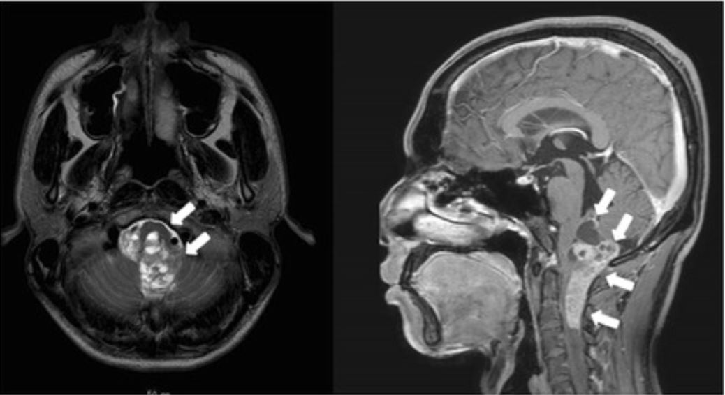
Magnetic resonance imaging scan reveals an anaplastic ependymoma extending from the brainstem to the 4th ventricle.

Image: “Giant duodenal ulcers after neurosurgery for brainstem tumors that required reoperation for gastric disconnection: a report of two cases” by BMC Surgery. License: CC BY 4.0

Histologic classification

Ependymoma is based on a histologic diagnosis; therefore, the gold standard for diagnosis is pathologic evaluation of a tissue specimen. Ependymomas are classified by histologic grades according to the WHO.

  • World Health Organization grade I: 
    • Subependymoma:
      • Rare, benign Benign Fibroadenoma tumors of the 4th or lateral ventricles Lateral ventricles Cavity in each of the cerebral hemispheres derived from the cavity of the embryonic neural tube. They are separated from each other by the septum pellucidum, and each communicates with the third ventricle by the foramen of monro, through which also the choroid plexuses (choroid plexus) of the lateral ventricles become continuous with that of the third ventricle. Ventricular System: Anatomy in adults
      • Hypocellular tissue consisting of a coarse, glial matrix containing clusters of cells with uniform nuclei and microcysts
    • Myxopapillary ependymoma:
      • Mostly benign Benign Fibroadenoma tumors arising in the conus medullaris Conus Medullaris Spinal Cord Injuries and filum terminale Filum terminale Spinal Cord: Anatomy of the spinal cord Spinal cord The spinal cord is the major conduction pathway connecting the brain to the body; it is part of the CNS. In cross section, the spinal cord is divided into an H-shaped area of gray matter (consisting of synapsing neuronal cell bodies) and a surrounding area of white matter (consisting of ascending and descending tracts of myelinated axons). Spinal Cord: Anatomy
      • Pseudopapillary structures with mucin-rich microcysts
      • Cuboidal cells surrounding a myxoid stroma
  • World Health Organization grade II: classic ependymoma:
    • May arise anywhere in the ventricular system Ventricular System The ventricular system is an extension of the subarachnoid space into the brain consisting of a series of interconnecting spaces and channels. Four chambers are filled with cerebrospinal fluid (CSF): the paired lateral ventricles, the unpaired 3rd ventricle, and the unpaired 4th ventricle. Ventricular System: Anatomy or spinal canal Spinal Canal The cavity within the spinal column through which the spinal cord passes. Spinal Cord Injuries
    • Potential variants include:
      • Papillary variant
      • Clear-cell variant
      • Tanycytic variant
    • Findings include:
      • Solid or papillary mass Mass Three-dimensional lesion that occupies a space within the breast Imaging of the Breast 
      • Perivascular pseudorosettes: tumor Tumor Inflammation cells appear as a gland surrounding the vasculature (diagnostic of ependymoma)
      • Cells with regular Regular Insulin, round-to-oval nuclei and an abundance of granular chromatin Chromatin The material of chromosomes. It is a complex of dna; histones; and nonhistone proteins found within the nucleus of a cell. DNA Types and Structure
      • Dense fibrillary background
  • World Health Organization grade III:anaplastic ependymoma:
    • Abundant mitotic cells
    • Pseudopalisading necrosis Necrosis The death of cells in an organ or tissue due to disease, injury or failure of the blood supply. Ischemic Cell Damage
  • RELA fusion-positive ependymoma:
    • A new piece of the WHO classification
    • Can include grades II and III
    • Determined based on genetic testing Genetic Testing Detection of a mutation; genotype; karyotype; or specific alleles associated with genetic traits, heritable diseases, or predisposition to a disease, or that may lead to the disease in descendants. It includes prenatal genetic testing. Myotonic Dystrophies
Who grade iii intracranial ependymoma

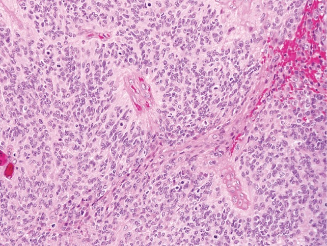
World Health Organization grade III intracranial ependymoma:
Perivascular pseudorosettes are seen as well as brisk, mitotic activity, which classifies the tumor as an anaplastic ependymoma.

Image: “Exomic sequencing of four rare central nervous system tumor types” by Bettegowda C., et al. License: CC BY 2.5

Other testing

Management and Prognosis

Management

  • Refer to specialized centers when possible.
  • Intracranial ependymoma:
    • Maximal safe resection
    • Adjuvant Adjuvant Substances that augment, stimulate, activate, potentiate, or modulate the immune response at either the cellular or humoral level. The classical agents (freund’s adjuvant, bcg, corynebacterium parvum, et al.) contain bacterial antigens. Some are endogenous (e.g., histamine, interferon, transfer factor, tuftsin, interleukin-1). Their mode of action is either non-specific, resulting in increased immune responsiveness to a wide variety of antigens, or antigen-specific, i.e., affecting a restricted type of immune response to a narrow group of antigens. The therapeutic efficacy of many biological response modifiers is related to their antigen-specific immunoadjuvanticity. Vaccination radiation Radiation Emission or propagation of acoustic waves (sound), electromagnetic energy waves (such as light; radio waves; gamma rays; or x-rays), or a stream of subatomic particles (such as electrons; neutrons; protons; or alpha particles). Osteosarcoma therapy
    • Chemotherapy Chemotherapy Osteosarcoma:
      • > 1 year of age: may be used post-radiation for some patients Patients Individuals participating in the health care system for the purpose of receiving therapeutic, diagnostic, or preventive procedures. Clinician–Patient Relationship
      • < 1 year of age: used to delay radiation Radiation Emission or propagation of acoustic waves (sound), electromagnetic energy waves (such as light; radio waves; gamma rays; or x-rays), or a stream of subatomic particles (such as electrons; neutrons; protons; or alpha particles). Osteosarcoma therapy (sparing adverse effects of radiation Radiation Emission or propagation of acoustic waves (sound), electromagnetic energy waves (such as light; radio waves; gamma rays; or x-rays), or a stream of subatomic particles (such as electrons; neutrons; protons; or alpha particles). Osteosarcoma)
  • Spinal ependymoma:
    • Optimal management includes complete surgical resection. 
    • The role of adjuvant Adjuvant Substances that augment, stimulate, activate, potentiate, or modulate the immune response at either the cellular or humoral level. The classical agents (freund’s adjuvant, bcg, corynebacterium parvum, et al.) contain bacterial antigens. Some are endogenous (e.g., histamine, interferon, transfer factor, tuftsin, interleukin-1). Their mode of action is either non-specific, resulting in increased immune responsiveness to a wide variety of antigens, or antigen-specific, i.e., affecting a restricted type of immune response to a narrow group of antigens. The therapeutic efficacy of many biological response modifiers is related to their antigen-specific immunoadjuvanticity. Vaccination radiation Radiation Emission or propagation of acoustic waves (sound), electromagnetic energy waves (such as light; radio waves; gamma rays; or x-rays), or a stream of subatomic particles (such as electrons; neutrons; protons; or alpha particles). Osteosarcoma therapy is unclear.
    • No proven role for chemotherapy Chemotherapy Osteosarcoma
  • Corticosteroids Corticosteroids Chorioretinitis: to reduce edema Edema Edema is a condition in which excess serous fluid accumulates in the body cavity or interstitial space of connective tissues. Edema is a symptom observed in several medical conditions. It can be categorized into 2 types, namely, peripheral (in the extremities) and internal (in an organ or body cavity). Edema surrounding the tumor Tumor Inflammation
  • Anticonvulsants: to treat seizures Seizures A seizure is abnormal electrical activity of the neurons in the cerebral cortex that can manifest in numerous ways depending on the region of the brain affected. Seizures consist of a sudden imbalance that occurs between the excitatory and inhibitory signals in cortical neurons, creating a net excitation. The 2 major classes of seizures are focal and generalized. Seizures in patients Patients Individuals participating in the health care system for the purpose of receiving therapeutic, diagnostic, or preventive procedures. Clinician–Patient Relationship with intracranial ependymoma

Prognosis Prognosis A prediction of the probable outcome of a disease based on a individual’s condition and the usual course of the disease as seen in similar situations. Non-Hodgkin Lymphomas

  • Survival rates: 
    • Children:
      • 10-year survival: up to 70% in patients Patients Individuals participating in the health care system for the purpose of receiving therapeutic, diagnostic, or preventive procedures. Clinician–Patient Relationship with totally resected tumors
      • 10-year survival: 30%–40% in patients Patients Individuals participating in the health care system for the purpose of receiving therapeutic, diagnostic, or preventive procedures. Clinician–Patient Relationship with partially resected tumors 
    • Adults:
      • 5-year survival: 67%–85%
      • 10-year survival: 50%–77%
  • Factors affecting survival:
    • Extent of resection (most important factor)
    • Histologic grade and molecular genetic subgroup
    • Location of the tumor Tumor Inflammation
    • Children diagnosed at an older age have a better prognosis Prognosis A prediction of the probable outcome of a disease based on a individual’s condition and the usual course of the disease as seen in similar situations. Non-Hodgkin Lymphomas.
    • Karnofsky performance status (measures functional status) in adults
  • Long-term survivors of treatment have increased risk of:

Differential Diagnosis

  • Medulloblastoma Medulloblastoma Medulloblastomas are malignant primitive neuroectodermal tumors that arise in the posterior fossa in children. Medulloblastomas are the most common malignant brain tumors in children. Patients with medulloblastoma present with symptoms of increased intracranial pressure and cerebellar signs, which both evolve and worsen over weeks to a few months. Medulloblastoma: a tumor Tumor Inflammation arising in the posterior fossa. Medulloblastoma Medulloblastoma Medulloblastomas are malignant primitive neuroectodermal tumors that arise in the posterior fossa in children. Medulloblastomas are the most common malignant brain tumors in children. Patients with medulloblastoma present with symptoms of increased intracranial pressure and cerebellar signs, which both evolve and worsen over weeks to a few months. Medulloblastoma is the most common malignant brain Brain The part of central nervous system that is contained within the skull (cranium). Arising from the neural tube, the embryonic brain is comprised of three major parts including prosencephalon (the forebrain); mesencephalon (the midbrain); and rhombencephalon (the hindbrain). The developed brain consists of cerebrum; cerebellum; and other structures in the brain stem. Nervous System: Anatomy, Structure, and Classification tumor Tumor Inflammation in children. Patients Patients Individuals participating in the health care system for the purpose of receiving therapeutic, diagnostic, or preventive procedures. Clinician–Patient Relationship present with symptoms of increased intracranial pressure Intracranial Pressure Idiopathic Intracranial Hypertension and cerebellar signs worsening over time. Treatment is with a combination of surgery, radiation Radiation Emission or propagation of acoustic waves (sound), electromagnetic energy waves (such as light; radio waves; gamma rays; or x-rays), or a stream of subatomic particles (such as electrons; neutrons; protons; or alpha particles). Osteosarcoma, and chemotherapy Chemotherapy Osteosarcoma. Unlike ependymomas, medulloblastomas rarely have foraminal extension Extension Examination of the Upper Limbs and primary tumors do not occur in the spinal cord Spinal cord The spinal cord is the major conduction pathway connecting the brain to the body; it is part of the CNS. In cross section, the spinal cord is divided into an H-shaped area of gray matter (consisting of synapsing neuronal cell bodies) and a surrounding area of white matter (consisting of ascending and descending tracts of myelinated axons). Spinal Cord: Anatomy.
  • Pilocytic astrocytoma Pilocytic Astrocytoma Astrocytoma: the 2nd most common brain Brain The part of central nervous system that is contained within the skull (cranium). Arising from the neural tube, the embryonic brain is comprised of three major parts including prosencephalon (the forebrain); mesencephalon (the midbrain); and rhombencephalon (the hindbrain). The developed brain consists of cerebrum; cerebellum; and other structures in the brain stem. Nervous System: Anatomy, Structure, and Classification tumor Tumor Inflammation in children. Magnetic resonance imaging can help distinguish pilocytic astrocytoma Pilocytic Astrocytoma Astrocytoma from other tumors. A pilocytic astrocytoma Pilocytic Astrocytoma Astrocytoma is usually cystic Cystic Fibrocystic Change with a mural module, or has central necrosis Necrosis The death of cells in an organ or tissue due to disease, injury or failure of the blood supply. Ischemic Cell Damage with a thick rim of enhancing tissue. In contrast, ependymomas often arise from the 4th ventricle and are usually accompanied by restricted diffusion Diffusion The tendency of a gas or solute to pass from a point of higher pressure or concentration to a point of lower pressure or concentration and to distribute itself throughout the available space. Diffusion, especially facilitated diffusion, is a major mechanism of biological transport. Peritoneal Dialysis and Hemodialysis on diffusion-weighted images. 
  • Choroid Choroid The thin, highly vascular membrane covering most of the posterior of the eye between the retina and sclera. Eye: Anatomy plexus tumors: rare, primary CNS tumors arising from the choroid Choroid The thin, highly vascular membrane covering most of the posterior of the eye between the retina and sclera. Eye: Anatomy plexus within the ventricles. Choroid Choroid The thin, highly vascular membrane covering most of the posterior of the eye between the retina and sclera. Eye: Anatomy plexus tumors usually present in children with signs and symptoms of hydrocephalus Hydrocephalus Excessive accumulation of cerebrospinal fluid within the cranium which may be associated with dilation of cerebral ventricles, intracranial. Subarachnoid Hemorrhage. Histologically, choroid Choroid The thin, highly vascular membrane covering most of the posterior of the eye between the retina and sclera. Eye: Anatomy plexus tumors resemble adenocarcinomas and may be associated with Li-Fraumeni syndrome.
  • Oligodendroglioma Oligodendroglioma Oligodendrogliomas are malignant CNS tumors arising from neural glial cell precursors. Oligodendrogliomas often arise in the frontal lobes of the brain and have a generally favorable prognosis when compared to other gliomas. Oligodendrogliomas are the 3rd most common CNS tumor. The most frequent presenting symptom is a seizure. Oligodendroglioma: a CNS tumor Tumor Inflammation arising from oligodendrocytes. Oligodendroglioma Oligodendroglioma Oligodendrogliomas are malignant CNS tumors arising from neural glial cell precursors. Oligodendrogliomas often arise in the frontal lobes of the brain and have a generally favorable prognosis when compared to other gliomas. Oligodendrogliomas are the 3rd most common CNS tumor. The most frequent presenting symptom is a seizure. Oligodendroglioma commonly develops in the cerebral hemisphere (usually the frontal lobe Frontal lobe The part of the cerebral hemisphere anterior to the central sulcus, and anterior and superior to the lateral sulcus. Cerebral Cortex: Anatomy). Presentation may include focal neurological deficits Focal Neurological Deficits Brain Abscess, seizures Seizures A seizure is abnormal electrical activity of the neurons in the cerebral cortex that can manifest in numerous ways depending on the region of the brain affected. Seizures consist of a sudden imbalance that occurs between the excitatory and inhibitory signals in cortical neurons, creating a net excitation. The 2 major classes of seizures are focal and generalized. Seizures, and personality changes. Diagnosis is by MRI and biopsy Biopsy Removal and pathologic examination of specimens from the living body. Ewing Sarcoma. Management involves surgical resection and may be accompanied by radiation Radiation Emission or propagation of acoustic waves (sound), electromagnetic energy waves (such as light; radio waves; gamma rays; or x-rays), or a stream of subatomic particles (such as electrons; neutrons; protons; or alpha particles). Osteosarcoma and/or chemotherapy Chemotherapy Osteosarcoma.

References

  1. Capodano, A.M. (2001). Nervous system: Ependymomas. Atlas Genet Cytogenet Oncol Haematol. 5(3), 208–210. 
  2. DeAngelis, L.M., Wen, P.Y. (2018). Primary and metastatic tumors of the nervous system. In Jameson, J., et al. (Eds.), Harrison’s Principles of Internal Medicine, 20e. McGraw-Hill. https://accessmedicine-mhmedical-com.aucmed.idm.oclc.org/content.aspx?bookid=2129&sectionid=192016511
  3. Genetic and Rare Disease Information Center. (2021). Ependymoma. Retrieved April 19, 2021, from https://rarediseases.info.nih.gov/diseases/6353/ependymoma
  4. Mayo Clinic. (2019). Embryonal tumors. Retrieved April 19, 2021, from https://www.mayoclinic.org/diseases-conditions/embryonal-tumor/
  5. National Cancer Institute Center for Cancer Research. (2021). Choroid Plexus Tumors Diagnosis and Treatment. Retrieved April 19, 2021, from https://www.cancer.gov/rare-brain-spine-tumor/tumors/choroid-plexus-tumors
  6. National Cancer Institute. (2020). Childhood Ependymoma Treatment (PDQ®)–Patient Version. Retrieved April 19, 2021, from https://www.cancer.gov/types/brain/patient/child-ependymoma-treatment-pdq
  7. National Cancer Institute. (2021). Ependymoma Diagnosis and Treatment. Retrieved April 19, 2021, from https://www.cancer.gov/rare-brain-spine-tumor/tumors/ependymoma
  8. Upadhyaya, S.A., Tinkle, C. (2020). Intracranial ependymoma and other ependymal tumors. UpToDate. Retrieved April 19, 2021, from https://www.uptodate.com/contents/intracranial-ependymoma-and-other-ependymal-tumors
  9. Welch, W.C., Schiff, D., Gerszten, P.C. (2020). Spinal cord tumors. UpToDate. Retrieved April 19, 2021, from https://www.uptodate.com/contents/spinal-cord-tumors

Create your free account or log in to continue reading!

Sign up now and get free access to Lecturio with concept pages, medical videos, and questions for your medical education.

User Reviews

Details