Achieve Mastery of Medical Concepts

Study for medical school and boards with Lecturio

Hemangioblastoma

Hemangioblastomas Hemangioblastomas A benign tumor of the nervous system that may occur sporadically or in association with von Hippel-Lindau disease. It accounts for approximately 2% of intracranial tumors, arising most frequently in the cerebellar hemispheres and vermis. Histologically, the tumors are composed of multiple capillary and sinusoidal channels lined with endothelial cells and clusters of lipid-laden pseudoxanthoma cells. Usually solitary, these tumors can be multiple and may also occur in the brain stem, spinal cord, retina, and supratentorial compartment. Cerebellar hemangioblastomas usually present in the third decade with intracranial hypertension, and ataxia. Von Hippel-Lindau Disease are vascular neoplasms Neoplasms New abnormal growth of tissue. Malignant neoplasms show a greater degree of anaplasia and have the properties of invasion and metastasis, compared to benign neoplasms. Benign Bone Tumors of the CNS. Hemangioblastomas Hemangioblastomas A benign tumor of the nervous system that may occur sporadically or in association with von Hippel-Lindau disease. It accounts for approximately 2% of intracranial tumors, arising most frequently in the cerebellar hemispheres and vermis. Histologically, the tumors are composed of multiple capillary and sinusoidal channels lined with endothelial cells and clusters of lipid-laden pseudoxanthoma cells. Usually solitary, these tumors can be multiple and may also occur in the brain stem, spinal cord, retina, and supratentorial compartment. Cerebellar hemangioblastomas usually present in the third decade with intracranial hypertension, and ataxia. Von Hippel-Lindau Disease are rare and are often associated with von Hippel-Lindau disease von Hippel-Lindau disease Von Hippel-Lindau (VHL) disease is an autosomal dominant genetic condition resulting from a deletion or mutation in the VHL gene. Individuals diagnosed with VHL disease have tumors and cysts in various parts of their bodies and may present with hemangioblastomas, renal cell carcinoma (RCC), pheochromocytoma, endolymphatic sac tumors of the middle ear, pancreatic tumors, and papillary cystadenomas of the epididymis or the broad ligament. Von Hippel-Lindau Disease (VHL). The most common presentation is a headache Headache The symptom of pain in the cranial region. It may be an isolated benign occurrence or manifestation of a wide variety of headache disorders. Brain Abscess and, depending on the size and location of the tumor Tumor Inflammation, patients Patients Individuals participating in the health care system for the purpose of receiving therapeutic, diagnostic, or preventive procedures. Clinician–Patient Relationship may present with sensory Sensory Neurons which conduct nerve impulses to the central nervous system. Nervous System: Histology deficits and motor Motor Neurons which send impulses peripherally to activate muscles or secretory cells. Nervous System: Histology weakness. Imaging is the main diagnostic method, with histopathologic evaluation required for definitive diagnosis. Surgery is often indicated in the management of hemangioblastomas Hemangioblastomas A benign tumor of the nervous system that may occur sporadically or in association with von Hippel-Lindau disease. It accounts for approximately 2% of intracranial tumors, arising most frequently in the cerebellar hemispheres and vermis. Histologically, the tumors are composed of multiple capillary and sinusoidal channels lined with endothelial cells and clusters of lipid-laden pseudoxanthoma cells. Usually solitary, these tumors can be multiple and may also occur in the brain stem, spinal cord, retina, and supratentorial compartment. Cerebellar hemangioblastomas usually present in the third decade with intracranial hypertension, and ataxia. Von Hippel-Lindau Disease, though depending on the size, number, and location of the tumors, radiation Radiation Emission or propagation of acoustic waves (sound), electromagnetic energy waves (such as light; radio waves; gamma rays; or x-rays), or a stream of subatomic particles (such as electrons; neutrons; protons; or alpha particles). Osteosarcoma therapy may also be warranted. Prognosis Prognosis A prediction of the probable outcome of a disease based on a individual's condition and the usual course of the disease as seen in similar situations. Non-Hodgkin Lymphomas is usually good in solitary hemangioblastomas Hemangioblastomas A benign tumor of the nervous system that may occur sporadically or in association with von Hippel-Lindau disease. It accounts for approximately 2% of intracranial tumors, arising most frequently in the cerebellar hemispheres and vermis. Histologically, the tumors are composed of multiple capillary and sinusoidal channels lined with endothelial cells and clusters of lipid-laden pseudoxanthoma cells. Usually solitary, these tumors can be multiple and may also occur in the brain stem, spinal cord, retina, and supratentorial compartment. Cerebellar hemangioblastomas usually present in the third decade with intracranial hypertension, and ataxia. Von Hippel-Lindau Disease, but tumors associated with VHL are often associated with a worse prognosis Prognosis A prediction of the probable outcome of a disease based on a individual's condition and the usual course of the disease as seen in similar situations. Non-Hodgkin Lymphomas and a higher risk of recurrence.

Last updated: Nov 3, 2022

Editorial responsibility: Stanley Oiseth, Lindsay Jones, Evelin Maza

Overview

Definition

Hemangioblastomas Hemangioblastomas A benign tumor of the nervous system that may occur sporadically or in association with von Hippel-Lindau disease. It accounts for approximately 2% of intracranial tumors, arising most frequently in the cerebellar hemispheres and vermis. Histologically, the tumors are composed of multiple capillary and sinusoidal channels lined with endothelial cells and clusters of lipid-laden pseudoxanthoma cells. Usually solitary, these tumors can be multiple and may also occur in the brain stem, spinal cord, retina, and supratentorial compartment. Cerebellar hemangioblastomas usually present in the third decade with intracranial hypertension, and ataxia. Von Hippel-Lindau Disease are rare, slow-growing, benign Benign Fibroadenoma, highly vascular neoplasms Neoplasms New abnormal growth of tissue. Malignant neoplasms show a greater degree of anaplasia and have the properties of invasion and metastasis, compared to benign neoplasms. Benign Bone Tumors of the CNS that have a high association with von Hippel-Lindau disease von Hippel-Lindau disease Von Hippel-Lindau (VHL) disease is an autosomal dominant genetic condition resulting from a deletion or mutation in the VHL gene. Individuals diagnosed with VHL disease have tumors and cysts in various parts of their bodies and may present with hemangioblastomas, renal cell carcinoma (RCC), pheochromocytoma, endolymphatic sac tumors of the middle ear, pancreatic tumors, and papillary cystadenomas of the epididymis or the broad ligament. Von Hippel-Lindau Disease (VHL), which is an autosomal dominant Autosomal dominant Autosomal inheritance, both dominant and recessive, refers to the transmission of genes from the 22 autosomal chromosomes. Autosomal dominant diseases are expressed when only 1 copy of the dominant allele is inherited. Autosomal Recessive and Autosomal Dominant Inheritance condition characterized by a variety of benign Benign Fibroadenoma and malignant tumors.

  • The tumors are most commonly found in the cerebellum Cerebellum The cerebellum, Latin for “little brain,” is located in the posterior cranial fossa, dorsal to the pons and midbrain, and its principal role is in the coordination of movements. The cerebellum consists of 3 lobes on either side of its 2 hemispheres and is connected in the middle by the vermis. Cerebellum: Anatomy.
  • Hemangioblastomas Hemangioblastomas A benign tumor of the nervous system that may occur sporadically or in association with von Hippel-Lindau disease. It accounts for approximately 2% of intracranial tumors, arising most frequently in the cerebellar hemispheres and vermis. Histologically, the tumors are composed of multiple capillary and sinusoidal channels lined with endothelial cells and clusters of lipid-laden pseudoxanthoma cells. Usually solitary, these tumors can be multiple and may also occur in the brain stem, spinal cord, retina, and supratentorial compartment. Cerebellar hemangioblastomas usually present in the third decade with intracranial hypertension, and ataxia. Von Hippel-Lindau Disease are often found in the parenchyma attached to or near the pia mater Pia mater The innermost layer of the three meninges covering the brain and spinal cord. It is the fine vascular membrane that lies under the arachnoid and the dura mater. Meninges: Anatomy, a part of the meninges Meninges The brain and the spinal cord are enveloped by 3 overlapping layers of connective tissue called the meninges. The layers are, from the most external layer to the most internal layer, the dura mater, arachnoid mater, and pia mater. Between these layers are 3 potential spaces called the epidural, subdural, and subarachnoid spaces. Meninges: Anatomy (made up of 3 fibrous Fibrous Fibrocystic Change layers) encasing the CNS:
    • Dura mater Dura mater The outermost of the three meninges, a fibrous membrane of connective tissue that covers the brain and the spinal cord. Meninges: Anatomy: the outer layer, which forms a tough, collagenous sheath
    • Arachnoid mater Arachnoid mater A delicate membrane enveloping the brain and spinal cord. It lies between the pia mater and the dura mater. It is separated from the pia mater by the subarachnoid cavity which is filled with cerebrospinal fluid. Meninges: Anatomy: the middle layer consisting of simple squamous epithelium Simple squamous epithelium Surface Epithelium: Histology, with a loose mesh of collagen Collagen A polypeptide substance comprising about one third of the total protein in mammalian organisms. It is the main constituent of skin; connective tissue; and the organic substance of bones (bone and bones) and teeth (tooth). Connective Tissue: Histology and elastic Elastic Connective Tissue: Histology fibers
    • Pia mater Pia mater The innermost layer of the three meninges covering the brain and spinal cord. It is the fine vascular membrane that lies under the arachnoid and the dura mater. Meninges: Anatomy: the thin, inner layer in contact with the brain Brain The part of central nervous system that is contained within the skull (cranium). Arising from the neural tube, the embryonic brain is comprised of three major parts including prosencephalon (the forebrain); mesencephalon (the midbrain); and rhombencephalon (the hindbrain). The developed brain consists of cerebrum; cerebellum; and other structures in the brain stem. Nervous System: Anatomy, Structure, and Classification and spinal cord Spinal cord The spinal cord is the major conduction pathway connecting the brain to the body; it is part of the CNS. In cross section, the spinal cord is divided into an H-shaped area of gray matter (consisting of synapsing neuronal cell bodies) and a surrounding area of white matter (consisting of ascending and descending tracts of myelinated axons). Spinal Cord: Anatomy
Table: Classification of nervous system Nervous system The nervous system is a small and complex system that consists of an intricate network of neural cells (or neurons) and even more glial cells (for support and insulation). It is divided according to its anatomical components as well as its functional characteristics. The brain and spinal cord are referred to as the central nervous system, and the branches of nerves from these structures are referred to as the peripheral nervous system. Nervous System: Anatomy, Structure, and Classification tumors
Categories Specific tumors
Neuroepithelial tumors in the CNS
  • Astrocytomas, including glioblastoma multiforme Glioblastoma multiforme Glioblastoma multiforme is a high-grade astrocytoma, an aggressive brain tumor arising from astrocytes, with an unknown cause and a poorly understood link to risk factors. There are two main types: primary, a more aggressive form seen more commonly in older patients, and secondary, developing from lower-grade astrocytomas and seen more commonly in younger patients. Glioblastoma Multiforme
  • Oligodendroglioma Oligodendroglioma Oligodendrogliomas are malignant CNS tumors arising from neural glial cell precursors. Oligodendrogliomas often arise in the frontal lobes of the brain and have a generally favorable prognosis when compared to other gliomas. Oligodendrogliomas are the 3rd most common CNS tumor. The most frequent presenting symptom is a seizure. Oligodendroglioma
  • Ependymoma Ependymoma Ependymomas are glial cell tumors arising from CSF-producing ependymal cells lining the ventricular system. Ependymomas most commonly occur within the posterior fossa in contact with the 4th ventricle, or within the intramedullary spinal cord. Ependymoma and choroid Choroid The thin, highly vascular membrane covering most of the posterior of the eye between the retina and sclera. Eye: Anatomy plexus tumors
  • Medulloblastomas (embryonal tumors)
Meningeal tumors
  • Meningiomas
  • Hemangioblastomas Hemangioblastomas A benign tumor of the nervous system that may occur sporadically or in association with von Hippel-Lindau disease. It accounts for approximately 2% of intracranial tumors, arising most frequently in the cerebellar hemispheres and vermis. Histologically, the tumors are composed of multiple capillary and sinusoidal channels lined with endothelial cells and clusters of lipid-laden pseudoxanthoma cells. Usually solitary, these tumors can be multiple and may also occur in the brain stem, spinal cord, retina, and supratentorial compartment. Cerebellar hemangioblastomas usually present in the third decade with intracranial hypertension, and ataxia. Von Hippel-Lindau Disease
Sellar region tumors
  • Craniopharyngioma Craniopharyngioma Craniopharyngiomas are rare squamous epithelial tumors with a solid and/or cystic structure that arise from the remnants of Rathke’s pouch along the pituitary stalk, in the suprasellar region. Craniopharyngiomas are histologically benign but tend to invade surrounding structures; thus, they should be treated as low-grade malignancies. Craniopharyngioma
  • Pituitary Pituitary A small, unpaired gland situated in the sella turcica. It is connected to the hypothalamus by a short stalk which is called the infundibulum. Hormones: Overview and Types adenoma
  • Pinealoma/pinealoblastoma
Primary CNS lymphoma Lymphoma A general term for various neoplastic diseases of the lymphoid tissue. Imaging of the Mediastinum Primary CNS lymphoma Lymphoma A general term for various neoplastic diseases of the lymphoid tissue. Imaging of the Mediastinum
Metastasis Metastasis The transfer of a neoplasm from one organ or part of the body to another remote from the primary site. Grading, Staging, and Metastasis to the brain Brain The part of central nervous system that is contained within the skull (cranium). Arising from the neural tube, the embryonic brain is comprised of three major parts including prosencephalon (the forebrain); mesencephalon (the midbrain); and rhombencephalon (the hindbrain). The developed brain consists of cerebrum; cerebellum; and other structures in the brain stem. Nervous System: Anatomy, Structure, and Classification (5x more common than primary brain Brain The part of central nervous system that is contained within the skull (cranium). Arising from the neural tube, the embryonic brain is comprised of three major parts including prosencephalon (the forebrain); mesencephalon (the midbrain); and rhombencephalon (the hindbrain). The developed brain consists of cerebrum; cerebellum; and other structures in the brain stem. Nervous System: Anatomy, Structure, and Classification tumors) Most commonly arising from:
  • Lung, breast, and renal cell carcinomas
  • Melanoma Melanoma Melanoma is a malignant tumor arising from melanocytes, the melanin-producing cells of the epidermis. These tumors are most common in fair-skinned individuals with a history of excessive sun exposure and sunburns. Melanoma
Peripheral tumors
  • Schwannomas, including acoustic neuromas
  • Neuroblastoma Neuroblastoma Neuroblastoma is a malignancy that arises from the neural crest cell derivatives along the sympathetic chain (neuroblasts) and is most commonly located in the adrenal medulla. The tumor often presents in childhood with a flank mass that crosses the midline. Neuroblastoma

Epidemiology

  • Sporadic Sporadic Selective IgA Deficiency: 75% 
  • Associated with VHL: 25%
  • Incidence Incidence The number of new cases of a given disease during a given period in a specified population. It also is used for the rate at which new events occur in a defined population. It is differentiated from prevalence, which refers to all cases in the population at a given time. Measures of Disease Frequency
    • Extremely rare 
    • 2.5% of all intracranial neoplasms Neoplasms New abnormal growth of tissue. Malignant neoplasms show a greater degree of anaplasia and have the properties of invasion and metastasis, compared to benign neoplasms. Benign Bone Tumors 
    • 5% of spinal cord Spinal cord The spinal cord is the major conduction pathway connecting the brain to the body; it is part of the CNS. In cross section, the spinal cord is divided into an H-shaped area of gray matter (consisting of synapsing neuronal cell bodies) and a surrounding area of white matter (consisting of ascending and descending tracts of myelinated axons). Spinal Cord: Anatomy tumors
  • Sex Sex The totality of characteristics of reproductive structure, functions, phenotype, and genotype, differentiating the male from the female organism. Gender Dysphoria: Incidence Incidence The number of new cases of a given disease during a given period in a specified population. It also is used for the rate at which new events occur in a defined population. It is differentiated from prevalence, which refers to all cases in the population at a given time. Measures of Disease Frequency is higher in men than in women (2:1).
  • Age: 
    • Children are rarely affected.
    • The peak age of incidence Incidence The number of new cases of a given disease during a given period in a specified population. It also is used for the rate at which new events occur in a defined population. It is differentiated from prevalence, which refers to all cases in the population at a given time. Measures of Disease Frequency is 20–50 years.

Etiology

  • The exact cause of hemangioblastoma is not known. 
  • A mutation Mutation Genetic mutations are errors in DNA that can cause protein misfolding and dysfunction. There are various types of mutations, including chromosomal, point, frameshift, and expansion mutations. Types of Mutations in the VHL gene Gene A category of nucleic acid sequences that function as units of heredity and which code for the basic instructions for the development, reproduction, and maintenance of organisms. Basic Terms of Genetics is being considered because of a high association with VHL.

Pathophysiology

Tumor Tumor Inflammation location

Hemangioblastomas Hemangioblastomas A benign tumor of the nervous system that may occur sporadically or in association with von Hippel-Lindau disease. It accounts for approximately 2% of intracranial tumors, arising most frequently in the cerebellar hemispheres and vermis. Histologically, the tumors are composed of multiple capillary and sinusoidal channels lined with endothelial cells and clusters of lipid-laden pseudoxanthoma cells. Usually solitary, these tumors can be multiple and may also occur in the brain stem, spinal cord, retina, and supratentorial compartment. Cerebellar hemangioblastomas usually present in the third decade with intracranial hypertension, and ataxia. Von Hippel-Lindau Disease grow attached to the pia mater Pia mater The innermost layer of the three meninges covering the brain and spinal cord. It is the fine vascular membrane that lies under the arachnoid and the dura mater. Meninges: Anatomy (innermost meningeal layer)

  • Parenchyma of the cerebellum Cerebellum The cerebellum, Latin for “little brain,” is located in the posterior cranial fossa, dorsal to the pons and midbrain, and its principal role is in the coordination of movements. The cerebellum consists of 3 lobes on either side of its 2 hemispheres and is connected in the middle by the vermis. Cerebellum: Anatomy
  • Brain Brain The part of central nervous system that is contained within the skull (cranium). Arising from the neural tube, the embryonic brain is comprised of three major parts including prosencephalon (the forebrain); mesencephalon (the midbrain); and rhombencephalon (the hindbrain). The developed brain consists of cerebrum; cerebellum; and other structures in the brain stem. Nervous System: Anatomy, Structure, and Classification stem
  • Spinal cord Spinal cord The spinal cord is the major conduction pathway connecting the brain to the body; it is part of the CNS. In cross section, the spinal cord is divided into an H-shaped area of gray matter (consisting of synapsing neuronal cell bodies) and a surrounding area of white matter (consisting of ascending and descending tracts of myelinated axons). Spinal Cord: Anatomy

Pathogenesis

Although the sporadic Sporadic Selective IgA Deficiency hemangiomas do not have a clear pathogenesis, the VHL-associated hemangiomas are thought to be caused by a mutation Mutation Genetic mutations are errors in DNA that can cause protein misfolding and dysfunction. There are various types of mutations, including chromosomal, point, frameshift, and expansion mutations. Types of Mutations in the VHL gene Gene A category of nucleic acid sequences that function as units of heredity and which code for the basic instructions for the development, reproduction, and maintenance of organisms. Basic Terms of Genetics. Up to 50% of sporadic Sporadic Selective IgA Deficiency hemangioblastomas Hemangioblastomas A benign tumor of the nervous system that may occur sporadically or in association with von Hippel-Lindau disease. It accounts for approximately 2% of intracranial tumors, arising most frequently in the cerebellar hemispheres and vermis. Histologically, the tumors are composed of multiple capillary and sinusoidal channels lined with endothelial cells and clusters of lipid-laden pseudoxanthoma cells. Usually solitary, these tumors can be multiple and may also occur in the brain stem, spinal cord, retina, and supratentorial compartment. Cerebellar hemangioblastomas usually present in the third decade with intracranial hypertension, and ataxia. Von Hippel-Lindau Disease have mutations or deletions in the VHL gene Gene A category of nucleic acid sequences that function as units of heredity and which code for the basic instructions for the development, reproduction, and maintenance of organisms. Basic Terms of Genetics as well.

  • VHL gene Gene A category of nucleic acid sequences that function as units of heredity and which code for the basic instructions for the development, reproduction, and maintenance of organisms. Basic Terms of Genetics:
    • Tumor-suppressor gene Gene A category of nucleic acid sequences that function as units of heredity and which code for the basic instructions for the development, reproduction, and maintenance of organisms. Basic Terms of Genetics found on chromosome Chromosome In a prokaryotic cell or in the nucleus of a eukaryotic cell, a structure consisting of or containing DNA which carries the genetic information essential to the cell. Basic Terms of Genetics 3p
    • Responsible for the inhibition of hypoxia-inducible factor-2α (HIF-2α) by ubiquitin-mediated proteasomal degradation 
  • HIF-2α:
    • Part of a larger transcription factor Transcription factor Generic term for proteins necessary for transcription Regulation of Transcription protein complex called HIF HIF Hypoxia-inducible factor 1, alpha subunit is a basic helix-loop-helix transcription factor that is regulated by oxygen availability and is targeted for degradation by VHL tumor suppressor protein. Von Hippel-Lindau Disease, which is involved in regulating the body’s ability to adapt to changing oxygen levels 
    • HIF HIF Hypoxia-inducible factor 1, alpha subunit is a basic helix-loop-helix transcription factor that is regulated by oxygen availability and is targeted for degradation by VHL tumor suppressor protein. Von Hippel-Lindau Disease can induce the expression of over 70 target genes Genes A category of nucleic acid sequences that function as units of heredity and which code for the basic instructions for the development, reproduction, and maintenance of organisms. DNA Types and Structure, including:
      • Vascular endothelial growth factor Vascular endothelial growth factor A family of angiogenic proteins that are closely-related to vascular endothelial growth factor a. They play an important role in the growth and differentiation of vascular as well as lymphatic endothelial cells. Wound Healing (VEGF)
      • Platelet-derived growth factor Platelet-derived growth factor Mitogenic peptide growth hormone carried in the alpha-granules of platelets. It is released when platelets adhere to traumatized tissues. Connective tissue cells near the traumatized region respond by initiating the process of replication. Hypertrophic and Keloid Scars (PDGF)
      • Erythropoietin Erythropoietin Glycoprotein hormone, secreted chiefly by the kidney in the adult and the liver in the fetus, that acts on erythroid stem cells of the bone marrow to stimulate proliferation and differentiation. Erythrocytes: Histology
      • Transforming growth factor α (TGF-α)  
  • When adequate oxygen is available, VHL protein VHL protein A ubiquitin-protein ligase that mediates oxygen-dependent polyubiquitination of hypoxia-inducible factor 1, alpha subunit. It is inactivated in von Hippel-Lindau syndrome. Von Hippel-Lindau Disease helps suppress HIF HIF Hypoxia-inducible factor 1, alpha subunit is a basic helix-loop-helix transcription factor that is regulated by oxygen availability and is targeted for degradation by VHL tumor suppressor protein. Von Hippel-Lindau Disease.
  • A dysfunctional VHL gene Gene A category of nucleic acid sequences that function as units of heredity and which code for the basic instructions for the development, reproduction, and maintenance of organisms. Basic Terms of Genetics (caused by mutation Mutation Genetic mutations are errors in DNA that can cause protein misfolding and dysfunction. There are various types of mutations, including chromosomal, point, frameshift, and expansion mutations. Types of Mutations or deletion) can lead to accumulation of HIF-α owing to the inability to degrade HIF-2α.
  • The stabilization of HIF-2α allows it to induce expression of its target genes Genes A category of nucleic acid sequences that function as units of heredity and which code for the basic instructions for the development, reproduction, and maintenance of organisms. DNA Types and Structure → ↑ angiogenic factors → tumor Tumor Inflammation growth

Symptom development

Symptom development is caused by:

Clinical Presentation and Complications

Clinical presentation

Symptoms depend on the location and progression of the tumor Tumor Inflammation. The most common symptoms observed are:

  • Headache Headache The symptom of pain in the cranial region. It may be an isolated benign occurrence or manifestation of a wide variety of headache disorders. Brain Abscess: most common
  • Local pain Pain An unpleasant sensation induced by noxious stimuli which are detected by nerve endings of nociceptive neurons. Pain: Types and Pathways (common in spinal cord Spinal cord The spinal cord is the major conduction pathway connecting the brain to the body; it is part of the CNS. In cross section, the spinal cord is divided into an H-shaped area of gray matter (consisting of synapsing neuronal cell bodies) and a surrounding area of white matter (consisting of ascending and descending tracts of myelinated axons). Spinal Cord: Anatomy lesions)
  • Cerebellar ataxia Cerebellar ataxia Incoordination of voluntary movements that occur as a manifestation of cerebellar diseases. Characteristic features include a tendency for limb movements to overshoot or undershoot a target (dysmetria), a tremor that occurs during attempted movements (intention tremor), impaired force and rhythm of diadochokinesis (rapidly alternating movements), and gait ataxia. Cerebellar Disorders and discoordination
  • Increased intracranial pressure Intracranial Pressure Idiopathic Intracranial Hypertension ( ICP ICP Normal intracranial pressure (ICP) is defined as < 15 mm Hg, whereas pathologically increased ICP is any pressure ≥ 20 mm Hg. Increased ICP may result from several etiologies, including trauma, intracranial hemorrhage, mass lesions, cerebral edema, increased CSF production, and decreased CSF absorption. Increased Intracranial Pressure (ICP)) due to obstructive hydrocephalus Obstructive Hydrocephalus Hydrocephalus in Children
  • Oculomotor nerve Oculomotor nerve The 3D cranial nerve. The oculomotor nerve sends motor fibers to the levator muscles of the eyelid and to the superior rectus, inferior rectus, and inferior oblique muscles of the eye. It also sends parasympathetic efferents (via the ciliary ganglion) to the muscles controlling pupillary constriction and accommodation. The motor fibers originate in the oculomotor nuclei of the midbrain. The 12 Cranial Nerves: Overview and Functions dysfunction
  • Motor Motor Neurons which send impulses peripherally to activate muscles or secretory cells. Nervous System: Histology weakness
  • Sensory Sensory Neurons which conduct nerve impulses to the central nervous system. Nervous System: Histology deficits

Sporadic Sporadic Selective IgA Deficiency versus VHL-associated hemangioblastomas Hemangioblastomas A benign tumor of the nervous system that may occur sporadically or in association with von Hippel-Lindau disease. It accounts for approximately 2% of intracranial tumors, arising most frequently in the cerebellar hemispheres and vermis. Histologically, the tumors are composed of multiple capillary and sinusoidal channels lined with endothelial cells and clusters of lipid-laden pseudoxanthoma cells. Usually solitary, these tumors can be multiple and may also occur in the brain stem, spinal cord, retina, and supratentorial compartment. Cerebellar hemangioblastomas usually present in the third decade with intracranial hypertension, and ataxia. Von Hippel-Lindau Disease

Table: Typical clinical presentations of sporadic Sporadic Selective IgA Deficiency versus VHL-associated hemangioblastomas Hemangioblastomas A benign tumor of the nervous system that may occur sporadically or in association with von Hippel-Lindau disease. It accounts for approximately 2% of intracranial tumors, arising most frequently in the cerebellar hemispheres and vermis. Histologically, the tumors are composed of multiple capillary and sinusoidal channels lined with endothelial cells and clusters of lipid-laden pseudoxanthoma cells. Usually solitary, these tumors can be multiple and may also occur in the brain stem, spinal cord, retina, and supratentorial compartment. Cerebellar hemangioblastomas usually present in the third decade with intracranial hypertension, and ataxia. Von Hippel-Lindau Disease
Sporadic Sporadic Selective IgA Deficiency hemangioblastoma VHL-associated hemangioblastoma
Presents as solitary tumor Tumor Inflammation Multiple tumors along the neuraxis
Usually occurs in the 3rd or 4th decade of life Diagnosed at a younger age (2nd decade)
Commonly presents as an isolated tumor Tumor Inflammation in the cerebellum Cerebellum The cerebellum, Latin for “little brain,” is located in the posterior cranial fossa, dorsal to the pons and midbrain, and its principal role is in the coordination of movements. The cerebellum consists of 3 lobes on either side of its 2 hemispheres and is connected in the middle by the vermis. Cerebellum: Anatomy 50% of the tumors are located at the spinal cord Spinal cord The spinal cord is the major conduction pathway connecting the brain to the body; it is part of the CNS. In cross section, the spinal cord is divided into an H-shaped area of gray matter (consisting of synapsing neuronal cell bodies) and a surrounding area of white matter (consisting of ascending and descending tracts of myelinated axons). Spinal Cord: Anatomy, 40% at the cerebellum Cerebellum The cerebellum, Latin for “little brain,” is located in the posterior cranial fossa, dorsal to the pons and midbrain, and its principal role is in the coordination of movements. The cerebellum consists of 3 lobes on either side of its 2 hemispheres and is connected in the middle by the vermis. Cerebellum: Anatomy, and 10% at the brain Brain The part of central nervous system that is contained within the skull (cranium). Arising from the neural tube, the embryonic brain is comprised of three major parts including prosencephalon (the forebrain); mesencephalon (the midbrain); and rhombencephalon (the hindbrain). The developed brain consists of cerebrum; cerebellum; and other structures in the brain stem. Nervous System: Anatomy, Structure, and Classification stem.

Complications

Complications usually occur because of an increase in the size of the tumor Tumor Inflammation (> 1.5 cm), causing compression Compression Blunt Chest Trauma, or because of spontaneous hemorrhage:

  • Symptoms of increased ICP Increased ICP Excessive accumulation of cerebrospinal fluid within the cranium which may be associated with dilation of cerebral ventricles, intracranial. Subarachnoid Hemorrhage/rapid obstructive hydrocephalus Obstructive Hydrocephalus Hydrocephalus in Children
  • Altered mental status Altered Mental Status Sepsis in Children 
  • Polycythemia Polycythemia An increase in the total red cell mass of the blood. Renal Cell Carcinoma due to ectopic production of erythropoietin Erythropoietin Glycoprotein hormone, secreted chiefly by the kidney in the adult and the liver in the fetus, that acts on erythroid stem cells of the bone marrow to stimulate proliferation and differentiation. Erythrocytes: Histology (paraneoplastic syndrome)
  • Quadriplegia Quadriplegia Severe or complete loss of motor function in all four limbs which may result from brain diseases; spinal cord diseases; peripheral nervous system diseases; neuromuscular diseases; or rarely muscular diseases. The locked-in syndrome is characterized by quadriplegia in combination with cranial muscle paralysis. Consciousness is spared and the only retained voluntary motor activity may be limited eye movements. This condition is usually caused by a lesion in the upper brain stem which injures the descending corticospinal and corticobulbar tracts. Locked-in Syndrome

Diagnosis

While the primary diagnostic tool is imaging, the gold standard of diagnosis requires histopathology from a biopsy Biopsy Removal and pathologic examination of specimens from the living body. Ewing Sarcoma specimen.

Imaging

The entire neural axis should be imaged to rule out multiple lesions, which are common in cases of VHL. 

  • Contrast MRI:
  • CT:
    • Not the preferred method of diagnosis, as small tumors may be concealed by bone Bone Bone is a compact type of hardened connective tissue composed of bone cells, membranes, an extracellular mineralized matrix, and central bone marrow. The 2 primary types of bone are compact and spongy. Bones: Structure and Types artifacts
    • A non-contrast-enhanced CT Non-contrast-enhanced CT Imaging of the Head and Brain shows an isointense nodule Nodule Chalazion.
    • With contrast: intense homogeneous enhancement Homogeneous Enhancement Imaging of the Spleen of the nodules
    • CT is also used as an adjuvant Adjuvant Substances that augment, stimulate, activate, potentiate, or modulate the immune response at either the cellular or humoral level. The classical agents (freund’s adjuvant, bcg, corynebacterium parvum, et al.) contain bacterial antigens. Some are endogenous (e.g., histamine, interferon, transfer factor, tuftsin, interleukin-1). Their mode of action is either non-specific, resulting in increased immune responsiveness to a wide variety of antigens, or antigen-specific, i.e., affecting a restricted type of immune response to a narrow group of antigens. The therapeutic efficacy of many biological response modifiers is related to their antigen-specific immunoadjuvanticity. Vaccination to angiography Angiography Radiography of blood vessels after injection of a contrast medium. Cardiac Surgery when the patient cannot undergo MRI.
  • Cerebral and spinal angiography Angiography Radiography of blood vessels after injection of a contrast medium. Cardiac Surgery
    • A high vascular tumor Tumor Inflammation blush is observed with enlarged arteries Arteries Arteries are tubular collections of cells that transport oxygenated blood and nutrients from the heart to the tissues of the body. The blood passes through the arteries in order of decreasing luminal diameter, starting in the largest artery (the aorta) and ending in the small arterioles. Arteries are classified into 3 types: large elastic arteries, medium muscular arteries, and small arteries and arterioles. Arteries: Histology and dilated veins Veins Veins are tubular collections of cells, which transport deoxygenated blood and waste from the capillary beds back to the heart. Veins are classified into 3 types: small veins/venules, medium veins, and large veins. Each type contains 3 primary layers: tunica intima, tunica media, and tunica adventitia. Veins: Histology.
    • Can detect blood supply to the tumor Tumor Inflammation aids AIDS Chronic HIV infection and depletion of CD4 cells eventually results in acquired immunodeficiency syndrome (AIDS), which can be diagnosed by the presence of certain opportunistic diseases called AIDS-defining conditions. These conditions include a wide spectrum of bacterial, viral, fungal, and parasitic infections as well as several malignancies and generalized conditions. HIV Infection and AIDS the surgeon in the proper resection of the tumor Tumor Inflammation
Hemangioblastoma

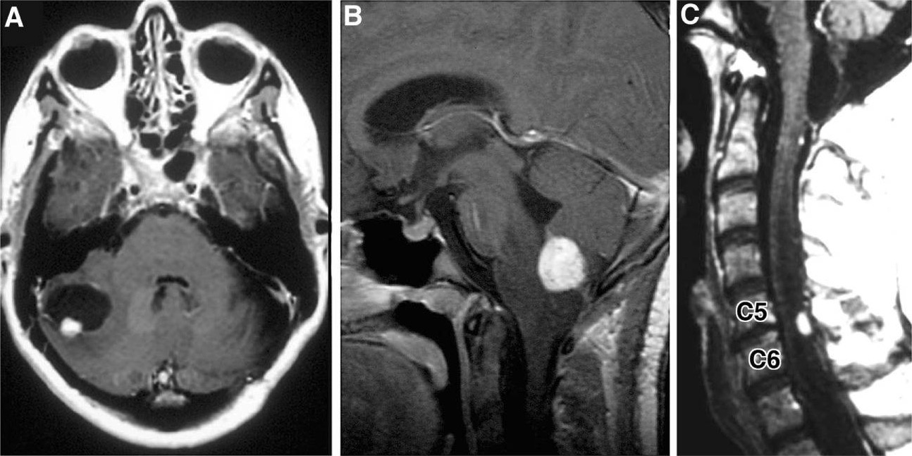
Radiographic images of hemangioblastomas:
A: Axial, contrast-enhanced, T1-weighted MRI showing cerebellar hemangioblastoma with contrast-enhancing mural nodule and peritumoral cyst
B: Sagittal, contrast-enhanced, T1-weighted MRI revealing contrast-enhancing medullary hemangioblastoma with surrounding vasogenic edema
C: Sagittal, contrast-enhanced, T1-weighted MRI with contrast-enhancing posterior/dorsal hemangioblastoma with associated syrinx

Image: “Hemangioblastoma” by Department of Neurological Surgery, Ohio State University Wexner Medical Center , Columbus, OH , USA. License: CC BY 4.0

Ophthalmic examination

  • Hemangioblastomas Hemangioblastomas A benign tumor of the nervous system that may occur sporadically or in association with von Hippel-Lindau disease. It accounts for approximately 2% of intracranial tumors, arising most frequently in the cerebellar hemispheres and vermis. Histologically, the tumors are composed of multiple capillary and sinusoidal channels lined with endothelial cells and clusters of lipid-laden pseudoxanthoma cells. Usually solitary, these tumors can be multiple and may also occur in the brain stem, spinal cord, retina, and supratentorial compartment. Cerebellar hemangioblastomas usually present in the third decade with intracranial hypertension, and ataxia. Von Hippel-Lindau Disease have a high association with VHL, which is associated with retinal hemangioblastomas Hemangioblastomas A benign tumor of the nervous system that may occur sporadically or in association with von Hippel-Lindau disease. It accounts for approximately 2% of intracranial tumors, arising most frequently in the cerebellar hemispheres and vermis. Histologically, the tumors are composed of multiple capillary and sinusoidal channels lined with endothelial cells and clusters of lipid-laden pseudoxanthoma cells. Usually solitary, these tumors can be multiple and may also occur in the brain stem, spinal cord, retina, and supratentorial compartment. Cerebellar hemangioblastomas usually present in the third decade with intracranial hypertension, and ataxia. Von Hippel-Lindau Disease.
  • Retinal angiography Angiography Radiography of blood vessels after injection of a contrast medium. Cardiac Surgery can be performed to rule out the presence of a retinal hemangioblastoma.
Von hippel-lindau disease with exudative maculopathy

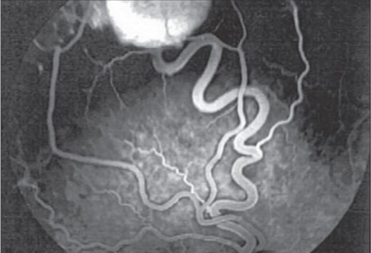
Fluorescein angiogram of right eye showing a temporal superior retinal hemangioblastoma with the feeder artery and draining vein

Image: “Fluorescein angiogram of right eye” by Vitreoretinal Division, Department of Ophthalmology, Al-Hussein Hospital, King Hussein Medical Center, Amman, Jordan. License: CC BY 2.0

Histopathology

  • Gold standard for diagnosis 
  • Gross examination: well-circumscribed, highly vascular red nodules
  • Microscopic examination:
    • Extensive vascular networks with structurally normal capillaries Capillaries Capillaries are the primary structures in the circulatory system that allow the exchange of gas, nutrients, and other materials between the blood and the extracellular fluid (ECF). Capillaries are the smallest of the blood vessels. Because a capillary diameter is so small, only 1 RBC may pass through at a time. Capillaries: Histology
    • Neoplastic stromal cells:
  • 2 distinct cellular components may appear in the same tumor Tumor Inflammation:
Optic nerve hemangioblastoma

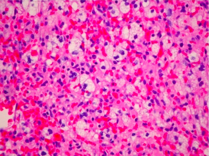
H&E photomicrograph of a hemangioblastoma of the optic nerve (unusual location) revealing a markedly vascular tumor with lipidized stromal cells (×200)

Image: “Optic nerve hemangioblastoma” by Department of Surgery, Division of Neurosurgery, University of Alabama at Birmingham, Birmingham, AL 35294, USA. License: CC BY 3.0

Management and Prognosis

Surgical resection is the primary approach to treating hemangioblastomas Hemangioblastomas A benign tumor of the nervous system that may occur sporadically or in association with von Hippel-Lindau disease. It accounts for approximately 2% of intracranial tumors, arising most frequently in the cerebellar hemispheres and vermis. Histologically, the tumors are composed of multiple capillary and sinusoidal channels lined with endothelial cells and clusters of lipid-laden pseudoxanthoma cells. Usually solitary, these tumors can be multiple and may also occur in the brain stem, spinal cord, retina, and supratentorial compartment. Cerebellar hemangioblastomas usually present in the third decade with intracranial hypertension, and ataxia. Von Hippel-Lindau Disease. Adjuvant Adjuvant Substances that augment, stimulate, activate, potentiate, or modulate the immune response at either the cellular or humoral level. The classical agents (freund’s adjuvant, bcg, corynebacterium parvum, et al.) contain bacterial antigens. Some are endogenous (e.g., histamine, interferon, transfer factor, tuftsin, interleukin-1). Their mode of action is either non-specific, resulting in increased immune responsiveness to a wide variety of antigens, or antigen-specific, i.e., affecting a restricted type of immune response to a narrow group of antigens. The therapeutic efficacy of many biological response modifiers is related to their antigen-specific immunoadjuvanticity. Vaccination therapies are commonly required in VHL-associated hemangioblastomas Hemangioblastomas A benign tumor of the nervous system that may occur sporadically or in association with von Hippel-Lindau disease. It accounts for approximately 2% of intracranial tumors, arising most frequently in the cerebellar hemispheres and vermis. Histologically, the tumors are composed of multiple capillary and sinusoidal channels lined with endothelial cells and clusters of lipid-laden pseudoxanthoma cells. Usually solitary, these tumors can be multiple and may also occur in the brain stem, spinal cord, retina, and supratentorial compartment. Cerebellar hemangioblastomas usually present in the third decade with intracranial hypertension, and ataxia. Von Hippel-Lindau Disease, and options include radiation Radiation Emission or propagation of acoustic waves (sound), electromagnetic energy waves (such as light; radio waves; gamma rays; or x-rays), or a stream of subatomic particles (such as electrons; neutrons; protons; or alpha particles). Osteosarcoma therapy, endovascular embolization Embolization A method of hemostasis utilizing various agents such as gelfoam, silastic, metal, glass, or plastic pellets, autologous clot, fat, and muscle as emboli. It has been used in the treatment of spinal cord and intracranial arteriovenous malformations, renal arteriovenous fistulas, gastrointestinal bleeding, epistaxis, hypersplenism, certain highly vascular tumors, traumatic rupture of blood vessels, and control of operative hemorrhage. Gastrointestinal Bleeding, and antiangiogenic Antiangiogenic Macular Degeneration therapy.

Surgery

Surgery is the primary definitive approach to treat hemangioblastomas Hemangioblastomas A benign tumor of the nervous system that may occur sporadically or in association with von Hippel-Lindau disease. It accounts for approximately 2% of intracranial tumors, arising most frequently in the cerebellar hemispheres and vermis. Histologically, the tumors are composed of multiple capillary and sinusoidal channels lined with endothelial cells and clusters of lipid-laden pseudoxanthoma cells. Usually solitary, these tumors can be multiple and may also occur in the brain stem, spinal cord, retina, and supratentorial compartment. Cerebellar hemangioblastomas usually present in the third decade with intracranial hypertension, and ataxia. Von Hippel-Lindau Disease, as they are benign Benign Fibroadenoma, noninvasive tumors.

  • Tumors are usually well demarcated from the surrounding structures but are highly vascular and located in neurologically sensitive areas.
  • Preoperative angiography Angiography Radiography of blood vessels after injection of a contrast medium. Cardiac Surgery is helpful to identify feeding arteries Arteries Arteries are tubular collections of cells that transport oxygenated blood and nutrients from the heart to the tissues of the body. The blood passes through the arteries in order of decreasing luminal diameter, starting in the largest artery (the aorta) and ending in the small arterioles. Arteries are classified into 3 types: large elastic arteries, medium muscular arteries, and small arteries and arterioles. Arteries: Histology.
  • Need for adjuvant Adjuvant Substances that augment, stimulate, activate, potentiate, or modulate the immune response at either the cellular or humoral level. The classical agents (freund’s adjuvant, bcg, corynebacterium parvum, et al.) contain bacterial antigens. Some are endogenous (e.g., histamine, interferon, transfer factor, tuftsin, interleukin-1). Their mode of action is either non-specific, resulting in increased immune responsiveness to a wide variety of antigens, or antigen-specific, i.e., affecting a restricted type of immune response to a narrow group of antigens. The therapeutic efficacy of many biological response modifiers is related to their antigen-specific immunoadjuvanticity. Vaccination therapy:

Other therapeutic modalities

  • Radiation Radiation Emission or propagation of acoustic waves (sound), electromagnetic energy waves (such as light; radio waves; gamma rays; or x-rays), or a stream of subatomic particles (such as electrons; neutrons; protons; or alpha particles). Osteosarcoma therapy:
    • Postoperative radiation Radiation Emission or propagation of acoustic waves (sound), electromagnetic energy waves (such as light; radio waves; gamma rays; or x-rays), or a stream of subatomic particles (such as electrons; neutrons; protons; or alpha particles). Osteosarcoma therapy reduces the recurrence of hemangioblastomas Hemangioblastomas A benign tumor of the nervous system that may occur sporadically or in association with von Hippel-Lindau disease. It accounts for approximately 2% of intracranial tumors, arising most frequently in the cerebellar hemispheres and vermis. Histologically, the tumors are composed of multiple capillary and sinusoidal channels lined with endothelial cells and clusters of lipid-laden pseudoxanthoma cells. Usually solitary, these tumors can be multiple and may also occur in the brain stem, spinal cord, retina, and supratentorial compartment. Cerebellar hemangioblastomas usually present in the third decade with intracranial hypertension, and ataxia. Von Hippel-Lindau Disease.
    • Indications:
      • Surgically inaccessible lesions
      • Multiple lesions
  • Endovascular embolization Embolization A method of hemostasis utilizing various agents such as gelfoam, silastic, metal, glass, or plastic pellets, autologous clot, fat, and muscle as emboli. It has been used in the treatment of spinal cord and intracranial arteriovenous malformations, renal arteriovenous fistulas, gastrointestinal bleeding, epistaxis, hypersplenism, certain highly vascular tumors, traumatic rupture of blood vessels, and control of operative hemorrhage. Gastrointestinal Bleeding:
    • Goal: to decrease vascularity to the tumor Tumor Inflammation and reduce intraoperative complications of hemorrhage
    • Large tumors can be considerably reduced to make the resection easier.
  • Angiogenic inhibitors: bevacizumab Bevacizumab An anti-vegf humanized murine monoclonal antibody. It inhibits vegf receptors and helps to prevent pathologic angiogenesis. Targeted and Other Nontraditional Antineoplastic Therapy
    • Mechanism of action: a monoclonal antibody that binds to and inhibits the effects of VEGF → ↓ angiogenesis Angiogenesis Bartonella
    • Uses: VHL patients Patients Individuals participating in the health care system for the purpose of receiving therapeutic, diagnostic, or preventive procedures. Clinician–Patient Relationship with tumors that do not respond to, or are not amenable to, surgery or radiation Radiation Emission or propagation of acoustic waves (sound), electromagnetic energy waves (such as light; radio waves; gamma rays; or x-rays), or a stream of subatomic particles (such as electrons; neutrons; protons; or alpha particles). Osteosarcoma
  • HIF-2ɑ inhibitor (belzutifan):
    • For individuals with known VHL disease VHL disease Von Hippel-Lindau (VHL) disease is an autosomal dominant genetic condition resulting from a deletion or mutation in the VHL gene. Individuals diagnosed with VHL disease have tumors and cysts in various parts of their bodies and may present with hemangioblastomas, renal cell carcinoma (RCC), pheochromocytoma, endolymphatic sac tumors of the middle ear, pancreatic tumors, and papillary cystadenomas of the epididymis or the broad ligament. Von Hippel-Lindau Disease
    • Used to delay surgery in those with rapidly enlarging tumors
    • Also beneficial for individuals with refractory or recurrent tumors

Prognosis Prognosis A prediction of the probable outcome of a disease based on a individual’s condition and the usual course of the disease as seen in similar situations. Non-Hodgkin Lymphomas

Prognosis Prognosis A prediction of the probable outcome of a disease based on a individual’s condition and the usual course of the disease as seen in similar situations. Non-Hodgkin Lymphomas is good in most treated hemangioblastomas Hemangioblastomas A benign tumor of the nervous system that may occur sporadically or in association with von Hippel-Lindau disease. It accounts for approximately 2% of intracranial tumors, arising most frequently in the cerebellar hemispheres and vermis. Histologically, the tumors are composed of multiple capillary and sinusoidal channels lined with endothelial cells and clusters of lipid-laden pseudoxanthoma cells. Usually solitary, these tumors can be multiple and may also occur in the brain stem, spinal cord, retina, and supratentorial compartment. Cerebellar hemangioblastomas usually present in the third decade with intracranial hypertension, and ataxia. Von Hippel-Lindau Disease:

  • Clean surgical resection (with negative surgical margins), especially in solitary sporadic Sporadic Selective IgA Deficiency tumors ensures a good prognosis Prognosis A prediction of the probable outcome of a disease based on a individual’s condition and the usual course of the disease as seen in similar situations. Non-Hodgkin Lymphomas.
  • Early detection and intervention is favorable.
  • Hemangioblastomas Hemangioblastomas A benign tumor of the nervous system that may occur sporadically or in association with von Hippel-Lindau disease. It accounts for approximately 2% of intracranial tumors, arising most frequently in the cerebellar hemispheres and vermis. Histologically, the tumors are composed of multiple capillary and sinusoidal channels lined with endothelial cells and clusters of lipid-laden pseudoxanthoma cells. Usually solitary, these tumors can be multiple and may also occur in the brain stem, spinal cord, retina, and supratentorial compartment. Cerebellar hemangioblastomas usually present in the third decade with intracranial hypertension, and ataxia. Von Hippel-Lindau Disease associated with VHL:

Differential Diagnosis

  • Meningioma Meningioma Meningiomas are slow-growing tumors that arise from the meninges of the brain and spinal cord. The vast majority are benign. These tumors commonly occur in individuals with a history of high doses of skull radiation, head trauma, and neurofibromatosis 2. Meningioma: tumors arising from the meninges Meninges The brain and the spinal cord are enveloped by 3 overlapping layers of connective tissue called the meninges. The layers are, from the most external layer to the most internal layer, the dura mater, arachnoid mater, and pia mater. Between these layers are 3 potential spaces called the epidural, subdural, and subarachnoid spaces. Meninges: Anatomy of the brain Brain The part of central nervous system that is contained within the skull (cranium). Arising from the neural tube, the embryonic brain is comprised of three major parts including prosencephalon (the forebrain); mesencephalon (the midbrain); and rhombencephalon (the hindbrain). The developed brain consists of cerebrum; cerebellum; and other structures in the brain stem. Nervous System: Anatomy, Structure, and Classification and spinal cord Spinal cord The spinal cord is the major conduction pathway connecting the brain to the body; it is part of the CNS. In cross section, the spinal cord is divided into an H-shaped area of gray matter (consisting of synapsing neuronal cell bodies) and a surrounding area of white matter (consisting of ascending and descending tracts of myelinated axons). Spinal Cord: Anatomy. Meningiomas are commonly asymptomatic, but they can present with headache Headache The symptom of pain in the cranial region. It may be an isolated benign occurrence or manifestation of a wide variety of headache disorders. Brain Abscess, seizures Seizures A seizure is abnormal electrical activity of the neurons in the cerebral cortex that can manifest in numerous ways depending on the region of the brain affected. Seizures consist of a sudden imbalance that occurs between the excitatory and inhibitory signals in cortical neurons, creating a net excitation. The 2 major classes of seizures are focal and generalized. Seizures, and visual disturbances. Diagnosis is through MRI and biopsy Biopsy Removal and pathologic examination of specimens from the living body. Ewing Sarcoma. Asymptomatic cases are typically kept under observation, while symptomatic patients Patients Individuals participating in the health care system for the purpose of receiving therapeutic, diagnostic, or preventive procedures. Clinician–Patient Relationship are treated surgically or with radiation Radiation Emission or propagation of acoustic waves (sound), electromagnetic energy waves (such as light; radio waves; gamma rays; or x-rays), or a stream of subatomic particles (such as electrons; neutrons; protons; or alpha particles). Osteosarcoma. Unlike hemangioblastomas Hemangioblastomas A benign tumor of the nervous system that may occur sporadically or in association with von Hippel-Lindau disease. It accounts for approximately 2% of intracranial tumors, arising most frequently in the cerebellar hemispheres and vermis. Histologically, the tumors are composed of multiple capillary and sinusoidal channels lined with endothelial cells and clusters of lipid-laden pseudoxanthoma cells. Usually solitary, these tumors can be multiple and may also occur in the brain stem, spinal cord, retina, and supratentorial compartment. Cerebellar hemangioblastomas usually present in the third decade with intracranial hypertension, and ataxia. Von Hippel-Lindau Disease, meningiomas are always near the meninges Meninges The brain and the spinal cord are enveloped by 3 overlapping layers of connective tissue called the meninges. The layers are, from the most external layer to the most internal layer, the dura mater, arachnoid mater, and pia mater. Between these layers are 3 potential spaces called the epidural, subdural, and subarachnoid spaces. Meninges: Anatomy and often have imaging findings of dural attachment (e.g., dural tail sign Dural tail sign Marginal dural thickening that tapers peripherally Meningioma).
  • Glioblastoma multiforme Glioblastoma multiforme Glioblastoma multiforme is a high-grade astrocytoma, an aggressive brain tumor arising from astrocytes, with an unknown cause and a poorly understood link to risk factors. There are two main types: primary, a more aggressive form seen more commonly in older patients, and secondary, developing from lower-grade astrocytomas and seen more commonly in younger patients. Glioblastoma Multiforme: rapidly progressive WHO grade IV astrocytoma Grade IV Astrocytoma Astrocytoma that arises from astrocytes Astrocytes A class of large neuroglial (macroglial) cells in the central nervous system – the largest and most numerous neuroglial cells in the brain and spinal cord. Astrocytes (from ‘star’ cells) are irregularly shaped with many long processes, including those with ‘end feet’ which form the glial (limiting) membrane and directly and indirectly contribute to the blood-brain barrier. They regulate the extracellular ionic and chemical environment, and ‘reactive astrocytes’ (along with microglia) respond to injury. Nervous System: Histology ( glial cells Glial Cells The non-neuronal cells of the nervous system. They not only provide physical support, but also respond to injury, regulate the ionic and chemical composition of the extracellular milieu, participate in the blood-brain barrier and blood-retinal barrier, form the myelin insulation of nervous pathways, guide neuronal migration during development, and exchange metabolites with neurons. Neuroglia have high-affinity transmitter uptake systems, voltage-dependent and transmitter-gated ion channels, and can release transmitters, but their role in signaling (as in many other functions) is unclear. Nervous System: Histology in the brain Brain The part of central nervous system that is contained within the skull (cranium). Arising from the neural tube, the embryonic brain is comprised of three major parts including prosencephalon (the forebrain); mesencephalon (the midbrain); and rhombencephalon (the hindbrain). The developed brain consists of cerebrum; cerebellum; and other structures in the brain stem. Nervous System: Anatomy, Structure, and Classification) and presents clinically as headache Headache The symptom of pain in the cranial region. It may be an isolated benign occurrence or manifestation of a wide variety of headache disorders. Brain Abscess, nausea Nausea An unpleasant sensation in the stomach usually accompanied by the urge to vomit. Common causes are early pregnancy, sea and motion sickness, emotional stress, intense pain, food poisoning, and various enteroviruses. Antiemetics, drowsiness, blurred vision Blurred Vision Retinal Detachment, personality changes, and seizures Seizures A seizure is abnormal electrical activity of the neurons in the cerebral cortex that can manifest in numerous ways depending on the region of the brain affected. Seizures consist of a sudden imbalance that occurs between the excitatory and inhibitory signals in cortical neurons, creating a net excitation. The 2 major classes of seizures are focal and generalized. Seizures. Imaging, clinical presentation, and biopsy Biopsy Removal and pathologic examination of specimens from the living body. Ewing Sarcoma are the mainstays for diagnosis. Management includes radiotherapy, chemotherapy Chemotherapy Osteosarcoma, and surgical excision. Prognosis Prognosis A prediction of the probable outcome of a disease based on a individual’s condition and the usual course of the disease as seen in similar situations. Non-Hodgkin Lymphomas is poor even with treatment. Unlike hemangioblastoma, glioblastoma multiforme Glioblastoma multiforme Glioblastoma multiforme is a high-grade astrocytoma, an aggressive brain tumor arising from astrocytes, with an unknown cause and a poorly understood link to risk factors. There are two main types: primary, a more aggressive form seen more commonly in older patients, and secondary, developing from lower-grade astrocytomas and seen more commonly in younger patients. Glioblastoma Multiforme is not associated with VHL.
  • Oligodendroglioma Oligodendroglioma Oligodendrogliomas are malignant CNS tumors arising from neural glial cell precursors. Oligodendrogliomas often arise in the frontal lobes of the brain and have a generally favorable prognosis when compared to other gliomas. Oligodendrogliomas are the 3rd most common CNS tumor. The most frequent presenting symptom is a seizure. Oligodendroglioma: CNS tumor Tumor Inflammation arising from oligodendrocytes. Oligodendroglioma Oligodendroglioma Oligodendrogliomas are malignant CNS tumors arising from neural glial cell precursors. Oligodendrogliomas often arise in the frontal lobes of the brain and have a generally favorable prognosis when compared to other gliomas. Oligodendrogliomas are the 3rd most common CNS tumor. The most frequent presenting symptom is a seizure. Oligodendroglioma can present with focal neurologic deficits Neurologic Deficits High-Risk Headaches, seizures Seizures A seizure is abnormal electrical activity of the neurons in the cerebral cortex that can manifest in numerous ways depending on the region of the brain affected. Seizures consist of a sudden imbalance that occurs between the excitatory and inhibitory signals in cortical neurons, creating a net excitation. The 2 major classes of seizures are focal and generalized. Seizures, and personality changes depending on the exact location. Diagnosis is made by MRI imaging and biopsy Biopsy Removal and pathologic examination of specimens from the living body. Ewing Sarcoma. Oligodendrogliomas commonly develop in the cerebral hemispheres, usually the frontal lobe Frontal lobe The part of the cerebral hemisphere anterior to the central sulcus, and anterior and superior to the lateral sulcus. Cerebral Cortex: Anatomy, and are rarely found in the infratentorial area or the spinal cord Spinal cord The spinal cord is the major conduction pathway connecting the brain to the body; it is part of the CNS. In cross section, the spinal cord is divided into an H-shaped area of gray matter (consisting of synapsing neuronal cell bodies) and a surrounding area of white matter (consisting of ascending and descending tracts of myelinated axons). Spinal Cord: Anatomy. Management involves surgical resection possibly accompanied by radiation Radiation Emission or propagation of acoustic waves (sound), electromagnetic energy waves (such as light; radio waves; gamma rays; or x-rays), or a stream of subatomic particles (such as electrons; neutrons; protons; or alpha particles). Osteosarcoma and/or chemotherapy Chemotherapy Osteosarcoma.
  • Brain Brain The part of central nervous system that is contained within the skull (cranium). Arising from the neural tube, the embryonic brain is comprised of three major parts including prosencephalon (the forebrain); mesencephalon (the midbrain); and rhombencephalon (the hindbrain). The developed brain consists of cerebrum; cerebellum; and other structures in the brain stem. Nervous System: Anatomy, Structure, and Classification aneurysm Aneurysm An aneurysm is a bulging, weakened area of a blood vessel that causes an abnormal widening of its diameter > 1.5 times the size of the native vessel. Aneurysms occur more often in arteries than in veins and are at risk of dissection and rupture, which can be life-threatening. Thoracic Aortic Aneurysms: weakness in the wall of a blood vessel supplying the brain Brain The part of central nervous system that is contained within the skull (cranium). Arising from the neural tube, the embryonic brain is comprised of three major parts including prosencephalon (the forebrain); mesencephalon (the midbrain); and rhombencephalon (the hindbrain). The developed brain consists of cerebrum; cerebellum; and other structures in the brain stem. Nervous System: Anatomy, Structure, and Classification that bulges and is at the risk of rupturing. An unruptured aneurysm Aneurysm An aneurysm is a bulging, weakened area of a blood vessel that causes an abnormal widening of its diameter > 1.5 times the size of the native vessel. Aneurysms occur more often in arteries than in veins and are at risk of dissection and rupture, which can be life-threatening. Thoracic Aortic Aneurysms is usually asymptomatic and the rupture of an aneurysm Aneurysm An aneurysm is a bulging, weakened area of a blood vessel that causes an abnormal widening of its diameter > 1.5 times the size of the native vessel. Aneurysms occur more often in arteries than in veins and are at risk of dissection and rupture, which can be life-threatening. Thoracic Aortic Aneurysms causes sudden, severe headache Headache The symptom of pain in the cranial region. It may be an isolated benign occurrence or manifestation of a wide variety of headache disorders. Brain Abscess. Brain Brain The part of central nervous system that is contained within the skull (cranium). Arising from the neural tube, the embryonic brain is comprised of three major parts including prosencephalon (the forebrain); mesencephalon (the midbrain); and rhombencephalon (the hindbrain). The developed brain consists of cerebrum; cerebellum; and other structures in the brain stem. Nervous System: Anatomy, Structure, and Classification aneurysms are diagnosed with CT, angiography Angiography Radiography of blood vessels after injection of a contrast medium. Cardiac Surgery, or ultrasonography. Management depends on the location and is typically treated with an endovascular stent graft Graft A piece of living tissue that is surgically transplanted Organ Transplantation.

References

  1. Wong, E.T., Joseph, J., Wu, J.K. Hemangioblastoma. UpToDate. Retrieved May 20, 2021, from https://www.uptodate.com/contents/hemangioblastoma
  2. Slavin, K.V. (2017). Hemangioblastoma. Medscape. Retrieved May 20, 2021, from https://emedicine.medscape.com/article/250670-overview
  3. Pernick, N. (2021). CNS tumors—other tumors—hemangioblastoma. Pathology Outlines. Retrieved May 20, 2021, fromhttps://www.pathologyoutlines.com/topic/cnstumorhemangioblastoma.html 
  4. Kim W.Y., Kaelin W.G. (2004). Role of VHL gene mutation in human cancer. J Clin Oncol. Retrieved June 1, 2021, from https://pubmed.ncbi.nlm.nih.gov/15611513/ 
  5. Dengler, V. L., Galbraith, M., Espinosa, J. M. (2014). Transcriptional regulation by hypoxia inducible factors. Critical reviews in biochemistry and molecular biology, 49(1):1–15. Retrieved June 1, 2021, from https://www.ncbi.nlm.nih.gov/pmc/articles/PMC4342852/

Create your free account or log in to continue reading!

Sign up now and get free access to Lecturio with concept pages, medical videos, and questions for your medical education.

User Reviews

Details