The spleen Spleen The spleen is the largest lymphoid organ in the body, located in the LUQ of the abdomen, superior to the left kidney and posterior to the stomach at the level of the 9th-11th ribs just below the diaphragm. The spleen is highly vascular and acts as an important blood filter, cleansing the blood of pathogens and damaged erythrocytes. Spleen: Anatomy is the largest ductless gland and largest single lymphatic organ in the human body. The main functions of the spleen Spleen The spleen is the largest lymphoid organ in the body, located in the LUQ of the abdomen, superior to the left kidney and posterior to the stomach at the level of the 9th-11th ribs just below the diaphragm. The spleen is highly vascular and acts as an important blood filter, cleansing the blood of pathogens and damaged erythrocytes. Spleen: Anatomy are immunologic surveillance Surveillance Developmental Milestones and Normal Growth and red blood cell breakdown. The spleen Spleen The spleen is the largest lymphoid organ in the body, located in the LUQ of the abdomen, superior to the left kidney and posterior to the stomach at the level of the 9th-11th ribs just below the diaphragm. The spleen is highly vascular and acts as an important blood filter, cleansing the blood of pathogens and damaged erythrocytes. Spleen: Anatomy can be affected by diseases of different origins, such as inflammatory, congenital Congenital Chorioretinitis, infectious, neoplastic, and vascular diseases. Ultrasonography (US) is usually used as the 1st-line imaging modality due to its easy accessibility and lack of ionizing radiation Radiation Emission or propagation of acoustic waves (sound), electromagnetic energy waves (such as light; radio waves; gamma rays; or x-rays), or a stream of subatomic particles (such as electrons; neutrons; protons; or alpha particles). Osteosarcoma, but CT and MRI with contrast can also be helpful. Contrasted imaging modalities can delineate lesions and help differentiate disorders.
Last updated: Dec 15, 2025
The common radiologic modalities used to evaluate the spleen Spleen The spleen is the largest lymphoid organ in the body, located in the LUQ of the abdomen, superior to the left kidney and posterior to the stomach at the level of the 9th-11th ribs just below the diaphragm. The spleen is highly vascular and acts as an important blood filter, cleansing the blood of pathogens and damaged erythrocytes. Spleen: Anatomy are the following:
Normal ultrasound appearance:
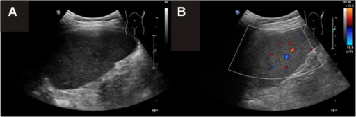
Normal ultrasonographic image of the spleen (A) with Doppler mode (B)
Image: “F3” by Jordan, A.J., Becker, K.P., et al. License: CC BY 2.0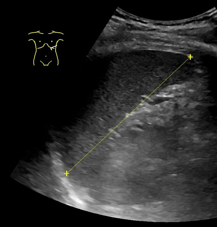
The maximum length of the spleen as measured by ultrasonography (US)
Image: “Maximum length of spleen on ultrasonography” by Mikael Häggström. License: CC0 1.0Standard CT scanning Standard CT scanning Imaging of the Liver and Biliary Tract:
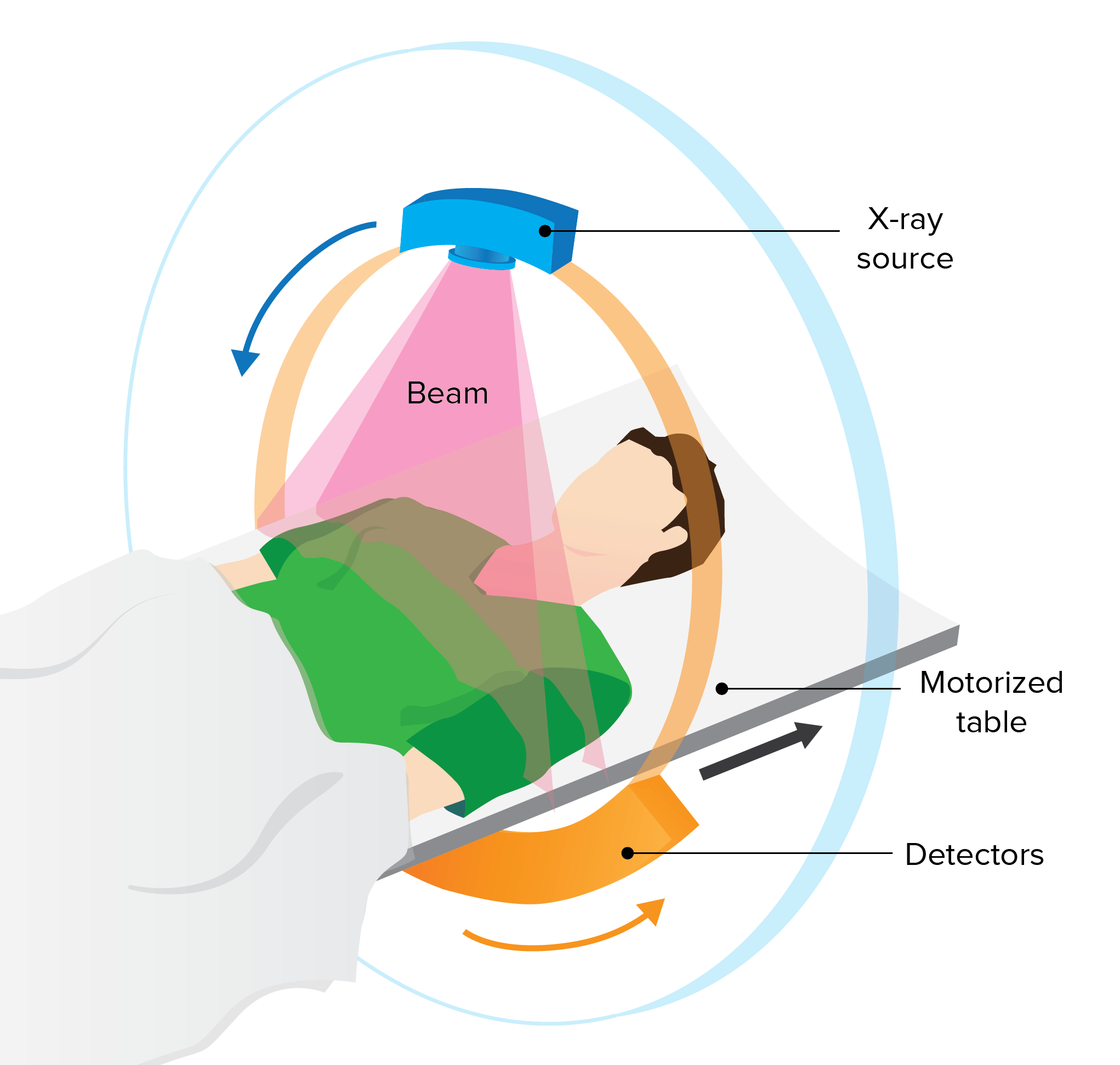
The patient is advanced into the CT machine and the scanner revolves around the patient.
Image by Lecturio.Interpretation should follow a systematic and reproducible pattern:
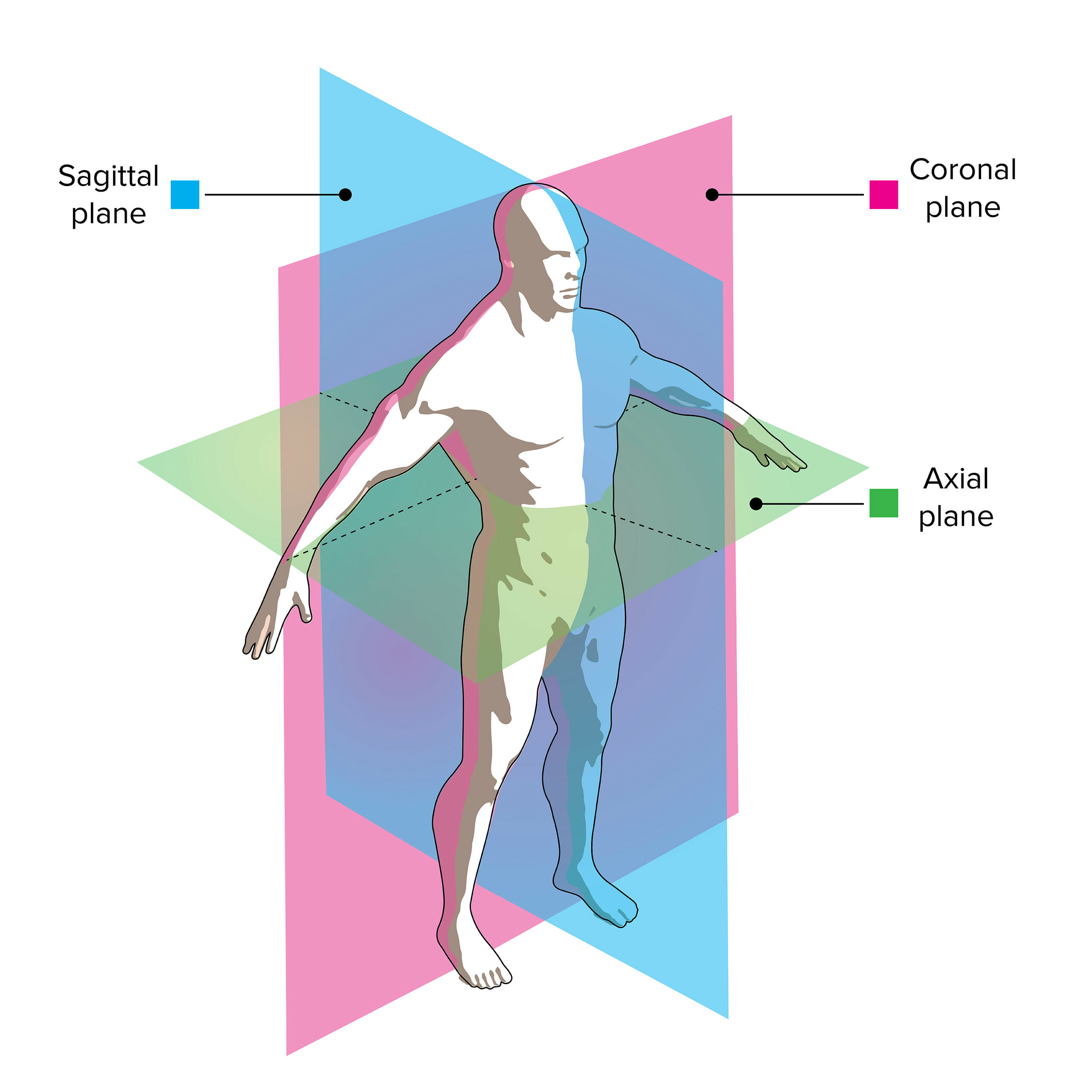
A CT scan uses multiple X-rays to create a 2- or 3-dimensional image:
The X-ray “slices” are taken in the axial plane and reconstructed into the sagittal and coronal planes by a computer to produce the final image.
Normal CT appearance:
| Tissue | T1-weighted images T1-Weighted Images Imaging of the Head and Brain | T2-weighted images T2-Weighted Images Imaging of the Head and Brain |
|---|---|---|
| Fluid (e.g., CSF) | Dark | Bright |
| Fat | Bright | Bright |
| Inflammation Inflammation Inflammation is a complex set of responses to infection and injury involving leukocytes as the principal cellular mediators in the body’s defense against pathogenic organisms. Inflammation is also seen as a response to tissue injury in the process of wound healing. The 5 cardinal signs of inflammation are pain, heat, redness, swelling, and loss of function. Inflammation | Dark | Bright |
Interpretation should follow a systematic and reproducible pattern:
Normal MRI appearance:
Computed tomography scans showing massive splenomegaly:
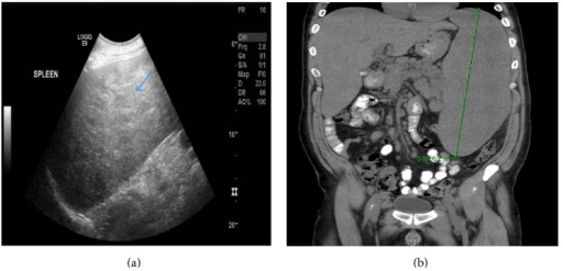
(a) Sagittal sonographic view of the spleen showing a markedly enlarged and diffuse heterogeneous spleen (blue arrow) measuring 30 cm in length
(b) Coronal non-contrast CT (NCCT) of the abdomen and pelvis showing enlarged spleen reaching 33.6 cm in length
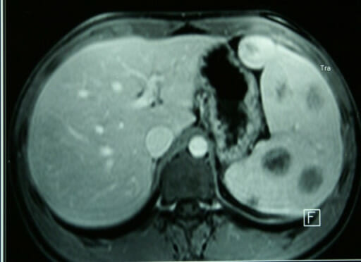
Magnetic resonance imaging showing splenomegaly and hemangiomas
Image: “F1” by Kosmidis, C., Efthimiadis, C., et al. License: CC BY 2.0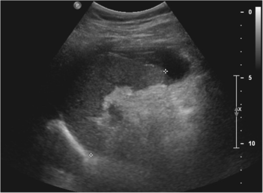
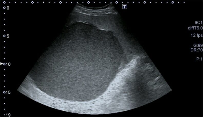
A 2D grayscale ultrasound image demonstrates a small subcapsular splenic hematoma.
Image: “F4” by Azar, F., Brownson, E., Dechert, T. License: CC BY 2.0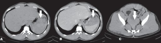
A CT scan showing subcapsular hematoma and hemoperitoneum:
A: A CT, axial slice, without contrast, demonstrating dense collections (arrow) adjacent to the spleen
B: Contrast-enhanced axial CT showing the dense collections adjacent to the spleen (arrow), without contrast enhancement, indicative of subcapsular hematoma
C: A CT, axial slice, without contrast, demonstrating dependently layering dense free fluid in the pelvis (arrow), indicative of hemoperitoneum
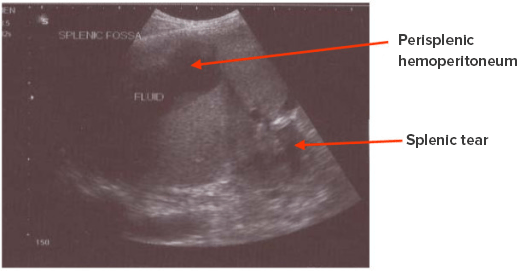
Rupture of spleen with hemoperitoneum in the perisplenic area
Image: “Chronic rupture of spleen with hemoperitonem in perisplenic area” by Sharma, D. License: CC BY 2.0, edited by Lecturio.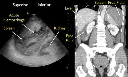
Splenic rupture:
Left image, abdominal ultrasound, left upper quadrant view
Right image, computed tomography scan (CT) of the abdomen and pelvis, coronal view
Abdominal ultrasound showing a large splenic cyst containing homogeneous internal echoes/debris
Image: “Fig. 1” by Sleiman, Y.A., Bohlok, A., et al. License: CC BY 4.0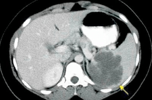
A CT scan of the splenic cyst (arrow)
Image: “fig-002” by Avgerinos, D.V., Kyriakopoulos, C.E., et al. License: CC BY 3.0
Preoperative abdominal CT, (a) axial and (b) coronal.
(c) A T2-weighted MRI shows a giant rounded cystic mass anterior to the spleen, which is significantly displaced posteriorly.
(d) An axial T1-weighted fat-saturated MRI, post–gadolinium administration, shows a thin peripheral enhancement of the splenic mass—possibly representing a capsule—and no internal enhancement consistent with its cystic nature.
Abdominal CT showing a splenule (arrow)
Image by Hetal Verma.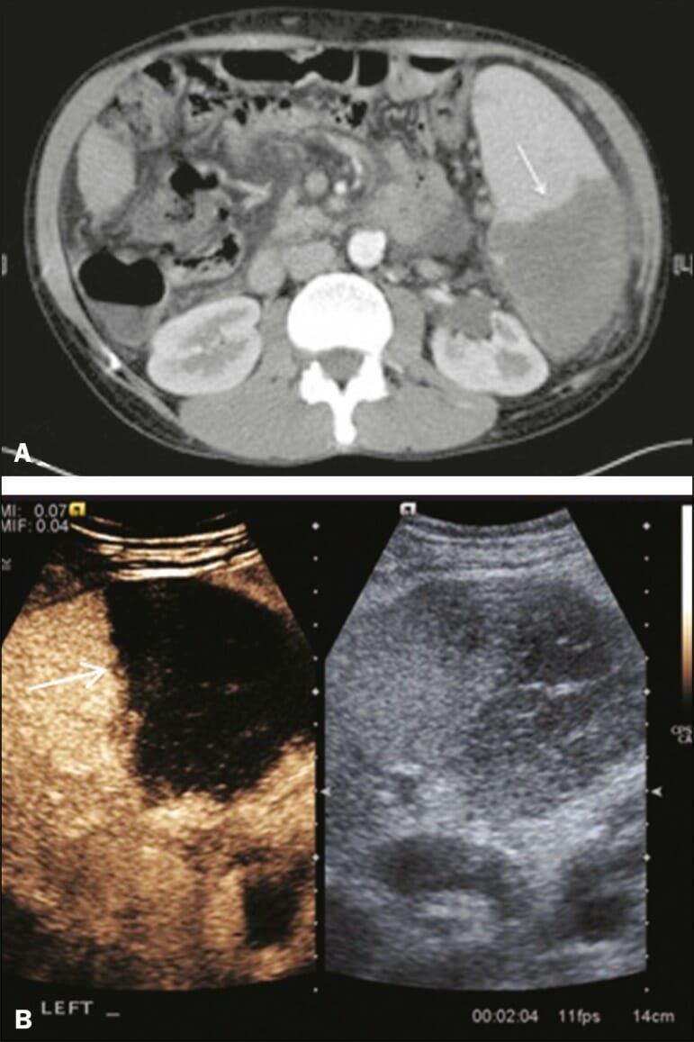
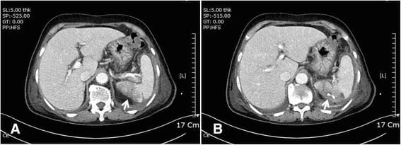
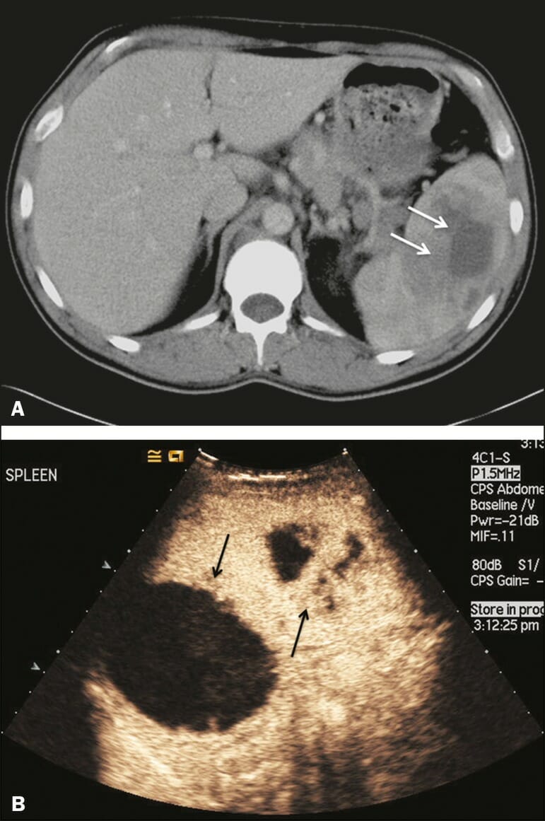
A: Contrast-enhanced CT scan in a pre-assessment for liver transplantation, showing splenomegaly with a sharply demarcated low-density area in the posterior–inferior pole of the spleen (arrow).
B: A split-screen view. Conventional ultrasound (right side of the screen) shows a hypoechoic, heterogeneous lower pole of the spleen, and contrast-enhanced ultrasound (CEUS) (left side of the screen) shows a total absence of enhancement of the lower pole of the spleen, confirming a large wedge-shaped infarction (arrow).
Computed tomography shows splenic infarction (A: white arrow) and abscess with percutaneous drainage (B: wedge-shaped, white arrow)
Image: “Fig5” by Kim, J.S., Kang, M.K., et al. License: CC0 1.0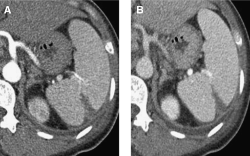
Abdominal CT with enhancement shows a low-attenuated, wedge-shaped lesion consistent with splenic infarction as well as splenomegaly in the arterial phase (A) and portal phase (B).
Image: “F2” by Hwang, J.H., Lee, C.S. License: CC BY 4.0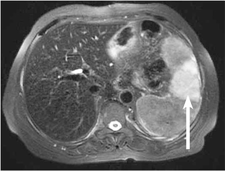
Axial T2-weighted fat-suppressed MRI shows a wedge-shaped hyperintense area of infarction in the spleen (arrow) as well as decreased signal intensity of the liver.
Image: “f1” by Usküdar Teke, H., Karahan, S., Gümüş, U. License: CC BY 2.5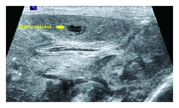
Ultrasound examination demonstrating solitary, small, well-defined splenic abscess
Image: “Figure 2” by Aslam, A., Ahmed Shatla E.S., et al. License: CC BY 4.0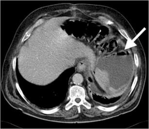
Splenic abscess:
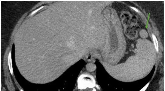
Axial contrast-enhanced CT of a 40-year-old woman demonstrates air (arrow) within a low attenuation splenic abscess. A culture test was positive for E. coli.
Abscess:
A: Contrast-enhanced CT scan showing a hypodense splenic lesion with a thick, irregular wall (arrows)
B: Contrast-enhanced ultrasound (CEUS) showing no internal enhancement and an irregular wall in the larger abscess, with thickened, enhancing septations in the smaller abscess (arrows)
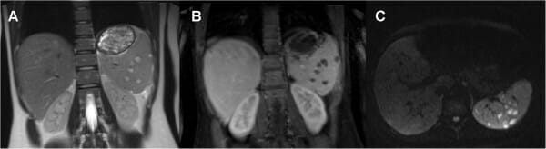
Follow-up MRI of the abdomen (24 days after admission):
A: The coronal T2-weighted image shows splenomegaly with hyperintense lesions with a hypointense rim in the spleen.
B and C: The coronal post-contrast T1-weighted image and transverse diffusion-weighted image (DWI) (b 800 sec/mm2) show multiple areas of intrasplenic abscess formation.