The mediastinum Mediastinum The mediastinum is the thoracic area between the 2 pleural cavities. The mediastinum contains vital structures of the circulatory, respiratory, digestive, and nervous systems including the heart and esophagus, and major thoracic vessels. Mediastinum and Great Vessels: Anatomy is the central part of the chest cavity containing many vital structures, such as the heart, great vessels, trachea Trachea The trachea is a tubular structure that forms part of the lower respiratory tract. The trachea is continuous superiorly with the larynx and inferiorly becomes the bronchial tree within the lungs. The trachea consists of a support frame of semicircular, or C-shaped, rings made out of hyaline cartilage and reinforced by collagenous connective tissue. Trachea: Anatomy, thoracic esophagus Esophagus The esophagus is a muscular tube-shaped organ of around 25 centimeters in length that connects the pharynx to the stomach. The organ extends from approximately the 6th cervical vertebra to the 11th thoracic vertebra and can be divided grossly into 3 parts: the cervical part, the thoracic part, and the abdominal part. Esophagus: Anatomy, lymph nodes Lymph Nodes They are oval or bean shaped bodies (1 - 30 mm in diameter) located along the lymphatic system. Lymphatic Drainage System: Anatomy, multiple nerves, sympathetic chains, and thoracic spine Spine The human spine, or vertebral column, is the most important anatomical and functional axis of the human body. It consists of 7 cervical vertebrae, 12 thoracic vertebrae, and 5 lumbar vertebrae and is limited cranially by the skull and caudally by the sacrum. Vertebral Column: Anatomy. Mediastinal pathology (e.g., masses) can be noted on conventional radiographs as part of evaluating chest-related symptoms, or it can be incidentally detected. To elucidate the characteristics of the mediastinal abnormality, further imaging studies are warranted. Common additional modalities are CT and MRI.
Last updated: Dec 15, 2025
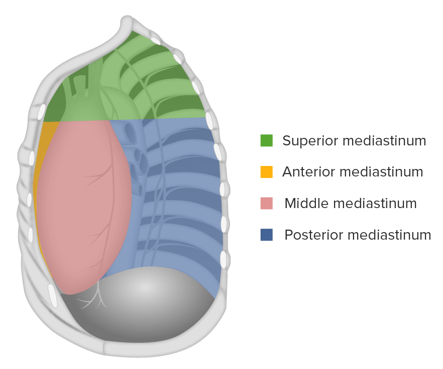
The mediastinum:
subdivided into the superior and inferior mediastinum, which is further divided into anterior, middle, and posterior thirds.
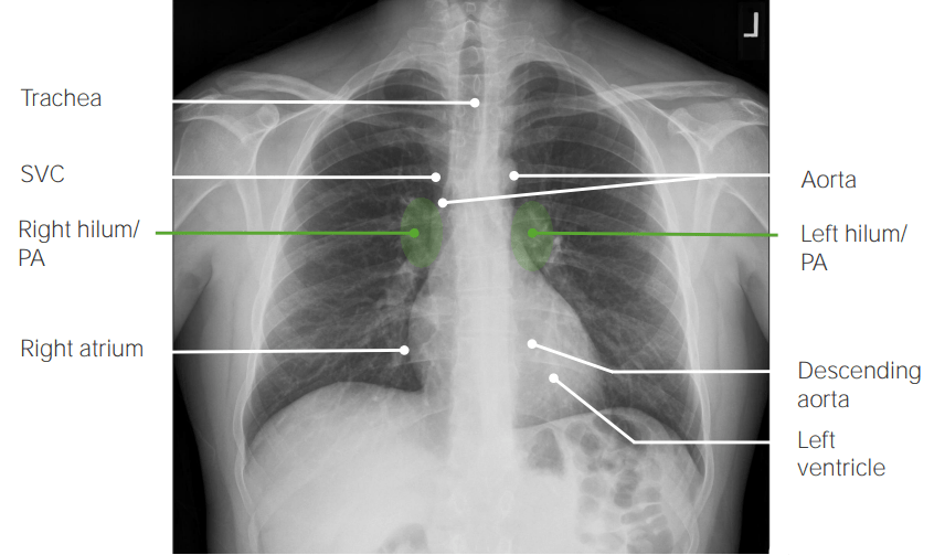
Posteroanterior (PA) view on chest X-ray showing normal findings:
The trachea is midline. The shadow of the superior vena cava (SVC) is immediately to the right of the mediastinum. Right and left hila are seen. The right (right atrium) and left (left ventricle) heart borders are clearly visible. Portions of ascending and descending aorta are visible.
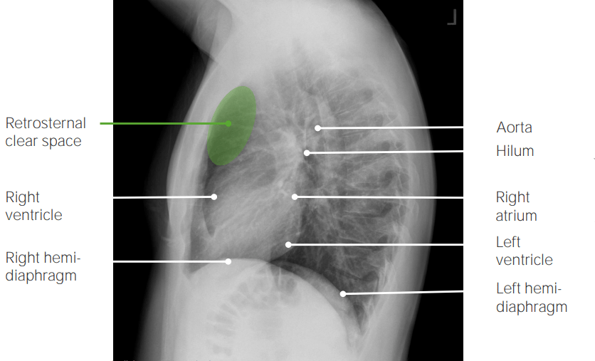
Lateral view on chest X-ray showing normal findings:
The retrosternal space is clear. The anterior (right ventricle) and posterior (right atrium) heart borders are clearly visible.
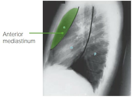
Chest X-ray lateral view illustrating the anterior mediastinum (area between the sternum and the anterior heart border)
Image by Hetal Verma.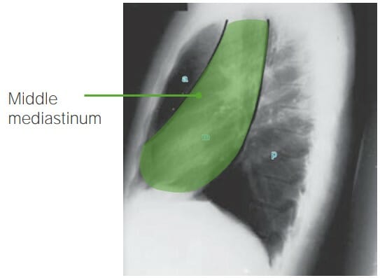
Chest X-ray lateral view illustrating the middle mediastinum (area bordered by the anterior and posterior margins of the heart)
Image by Hetal Verma.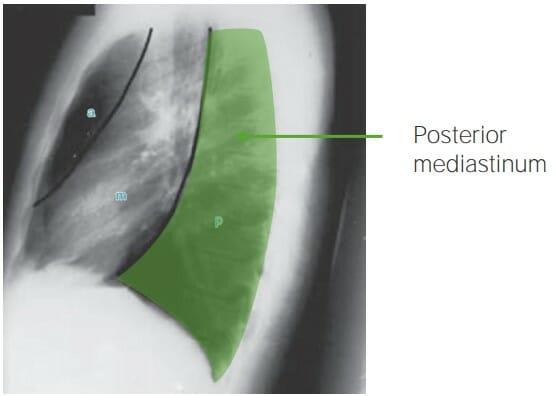
Chest X-ray lateral view illustrating the posterior mediastinum (area from the posterior margin of the heart to the spinous processes)
Image by Hetal Verma.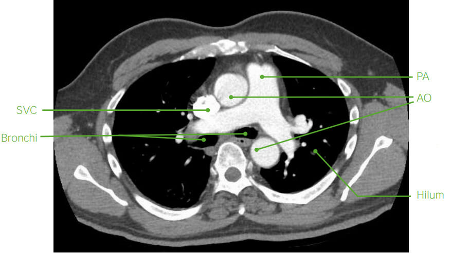
Axial mediastinal anatomy on CT (post-contrast):
the blood vessels (SVC: superior vena cava; PA: pulmonary artery, AO: aorta) are filled with contrast. The bronchi are air-filled. Bone structures are noted to be bright on CT.
MRI
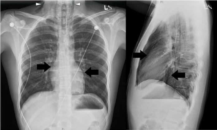
Chest radiograph, posteroanterior (PA) and lateral views:
Pneumomediastinum was confirmed on chest X-ray, based on the presence of air dissecting along the mediastinum (arrows) and into the lower neck region bilaterally (arrowheads).
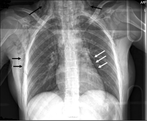
Chest X-ray showing SC emphysema (black arrows) and pneumomediastinum (white arrows) noted in an individual with esophageal rupture
Image: “F1” by van Heijl, M., Saltzherr, T.P., van Berge Henegouwen, M.I., Goslings, J.C. License: CC BY 2.0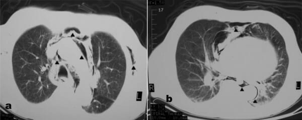
Chest CT demonstrating SC emphysema and pneumomediastinum (arrows)
Image: “F2” by Alexiou, K., Sakellaridis, T., Sikalias, N., Karanikas, I., Economou, N., Antsaklis, G. License: CC BY 2.0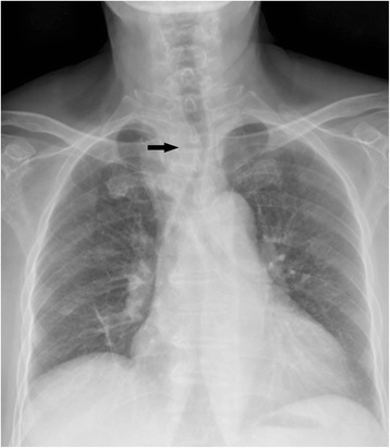
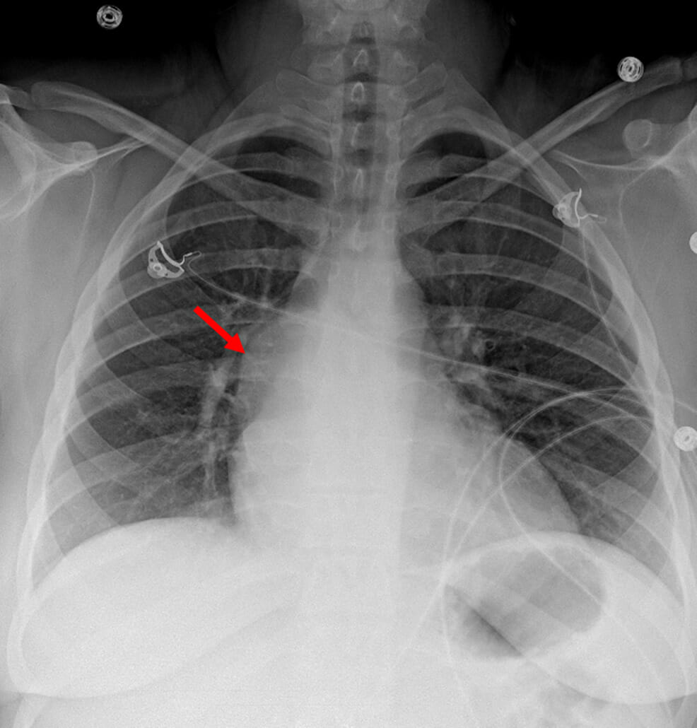
Chest radiograph illustrating the trachea was shifted to the left in an individual found to have primary ectopic substernal thyroid cancer
Image: “Fig1” by Ma, R.M., Lv, L., Zheng, S.R., You, J., Huang, D.P., Guo, G.L. License: CC BY 4.0
A CT scan showing a 5.9 cm × 4.3 cm lesion involving the left lobe of thyroid and isthmus encasing the left common carotid artery and subclavian artery and extending into the left carotid space and anterosuperior mediastinum
Image: “fig2” by Ghosh, S., Rao, P.B., Sarkar, S., Kotne, S., Turlapati, S.P., Mishra, A. License: CC BY 3.0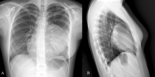
A CT scan of the thorax shows calcified lesions in a cyst in the mediastinum, suggestive of bone and teeth in the mass.
Image: “F2” by Reddy, K.B., Murthy, G.R., Ks, S. License: CC BY 3.0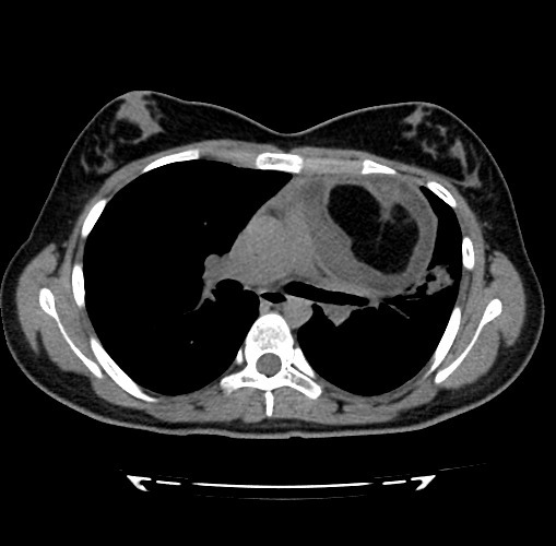
Axial CT scan showing a well-demarcated multi-septated cystic mass compatible with mediastinal teratoma
Image: “F2” by Vaccaro, A., Vierucci, F., Dini, F., Ruggieri, S., Crespin, L., Matteucci, L., Domenici, R. License: CC BY 3.0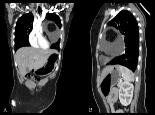
Coronal (A) and sagittal (B) CT reconstructions of mediastinal cystic teratoma. The mass appears to compress the adjacent structures without invasion. A calcification is evident in coronal reconstruction.
Image: “F3” by Vaccaro, A., Vierucci, F., Dini, F., Ruggieri, S., Crespin, L., Matteucci, L., Domenici, R. License: CC BY 3.0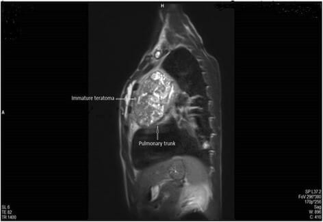
A T2-weighted sagittal MRI showing a multilocular cystic lesion compressing the pulmonary trunk. Biopsy proved that the mass was an immature teratoma (IT).
Image: “Fig. 4” by Koçinaj, D., Krasniqi, X., and Bakalli, A. License: CC BY 4.0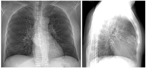
Image (chest X-ray):
Frontal chest radiograph shows a rim calcified lesion (left panel).
Lateral chest radiograph confirms that the rim calcified mass resides within the anterior mediastinum (right panel).
Chest CT demonstrating a mass, representing a thymic carcinoma, with involvement of aortic arch
Image: “f2-cmo-2-2008-477” by Kobrinsky, B., Khaykis, I., Hill, D., Petrovic, L., Yee, H., Chandra, A., Diehl, D.L. License: CC BY 3.0
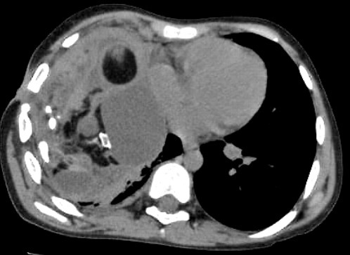
Enhanced chest CT showing a large mediastinal mass outlined in red and found subsequently to be a thymoma
Image: “Fig2” by Downes, K.M., Tarasewicz, D., Weisberg, L.J., Cunningham, E.T. License: CC BY 4.0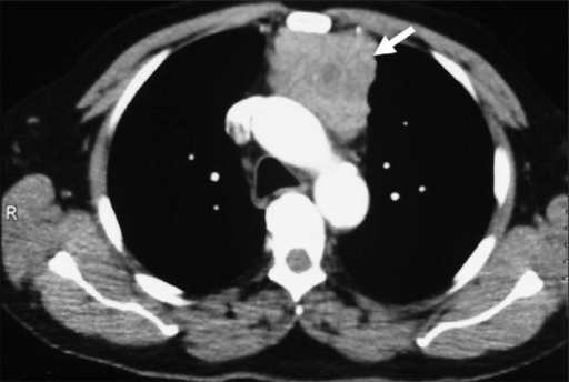
A contrast-enhanced thorax CT shows a soft -issue mass (arrow) in the anterior mediastinum, later confirmed to be a thymoma.
Image: “f3” by Aydın, F., Sürer Budak, E., Dertsiz, L., Belgi, A., Arslan, G., Güngör, F. License: CC BY 2.5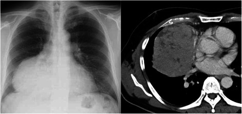
A chest X-ray shows a giant mass in the right lower lung field (left). Chest CT shows a mass measuring 13 x 10 cm in diameter, in contact with the right inferior pulmonary vein (right). Resected tumor was confirmed to be a thymoma.
Image: “Fig1” by Saito, T., Makino, T., Hata, Y., Koezuka, S., Otsuka, H., Isobe, K., Tochigi, N., Shibuya, K., Homma, S., Iyoda, A. License: CC BY 4.0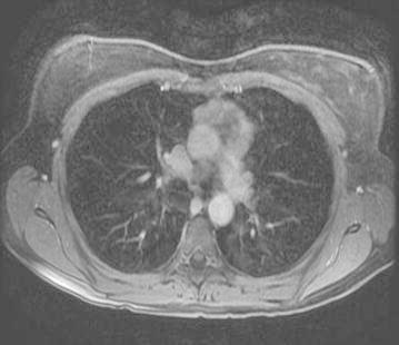
Axial-enhanced T2 image showing an anterior mediastinal mass representing a thymoma
Image: “Fig4” by Bano, G., Sennik, D., Kenchaiah, M., Kyaw, Y., Snape, K., Tripathi, V., Wilson, P., Vlahos, I., Hunt, I., Hodgson, S. License: CC BY 4.0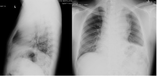
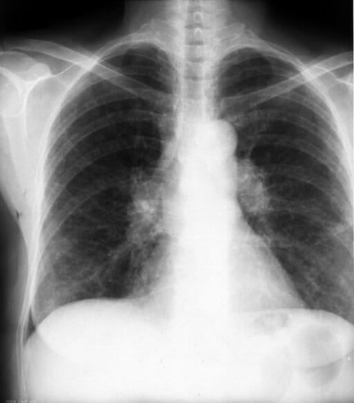
Chest radiographs of an individual with large B cell lymphoma revealed a large anterior mediastinal mass.
Note the widened mediastinum (right) and obscured retrosternal space on the lateral view (left)
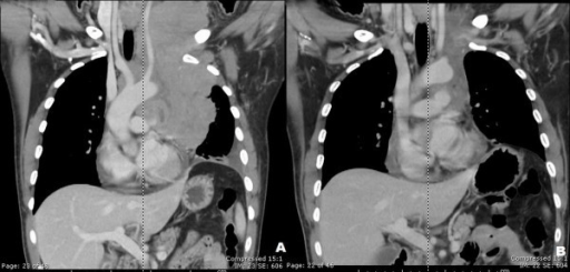
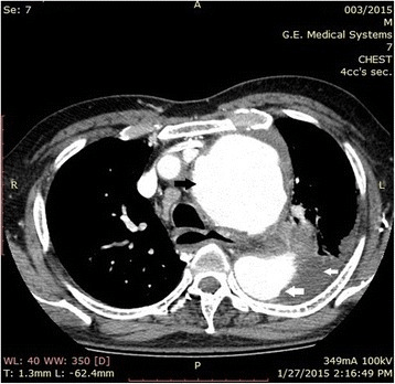
A CT of the neck and chest showed a large lobulated heterogeneous-enhancing mass with central necrosis at the anterior and the middle mediastinum with some parts extending into the left anterior neck (A).
Significant improvement of lymphomatous involvement at the prevascular region was seen after 8 cycles of CHOP chemotherapy (B).
CHOP: cyclophosphamide (cytoxan), hydroxyrubicin (adriamycin), oncovin (vincristine), prednisone
Chest X-ray showing a bulge in the right mediastinal contour (red arrow), which represents a mediastinal bronchogenic cyst
Image: “Figure 1” by Ramireddy, K., Golamari, R. R., Minupuri, A., et al. License: CC BY 4.0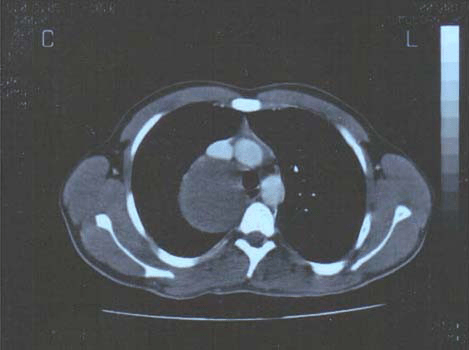
A CT scan post–contrast media injection reveals a huge low-density compressive lesion (cyst) in the posteromedial mediastinum.
Image: “F1” by Kouerinis, I.A., Zografos, G.C., et al. License: CC BY 2.0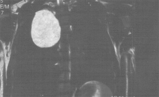
Posteroanterior (PA) chest radiograph showing bilateral hilar lymphadenopathy (BHL) together with lower bilateral interstitial lung densities (sarcoidosis)
Image: “F1” by Papaetis, G.S., Pefanis, A., et al. License: CC BY 2.0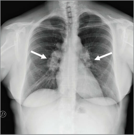
Hilar lymphadenopathy (LAD) (arrows) seen on chest X-ray
Image: “figure1” by Conte, G., Zugni, F., et al. License: CC BY 3.0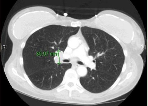
A CT scan demonstrating unilateral (right) hilar lymphadenopathy (LAD) in an individual with small cell lung cancer
Image: “F1” by Hudhud, K.H., Masood, A., Oh, Y., Hegazi, A. License: CC BY 2.0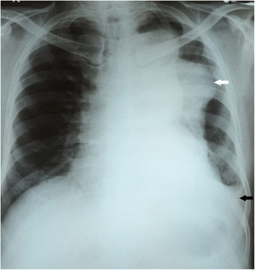
Chest X-ray showing a left-sided mediastinal mass (thoracic aortic aneurysm):
A homogeneous, soft-tissue, dense, well-defined lesion in the left lung (white arrow) extending from the aortic knuckle to the left hemidiaphragm.
Its medial margin merges with the mediastinum and there is a left-sided mild-to-moderate pleural effusion (PE) (black arrow).
A CT aortography showing a dilated portion of the aorta consistent with an ascending (black arrow) and descending (large white arrow) aortic aneurysm. A left-sided pleural effusion (PE) (small white arrow) is also seen.
Image: “Fig2” by Pathirana, U., Kularatne, S., Handagala, S., Ranasinghe, G., Samarasinghe, R. License: CC BY 4.0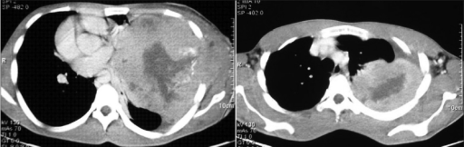
A CT scan of the thorax showing a large, well-defined, heterogeneous mass in the left hemithorax (thoracic schwannoma):
The mass extends peripherally up to the pleura and chest wall and shows infiltrations into the SC tissue.
The mass also contains areas of low attenuation, corresponding to hemorrhage and necrosis, and extends up to the pleura and the chest wall. There is a mediastinal shift to the right side.
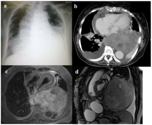
(a) Chest radiograph shows bilateral pleural effusion, mediastinal, widening and cardiac enlargement.
(b) Contrast-enhanced chest CT image (lung window) demonstrates a giant posterior mediastinal tumor, pericardial effusion, and bilateral pleural effusion (PE).
(c) The individual underwent pericardial drainage, and a transversal T1-weighted MRI scan taken after shows a giant encapsulating tumor in the posterior mediastinum compressing the heart.
(d) In a sagittal true steady-state free precession (SSFP) MRI image, a tumor occupying most of left thoracic cavity is seen.