Ovarian cancer is a malignant tumorTumorInflammation arising from the ovarian tissue and is classified according to the type of tissue from which it originates. The 3 major types of ovarian cancer are epithelial ovarian carcinomas (EOCs), ovarian germ cell tumors (OGCTs), and sexSexThe totality of characteristics of reproductive structure, functions, phenotype, and genotype, differentiating the male from the female organism.Gender Dysphoria cord-stromal tumors (SCSTs). By far, EOCs are the most common, tend to present in postmenopausal women with advanced disease, and carry a poor prognosisPrognosisA prediction of the probable outcome of a disease based on a individual's condition and the usual course of the disease as seen in similar situations.Non-Hodgkin Lymphomas. On the other handHandThe hand constitutes the distal part of the upper limb and provides the fine, precise movements needed in activities of daily living. It consists of 5 metacarpal bones and 14 phalanges, as well as numerous muscles innervated by the median and ulnar nerves. Hand: Anatomy, OGCTs and SCSTs frequently affect younger women, tend to present earlier, and carry a better prognosisPrognosisA prediction of the probable outcome of a disease based on a individual's condition and the usual course of the disease as seen in similar situations.Non-Hodgkin Lymphomas. Affected individuals are frequently asymptomatic, although they may present with nonspecific symptoms such as fatigueFatigueThe state of weariness following a period of exertion, mental or physical, characterized by a decreased capacity for work and reduced efficiency to respond to stimuli.Fibromyalgia, increasing abdominal girth, GI symptoms, and pelvic painPainAn unpleasant sensation induced by noxious stimuli which are detected by nerve endings of nociceptive neurons.Pain: Types and Pathways. Moreover, if the tumorTumorInflammation secretes hormonesHormonesHormones are messenger molecules that are synthesized in one part of the body and move through the bloodstream to exert specific regulatory effects on another part of the body. Hormones play critical roles in coordinating cellular activities throughout the body in response to the constant changes in both the internal and external environments. Hormones: Overview and Types, abnormal bleeding may be a presenting symptom. Diagnosis is suspected based on imaging studies and confirmed with histologic examination. Treatment is primarily surgical and often with adjuvantAdjuvantSubstances that augment, stimulate, activate, potentiate, or modulate the immune response at either the cellular or humoral level. The classical agents (freund's adjuvant, bcg, corynebacterium parvum, et al.) contain bacterial antigens. Some are endogenous (e.g., histamine, interferon, transfer factor, tuftsin, interleukin-1). Their mode of action is either non-specific, resulting in increased immune responsiveness to a wide variety of antigens, or antigen-specific, i.e., affecting a restricted type of immune response to a narrow group of antigens. The therapeutic efficacy of many biological response modifiers is related to their antigen-specific immunoadjuvanticity.VaccinationchemotherapyChemotherapyOsteosarcoma.
Ovarian cancer is a malignant tumorTumorInflammation arising from the ovarian tissues.
Epidemiology
IncidenceIncidenceThe number of new cases of a given disease during a given period in a specified population. It also is used for the rate at which new events occur in a defined population. It is differentiated from prevalence, which refers to all cases in the population at a given time.Measures of Disease Frequency:
11.4 per 100,000 women annually
The lifetime risk of ovarian cancer among women in the US is approximately 1.3%.
3rd-most common gynecologic cancer (after cervical and uterine)
Leading cause of gynecologic cancer death in women
5th-most common cause of cancer-related deaths in women
Risk factors
Postmenopausal women
Family historyFamily HistoryAdult Health Maintenance of breast cancerBreast cancerBreast cancer is a disease characterized by malignant transformation of the epithelial cells of the breast. Breast cancer is the most common form of cancer and 2nd most common cause of cancer-related death among women. Breast Cancer and/or ovarian cancer
Increasing age
SmokingSmokingWillful or deliberate act of inhaling and exhaling smoke from burning substances or agents held by hand.Interstitial Lung Diseases
Early menarcheMenarcheThe first menstrual cycle marked by the initiation of menstruation.Menstrual Cycle and/or late menopauseMenopauseMenopause is a physiologic process in women characterized by the permanent cessation of menstruation that occurs after the loss of ovarian activity. Menopause can only be diagnosed retrospectively, after 12 months without menstrual bleeding. Menopause (↑ number of menstrual cycles)
Nulliparity
EndometriosisEndometriosisEndometriosis is a common disease in which patients have endometrial tissue implanted outside of the uterus. Endometrial implants can occur anywhere in the pelvis, including the ovaries, the broad and uterosacral ligaments, the pelvic peritoneum, and the urinary and gastrointestinal tracts.Endometriosis
Protective factors
Several common factors significantly reduce the risk of ovarian cancer, including:
BreastfeedingBreastfeedingBreastfeeding is often the primary source of nutrition for the newborn. During pregnancy, hormonal stimulation causes the number and size of mammary glands in the breast to significantly increase. After delivery, prolactin stimulates milk production, while oxytocin stimulates milk expulsion through the lactiferous ducts, where it is sucked out through the nipple by the infant. Breastfeeding
ParityParityThe number of offspring a female has borne. It is contrasted with gravidity, which refers to the number of pregnancies, regardless of outcome.Pregnancy: Diagnosis, Physiology, and Care
Bilateral salpingo-oophorectomy (BSO) is the removal of fallopian tubesFallopian tubesThe uterus, cervix, and fallopian tubes are part of the internal female reproductive system. The fallopian tubes receive an ovum after ovulation and help move it and/or a fertilized embryo toward the uterus via ciliated cells lining the tubes and peristaltic movements of its smooth muscle. Uterus, Cervix, and Fallopian Tubes: Anatomy and ovariesOvariesOvaries are the paired gonads of the female reproductive system that contain haploid gametes known as oocytes. The ovaries are located intraperitoneally in the pelvis, just posterior to the broad ligament, and are connected to the pelvic sidewall and to the uterus by ligaments. These organs function to secrete hormones (estrogen and progesterone) and to produce the female germ cells (oocytes).Ovaries: Anatomy:
Performed prophylactically in women with known mutations, such as BRCA mutations, which put them at high risk for ovarian cancer
Cancer risk is not entirely eliminated despite surgical excision.
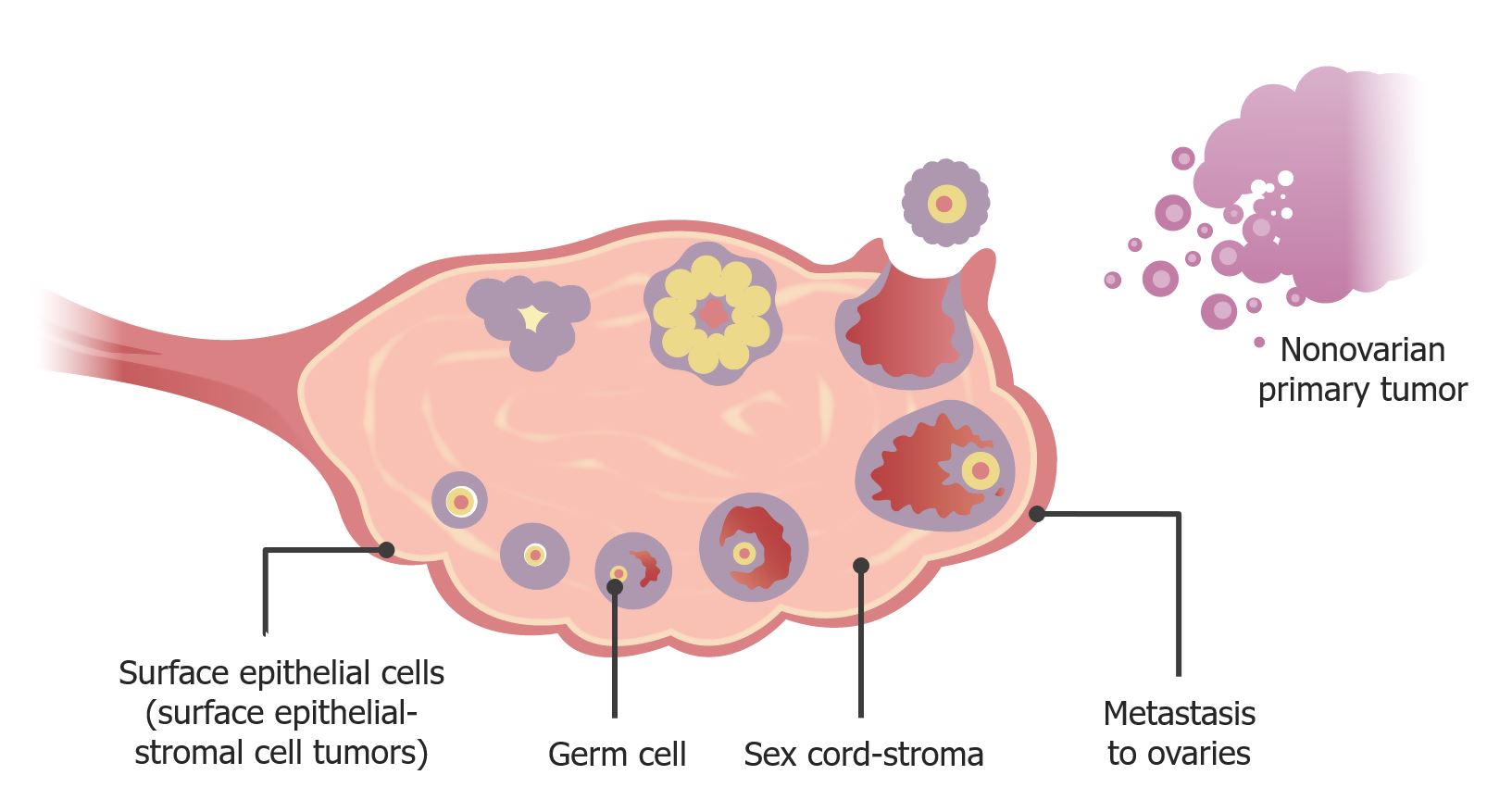
Ovarian tumors are classified according to the type of cell from which they originate:
Epithelial tumors
Germ cell tumors
SexSexThe totality of characteristics of reproductive structure, functions, phenotype, and genotype, differentiating the male from the female organism.Gender Dysphoria cord-stromal tumors
Metastatic tumors (originating in other tissues and metastasizing to the ovary)
There are multiple histologic subtypes within each major tumorTumorInflammation class.
Tumors may be classified as either:
BenignBenignFibroadenoma(adenomas): neoplasmsNeoplasmsNew abnormal growth of tissue. Malignant neoplasms show a greater degree of anaplasia and have the properties of invasion and metastasis, compared to benign neoplasms.Benign Bone Tumors exhibiting abnormal growth but not invading into the surrounding tissue
Borderline/low malignant potential (LMPLMPThe 1st day of a woman’s LMPPregnancy: Diagnosis, Physiology, and Care) tumors: malignant neoplasmsNeoplasmsNew abnormal growth of tissue. Malignant neoplasms show a greater degree of anaplasia and have the properties of invasion and metastasis, compared to benign neoplasms.Benign Bone Tumors that are not very aggressive and have excellent prognosisPrognosisA prediction of the probable outcome of a disease based on a individual’s condition and the usual course of the disease as seen in similar situations.Non-Hodgkin Lymphomas
Malignant (carcinomas or adenocarcinomas): tumors that invade into the surrounding tissue
Image depicting different types of ovarian cancer and their location of origin
Image by Lecturio.
Table: Summary of ovarian cancer classification
Proportion of primary malignant ovarian tumors
Commonly affected age group
Major types
Epithelial tumors
90%
Postmenopausal women
Serous
Mucinous
Endometrioid
Clear cell
Germ cell tumors
5%
10‒30 years
TeratomaTeratomaA true neoplasm composed of a number of different types of tissue, none of which is native to the area in which it occurs. It is composed of tissues that are derived from three germinal layers, the endoderm, mesoderm, and ectoderm. They are classified histologically as mature (benign) or immature (malignant).Imaging of the Mediastinum
Dysgerminoma
Yolk sacYolk SacThe first of four extra-embryonic membranes to form during embryogenesis. In reptiles and birds, it arises from endoderm and mesoderm to incorporate the egg yolk into the digestive tract for nourishing the embryo. In placental mammals, its nutritional function is vestigial; however, it is the source of intestinal mucosa; blood cells; and germ cells. It is sometimes called the vitelline sac, which should not be confused with the vitelline membrane of the egg.Embryoblast and Trophoblast Development tumors
ChoriocarcinomaChoriocarcinomaA malignant metastatic form of trophoblastic tumors. Unlike the hydatidiform mole, choriocarcinoma contains no chorionic villi but rather sheets of undifferentiated cytotrophoblasts and syncytiotrophoblasts (trophoblasts). It is characterized by the large amounts of chorionic gonadotropin produced. Tissue origins can be determined by DNA analyses: placental (fetal) origin or non-placental origin.Gestational Trophoblastic Disease
SexSexThe totality of characteristics of reproductive structure, functions, phenotype, and genotype, differentiating the male from the female organism.Gender Dysphoria cord-stroma
5%
Perimenopausal women
Granulosa cellGranulosa cellSupporting cells for the developing female gamete in the ovary. They are derived from the coelomic epithelial cells of the gonadal ridge. Granulosa cells form a single layer around the oocyte in the primordial ovarian follicle and advance to form a multilayered cumulus oophorus surrounding the ovum in the graafian follicle. The major functions of granulosa cells include the production of steroids and LH receptors.Puberty tumors
Theca cell tumors
Fibroma
Fibrosarcoma
MetastasisMetastasisThe transfer of a neoplasm from one organ or part of the body to another remote from the primary site.Grading, Staging, and Metastasis to ovariesOvariesOvaries are the paired gonads of the female reproductive system that contain haploid gametes known as oocytes. The ovaries are located intraperitoneally in the pelvis, just posterior to the broad ligament, and are connected to the pelvic sidewall and to the uterus by ligaments. These organs function to secrete hormones (estrogen and progesterone) and to produce the female germ cells (oocytes).Ovaries: Anatomy
–
VariableVariableVariables represent information about something that can change. The design of the measurement scales, or of the methods for obtaining information, will determine the data gathered and the characteristics of that data. As a result, a variable can be qualitative or quantitative, and may be further classified into subgroups.Types of Variables
From:
EndometriumEndometriumThe mucous membrane lining of the uterine cavity that is hormonally responsive during the menstrual cycle and pregnancy. The endometrium undergoes cyclic changes that characterize menstruation. After successful fertilization, it serves to sustain the developing embryo.Embryoblast and Trophoblast Development
CervixCervixThe uterus, cervix, and fallopian tubes are part of the internal female reproductive system. The most inferior portion of the uterus is the cervix, which connects the uterine cavity to the vagina. Externally, the cervix is lined by stratified squamous cells; however, the cervical canal is lined by columnar epithelium.Uterus, Cervix, and Fallopian Tubes: Anatomy
Breast
StomachStomachThe stomach is a muscular sac in the upper left portion of the abdomen that plays a critical role in digestion. The stomach develops from the foregut and connects the esophagus with the duodenum. Structurally, the stomach is C-shaped and forms a greater and lesser curvature and is divided grossly into regions: the cardia, fundus, body, and pylorus. Stomach: Anatomy (Krukenberg tumors)
ColonColonThe large intestines constitute the last portion of the digestive system. The large intestine consists of the cecum, appendix, colon (with ascending, transverse, descending, and sigmoid segments), rectum, and anal canal. The primary function of the colon is to remove water and compact the stool prior to expulsion from the body via the rectum and anal canal. Colon, Cecum, and Appendix: Anatomy
Epithelial ovarian tumors
Epithelial ovarian carcinomas (EOCs) originate from surface epithelial cells and account for 90%‒95% of ovarian malignancies. Major types of EOCs include:
Serous adenocarcinoma:
Most common type accounting for 50% of malignant EOCs
Intracellular calciumCalciumA basic element found in nearly all tissues. It is a member of the alkaline earth family of metals with the atomic symbol ca, atomic number 20, and atomic weight 40. Calcium is the most abundant mineral in the body and combines with phosphorus to form calcium phosphate in the bones and teeth. It is essential for the normal functioning of nerves and muscles and plays a role in blood coagulation (as factor IV) and in many enzymatic processes.Electrolytes deposits
Key feature of serous EOCs
Usually high grade at diagnosis
Generally associated with a poor prognosisPrognosisA prediction of the probable outcome of a disease based on a individual’s condition and the usual course of the disease as seen in similar situations.Non-Hodgkin Lymphomas
Complex glandular architecture with back-to-back glandular growth
Better prognosisPrognosisA prediction of the probable outcome of a disease based on a individual’s condition and the usual course of the disease as seen in similar situations.Non-Hodgkin Lymphomas compared with serous types:
Typically low grade at diagnosis
More chemosensitive
Associated with:
Coexisting endometrial adenocarcinoma (15%‒20% of cases)
EndometriosisEndometriosisEndometriosis is a common disease in which patients have endometrial tissue implanted outside of the uterus. Endometrial implants can occur anywhere in the pelvis, including the ovaries, the broad and uterosacral ligaments, the pelvic peritoneum, and the urinary and gastrointestinal tracts.Endometriosis (40%‒50% of cases)
Associated with endometriosisEndometriosisEndometriosis is a common disease in which patients have endometrial tissue implanted outside of the uterus. Endometrial implants can occur anywhere in the pelvis, including the ovaries, the broad and uterosacral ligaments, the pelvic peritoneum, and the urinary and gastrointestinal tracts.Endometriosis
Clinical syndrome of abundant mucoid material in the abdominopelvic cavity
While possible in mucinous EOCs, pseudomyxoma peritonei is typically associated with mucinous tumors of the appendixAppendixA worm-like blind tube extension from the cecum.Colon, Cecum, and Appendix: Anatomy
Less common types of EOC:
Squamous cell carcinomaSquamous cell carcinomaCutaneous squamous cell carcinoma (cSCC) is caused by malignant proliferation of atypical keratinocytes. This condition is the 2nd most common skin malignancy and usually affects sun-exposed areas of fair-skinned patients. The cancer presents as a firm, erythematous, keratotic plaque or papule. Squamous Cell Carcinoma (SCC)
Small cell carcinomaSmall cell carcinomaAn anaplastic, highly malignant, and usually bronchogenic carcinoma composed of small ovoid cells with scanty neoplasm. It is characterized by a dominant, deeply basophilic nucleus, and absent or indistinct nucleoli.Lung Cancer
Transitional cell tumors: Brenner tumors
Mixed carcinoma
Undifferentiated carcinoma
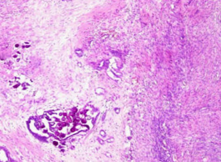
Psammoma bodies in an ovarian adenocarcinoma
Image: “Moderately differentiated ovarian adenocarcinoma” by Pusiol T, Parolari AM, Piscioli I, Morelli L, Del Nonno F, Licci S. License: CC BY 2.0, cropped by Lecturio.
Germ cell tumors
Ovarian germ cell tumors (OGCTs) originate from primordial germ cellsPrimordial germ cellsGametogenesis (i.e., primordial oocytesOocytesFemale germ cells derived from oogonia and termed oocytes when they enter meiosis. The primary oocytes begin meiosis but are arrested at the diplotene state until ovulation at puberty to give rise to haploid secondary oocytes or ova (ovum).Ovaries: Anatomy).
Malignant OGCTs account for 5% of malignant ovarian cancers.
Typically arise in young women aged 10‒30 years
Excellent prognosisPrognosisA prediction of the probable outcome of a disease based on a individual’s condition and the usual course of the disease as seen in similar situations.Non-Hodgkin Lymphomas because:
Most common malignant OGCT accounting for approximately 30% of cases
Less aggressive than many other ovarian cancers
Composed of undifferentiated germ cellsGerm CellsThe reproductive cells in multicellular organisms at various stages during gametogenesis.Gametogenesis
Associated with gonadal dysgenesisGonadal dysgenesisA number of syndromes with defective gonadal developments such as streak gonads and dysgenetic testes or ovaries. The spectrum of gonadal and sexual abnormalities is reflected in their varied sex chromosome (sex chromosomes) constitution as shown by the karyotypes of 45, X monosomy (Turner syndrome); 46, XX (gonadal dysgenesis, 46xx); 46, XY (gonadal dysgenesis, 46, xy); and sex chromosome mosaicism. Their phenotypes range from female, through ambiguous, to male. This concept includes gonadal agenesis.Wilms Tumor (which occurs in phenotypic females with a Y chromosomeY chromosomeThe male sex chromosome, being the differential sex chromosome carried by half the male gametes and none of the female gametes in humans and in some other male-heterogametic species in which the homologue of the X chromosome has been retained.Basic Terms of Genetics):
46XY pure gonadal dysgenesisGonadal dysgenesisA number of syndromes with defective gonadal developments such as streak gonads and dysgenetic testes or ovaries. The spectrum of gonadal and sexual abnormalities is reflected in their varied sex chromosome (sex chromosomes) constitution as shown by the karyotypes of 45, X monosomy (Turner syndrome); 46, XX (gonadal dysgenesis, 46xx); 46, XY (gonadal dysgenesis, 46, xy); and sex chromosome mosaicism. Their phenotypes range from female, through ambiguous, to male. This concept includes gonadal agenesis.Wilms Tumor
45X/46XY Turner-mosaic mixed gonadal dysgenesisGonadal dysgenesisA number of syndromes with defective gonadal developments such as streak gonads and dysgenetic testes or ovaries. The spectrum of gonadal and sexual abnormalities is reflected in their varied sex chromosome (sex chromosomes) constitution as shown by the karyotypes of 45, X monosomy (Turner syndrome); 46, XX (gonadal dysgenesis, 46xx); 46, XY (gonadal dysgenesis, 46, xy); and sex chromosome mosaicism. Their phenotypes range from female, through ambiguous, to male. This concept includes gonadal agenesis.Wilms Tumor
Develop from malignant transformationTransformationChange brought about to an organism’s genetic composition by unidirectional transfer (transfection; transduction, genetic; conjugation, genetic, etc.) and incorporation of foreign DNA into prokaryotic or eukaryotic cells by recombination of part or all of that DNA into the cell’s genome.Bacteriology of gonadoblastomas (benignBenignFibroadenoma OGCTs)
↑ Risk of torsion due to the “lopsided” nature of masses
Develop from malignant transformationTransformationChange brought about to an organism’s genetic composition by unidirectional transfer (transfection; transduction, genetic; conjugation, genetic, etc.) and incorporation of foreign DNA into prokaryotic or eukaryotic cells by recombination of part or all of that DNA into the cell’s genome.Bacteriology of mature teratomas (benignBenignFibroadenoma OGCTs that are also known as dermoid cystsCystsAny fluid-filled closed cavity or sac that is lined by an epithelium. Cysts can be of normal, abnormal, non-neoplastic, or neoplastic tissues.Fibrocystic Change)
Yolk sacYolk SacThe first of four extra-embryonic membranes to form during embryogenesis. In reptiles and birds, it arises from endoderm and mesoderm to incorporate the egg yolk into the digestive tract for nourishing the embryo. In placental mammals, its nutritional function is vestigial; however, it is the source of intestinal mucosa; blood cells; and germ cells. It is sometimes called the vitelline sac, which should not be confused with the vitelline membrane of the egg.Embryoblast and Trophoblast Development tumors:
Previously known as endodermal sinus tumors
Approximately 15% of malignant OGCTs
Derived from cells of the primitive yolk sacYolk SacThe first of four extra-embryonic membranes to form during embryogenesis. In reptiles and birds, it arises from endoderm and mesoderm to incorporate the egg yolk into the digestive tract for nourishing the embryo. In placental mammals, its nutritional function is vestigial; however, it is the source of intestinal mucosa; blood cells; and germ cells. It is sometimes called the vitelline sac, which should not be confused with the vitelline membrane of the egg.Embryoblast and Trophoblast Development
Schiller-Duval bodies: glomerulus-like structure composed of central blood vessels surrounded by germ cellsGerm CellsThe reproductive cells in multicellular organisms at various stages during gametogenesis.Gametogenesis
Most dangerous OGCT
ChoriocarcinomaChoriocarcinomaA malignant metastatic form of trophoblastic tumors. Unlike the hydatidiform mole, choriocarcinoma contains no chorionic villi but rather sheets of undifferentiated cytotrophoblasts and syncytiotrophoblasts (trophoblasts). It is characterized by the large amounts of chorionic gonadotropin produced. Tissue origins can be determined by DNA analyses: placental (fetal) origin or non-placental origin.Gestational Trophoblastic Disease:
Derived from germ cellsGerm CellsThe reproductive cells in multicellular organisms at various stages during gametogenesis.Gametogenesis, but have an appearance similar to gestational choriocarcinomaChoriocarcinomaA malignant metastatic form of trophoblastic tumors. Unlike the hydatidiform mole, choriocarcinoma contains no chorionic villi but rather sheets of undifferentiated cytotrophoblasts and syncytiotrophoblasts (trophoblasts). It is characterized by the large amounts of chorionic gonadotropin produced. Tissue origins can be determined by DNA analyses: placental (fetal) origin or non-placental origin.Gestational Trophoblastic Disease (derived from trophoblastic tissue found in pregnancies)
Worse prognosisPrognosisA prediction of the probable outcome of a disease based on a individual’s condition and the usual course of the disease as seen in similar situations.Non-Hodgkin Lymphomas compared with that of gestational choriocarcinomaChoriocarcinomaA malignant metastatic form of trophoblastic tumors. Unlike the hydatidiform mole, choriocarcinoma contains no chorionic villi but rather sheets of undifferentiated cytotrophoblasts and syncytiotrophoblasts (trophoblasts). It is characterized by the large amounts of chorionic gonadotropin produced. Tissue origins can be determined by DNA analyses: placental (fetal) origin or non-placental origin.Gestational Trophoblastic Disease
Other less common types:
Embryonal carcinoma
Mixed germ cell tumors
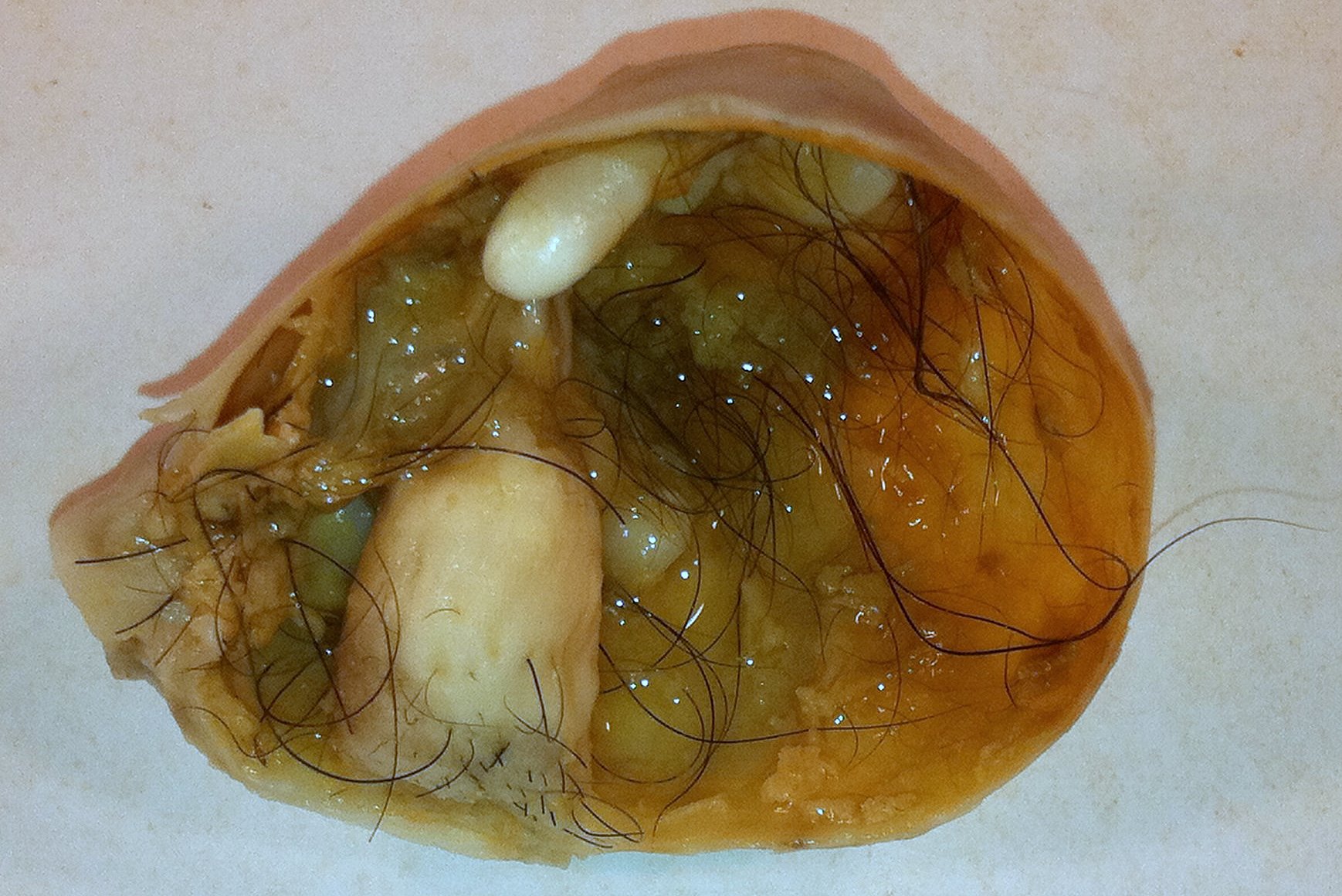
Mature cystic teratoma (benign ovarian germ cell tumor (OGCT)): Note the hair, sebaceous material, and tooth. Mature cystic teratomas are the most common type of benign OGCTs and can undergo malignant transformation into immature teratomas.
Image: “Mature Cystic Teratoma of the Ovary” by Ed Uthman. License: CC BY 2.0
SexSexThe totality of characteristics of reproductive structure, functions, phenotype, and genotype, differentiating the male from the female organism.Gender Dysphoria cord-stromal tumors (SCSTs)
Ovarian SCSTs originate from sexSexThe totality of characteristics of reproductive structure, functions, phenotype, and genotype, differentiating the male from the female organism.Gender Dysphoria cord cells, stromal cells, or both.
Malignant SCSTs account for approximately 5% of malignant ovarian cancers.
Most common in perimenopausal women (average age at diagnosis: 50 years)
Histologic types of SCSTs:
Granulosa cellGranulosa cellSupporting cells for the developing female gamete in the ovary. They are derived from the coelomic epithelial cells of the gonadal ridge. Granulosa cells form a single layer around the oocyte in the primordial ovarian follicle and advance to form a multilayered cumulus oophorus surrounding the ovum in the graafian follicle. The major functions of granulosa cells include the production of steroids and LH receptors.Puberty tumors (often malignant):
90% (most common type) of malignant SCSTs
Made up of granulosa cells (cells that normally line the developing follicles)
Usually secrete estrogens; occasionally secrete androgensAndrogensAndrogens are naturally occurring steroid hormones responsible for development and maintenance of the male sex characteristics, including penile, scrotal, and clitoral growth, development of sexual hair, deepening of the voice, and musculoskeletal growth. Androgens and Antiandrogens → present with hormonally driven symptoms (e.g., abnormal bleeding, virilization)
Call-Exner bodies: small follicles with eosinophilic material
Much rarer than granulosa cellGranulosa cellSupporting cells for the developing female gamete in the ovary. They are derived from the coelomic epithelial cells of the gonadal ridge. Granulosa cells form a single layer around the oocyte in the primordial ovarian follicle and advance to form a multilayered cumulus oophorus surrounding the ovum in the graafian follicle. The major functions of granulosa cells include the production of steroids and LH receptors.Puberty tumors
Composed of theca cellsTheca cellsThe flattened stroma cells forming a sheath or theca outside the basal lamina lining the mature ovarian follicle. Thecal interstitial or stromal cells are steroidogenic, and produce primarily androgens which serve as precursors of estrogens in the granulosa cells.Puberty (supportive cells that produce hormonesHormonesHormones are messenger molecules that are synthesized in one part of the body and move through the bloodstream to exert specific regulatory effects on another part of the body. Hormones play critical roles in coordinating cellular activities throughout the body in response to the constant changes in both the internal and external environments. Hormones: Overview and Types)
Often secrete estrogens and/or androgensAndrogensAndrogens are naturally occurring steroid hormones responsible for development and maintenance of the male sex characteristics, including penile, scrotal, and clitoral growth, development of sexual hair, deepening of the voice, and musculoskeletal growth. Androgens and Antiandrogens
BenignBenignFibroadenomatumorTumorInflammation arising from fibroblastsFibroblastsConnective tissue cells which secrete an extracellular matrix rich in collagen and other macromolecules.Sarcoidosis in connective tissueConnective tissueConnective tissues originate from embryonic mesenchyme and are present throughout the body except inside the brain and spinal cord. The main function of connective tissues is to provide structural support to organs. Connective tissues consist of cells and an extracellular matrix.Connective Tissue: Histology
The most common SCST overall
Fibrosarcoma (malignant):
Malignant soft-tissue tumorTumorInflammation (i.e., sarcoma) arising from fibroblastsFibroblastsConnective tissue cells which secrete an extracellular matrix rich in collagen and other macromolecules.Sarcoidosis in the connective tissueConnective tissueConnective tissues originate from embryonic mesenchyme and are present throughout the body except inside the brain and spinal cord. The main function of connective tissues is to provide structural support to organs. Connective tissues consist of cells and an extracellular matrix.Connective Tissue: Histology
Extremely rare
Sertoli-Leydig cell tumors:
Abnormal cells that are typically only found in men (but found in women with this condition)
Secrete androgensAndrogensAndrogens are naturally occurring steroid hormones responsible for development and maintenance of the male sex characteristics, including penile, scrotal, and clitoral growth, development of sexual hair, deepening of the voice, and musculoskeletal growth. Androgens and Antiandrogens → present with virilization
Ovarian cancer develops as a result of cellular mutations → leads to dysregulated cellular growth → ↑ proliferation → tumorTumorInflammation
Mutations may:
Arise de novo (a majority of mutations)
Be genetically inherited
Some tumors undergo progression from benignBenignFibroadenomacystsCystsAny fluid-filled closed cavity or sac that is lined by an epithelium. Cysts can be of normal, abnormal, non-neoplastic, or neoplastic tissues.Fibrocystic Change → borderline/LMPLMPThe 1st day of a woman’s LMPPregnancy: Diagnosis, Physiology, and Care tumors → invasive carcinoma
Many EOCs are actually believed to originate from the epitheliumEpitheliumThe epithelium is a complex of specialized cellular organizations arranged into sheets and lining cavities and covering the surfaces of the body. The cells exhibit polarity, having an apical and a basal pole. Structures important for the epithelial integrity and function involve the basement membrane, the semipermeable sheet on which the cells rest, and interdigitations, as well as cellular junctions. Surface Epithelium: Histology of adjacent fallopian tubeFallopian TubeA pair of highly specialized canals extending from the uterus to its corresponding ovary. They provide the means for ovum transport from the ovaries and they are the site of the ovum’s final maturation and fertilization. The fallopian tube consists of an interstitium, an isthmus, an ampulla, an infundibulum, and fimbriae. Its wall consists of three layers: serous, muscular, and an internal mucosal layer lined with both ciliated and secretory cells.Uterus, Cervix, and Fallopian Tubes: AnatomyfimbriaeFimbriaeThin, hairlike appendages, 1 to 20 microns in length and often occurring in large numbers, present on the cells of gram-negative bacteria, particularly enterobacteriaceae and Neisseria. Unlike flagella, they do not possess motility, but being protein (pilin) in nature, they possess antigenic and hemagglutinating properties. They are of medical importance because some fimbriae mediate the attachment of bacteria to cells via adhesins. Bacterial fimbriae refer to common pili, to be distinguished from the preferred use of ‘pili’.Escherichia coli (thus, tubal ligationTubal LigationNonhormonal Contraception and salpingectomies ↓ risk of EOC).
Theory: regularRegularInsulinovulationOvulationThe discharge of an ovum from a rupturing follicle in the ovary.Menstrual Cycle over a prolonged period leads to an increased need to repair the surface of the ovary → constant repair, inflammationInflammationInflammation is a complex set of responses to infection and injury involving leukocytes as the principal cellular mediators in the body’s defense against pathogenic organisms. Inflammation is also seen as a response to tissue injury in the process of wound healing. The 5 cardinal signs of inflammation are pain, heat, redness, swelling, and loss of function. Inflammation, and proliferation can lead to the accumulation of genetic mutationsGenetic MutationsCarcinogenesis responsible for cancer development
Some mutations are commonly associated with ovarian cancers, including:
BRCA1 and 2:
Tumor-suppressor genesGenesA category of nucleic acid sequences that function as units of heredity and which code for the basic instructions for the development, reproduction, and maintenance of organisms.DNA Types and Structure involved in DNADNAA deoxyribonucleotide polymer that is the primary genetic material of all cells. Eukaryotic and prokaryotic organisms normally contain DNA in a double-stranded state, yet several important biological processes transiently involve single-stranded regions. DNA, which consists of a polysugar-phosphate backbone possessing projections of purines (adenine and guanine) and pyrimidines (thymine and cytosine), forms a double helix that is held together by hydrogen bonds between these purines and pyrimidines (adenine to thymine and guanine to cytosine).DNA Types and Structure repair
Most commonly inherited mutations associated with ovarian cancer
Inherited in an autosomal dominantAutosomal dominantAutosomal inheritance, both dominant and recessive, refers to the transmission of genes from the 22 autosomal chromosomes. Autosomal dominant diseases are expressed when only 1 copy of the dominant allele is inherited. Autosomal Recessive and Autosomal Dominant Inheritance pattern
BRCA1: approximately 40% lifetime risk of ovarian cancer
BRCA2: approximately 20% lifetime risk of ovarian cancer
P53 (de novo or as part of Li-Fraumeni syndrome)
Some of the mismatch repairMismatch repairA DNA repair pathway involved in correction of errors introduced during DNA replication when an incorrect base, which cannot form hydrogen bonds with the corresponding base in the parent strand, is incorporated into the daughter strand. Exonucleases recognize the base pair mismatch and cause a segment of polynucleotide chain to be excised from the daughter strand, thereby removing the mismatched base.Lynch syndromegenesGenesA category of nucleic acid sequences that function as units of heredity and which code for the basic instructions for the development, reproduction, and maintenance of organisms.DNA Types and Structure that cause Lynch syndromeLynch syndromeLynch syndrome, also called hereditary non-polyposis colorectal cancer (HNPCC), is the most common inherited colon cancer syndrome, and carries a significantly increased risk for endometrial cancer and other malignancies. Lynch syndrome has an autosomal dominant inheritance pattern involving pathogenic variants in one of the mismatch repair (MMR) genes or epithelial cell adhesion molecule (EpCAM). Lynch syndrome:
MLH1
MSH2
MSH6
Other genesGenesA category of nucleic acid sequences that function as units of heredity and which code for the basic instructions for the development, reproduction, and maintenance of organisms.DNA Types and Structure involved in the double-strand break repair system:
MetastasisMetastasisThe transfer of a neoplasm from one organ or part of the body to another remote from the primary site.Grading, Staging, and Metastasis within the peritoneal cavityPeritoneal CavityThe space enclosed by the peritoneum. It is divided into two portions, the greater sac and the lesser sac or omental bursa, which lies behind the stomach. The two sacs are connected by the foramen of winslow, or epiploic foramen.Peritoneum: Anatomy:
On the mesenteryMesenteryA layer of the peritoneum which attaches the abdominal viscera to the abdominal wall and conveys their blood vessels and nerves.Peritoneum: Anatomy and serosa of the bowel
Undersurface of the diaphragmDiaphragmThe diaphragm is a large, dome-shaped muscle that separates the thoracic cavity from the abdominal cavity. The diaphragm consists of muscle fibers and a large central tendon, which is divided into right and left parts. As the primary muscle of inspiration, the diaphragm contributes 75% of the total inspiratory muscle force.Diaphragm: Anatomy or surface of the liverLiverThe liver is the largest gland in the human body. The liver is found in the superior right quadrant of the abdomen and weighs approximately 1.5 kilograms. Its main functions are detoxification, metabolism, nutrient storage (e.g., iron and vitamins), synthesis of coagulation factors, formation of bile, filtration, and storage of blood. Liver: Anatomy
Lymphatic invasion:
Para-aortic lymph nodesLymph NodesThey are oval or bean shaped bodies (1 – 30 mm in diameter) located along the lymphatic system.Lymphatic Drainage System: Anatomy (Recall: Primary blood supply to the ovariesOvariesOvaries are the paired gonads of the female reproductive system that contain haploid gametes known as oocytes. The ovaries are located intraperitoneally in the pelvis, just posterior to the broad ligament, and are connected to the pelvic sidewall and to the uterus by ligaments. These organs function to secrete hormones (estrogen and progesterone) and to produce the female germ cells (oocytes).Ovaries: Anatomy is via the ovarian arteriesArteriesArteries are tubular collections of cells that transport oxygenated blood and nutrients from the heart to the tissues of the body. The blood passes through the arteries in order of decreasing luminal diameter, starting in the largest artery (the aorta) and ending in the small arterioles. Arteries are classified into 3 types: large elastic arteries, medium muscular arteries, and small arteries and arterioles. Arteries: Histology, directly off the aortaAortaThe main trunk of the systemic arteries.Mediastinum and Great Vessels: Anatomy.)
HematogenousHematogenousHepatocellular Carcinoma (HCC) and Liver Metastases dissemination to distant sites (most commonly the lungsLungsLungs are the main organs of the respiratory system. Lungs are paired viscera located in the thoracic cavity and are composed of spongy tissue. The primary function of the lungs is to oxygenate blood and eliminate CO2. Lungs: Anatomy)
Clinical Presentation
Women with early disease (especially EOCs) tend to be asymptomatic or present with vague, nonspecific complaints. Most women with EOCs especially tend to present later with signs/symptoms associated with metastasisMetastasisThe transfer of a neoplasm from one organ or part of the body to another remote from the primary site.Grading, Staging, and Metastasis.
FatigueFatigueThe state of weariness following a period of exertion, mental or physical, characterized by a decreased capacity for work and reduced efficiency to respond to stimuli.Fibromyalgia
Gynecologic symptoms:
Pelvic painPainAn unpleasant sensation induced by noxious stimuli which are detected by nerve endings of nociceptive neurons.Pain: Types and Pathways
DyspareuniaDyspareuniaRecurrent genital pain occurring during, before, or after sexual intercourse in either the male or the female.Primary Ovarian Insufficiency (painPainAn unpleasant sensation induced by noxious stimuli which are detected by nerve endings of nociceptive neurons.Pain: Types and Pathways with intercourse)
Palpable massMassThree-dimensional lesion that occupies a space within the breastImaging of the Breast on pelvic exam
NauseaNauseaAn unpleasant sensation in the stomach usually accompanied by the urge to vomit. Common causes are early pregnancy, sea and motion sickness, emotional stress, intense pain, food poisoning, and various enteroviruses.Antiemetics/vomitingVomitingThe forcible expulsion of the contents of the stomach through the mouth.Hypokalemia
DiarrheaDiarrheaDiarrhea is defined as ≥ 3 watery or loose stools in a 24-hour period. There are a multitude of etiologies, which can be classified based on the underlying mechanism of disease. The duration of symptoms (acute or chronic) and characteristics of the stools (e.g., watery, bloody, steatorrheic, mucoid) can help guide further diagnostic evaluation. Diarrhea or constipationConstipationConstipation is common and may be due to a variety of causes. Constipation is generally defined as bowel movement frequency < 3 times per week. Patients who are constipated often strain to pass hard stools. The condition is classified as primary (also known as idiopathic or functional constipation) or secondary, and as acute or chronic. Constipation
Large palpable massMassThree-dimensional lesion that occupies a space within the breastImaging of the Breast on abdominal exam (especially with mucinous and clear cell EOCs)
AscitesAscitesAscites is the pathologic accumulation of fluid within the peritoneal cavity that occurs due to an osmotic and/or hydrostatic pressure imbalance secondary to portal hypertension (cirrhosis, heart failure) or non-portal hypertension (hypoalbuminemia, malignancy, infection).Ascites
Bowel obstructionBowel obstructionAny impairment, arrest, or reversal of the normal flow of intestinal contents toward the anal canal.Ascaris/Ascariasis
Back painPainAn unpleasant sensation induced by noxious stimuli which are detected by nerve endings of nociceptive neurons.Pain: Types and Pathways
Weakness
Endocrine (common with hormone-secreting SCSTs):
Breast development in young girls/breast growth in adults
Abnormal uterine bleedingAbnormal Uterine BleedingAbnormal uterine bleeding is the medical term for abnormalities in the frequency, volume, duration, and regularity of the menstrual cycle. Abnormal uterine bleeding is classified using the acronym PALM-COEIN, with PALM representing the structural causes and COEIN indicating the non-structural causes. Abnormal Uterine Bleeding
InfertilityInfertilityInfertility is the inability to conceive in the context of regular intercourse. The most common causes of infertility in women are related to ovulatory dysfunction or tubal obstruction, whereas, in men, abnormal sperm is a common cause. Infertility due to anovulationAnovulationSuspension or cessation of ovulation in animals or humans with follicle-containing ovaries (ovarian follicle). Depending on the etiology, ovulation may be induced with appropriate therapy.Polycystic Ovarian Syndrome, as estrogenEstrogenCompounds that interact with estrogen receptors in target tissues to bring about the effects similar to those of estradiol. Estrogens stimulate the female reproductive organs, and the development of secondary female sex characteristics. Estrogenic chemicals include natural, synthetic, steroidal, or non-steroidal compounds.Ovaries: Anatomy and testosteroneTestosteroneA potent androgenic steroid and major product secreted by the leydig cells of the testis. Its production is stimulated by luteinizing hormone from the pituitary gland. In turn, testosterone exerts feedback control of the pituitary LH and FSH secretion. Depending on the tissues, testosterone can be further converted to dihydrotestosterone or estradiol.Androgens and Antiandrogens suppress ovulationOvulationThe discharge of an ovum from a rupturing follicle in the ovary.Menstrual Cycle
Androgenic symptoms:
Acne
HirsutismHirsutismA condition observed in women and children when there is excess coarse body hair of an adult male distribution pattern, such as facial and chest areas. It is the result of elevated androgens from the ovaries, the adrenal glands, or exogenous sources. The concept does not include hypertrichosis, which is an androgen-independent excessive hair growth.Polycystic Ovarian Syndrome
Virilization in young girls
Other signs of advanced disease:
Pleural effusionPleural EffusionPleural effusion refers to the accumulation of fluid between the layers of the parietal and visceral pleura. Common causes of this condition include infection, malignancy, autoimmune disorders, or volume overload. Clinical manifestations include chest pain, cough, and dyspnea. Pleural Effusion
Palpable lymphadenopathyLymphadenopathyLymphadenopathy is lymph node enlargement (> 1 cm) and is benign and self-limited in most patients. Etiologies include malignancy, infection, and autoimmune disorders, as well as iatrogenic causes such as the use of certain medications. Generalized lymphadenopathy often indicates underlying systemic disease. Lymphadenopathy in the groinGroinThe external junctural region between the lower part of the abdomen and the thigh.Male Genitourinary Examination
The diagnosis of ovarian cancer relies upon tissue biopsyBiopsyRemoval and pathologic examination of specimens from the living body.Ewing Sarcoma. The initial detection of cancer is based on physical examination and imaging techniques, including ultrasound.
Physical exam
MassMassThree-dimensional lesion that occupies a space within the breastImaging of the Breast characteristics that are particularly suspicious for ovarian cancer:
Nonmobile (i.e., “fixed”)
Nodular
Grossly enlarged
Exam should include:
Pelvic examination
Rectovaginal examination
Detection of a massMassThree-dimensional lesion that occupies a space within the breastImaging of the Breast is not indicative of malignant cancer, but should prompt imaging.
Imaging
Imaging is typically ordered 1st if signs/symptoms suggestive of a pelvic massMassThree-dimensional lesion that occupies a space within the breastImaging of the Breast are present. Discovery of concerning masses is often 1st identified on imaging and can help narrow differential diagnoses, assess the extent of disease, and assist in treatment planning. Modalities include:
Pelvic MRI: ↑ specificity when ultrasound findings are indeterminate
CT of the abdomen and pelvisPelvisThe pelvis consists of the bony pelvic girdle, the muscular and ligamentous pelvic floor, and the pelvic cavity, which contains viscera, vessels, and multiple nerves and muscles. The pelvic girdle, composed of 2 “hip” bones and the sacrum, is a ring-like bony structure of the axial skeleton that links the vertebral column with the lower extremities.Pelvis: Anatomy:
Imaging findings that are a cause of concern for malignancyMalignancyHemothorax include:
Thick septa, especially if vascularized
Solid components
Wall nodularity/papillary excrescences
AscitesAscitesAscites is the pathologic accumulation of fluid within the peritoneal cavity that occurs due to an osmotic and/or hydrostatic pressure imbalance secondary to portal hypertension (cirrhosis, heart failure) or non-portal hypertension (hypoalbuminemia, malignancy, infection).Ascites
Additional imaging:
Chest X-rayX-rayPenetrating electromagnetic radiation emitted when the inner orbital electrons of an atom are excited and release radiant energy. X-ray wavelengths range from 1 pm to 10 nm. Hard x-rays are the higher energy, shorter wavelength x-rays. Soft x-rays or grenz rays are less energetic and longer in wavelength. The short wavelength end of the x-ray spectrum overlaps the gamma rays wavelength range. The distinction between gamma rays and x-rays is based on their radiation source.Pulmonary Function Tests: to detect an effusion and evaluate for metastases in the lungsLungsLungs are the main organs of the respiratory system. Lungs are paired viscera located in the thoracic cavity and are composed of spongy tissue. The primary function of the lungs is to oxygenate blood and eliminate CO2. Lungs: Anatomy
MammogramMammogramFibrocystic Change: should be up to date, especially in women with estrogen-producing tumors
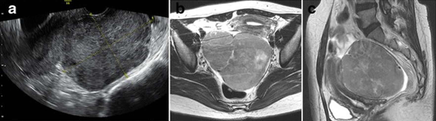
Preoperative transvaginal ultrasonography (a) showing an isoechoic solid mass in the right pelvic cavity (approximately 10 cm in size) with an ill-defined boundary between the lesion and the uterus, and T2-weighted MRI of the ovarian tumor in (b) the horizontal and (c) sagittal planes. The mass was later identified as a fibroma.
Image: “Preoperative transvaginal ultrasonography” by Diagnostic Pathology. License: CC BY 4.0
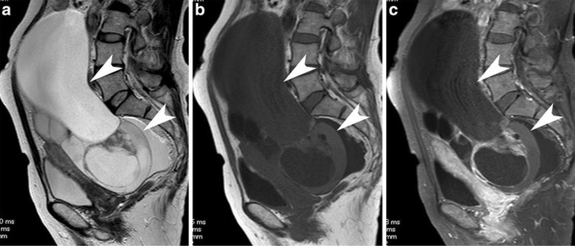
MRI of a mucinous cystadenomcarcinoma demonstrating a classically large multiloculated cystic mass: a: T2- weighted image; after administration of contrast material, we can see some small solid parts along the septa enhanced by contrast material (arrow heads). b: sagittal T1-weighted image c: contrast-enhanced and fat saturated T1-weighted image
Image: “A case with mucinous carcinoma” by Tanaka YO, Okada S, Satoh T, Matsumoto K, Oki A, Saida T, Yoshikawa H, Minami M. License: CC BY 4.0
Laboratory
TumorTumorInflammation markers are generally nonspecific but can aid in diagnosis and assessing treatment response if imaging is suggestive of ovarian cancer. Commonly obtained serum tumorTumorInflammation markers include:
CA-125CA-125A carbohydrate antigen that occurs in tumors of the ovary as well as in breast, kidney, and gastrointestinal tract tumors and normal tissue. While it is tumor-associated, it is not tumor-specific and may have a protective function against particles and infectious agents at mucosal surfaces.Serum Tumor Markers:
Associated with several ovarian tumors, especially epithelial tumors
Only ↑ in 50% of early-stage EOCs
Also ↑ in benignBenignFibroadenoma conditions, including endometriosisEndometriosisEndometriosis is a common disease in which patients have endometrial tissue implanted outside of the uterus. Endometrial implants can occur anywhere in the pelvis, including the ovaries, the broad and uterosacral ligaments, the pelvic peritoneum, and the urinary and gastrointestinal tracts.Endometriosis, pregnancyPregnancyThe status during which female mammals carry their developing young (embryos or fetuses) in utero before birth, beginning from fertilization to birth.Pregnancy: Diagnosis, Physiology, and Care, and cystsCystsAny fluid-filled closed cavity or sac that is lined by an epithelium. Cysts can be of normal, abnormal, non-neoplastic, or neoplastic tissues.Fibrocystic Change
CA19-9CA19-9Sialylated lewis(a) blood group carbohydrate antigen found in many adenocarcinomas of the digestive tract, especially pancreatic tumors.Serum Tumor Markers
Carcinoembryonic antigenCarcinoembryonic antigenA glycoprotein that is secreted into the luminal surface of the epithelia in the gastrointestinal tract. It is found in the feces and pancreaticobiliary secretions and is used to monitor the response to colon cancer treatment.Serum Tumor Markers (CEACEAA glycoprotein that is secreted into the luminal surface of the epithelia in the gastrointestinal tract. It is found in the feces and pancreaticobiliary secretions and is used to monitor the response to colon cancer treatment.Serum Tumor Markers)
α-Fetoprotein (AFPAFPThe first alpha-globulins to appear in mammalian sera during fetal development and the dominant serum proteins in early embryonic life.Hepatocellular Carcinoma (HCC) and Liver Metastases): associated with germ cell tumors
Inhibins A and B: associated with granulosa tumors
β-hCG: ↑ in pregnancyPregnancyThe status during which female mammals carry their developing young (embryos or fetuses) in utero before birth, beginning from fertilization to birth.Pregnancy: Diagnosis, Physiology, and Care and choriocarcinomas
Human epididymisEpididymisThe convoluted cordlike structure attached to the posterior of the testis. Epididymis consists of the head (caput), the body (corpus), and the tail (cauda). A network of ducts leaving the testis joins into a common epididymal tubule proper which provides the transport, storage, and maturation of spermatozoa.Testicles: Anatomy protein 4 (HE4): a new marker associated with epithelial tumors
Histopathology and cytology
Histopathologic evaluation is ultimately required for diagnosis (gold standard).
Histopathology determines the subtype of ovarian cancer.
Recommended:
Specimen is generally obtained during the surgical excision of a concerning massMassThree-dimensional lesion that occupies a space within the breastImaging of the Breast.
LaparotomyLaparotomyIncision into the side of the abdomen between the ribs and pelvis.Laparotomy and Laparoscopy or laparoscopyLaparoscopyLaparoscopy is surgical exploration and interventions performed through small incisions with a camera and long instruments. Laparotomy and Laparoscopy are both acceptable surgical techniques.
Not recommended:
Fine-needle aspiration (FNA)
Percutaneous biopsyBiopsyRemoval and pathologic examination of specimens from the living body.Ewing Sarcoma
Pelvic washings:
Fill the pelvic cavity with fluid, then suction fluid → send for cytology
Ovarian cancer is staged surgically. There are 4 primary stages (each also contains several substages, which are beyond the scope required for medical school).
Since stagingStagingMethods which attempt to express in replicable terms the extent of the neoplasm in the patient.Grading, Staging, and Metastasis depends on the extent of disease and presence of metastasisMetastasisThe transfer of a neoplasm from one organ or part of the body to another remote from the primary site.Grading, Staging, and Metastasis, stagingStagingMethods which attempt to express in replicable terms the extent of the neoplasm in the patient.Grading, Staging, and Metastasis procedures involve evaluation of:
Peritoneal cytology
Multiple peritoneal biopsies
Omental biopsies
Pelvic and para-aortic lymphLymphThe interstitial fluid that is in the lymphatic system.Secondary Lymphatic Organs node sampling
Ovarian cancer stages:
Individuals are staged based on their “highest” findings. For example, an individual with a tumorTumorInflammation confined to the ovary but with positive para-aortic lymph nodesLymph NodesThey are oval or bean shaped bodies (1 – 30 mm in diameter) located along the lymphatic system.Lymphatic Drainage System: Anatomy is classified as stage III.
Table: Summary of ovarian cancer stagingStagingMethods which attempt to express in replicable terms the extent of the neoplasm in the patient.Grading, Staging, and Metastasis
TumorTumorInflammation is confined to the ovariesOvariesOvaries are the paired gonads of the female reproductive system that contain haploid gametes known as oocytes. The ovaries are located intraperitoneally in the pelvis, just posterior to the broad ligament, and are connected to the pelvic sidewall and to the uterus by ligaments. These organs function to secrete hormones (estrogen and progesterone) and to produce the female germ cells (oocytes).Ovaries: Anatomy (may be bilateral).
None
II
Direct tumorTumorInflammation invasion into other tissues within the pelvic cavity (i.e., below the pelvic brim), which typically includes:
UterusUterusThe uterus, cervix, and fallopian tubes are part of the internal female reproductive system. The uterus has a thick wall made of smooth muscle (the myometrium) and an inner mucosal layer (the endometrium). The most inferior portion of the uterus is the cervix, which connects the uterine cavity to the vagina.Uterus, Cervix, and Fallopian Tubes: Anatomy
Fallopian tubesFallopian tubesThe uterus, cervix, and fallopian tubes are part of the internal female reproductive system. The fallopian tubes receive an ovum after ovulation and help move it and/or a fertilized embryo toward the uterus via ciliated cells lining the tubes and peristaltic movements of its smooth muscle. Uterus, Cervix, and Fallopian Tubes: Anatomy
PeritoneumPeritoneumThe peritoneum is a serous membrane lining the abdominopelvic cavity. This lining is formed by connective tissue and originates from the mesoderm. The membrane lines both the abdominal walls (as parietal peritoneum) and all of the visceral organs (as visceral peritoneum).Peritoneum: Anatomy
None
III
TumorTumorInflammation invades outside the pelvisPelvisThe pelvis consists of the bony pelvic girdle, the muscular and ligamentous pelvic floor, and the pelvic cavity, which contains viscera, vessels, and multiple nerves and muscles. The pelvic girdle, composed of 2 “hip” bones and the sacrum, is a ring-like bony structure of the axial skeleton that links the vertebral column with the lower extremities.Pelvis: Anatomy/peritoneal cavityPeritoneal CavityThe space enclosed by the peritoneum. It is divided into two portions, the greater sac and the lesser sac or omental bursa, which lies behind the stomach. The two sacs are connected by the foramen of winslow, or epiploic foramen.Peritoneum: Anatomy, potentially including:
CapsuleCapsuleAn envelope of loose gel surrounding a bacterial cell which is associated with the virulence of pathogenic bacteria. Some capsules have a well-defined border, whereas others form a slime layer that trails off into the medium. Most capsules consist of relatively simple polysaccharides but there are some bacteria whose capsules are made of polypeptides.Bacteroides of the liverLiverThe liver is the largest gland in the human body. The liver is found in the superior right quadrant of the abdomen and weighs approximately 1.5 kilograms. Its main functions are detoxification, metabolism, nutrient storage (e.g., iron and vitamins), synthesis of coagulation factors, formation of bile, filtration, and storage of blood. Liver: Anatomy/spleenSpleenThe spleen is the largest lymphoid organ in the body, located in the LUQ of the abdomen, superior to the left kidney and posterior to the stomach at the level of the 9th-11th ribs just below the diaphragm. The spleen is highly vascular and acts as an important blood filter, cleansing the blood of pathogens and damaged erythrocytes. Spleen: Anatomy
MetastasisMetastasisThe transfer of a neoplasm from one organ or part of the body to another remote from the primary site.Grading, Staging, and Metastasis to extraabdominal organs (e.g., lungsLungsLungs are the main organs of the respiratory system. Lungs are paired viscera located in the thoracic cavity and are composed of spongy tissue. The primary function of the lungs is to oxygenate blood and eliminate CO2. Lungs: Anatomy)
Management
The primary treatment for ovarian cancer is surgery; however, specific treatment depends upon the stage of cancer. Other treatment modalities include chemotherapyChemotherapyOsteosarcoma and immunotherapy.
Surgical management
Surgical excision is typically the treatment of choice.
Procedures may be conducted to remove:
1 or both ovariesOvariesOvaries are the paired gonads of the female reproductive system that contain haploid gametes known as oocytes. The ovaries are located intraperitoneally in the pelvis, just posterior to the broad ligament, and are connected to the pelvic sidewall and to the uterus by ligaments. These organs function to secrete hormones (estrogen and progesterone) and to produce the female germ cells (oocytes).Ovaries: Anatomy
Ipsilateral or bilateral fallopian tubesFallopian tubesThe uterus, cervix, and fallopian tubes are part of the internal female reproductive system. The fallopian tubes receive an ovum after ovulation and help move it and/or a fertilized embryo toward the uterus via ciliated cells lining the tubes and peristaltic movements of its smooth muscle. Uterus, Cervix, and Fallopian Tubes: Anatomy
UterusUterusThe uterus, cervix, and fallopian tubes are part of the internal female reproductive system. The uterus has a thick wall made of smooth muscle (the myometrium) and an inner mucosal layer (the endometrium). The most inferior portion of the uterus is the cervix, which connects the uterine cavity to the vagina.Uterus, Cervix, and Fallopian Tubes: Anatomy
Removal of other tissues (e.g., bowel, bladderBladderA musculomembranous sac along the urinary tract. Urine flows from the kidneys into the bladder via the ureters, and is held there until urination.Pyelonephritis and Perinephric Abscess, liverLiverThe liver is the largest gland in the human body. The liver is found in the superior right quadrant of the abdomen and weighs approximately 1.5 kilograms. Its main functions are detoxification, metabolism, nutrient storage (e.g., iron and vitamins), synthesis of coagulation factors, formation of bile, filtration, and storage of blood. Liver: Anatomy, spleenSpleenThe spleen is the largest lymphoid organ in the body, located in the LUQ of the abdomen, superior to the left kidney and posterior to the stomach at the level of the 9th-11th ribs just below the diaphragm. The spleen is highly vascular and acts as an important blood filter, cleansing the blood of pathogens and damaged erythrocytes. Spleen: Anatomy, omentumOmentumPeritoneum: Anatomy) affected by the tumorTumorInflammation
The goal is to reduce the tumorTumorInflammation burden when complete excision is not possible.
Pelvic washings are usually obtained 1st to help with stagingStagingMethods which attempt to express in replicable terms the extent of the neoplasm in the patient.Grading, Staging, and Metastasis.
Procedures typical for malignant tumors:
Standard procedure is total hysterectomy with BSO and lymphadenectomy.
Fertility-sparing procedures may be considered in young individuals depending on the situation.
Issues for young women:
BSO → surgically-induced menopauseMenopauseMenopause is a physiologic process in women characterized by the permanent cessation of menstruation that occurs after the loss of ovarian activity. Menopause can only be diagnosed retrospectively, after 12 months without menstrual bleeding. Menopause → ↑ risk for early osteoporosisOsteoporosisOsteoporosis refers to a decrease in bone mass and density leading to an increased number of fractures. There are 2 forms of osteoporosis: primary, which is commonly postmenopausal or senile; and secondary, which is a manifestation of immobilization, underlying medical disorders, or long-term use of certain medications. Osteoporosis
Fertility: Egg retrieval can be considered prior to BSO.
Indicated in almost all women except in those with early stage-I disease
Typical regimen:
For EOCs: Taxane-platinum combination
TaxanesTaxanesA group of diterpenoid cyclodecanes named for the taxanes that were discovered in the taxus tree. The action on microtubules has made some of them useful as antineoplastic agents.Microtubule and Topoisomerase Inhibitors: paclitaxelPaclitaxelA cyclodecane isolated from the bark of the pacific yew tree, taxus brevifolia. It stabilizes microtubules in their polymerized form leading to cell death.Microtubule and Topoisomerase Inhibitors (preferred) or docetaxelDocetaxelA semisynthetic analog of paclitaxel used in the treatment of locally advanced or metastatic breast neoplasms and non-small cell lung cancer.Microtubule and Topoisomerase Inhibitors
Platinums: carboplatinCarboplatinAn organoplatinum compound that possesses antineoplastic activity.Alkylating Agents and Platinum (preferred) or cisplatinCisplatinAn inorganic and water-soluble platinum complex. After undergoing hydrolysis, it reacts with DNA to produce both intra and interstrand crosslinks. These cross links appear to impair replication and transcription of DNA. The cytotoxicity of cisplatin correlates with cellular arrest in the g2 phase of the cell cycle.Alkylating Agents and Platinum
For OGCTs and SCSTs: platinum-based therapy, usually bleomycinBleomycinA complex of related glycopeptide antibiotics from streptomyces verticillus consisting of bleomycin a2 and b2. It inhibits DNA metabolism and is used as an antineoplastic, especially for solid tumors.Antitumor Antibiotics + cisplatinCisplatinAn inorganic and water-soluble platinum complex. After undergoing hydrolysis, it reacts with DNA to produce both intra and interstrand crosslinks. These cross links appear to impair replication and transcription of DNA. The cytotoxicity of cisplatin correlates with cellular arrest in the g2 phase of the cell cycle.Alkylating Agents and Platinum + etoposideEtoposideA semisynthetic derivative of podophyllotoxin that exhibits antitumor activity. Etoposide inhibits DNA synthesis by forming a complex with topoisomerase II and DNA. This complex induces breaks in double stranded DNA and prevents repair by topoisomerase II binding. Accumulated breaks in DNA prevent entry into the mitotic phase of cell division, and lead to cell death. Etoposide acts primarily in the g2 and s phases of the cell cycle.Microtubule and Topoisomerase Inhibitors
Timing of administration:
May be given after surgery (termed adjuvantAdjuvantSubstances that augment, stimulate, activate, potentiate, or modulate the immune response at either the cellular or humoral level. The classical agents (freund’s adjuvant, bcg, corynebacterium parvum, et al.) contain bacterial antigens. Some are endogenous (e.g., histamine, interferon, transfer factor, tuftsin, interleukin-1). Their mode of action is either non-specific, resulting in increased immune responsiveness to a wide variety of antigens, or antigen-specific, i.e., affecting a restricted type of immune response to a narrow group of antigens. The therapeutic efficacy of many biological response modifiers is related to their antigen-specific immunoadjuvanticity.Vaccination therapy)
May be given before surgery to reduce the size of a tumorTumorInflammation (termed neoadjuvant therapy)
May be used as primary treatment in individuals who are poor surgical candidates
The exact number and timing of cycles are individualized.
Example: bevacizumabBevacizumabAn anti-vegf humanized murine monoclonal antibody. It inhibits vegf receptors and helps to prevent pathologic angiogenesis.Targeted and Other Nontraditional Antineoplastic Therapy (Avastin), which binds to and inhibits vascular endothelial growth factorVascular endothelial growth factorA family of angiogenic proteins that are closely-related to vascular endothelial growth factor a. They play an important role in the growth and differentiation of vascular as well as lymphatic endothelial cells.Wound Healing (VEGF)
RadiationRadiationEmission or propagation of acoustic waves (sound), electromagnetic energy waves (such as light; radio waves; gamma rays; or x-rays), or a stream of subatomic particles (such as electrons; neutrons; protons; or alpha particles).Osteosarcoma therapy may be considered but is rarely used in ovarian cancer.
Psychosocial support is an important aspect of treatment. Affected individuals should be offered services to improve their qualityQualityActivities and programs intended to assure or improve the quality of care in either a defined medical setting or a program. The concept includes the assessment or evaluation of the quality of care; identification of problems or shortcomings in the delivery of care; designing activities to overcome these deficiencies; and follow-up monitoring to ensure effectiveness of corrective steps.Quality Measurement and Improvement of life via counseling and social support.
Monitor relevant tumorTumorInflammation markers (e.g., CA-125CA-125A carbohydrate antigen that occurs in tumors of the ovary as well as in breast, kidney, and gastrointestinal tract tumors and normal tissue. While it is tumor-associated, it is not tumor-specific and may have a protective function against particles and infectious agents at mucosal surfaces.Serum Tumor Markers for EOC).
Obtain imaging for any concerns about recurrence (e.g., symptoms, ↑ tumorTumorInflammation markers, exam findings).
Recurrent disease is often managed with a repeat surgical procedure and chemotherapyChemotherapyOsteosarcoma.
Prognosis
The prognosisPrognosisA prediction of the probable outcome of a disease based on a individual’s condition and the usual course of the disease as seen in similar situations.Non-Hodgkin Lymphomas depends primarily on the stage at diagnosis and the specific histology. General 5-year survival rates are noted in the table.
Table: PrognosisPrognosisA prediction of the probable outcome of a disease based on a individual’s condition and the usual course of the disease as seen in similar situations.Non-Hodgkin Lymphomas in ovarian cancer
Stage
Epithelial ovarian carcinomas
Ovarian germ cell tumors
SexSexThe totality of characteristics of reproductive structure, functions, phenotype, and genotype, differentiating the male from the female organism.Gender Dysphoria cord-stromal tumors
Stage I
Approximately 85%
Approximately 100%
Approximately 90%‒100%
Stage II
Approximately 70%
Approximately 85%
Approximately 55%‒75%
Stage III
Approximately 40%
Approximately 80%
Approximately 25%‒50% (combined data)
Stage IV
Approximately 20%
Approximately 70%
Differential Diagnosis
The differential diagnosis of a pelvic massMassThree-dimensional lesion that occupies a space within the breastImaging of the Breast includes:
Follicular cystsCystsAny fluid-filled closed cavity or sac that is lined by an epithelium. Cysts can be of normal, abnormal, non-neoplastic, or neoplastic tissues.Fibrocystic Change (physiologic cystsCystsAny fluid-filled closed cavity or sac that is lined by an epithelium. Cysts can be of normal, abnormal, non-neoplastic, or neoplastic tissues.Fibrocystic Change): As an oocyte develops in the 1st half of the menstrual cycleMenstrual cycleThe menstrual cycle is the cyclic pattern of hormonal and tissular activity that prepares a suitable uterine environment for the fertilization and implantation of an ovum. The menstrual cycle involves both an endometrial and ovarian cycle that are dependent on one another for proper functioning. There are 2 phases of the ovarian cycle and 3 phases of the endometrial cycle.Menstrual Cycle, the follicle grows into a small cyst, approximately 2‒3 cm in size, prior to ovulationOvulationThe discharge of an ovum from a rupturing follicle in the ovary.Menstrual Cycle. Occasionally, an oocyte may not ovulate and the follicle can enlarge (typically < 10 cm) and persist. On ultrasound, these follicles appear as simple cystsCystsAny fluid-filled closed cavity or sac that is lined by an epithelium. Cysts can be of normal, abnormal, non-neoplastic, or neoplastic tissues.Fibrocystic Change with smooth walls. The cystsCystsAny fluid-filled closed cavity or sac that is lined by an epithelium. Cysts can be of normal, abnormal, non-neoplastic, or neoplastic tissues.Fibrocystic Change typically resolve spontaneously, although large, persistent cystsCystsAny fluid-filled closed cavity or sac that is lined by an epithelium. Cysts can be of normal, abnormal, non-neoplastic, or neoplastic tissues.Fibrocystic Change may be treated surgically.
Corpus luteal cystsCystsAny fluid-filled closed cavity or sac that is lined by an epithelium. Cysts can be of normal, abnormal, non-neoplastic, or neoplastic tissues.Fibrocystic Change: After ovulationOvulationThe discharge of an ovum from a rupturing follicle in the ovary.Menstrual Cycle, the follicles transform into corpus luteal cystsCystsAny fluid-filled closed cavity or sac that is lined by an epithelium. Cysts can be of normal, abnormal, non-neoplastic, or neoplastic tissues.Fibrocystic Change, secreting both estrogenEstrogenCompounds that interact with estrogen receptors in target tissues to bring about the effects similar to those of estradiol. Estrogens stimulate the female reproductive organs, and the development of secondary female sex characteristics. Estrogenic chemicals include natural, synthetic, steroidal, or non-steroidal compounds.Ovaries: Anatomy and progesteroneProgesteroneThe major progestational steroid that is secreted primarily by the corpus luteum and the placenta. Progesterone acts on the uterus, the mammary glands and the brain. It is required in embryo implantation; pregnancy maintenance, and the development of mammary tissue for milk production. Progesterone, converted from pregnenolone, also serves as an intermediate in the biosynthesis of gonadal steroid hormones and adrenal corticosteroids.Gonadal Hormones. On ultrasound, corpus luteal cystsCystsAny fluid-filled closed cavity or sac that is lined by an epithelium. Cysts can be of normal, abnormal, non-neoplastic, or neoplastic tissues.Fibrocystic Change may show slightly thicker walls and contain some internal debris. The cystsCystsAny fluid-filled closed cavity or sac that is lined by an epithelium. Cysts can be of normal, abnormal, non-neoplastic, or neoplastic tissues.Fibrocystic Change may also enlarge (< 10 cm) but still usually resolve spontaneously. A corpus luteumCorpus LuteumThe yellow body derived from the ruptured ovarian follicle after ovulation. The process of corpus luteum formation, luteinization, is regulated by luteinizing hormone.Ovaries: Anatomy is critical to sustaining an early pregnancyPregnancyThe status during which female mammals carry their developing young (embryos or fetuses) in utero before birth, beginning from fertilization to birth.Pregnancy: Diagnosis, Physiology, and Care and should not be confused with an ectopic pregnancyEctopic pregnancyEctopic pregnancy refers to the implantation of a fertilized egg (embryo) outside the uterine cavity. The main cause is disruption of the normal anatomy of the fallopian tube. Ectopic Pregnancy.
Hemorrhagic cystsCystsAny fluid-filled closed cavity or sac that is lined by an epithelium. Cysts can be of normal, abnormal, non-neoplastic, or neoplastic tissues.Fibrocystic Change: Follicular or corpus luteal cystsCystsAny fluid-filled closed cavity or sac that is lined by an epithelium. Cysts can be of normal, abnormal, non-neoplastic, or neoplastic tissues.Fibrocystic Change bleed occasionally and are known as hemorrhagic cystsCystsAny fluid-filled closed cavity or sac that is lined by an epithelium. Cysts can be of normal, abnormal, non-neoplastic, or neoplastic tissues.Fibrocystic Change. These cystsCystsAny fluid-filled closed cavity or sac that is lined by an epithelium. Cysts can be of normal, abnormal, non-neoplastic, or neoplastic tissues.Fibrocystic Change typically present with sudden-onset pelvic painPainAn unpleasant sensation induced by noxious stimuli which are detected by nerve endings of nociceptive neurons.Pain: Types and Pathways in ovulating women. On ultrasound, they appear as simple cystsCystsAny fluid-filled closed cavity or sac that is lined by an epithelium. Cysts can be of normal, abnormal, non-neoplastic, or neoplastic tissues.Fibrocystic Change with internal echoes (representing blood and clot). Hemorrhagic cystsCystsAny fluid-filled closed cavity or sac that is lined by an epithelium. Cysts can be of normal, abnormal, non-neoplastic, or neoplastic tissues.Fibrocystic Change resolve spontaneously over 1‒2 menstrual cycles. Emergency surgery may be warranted to stop the bleeding if it is significant at presentation.
EndometriosisEndometriosisEndometriosis is a common disease in which patients have endometrial tissue implanted outside of the uterus. Endometrial implants can occur anywhere in the pelvis, including the ovaries, the broad and uterosacral ligaments, the pelvic peritoneum, and the urinary and gastrointestinal tracts.Endometriosis: a condition in which endometrial cells implant outside the uterine cavity. Implants on the ovariesOvariesOvaries are the paired gonads of the female reproductive system that contain haploid gametes known as oocytes. The ovaries are located intraperitoneally in the pelvis, just posterior to the broad ligament, and are connected to the pelvic sidewall and to the uterus by ligaments. These organs function to secrete hormones (estrogen and progesterone) and to produce the female germ cells (oocytes).Ovaries: Anatomy can grow into endometriomas or chocolate cystsChocolate CystsEndometriosis, which are cystsCystsAny fluid-filled closed cavity or sac that is lined by an epithelium. Cysts can be of normal, abnormal, non-neoplastic, or neoplastic tissues.Fibrocystic Change filled with endometrial fluid.Endometriomas and chocolate cystsChocolate CystsEndometriosis appear almost identical to hemorrhagic cystsCystsAny fluid-filled closed cavity or sac that is lined by an epithelium. Cysts can be of normal, abnormal, non-neoplastic, or neoplastic tissues.Fibrocystic Change on ultrasound, but will not resolve spontaneously and must be surgically excised. Affected individuals typically present with dysmenorrhea, dyspareuniaDyspareuniaRecurrent genital pain occurring during, before, or after sexual intercourse in either the male or the female.Primary Ovarian Insufficiency, and/or infertilityInfertilityInfertility is the inability to conceive in the context of regular intercourse. The most common causes of infertility in women are related to ovulatory dysfunction or tubal obstruction, whereas, in men, abnormal sperm is a common cause. Infertility.
Leiomyomas (uterine fibroidsUterine FibroidsGynecological Imaging): common benignBenignFibroadenoma tumors arising from smooth muscle cells in the uterine myometrium. Leiomyomas typically present with abnormal bleeding, pelvic painPainAn unpleasant sensation induced by noxious stimuli which are detected by nerve endings of nociceptive neurons.Pain: Types and Pathways, and/or bulk symptoms. FibroidsFibroidsA benign tumor derived from smooth muscle tissue, also known as a fibroid tumor. They rarely occur outside of the uterus and the gastrointestinal tract but can occur in the skin and subcutaneous tissue, probably arising from the smooth muscle of small blood vessels in these tissues.Infertility are identified as hypoechoicHypoechoicA structure that produces a low-amplitude echo (darker grays)Ultrasound (Sonography), well-circumscribed, round masses on pelvic ultrasound and may be confused with solid adnexal massesAdnexal MassesFemale Genitourinary Examination if they are pedunculated. Management is surgical if the affected individuals are symptomatic.
Ectopic pregnancyEctopic pregnancyEctopic pregnancy refers to the implantation of a fertilized egg (embryo) outside the uterine cavity. The main cause is disruption of the normal anatomy of the fallopian tube. Ectopic Pregnancy:implantationImplantationEndometrial implantation of embryo, mammalian at the blastocyst stage.Fertilization and First Week of the fertilized egg (embryoEmbryoThe entity of a developing mammal, generally from the cleavage of a zygote to the end of embryonic differentiation of basic structures. For the human embryo, this represents the first two months of intrauterine development preceding the stages of the fetus.Fertilization and First Week) outside the uterine cavity. Affected individuals may present with abdominal painAbdominal PainAcute Abdomen and/or bleeding as the developing embryoEmbryoThe entity of a developing mammal, generally from the cleavage of a zygote to the end of embryonic differentiation of basic structures. For the human embryo, this represents the first two months of intrauterine development preceding the stages of the fetus.Fertilization and First Week increases in size. An ectopic pregnancyEctopic pregnancyEctopic pregnancy refers to the implantation of a fertilized egg (embryo) outside the uterine cavity. The main cause is disruption of the normal anatomy of the fallopian tube. Ectopic Pregnancy is diagnosed by pelvic ultrasound and by monitoring β-hCG levels. Ectopic pregnancies are dangerous because a life-threatening hemorrhage may result if the fallopian tubeFallopian TubeA pair of highly specialized canals extending from the uterus to its corresponding ovary. They provide the means for ovum transport from the ovaries and they are the site of the ovum’s final maturation and fertilization. The fallopian tube consists of an interstitium, an isthmus, an ampulla, an infundibulum, and fimbriae. Its wall consists of three layers: serous, muscular, and an internal mucosal layer lined with both ciliated and secretory cells.Uterus, Cervix, and Fallopian Tubes: Anatomy ruptures with the expanding pregnancyPregnancyThe status during which female mammals carry their developing young (embryos or fetuses) in utero before birth, beginning from fertilization to birth.Pregnancy: Diagnosis, Physiology, and Care.
Doubeni, C.A., Doubeni, A.R., Myers, A.E. (2016). Diagnosis and Management of Ovarian Cancer. Am Fam Physician. 93, 937–44. https://pubmed.ncbi.nlm.nih.gov/27281838/
Momenimovahed, Z., Tiznobaik, A., Taheri, S., Salehiniya, H. (2019). Ovarian cancer in the world: Epidemiology and risk factors. Int J Womens Health. 11, 287–99. https://doi.org/10.2147/IJWH.S197604
Committee on the State of the Science in Ovarian Cancer Research; Board on Health Care Services; Institute of Medicine; National Academies of Sciences, Engineering, and Medicine. (2016). Ovarian Cancers: Evolving Paradigms in Research and Care. National Academies Press (US), Washington (DC). https://www.ncbi.nlm.nih.gov/books/NBK367619/
Create your free account or log in to continue reading!