Overview
Definition
Age-related macular degeneration (AMD):
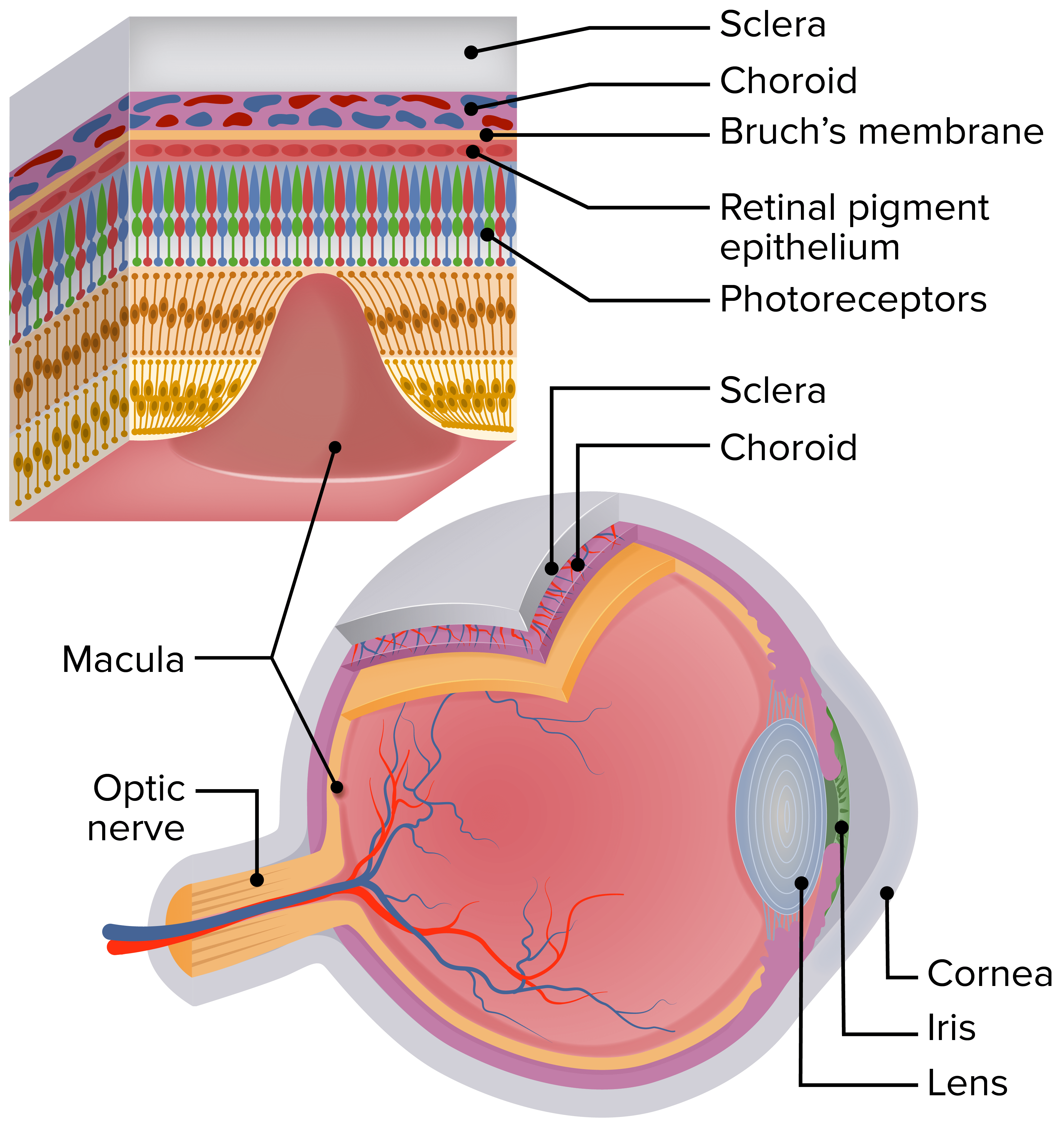
- Degenerative changes involving the macula, the central part of the retina, that cause central vision impairment
- Often affects bilateral vision, but can be asymmetrical
- Peripheral vision is rarely lost.
Anatomy
- Macula:
- An oval area at the center of the retina, where light is focused by the lens and cornea
- Contains the fovea, which holds the highest density of cone cells (photoreceptors sensitive to colors under bright light conditions)
- Function:
- Central, high-acuity, and color vision
- Provides the ability to read, drive, and see in great detail
- Other structures:
- Photoreceptor cells in outer retina: light-sensing rods and cones
- Retinal pigment epithelium (RPE)
- Protects from damaging effects of sunlight
- Eliminates waste products and damaged cells
- Regulates transport of nutrients
- Bruch’s membrane:
- Barrier and filter separating the retina from blood vessels
- A defect can be a site of blood vessel growth.
- Capillary bed in the inner choroid (choriocapillaris):
- Nourishment and oxygen to RPE and outer layers of the retina
- Main blood supply for fovea
Schematic of the human eye showing the location of the macula. The enlarged section of the retina shows the retinal layers and the relationship of the photoreceptors with the RPE, Bruch’s membrane, and the choriocapillaris.
Image: “Schematic diagram of the human eye” by Institute of Human Genetics, Newcastle University, International Centre for Life, Newcastle Upon Tyne NE1 3BZ, United Kingdom. License: CC BY 2.5
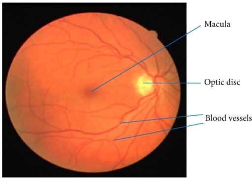
Retinal image showing the macula, blood vessels, and optic disc
Image: “Retinal image showing blood vessels and OD” by Department of ECE, SACS MAVMM Engineering College, Madurai, Tamil Nadu 625 301, India. License: CC BY 3.0
Classification
- Dry AMD (non-exudative or atrophic)
- 90% of all cases
- Often asymptomatic with gradual onset of visual changes
- In a minority of patients: advances to central visual-field loss or wet AMD
- Associated with drusen:
- Deposits of extracellular material or waste beneath the RPE
- Few are found in people > 50 years old as part of aging.
- Increased size and/or number of drusen → AMD
- Wet AMD (exudative or neovascular)
- 10% of cases but accounts for majority of patients with advanced AMD
- Presents as insidious or rapid progression of central vision loss
- Caused by growth of abnormal blood vessels in the subretinal space, frequently from the choroidal circulation (choroidal neovascularization (CNV))
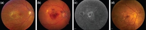
Fundus photographs showing different stages of AMD progression.
Image: “Fundus photographs” by Neurobiology-Neurodegeneration and Repair Laboratory, National Institutes of Health, Bethesda, MD, USA. License: CC BY 2.5
(a) Large and intermediate drusen at intermediate stage of AMD
(b) Neovascular AMD: right eye with subretinal fluid, hemorrhage, and hard exudate in the presence of choroidal neovascularization
(c) Fluorescein angiography of the neovascular AMD: left eye showing hyperfluorescence of the fluorescein angiogram corresponding to the area of the choroidal neovascularization
(d) Central geographic atrophy: right eye with evidence of geographic atrophy involving the center of the fovea with evidence of large drusen temporally
Epidemiology
- The leading cause of irreversible blindness in developed countries
- Worldwide prevalence of 8.7%
- Women are affected more than men.
- Risk factors:
- Advanced age: ↑ incidence in > 60 years of age
- Caucasians > African Americans or Hispanics
- Smoking
- Heavy alcohol use (> 3 drinks per day)
- Family history
- Genetic predisposition (complement H gene)
- Obesity
- Hypercholesterolemia
- Cardiovascular disease
- HIV/AIDS
- Chronic myeloproliferative disease
- Cataract surgery
- Decrease the risk of AMD: ↑ fish intake, ↑ omega-3, and omega-6 polyunsaturated fatty acids
Related videos
Pathophysiology
Dry AMD
- Exact mechanism is still unclear.
- Known risk factors (such as aging and genetics), oxidative stress, and inflammation lead to pathologic changes:
- Aging Bruch’s membrane: membrane thickens and accumulates apolipoproteins → ↑ inflammation and debris → nodular basal deposits become soft drusen
- RPE degeneration → dissociates from Bruch’s membrane → RPE dysfunction → photoreceptor loss
- Loss of choriocapillaris:
- Correlates with ↑ size and number of soft drusen
- Precedes the damage of RPE (geographic atrophy)
Wet AMD
- Risk factors + oxidative stress + activated complement → inflammatory changes
- Inflammation → RPE and other retinal cells secrete vascular endothelial growth factor (VEGF)-A and vasculogenic molecules → CNV and RPE dysfunction
- New immature blood vessels grow from the choroid → perforate Bruch’s membrane and extend toward the retina
- These blood vessels leak fluid and blood below or within the retina → RPE detachment and hemorrhage
Clinical Presentation
Dry AMD
- Symptoms
- Asymptomatic in most cases
- As disease progresses:
- Difficulty with night vision
- Visual fluctuation
- Reading difficulty
- Cannot make out faces
- Prolonged time to adapt to low light levels
- Metamorphopsia (distorted straight lines) in advanced disease
- Signs (funduscopic examination and slit-lamp biomicroscopy)
- Yellow-white deposits (drusen) between Bruch’s membrane and RPE
- RPE pigmentary mottling: increased pigmentation
- Geographic (RPE) atrophy:
- Seen in advanced dry AMD
- Hypopigmentation or depigmentation of the RPE
- Represents cell death in adjacent areas of RPE and photoreceptors
- Stages
- Early stage:
- > 15 small drusen or < 20 medium-sized drusen
- No vision loss
- Intermediate stage:
- At least 1 large druse, ≥ 20 drusen or non-central geographic atrophy (fovea not involved)
- May have vision loss
- Advanced stage:
- Central geographic atrophy (fovea involved)
- Noticeable vision loss
- Early stage:
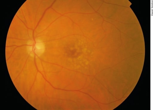
Early AMD: There are irregular pale dots at the macula, which are called drusen. They are caused by a build-up of waste products from photoreceptor metabolism. Although drusen are associated with AMD, most patients with drusen will not develop severe AMD.
Image: “Early AMD” by Africa Regional Medical Advisor: Fred Hollows Foundation, Kigali, Rwanda. License: CC BY 2.0
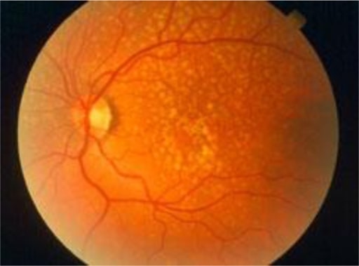
Drusen are yellow deposits under the retina, the light-sensitive tissue at the back of the eye. Drusen consist of lipids and fatty protein.
Image: “Oxidative stress, innate immunity, and age-related macular degeneration” by Department of Ophthalmology and Shiley Eye Institute, University of California San Diego, San Diego, CA, USA. License: CC BY 4.0
Wet AMD
- Symptoms
- Often bilateral, painless central visual distortion or loss
- Acuity below reading and driving level
- Can be gradual depending on pathology
- Subretinal hemorrhage causes acute vision change.
- Metamorphopsia: one of the earliest vision changes
- Scotoma: partial alteration of vision in one spot but with normal surrounding visual field
- Signs (funduscopic examination and slit-lamp biomicroscopy)
- Neovascularization in the macula: gray-green discoloration
- Subretinal hemorrhage
- Retinal pigment epithelial detachments (PEDs): separation of RPE from Bruch’s membrane
- Subretinal lipid deposits on occasions
- Disciform scars
Example of normal vision
Image: “An example of normal vision” by National Eye Institute. License: Public Domain
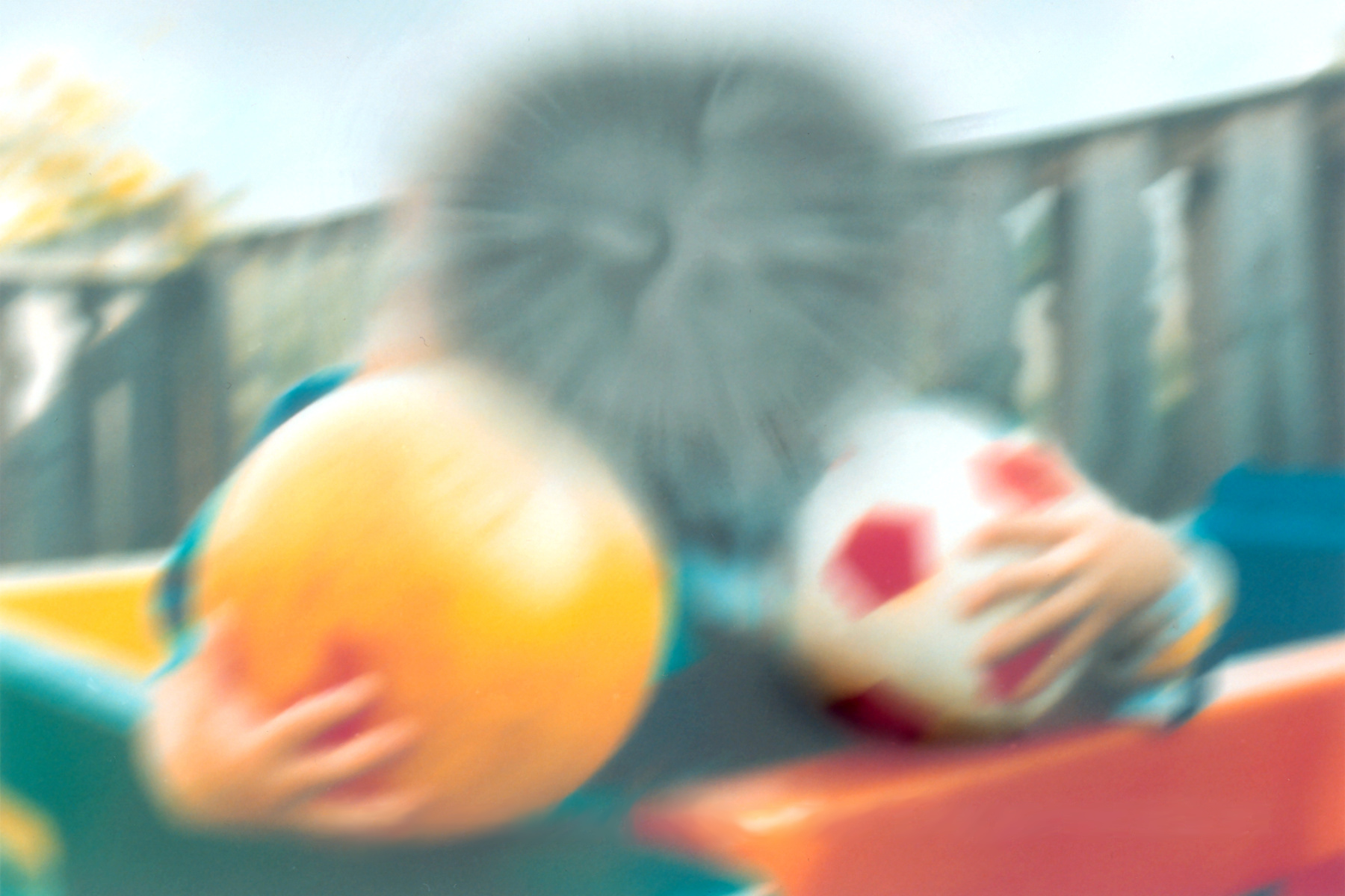
Vision affected by AMD
Image: “The same view with age-related macular degeneration” by National Eye Institute. License: Public Domain
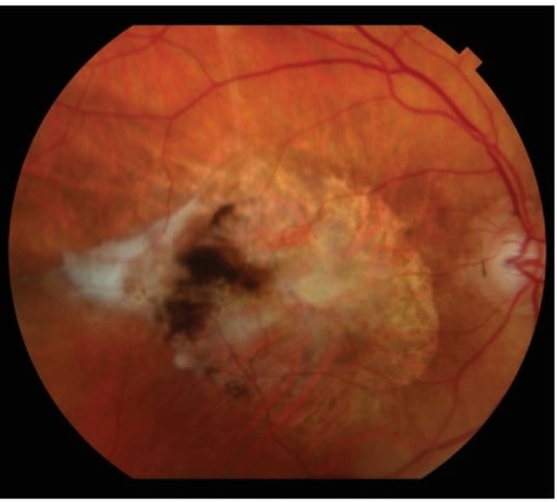
Subretinal fibrotic scarring is the end-stage manifestation of neovascular AMD.
Image: “Subretinal fibrotic scarring” by Retina Service, Department of Ophthalmology, Massachusetts Eye and Ear Infirmary, Harvard Medical School, Boston, MA 02114, USA. License: CC BY 4.0
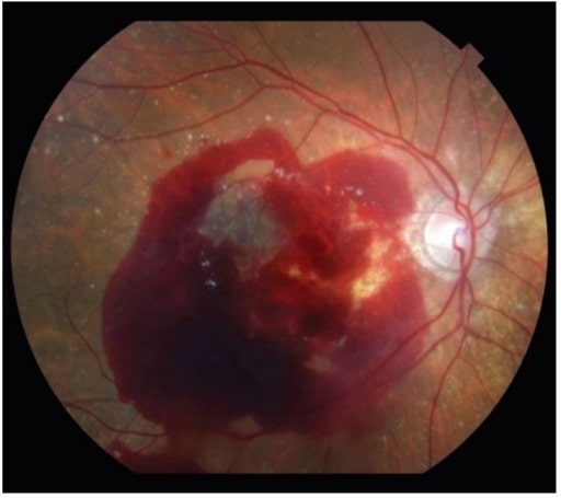
Neovascular AMD can also present with significant retinal hemorrhage.
Image: “Neovascular age-related macular degeneration” by Retina Service, Department of Ophthalmology, Massachusetts Eye and Ear Infirmary, Harvard Medical School, Boston, MA 02114, USA. License: CC BY 4.0
Diagnosis
- Clinical findings, dilated funduscopic examination with slit-lamp biomicroscopy
- Amsler grid test: detects metamorphopsias and scotomas
- Angiography
- Fundus fluorescein angiography (FFA):
- IV injection of fluorescein dye, which follows the course of choroid and retinal vessels
- Hypofluorescence: hemorrhage
- Hyperfluorescence: drusen, RPE atrophy, CNV membranes, serous PEDs
- Indocyanine green angiography
- Uses a water-soluble tricarbocyanine dye
- Detects choroidal neovascularization and PEDs
- Fundus fluorescein angiography (FFA):
- Ocular coherence tomography (OCT)
- Non-invasive imaging test utilizing light waves
- Produces high-resolution cross-sectional retinal images
- Detection of:
- Drusen as nodular elevation of RPE
- Retinal edema and subretinal fluid
- Monitors treatment response in wet AMD
- Color fundus photography: used to monitor AMD
- Fundus autofluorescence
- No contrast
- Identifies lipofuscin accumulation (↑ in cellular aging)
- Defines areas of geographic atrophy
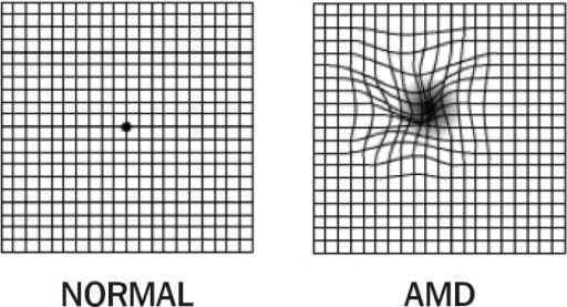
1. Amsler grid: normal vision (left); 2. Amsler grid: AMD with metamorphopsia (right). Note the distorted lines.
Image: “Amsler Grid” by Africa Regional Medical Advisor: Fred Hollows Foundation, Kigali, Rwanda. License: CC BY 2.0
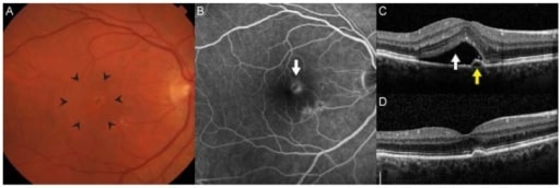
Choroidal neovascularization is the hallmark of neovascular AMD. (A): There is often thickening or elevation of the retina seen clinically through stereoscopic biomicroscopy (area within arrowheads). (B): On fluorescein angiography, neovascular membranes appear as hyperfluorescent lesions deep in the retina (arrow) that leak over time. (C): Spectral-domain optical coherence tomography allows for detailed cross-sectional imaging of retinal anatomy. In this patient, there was subretinal fluid (white arrow) and a small adjacent pigment epithelial detachment. Visual acuity was 20/32. (D): After 3 monthly intravitreal injections of ranibizumab, the fluid resolved, and visual acuity improved to 20/20.
Image: “Age-Related Macular Degeneration” by Retina Service, Department of Ophthalmology, Massachusetts Eye and Ear Infirmary, Harvard Medical School, Boston, MA 02114, USA. License: CC BY 4.0
Management
Dry AMD
- For all patients: smoking cessation
- Monitoring of progression with regular follow-up
- For intermediate AMD and advanced AMD: AREDS 2 (Age-Related Eye Disease Study 2) supplement daily
- Vitamin C 500 mg
- Vitamin E 400 international units (IU)
- Zinc oxide 80 mg
- Cupric oxide 2 mg
- Lutein 10 mg
- Zeaxanthin 2 mg
Wet AMD
- AREDS 2
- Intravitreal anti-VEGF
- First-line therapy; antiangiogenic
- Ranibizumab:
- Recombinant humanized antibody fragment (Fab) that binds VEGF
- 0.5 mg monthly for 3 months
- Bevacizumab:
- Monoclonal antibody against VEGF
- 1.25 mg monthly for 3 months
- Lower cost than ranibizumab
- Aflibercept:
- Recombinant fusion protein that acts as a VEGF receptor decoy
- 2 mg every month for 3 months then every 2 months
- Adverse effects:
- Endophthalmitis
- Increased intraocular pressure
- Retinal detachment
- Thrombotic events reported
- Thermal laser photocoagulation
- Rarely used due to recurrence of disease and poor visual outcomes
- Photodynamic therapy
- Application of light of specific wavelength and IV administration of verteporfin
- Results in slowed CNV progression
- Rarely used but an option in cases of failed anti-VEGF treatment
- Surgery
- Intravitreal tissue plasminogen activator with pneumatic displacement
- Required in rare cases of submacular hemorrhage
Differential Diagnosis
- Cataracts: a decrease in vision due to clouding of the lens, which presents as painless, blurry vision and glare problems. Cataracts are the leading cause of blindness worldwide. The condition can occur at any age, but most cases affect those > 60 years old.
- Diabetic retinopathy: a visual impairment due to microvascular end-organ damage from diabetes mellitus. Diabetic retinopathy is classified as non-proliferative and proliferative retinopathy. Non-proliferative retinopathy is characterized by microaneurysms, intraretinal hemorrhages, exudates, and macular edema. The distinguishing feature of the proliferative type is the presence of retinal or optic disc neovascularization. Patients are initially asymptomatic but in the late stages present with decreased or fluctuating vision, possibly with floaters.
- Retinal detachment: separation of the retina from the retinal pigment epithelium. This results in rapid photoreceptor damage. Symptoms include painless vision changes such as sudden flashes of light, floaters, worsening peripheral vision, or having a shadow in the field of vision. Retinal detachment is a medical emergency requiring corrective surgery.
References
- Ambati, J., Fowler, B. (2012 ) Mechanism of age-related Macular Degeneration. https://pubmed.ncbi.nlm.nih.gov/22794258/
- Arroyo, J., Gardiner, M., Schmader, K., Givens, J. (2020). Age-related macular degeneration: Treatment and prevention. UpToDate. Retrieved September 18, 2020, from https://www.uptodate.com/contents/age-related-macular-degeneration-treatment-and-prevention?search=macular%20degeneration&source=search_result&selectedTitle=1~66&usage_type=default&display_rank=1
- Arroyo, J., Gardiner, M., Schmader, K., Givens, J. (2020). Age-related macular degeneration: Clinical presentation, etiology, and diagnosis. UpToDate. Retrieved September 18, 2020, from https://www.uptodate.com/contents/age-related-macular-degeneration-clinical-presentation-etiology-and-diagnosis?search=macular%20degeneration&source=search_result&selectedTitle=2~66&usage_type=default&display_rank=2
- Handa, J., Rickman, C., Dick, A., Gorin, M., Miller, J., Toth, C.,Ueffing, M., Zarbin, M., Farrer, L. (2019). A systems biology approach towards understanding and treating non-neovascular age-related macular degeneration. Nature 10, 3347. https://doi.org/10.1038/s41467-019-11262-1
- Jonas, JB, Cheung, C., Panda-Jonas, S. (2017). Updates on the Epidemiology of Age-related Macular Degeneration. Asia Pacific J Ophthalmol 6 (6):493-497. doi: 10.22608/APO.2017251
- Maturi, R., Franklin, A. (2018). Nonexudative (Dry) Age-Related Macular Degeneration. Medscape. Retrieved September 19, 2020, from https://emedicine.medscape.com/article/1223154-overview
- Prall, F.R., Ciulla, T., Dahl, A. (2019). Exudative (Wet) Age-Related Macular Degeneration. Medscape. Retrieved September 19, 2020, from https://emedicine.medscape.com/article/1226030-overview