Achieve Mastery of Medical Concepts

Study for medical school and boards with Lecturio

Spinal Stenosis

Spinal stenosis Stenosis Hypoplastic Left Heart Syndrome (HLHS) is the progressive narrowing of the central spinal canal Spinal Canal The cavity within the spinal column through which the spinal cord passes. Spinal Cord Injuries, intervertebral foramen, and lateral recess, leading to compression Compression Blunt Chest Trauma of the nerve root. Spinal stenosis Stenosis Hypoplastic Left Heart Syndrome (HLHS) can occur in the cervical, thoracic, and lumbar spine Spine The human spine, or vertebral column, is the most important anatomical and functional axis of the human body. It consists of 7 cervical vertebrae, 12 thoracic vertebrae, and 5 lumbar vertebrae and is limited cranially by the skull and caudally by the sacrum. Vertebral Column: Anatomy and is commonly caused by degenerative bone Bone Bone is a compact type of hardened connective tissue composed of bone cells, membranes, an extracellular mineralized matrix, and central bone marrow. The 2 primary types of bone are compact and spongy. Bones: Structure and Types disease (mostly affecting the elderly). Patients Patients Individuals participating in the health care system for the purpose of receiving therapeutic, diagnostic, or preventive procedures. Clinician–Patient Relationship present with pain Pain An unpleasant sensation induced by noxious stimuli which are detected by nerve endings of nociceptive neurons. Pain: Types and Pathways, weakness, numbness, and tingling Tingling Posterior Cord Syndrome in the neck Neck The part of a human or animal body connecting the head to the rest of the body. Peritonsillar Abscess or lower back, which may radiate to the legs and buttocks. Clinical examination shows symptom improvement with spinal flexion Flexion Examination of the Upper Limbs and symptom deterioration with spinal extension Extension Examination of the Upper Limbs. The diagnosis is confirmed with MRI. Conservative management includes analgesic/antiinflammatory medications and physiotherapy. Decompression surgery Decompression surgery A surgical operation for the relief of pressure in a body compartment or on a body part. Cranial Nerve Palsies ( laminectomy Laminectomy A surgical procedure that entails removing all (laminectomy) or part (laminotomy) of selected vertebral lamina to relieve pressure on the spinal cord and/or spinal nerve roots. Vertebral lamina is the thin flattened posterior wall of vertebral arch that forms the vertebral foramen through which pass the spinal cord and nerve roots. Neurosurgery) is considered in advanced cases, or if conservative management fails.

Last updated: Mar 29, 2023

Editorial responsibility: Stanley Oiseth, Lindsay Jones, Evelin Maza

Overview

Definition

Epidemiology

  • Incidence Incidence The number of new cases of a given disease during a given period in a specified population. It also is used for the rate at which new events occur in a defined population. It is differentiated from prevalence, which refers to all cases in the population at a given time. Measures of Disease Frequency: 5 per 1,000 individuals > 50 years of age
  • Most common age groups: middle and elderly
  • More common in women
  • Risk factors:
    • Obesity Obesity Obesity is a condition associated with excess body weight, specifically with the deposition of excessive adipose tissue. Obesity is considered a global epidemic. Major influences come from the western diet and sedentary lifestyles, but the exact mechanisms likely include a mixture of genetic and environmental factors. Obesity
    • Family history Family History Adult Health Maintenance
    • Trauma

Etiology

  • Acquired causes:
    • Degenerative changes (e.g., spondylosis Spondylosis A degenerative spinal disease that can involve any part of the vertebra, the intervertebral disk, and the surrounding soft tissue. Central Cord Syndrome, degenerative arthritis Arthritis Acute or chronic inflammation of joints. Osteoarthritis)
    • Trauma
    • Iatrogenic Iatrogenic Any adverse condition in a patient occurring as the result of treatment by a physician, surgeon, or other health professional, especially infections acquired by a patient during the course of treatment. Anterior Cord Syndrome (postoperative)
    • Lipoma Lipoma A lipoma is a benign neoplasm of fat cells (adipocytes) and the most common soft tissue tumor in adults. The etiology is unknown, but obesity is a predisposing factor; genetics also play a role, with multiple lipomas occurring in various inherited disorders. Lipoma, synovial cysts Cysts Any fluid-filled closed cavity or sac that is lined by an epithelium. Cysts can be of normal, abnormal, non-neoplastic, or neoplastic tissues. Fibrocystic Change, neural cysts Cysts Any fluid-filled closed cavity or sac that is lined by an epithelium. Cysts can be of normal, abnormal, non-neoplastic, or neoplastic tissues. Fibrocystic Change, and neoplasms Neoplasms New abnormal growth of tissue. Malignant neoplasms show a greater degree of anaplasia and have the properties of invasion and metastasis, compared to benign neoplasms. Benign Bone Tumors
    • Systemic disorders (e.g., Paget disease, ankylosing spondylitis Ankylosing spondylitis Ankylosing spondylitis (also known as Bechterew’s disease or Marie-Strümpell disease) is a seronegative spondyloarthropathy characterized by chronic and indolent inflammation of the axial skeleton. Severe disease can lead to fusion and rigidity of the spine. Ankylosing Spondylitis)
  • Congenital Congenital Chorioretinitis causes:

Pathophysiology

Clinical Presentation

  • Bilateral pain Pain An unpleasant sensation induced by noxious stimuli which are detected by nerve endings of nociceptive neurons. Pain: Types and Pathways (most commonly in the lower back)
  • Radiation Radiation Emission or propagation of acoustic waves (sound), electromagnetic energy waves (such as light; radio waves; gamma rays; or x-rays), or a stream of subatomic particles (such as electrons; neutrons; protons; or alpha particles). Osteosarcoma to the buttocks and/or legs
  • Associated symptoms:
    • Cramping
    • Numbness
    • Weakness
    • Paresthesia
  • Symptoms ↓ with spinal flexion Flexion Examination of the Upper Limbs:
    • Widens the spinal canal Spinal Canal The cavity within the spinal column through which the spinal cord passes. Spinal Cord Injuries
    • Examples:
      • Sitting
      • Cycling
      • Walking uphill
      • Bending forward
  • Symptoms ↑ with spinal extension Extension Examination of the Upper Limbs:
    • Narrows the spinal canal Spinal Canal The cavity within the spinal column through which the spinal cord passes. Spinal Cord Injuries
    • Examples:
      • Standing
      • Walking downhill

Diagnosis and Management

Diagnosis

  • Detailed history and physical examination is necessary to localize the level of spine Spine The human spine, or vertebral column, is the most important anatomical and functional axis of the human body. It consists of 7 cervical vertebrae, 12 thoracic vertebrae, and 5 lumbar vertebrae and is limited cranially by the skull and caudally by the sacrum. Vertebral Column: Anatomy affected. 
  • Imaging:
    • MRI: the diagnostic gold standard (CT alternatively)
    • X-ray X-ray Penetrating electromagnetic radiation emitted when the inner orbital electrons of an atom are excited and release radiant energy. X-ray wavelengths range from 1 pm to 10 nm. Hard x-rays are the higher energy, shorter wavelength x-rays. Soft x-rays or grenz rays are less energetic and longer in wavelength. The short wavelength end of the x-ray spectrum overlaps the gamma rays wavelength range. The distinction between gamma rays and x-rays is based on their radiation source. Pulmonary Function Tests: to assess for degenerative bone Bone Bone is a compact type of hardened connective tissue composed of bone cells, membranes, an extracellular mineralized matrix, and central bone marrow. The 2 primary types of bone are compact and spongy. Bones: Structure and Types changes 
  • Nerve conduction studies: to differentiate lumbar stenosis Stenosis Hypoplastic Left Heart Syndrome (HLHS) from lumbosacral plexopathy Plexopathy Neuropathy is a nerve pathology presenting with sensory, motor, or autonomic impairment secondary to dysfunction of the affected nerve. The peripheral nerves are derived from several plexuses, with the brachial and lumbosacral plexuses supplying the major innervation to the extremities. Mononeuropathy (affecting a single nerve) and plexopathy (affecting the plexus) can occur from trauma, compression, and systemic diseases. Mononeuropathy and Plexopathy and generalized peripheral neuropathy Neuropathy Leprosy
Spinal stenosis on mri

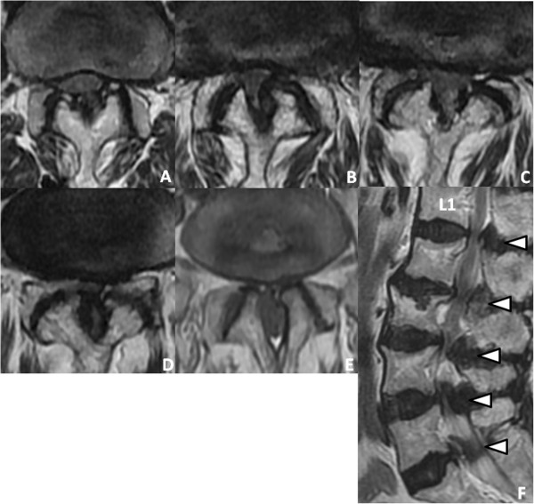
Magnetic resonance imaging (MRI) showing extensive spinal stenosis:
(A): L1–L2
(B): L2–L3
(C): L3–L4
(D): L4–L5
(E): L5–S1
(F): Sagittal T2-weighted image reveals stenosis from L1–L2 through L5–S1 (arrowheads).

Image: “Preoperative magnetic resonance images” by Saito K, Miyakoshi N, Hongo M, Kasukawa Y, Ishikawa Y, Shimada Y. License: CC BY 2.0

Management

The goal of spinal stenosis Stenosis Hypoplastic Left Heart Syndrome (HLHS) management is to reduce symptoms and increase the individual’s functional status. 

Conservative management: 

Surgery:

  • Failure of conservative therapy
  • Presence of significant myelopathy, radiculopathy Radiculopathy Disease involving a spinal nerve root which may result from compression related to intervertebral disk displacement; spinal cord injuries; spinal diseases; and other conditions. Clinical manifestations include radicular pain, weakness, and sensory loss referable to structures innervated by the involved nerve root. Rheumatoid Arthritis, and/or neurogenic claudication
  • Laminectomy Laminectomy A surgical procedure that entails removing all (laminectomy) or part (laminotomy) of selected vertebral lamina to relieve pressure on the spinal cord and/or spinal nerve roots. Vertebral lamina is the thin flattened posterior wall of vertebral arch that forms the vertebral foramen through which pass the spinal cord and nerve roots. Neurosurgery is the most frequently employed surgery.

Differential Diagnosis

  • Spinal disk herniation Herniation Omphalocele: the expulsion of the nucleus pulposus Nucleus Pulposus Fibrocartilage inner core of the intervertebral disc. Prolapsed or bulged nucleus pulposus leads to intervertebral disc displacement while proliferation of cells in the nucleus pulposus is associated with intervertebral disc degeneration. Spinal Disk Herniation through a perforation Perforation A pathological hole in an organ, blood vessel or other soft part of the body, occurring in the absence of external force. Esophagitis in the anulus fibrosus of the intervertebral disc Intervertebral disc Any of the 23 plates of fibrocartilage found between the bodies of adjacent vertebrae. Vertebral Column: Anatomy. Spinal disk herniation Herniation Omphalocele is most commonly caused by degenerative disk disease Disk Disease Examination of the Lower Limbs. Clinical presentation includes pain Pain An unpleasant sensation induced by noxious stimuli which are detected by nerve endings of nociceptive neurons. Pain: Types and Pathways, paresthesia, and, in severe cases, muscle weakness and bladder Bladder A musculomembranous sac along the urinary tract. Urine flows from the kidneys into the bladder via the ureters, and is held there until urination. Pyelonephritis and Perinephric Abscess/bowel dysfunction. Diagnosis is clinical and can be confirmed with diagnostic imaging (i.e., MRI). Depending on the severity of the symptoms, management‌ ‌can be conservative or surgical. 
  • Radiculitis Radiculitis Ankylosing Spondylitis: the compression Compression Blunt Chest Trauma of a nerve root exiting the spinal column Spinal column The human spine, or vertebral column, is the most important anatomical and functional axis of the human body. It consists of 7 cervical vertebrae, 12 thoracic vertebrae, and 5 lumbar vertebrae and is limited cranially by the skull and caudally by the sacrum. Vertebral Column: Anatomy. Radiculitis Radiculitis Ankylosing Spondylitis is caused by either a bacterial/viral infection (mainly borreliosis or herpes zoster Herpes Zoster Varicella-zoster virus (VZV) is a linear, double-stranded DNA virus in the Herpesviridae family. Shingles (also known as herpes zoster) is more common in adults and occurs due to the reactivation of VZV. Varicella-Zoster Virus/Chickenpox) or autoimmune/cryptogenic inflammation Inflammation Inflammation is a complex set of responses to infection and injury involving leukocytes as the principal cellular mediators in the body’s defense against pathogenic organisms. Inflammation is also seen as a response to tissue injury in the process of wound healing. The 5 cardinal signs of inflammation are pain, heat, redness, swelling, and loss of function. Inflammation. Individuals present with pain Pain An unpleasant sensation induced by noxious stimuli which are detected by nerve endings of nociceptive neurons. Pain: Types and Pathways radiating from the affected nerve to the spinal cord Spinal cord The spinal cord is the major conduction pathway connecting the brain to the body; it is part of the CNS. In cross section, the spinal cord is divided into an H-shaped area of gray matter (consisting of synapsing neuronal cell bodies) and a surrounding area of white matter (consisting of ascending and descending tracts of myelinated axons). Spinal Cord: Anatomy without any muscle weakness. Diagnostic studies and management depend on the suspected etiology.
  • Lumbar compression Compression Blunt Chest Trauma fracture Fracture A fracture is a disruption of the cortex of any bone and periosteum and is commonly due to mechanical stress after an injury or accident. Open fractures due to trauma can be a medical emergency. Fractures are frequently associated with automobile accidents, workplace injuries, and trauma. Overview of Bone Fractures: may occur as a result of trauma or pathological weakening of the bone Bone Bone is a compact type of hardened connective tissue composed of bone cells, membranes, an extracellular mineralized matrix, and central bone marrow. The 2 primary types of bone are compact and spongy. Bones: Structure and Types. Midline back pain Pain An unpleasant sensation induced by noxious stimuli which are detected by nerve endings of nociceptive neurons. Pain: Types and Pathways is the hallmark of lumbar compression Compression Blunt Chest Trauma fracture Fracture A fracture is a disruption of the cortex of any bone and periosteum and is commonly due to mechanical stress after an injury or accident. Open fractures due to trauma can be a medical emergency. Fractures are frequently associated with automobile accidents, workplace injuries, and trauma. Overview of Bone Fractures. X-ray X-ray Penetrating electromagnetic radiation emitted when the inner orbital electrons of an atom are excited and release radiant energy. X-ray wavelengths range from 1 pm to 10 nm. Hard x-rays are the higher energy, shorter wavelength x-rays. Soft x-rays or grenz rays are less energetic and longer in wavelength. The short wavelength end of the x-ray spectrum overlaps the gamma rays wavelength range. The distinction between gamma rays and x-rays is based on their radiation source. Pulmonary Function Tests, CT, or MRI is used for diagnosis. Decompression surgery Decompression surgery A surgical operation for the relief of pressure in a body compartment or on a body part. Cranial Nerve Palsies is often performed along with physiotherapy to provide long-term care. 
  • Rheumatoid arthritis Arthritis Acute or chronic inflammation of joints. Osteoarthritis: both a symmetric, inflammatory polyarthritis Polyarthritis Rheumatoid Arthritis and a chronic, progressive autoimmune disorder Autoimmune Disorder Septic Arthritis. Rheumatoid arthritis Arthritis Acute or chronic inflammation of joints. Osteoarthritis occurs most commonly in middle-aged women with joint swelling Swelling Inflammation, pain Pain An unpleasant sensation induced by noxious stimuli which are detected by nerve endings of nociceptive neurons. Pain: Types and Pathways, and morning stiffness. Prolonged and severe disease can lead to irreversible joint deformities. Diagnosis is based on strong clinical suspicion and confirmed by blood tests (e.g., rheumatoid factor Rheumatoid factor Antibodies found in adult rheumatoid arthritis patients that are directed against gamma-chain immunoglobulins. Autoimmune Hepatitis, anti-cyclic citrullinated peptide antibodies Antibodies Immunoglobulins (Igs), also known as antibodies, are glycoprotein molecules produced by plasma cells that act in immune responses by recognizing and binding particular antigens. The various Ig classes are IgG (the most abundant), IgM, IgE, IgD, and IgA, which differ in their biologic features, structure, target specificity, and distribution. Immunoglobulins: Types and Functions) and imaging. Management involves long-term disease-modifying antirheumatic drugs Disease-modifying antirheumatic drugs Disease-modifying antirheumatic drugs are antiinflammatory medications used to manage rheumatoid arthritis. The medications slow, but do not cure, the progression of the disease. The medications are classified as either synthetic or biologic agents and each has unique mechanisms of action and side effects. Disease-Modifying Antirheumatic Drugs (DMARDs), biologic agents Biologic Agents Immunosuppressants, and physical therapy Physical Therapy Becker Muscular Dystrophy
  • Osteoarthritis Osteoarthritis Osteoarthritis (OA) is the most common form of arthritis, and is due to cartilage destruction and changes of the subchondral bone. The risk of developing this disorder increases with age, obesity, and repetitive joint use or trauma. Patients develop gradual joint pain, stiffness lasting < 30 minutes, and decreased range of motion. Osteoarthritis: degenerative joint disease occurring most frequently in the hands, hips, and knees. Individuals present with joint pain Pain An unpleasant sensation induced by noxious stimuli which are detected by nerve endings of nociceptive neurons. Pain: Types and Pathways, stiffness lasting < 30 minutes, and decreased range of motion Range of motion The distance and direction to which a bone joint can be extended. Range of motion is a function of the condition of the joints, muscles, and connective tissues involved. Joint flexibility can be improved through appropriate muscle strength exercises. Examination of the Upper Limbs. Physical exam may reveal crepitus Crepitus Osteoarthritis with joint motion and Heberden/Bouchard nodes. The diagnosis is confirmed by radiographic joint findings. Management includes conservative measures, analgesic medications, glucocorticoid intra-articular injections, and surgery for advanced disease.

References

  1. Levin, K. (2019). Lumbar spinal stenosis: Pathophysiology, clinical features, and diagnosis. UpToDate. Retrieved August 16, 2021, from https://www.uptodate.com/contents/lumbar-spinal-stenosis-pathophysiology-clinical-features-and-diagnosis
  2. Raja, A., Hoang, S., Patel, P., Mesfin, F. B. (2021). Spinal Stenosis. https://www.ncbi.nlm.nih.gov/books/NBK441989/
  3. Lee, B. H., Moon, S. H., Suk, K. S., Kim, H. S., Yang, J. H. (2020). Lumbar Spinal Stenosis: Pathophysiology and Treatment Principle: A Narrative Review. https://www.ncbi.nlm.nih.gov/pmc/articles/PMC7595829/
  4. Wu, L., Cruz, R. (2020). Lumbar Spinal Stenosis. https://www.ncbi.nlm.nih.gov/books/NBK531493/
  5. Katz, J. N., & Harris, M. B. (2008). Clinical practice. Lumbar spinal stenosis. The New England journal of medicine. 358(8), 818–825. https://doi.org/10.1056/NEJMcp0708097

Create your free account or log in to continue reading!

Sign up now and get free access to Lecturio with concept pages, medical videos, and questions for your medical education.

User Reviews

Details