Examination of the lower limbs involves assessment of the hips, knees, ankles, and feet to evaluate for signs of pathology. The examination includes inspection, palpation, assessment of range of movement, and provocative maneuvers. A good history should be taken and concurrently used with the exam findings to obtain a presumptive diagnosis.
Briefly explain each step of the examination to the individual and obtain consent.
Place the individual in the appropriate position.
Expose the legLegThe lower leg, or just “leg” in anatomical terms, is the part of the lower limb between the knee and the ankle joint. The bony structure is composed of the tibia and fibula bones, and the muscles of the leg are grouped into the anterior, lateral, and posterior compartments by extensions of fascia.Leg: Anatomy completely, especially the thighThighThe thigh is the region of the lower limb found between the hip and the knee joint. There is a single bone in the thigh called the femur, which is surrounded by large muscles grouped into 3 fascial compartments. Thigh: Anatomy, knee, ankle, and footFootThe foot is the terminal portion of the lower limb, whose primary function is to bear weight and facilitate locomotion. The foot comprises 26 bones, including the tarsal bones, metatarsal bones, and phalanges. The bones of the foot form longitudinal and transverse arches and are supported by various muscles, ligaments, and tendons.Foot: Anatomy.
Note any legLegThe lower leg, or just “leg” in anatomical terms, is the part of the lower limb between the knee and the ankle joint. The bony structure is composed of the tibia and fibula bones, and the muscles of the leg are grouped into the anterior, lateral, and posterior compartments by extensions of fascia.Leg: Anatomylength discrepancyLength DiscrepancyBlount’s Disease.
Test both active and passive range of motionRange of motionThe distance and direction to which a bone joint can be extended. Range of motion is a function of the condition of the joints, muscles, and connective tissues involved. Joint flexibility can be improved through appropriate muscle strength exercises.Examination of the Upper Limbs.
Perform special tests.
Test strength against resistanceResistancePhysiologically, the opposition to flow of air caused by the forces of friction. As a part of pulmonary function testing, it is the ratio of driving pressure to the rate of air flow.Ventilation: Mechanics of Breathing.
ThighThighThe thigh is the region of the lower limb found between the hip and the knee joint. There is a single bone in the thigh called the femur, which is surrounded by large muscles grouped into 3 fascial compartments. Thigh: Anatomy
LegLegThe lower leg, or just “leg” in anatomical terms, is the part of the lower limb between the knee and the ankle joint. The bony structure is composed of the tibia and fibula bones, and the muscles of the leg are grouped into the anterior, lateral, and posterior compartments by extensions of fascia.Leg: Anatomy
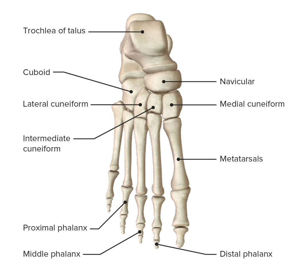
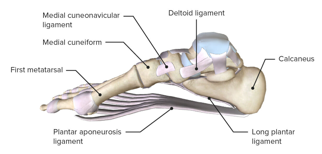
FootFootThe foot is the terminal portion of the lower limb, whose primary function is to bear weight and facilitate locomotion. The foot comprises 26 bones, including the tarsal bones, metatarsal bones, and phalanges. The bones of the foot form longitudinal and transverse arches and are supported by various muscles, ligaments, and tendons.Foot: Anatomy
The joints involved include:
Hip jointHip jointThe hip joint is a ball-and-socket joint formed by the head of the femur and the acetabulum of the pelvis. The hip joint is the most stable joint in the body and is supported by a very strong capsule and several ligaments, allowing the joint to sustain forces that can be multiple times the total body weight. Hip Joint: Anatomy
Knee jointKnee jointThe knee joint is made up of the articulations between the femur, tibia, and patella bones, and is one of the largest and most complex joints of the human body. The knee is classified as a synovial hinge joint, which primarily allows for flexion and extension with a more limited degree of translation and rotation. Knee Joint: Anatomy
Ankle jointAnkle jointThe ankle is a hinged synovial joint formed between the articular surfaces of the distal tibia, distal fibula, and talus. The ankle primarily allows plantar flexion and dorsiflexion of the foot.
These joints are surrounded and supported by many muscles, tendons, ligaments, and fibrocartilaginous structures to ensure support and stability and to absorb shockShockShock is a life-threatening condition associated with impaired circulation that results in tissue hypoxia. The different types of shock are based on the underlying cause: distributive (↑ cardiac output (CO), ↓ systemic vascular resistance (SVR)), cardiogenic (↓ CO, ↑ SVR), hypovolemic (↓ CO, ↑ SVR), obstructive (↓ CO), and mixed. Types of Shock during locomotion.
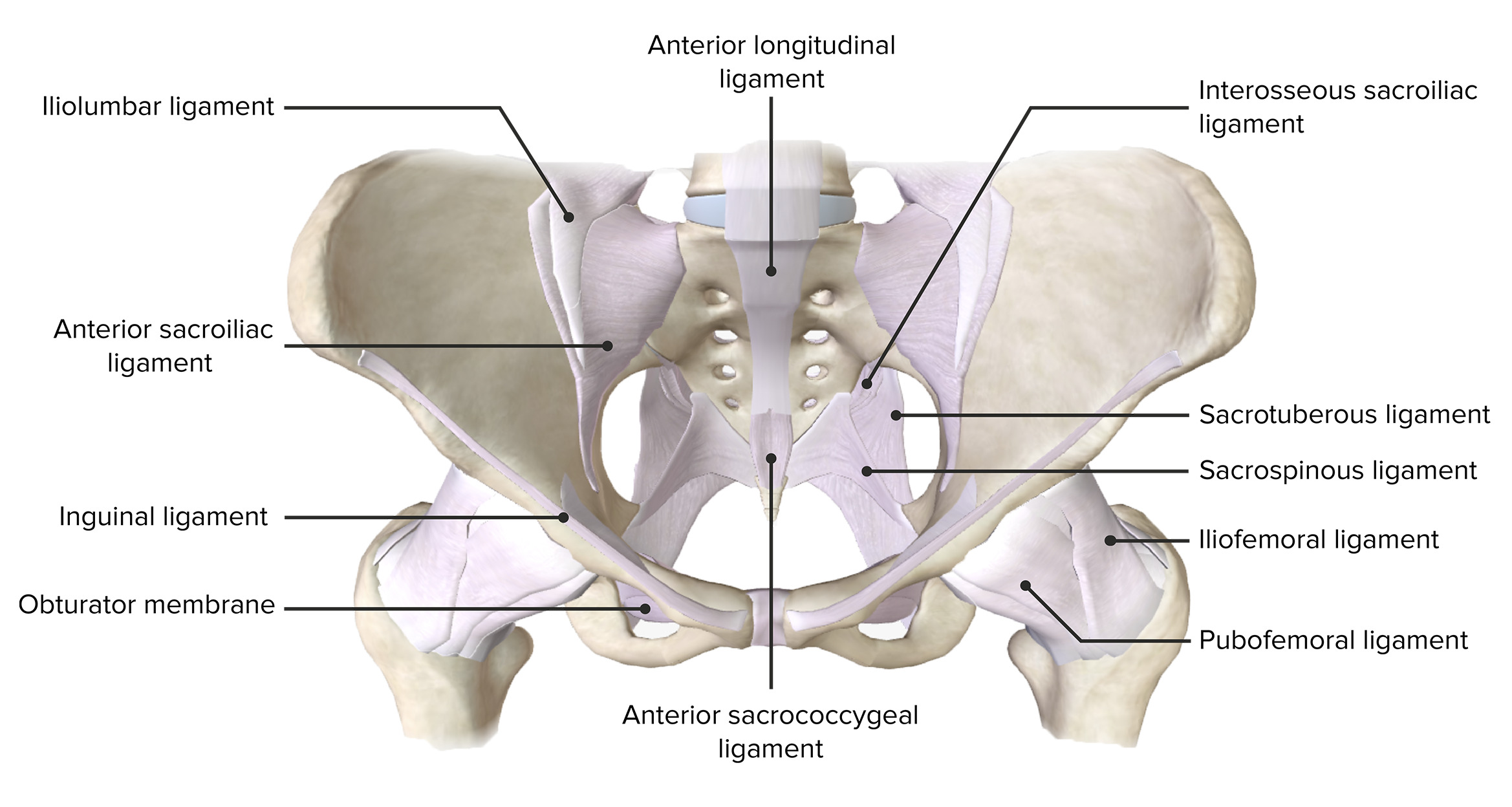
Anterior view of the pelvis, featuring the supporting ligaments of the joints of the pelvic girdle
Determined by measurement of the angle formed between the head and neckNeckThe part of a human or animal body connecting the head to the rest of the body.Peritonsillar Abscess of the femur:
A line is extended through the center of the shaft of the femoral neckNeckThe part of a human or animal body connecting the head to the rest of the body.Peritonsillar Abscess.
Another line is extended through the center of the shaft of the long axis of the femur.
The intersection of these 2 lines is normally approximately 120‒135 degrees.
SwellingSwellingInflammation or ecchymosisEcchymosisExtravasation of blood into the skin, resulting in a nonelevated, rounded or irregular, blue or purplish patch, larger than a petechia.Orbital Fractures
Muscle (quadriceps) wasting
Observe the inguinal regionInguinal regionAnterior Abdominal Wall: Anatomy for swellingSwellingInflammation (e.g., lymphadenopathyLymphadenopathyLymphadenopathy is lymph node enlargement (> 1 cm) and is benign and self-limited in most patients. Etiologies include malignancy, infection, and autoimmune disorders, as well as iatrogenic causes such as the use of certain medications. Generalized lymphadenopathy often indicates underlying systemic disease. Lymphadenopathy, inguinal herniaHerniaProtrusion of tissue, structure, or part of an organ through the bone, muscular tissue, or the membrane by which it is normally contained. Hernia may involve tissues such as the abdominal wall or the respiratory diaphragm. Hernias may be internal, external, congenital, or acquired.Abdominal Hernias).
Observe the hip capsuleCapsuleAn envelope of loose gel surrounding a bacterial cell which is associated with the virulence of pathogenic bacteria. Some capsules have a well-defined border, whereas others form a slime layer that trails off into the medium. Most capsules consist of relatively simple polysaccharides but there are some bacteria whose capsules are made of polypeptides.Bacteroides for effusion.
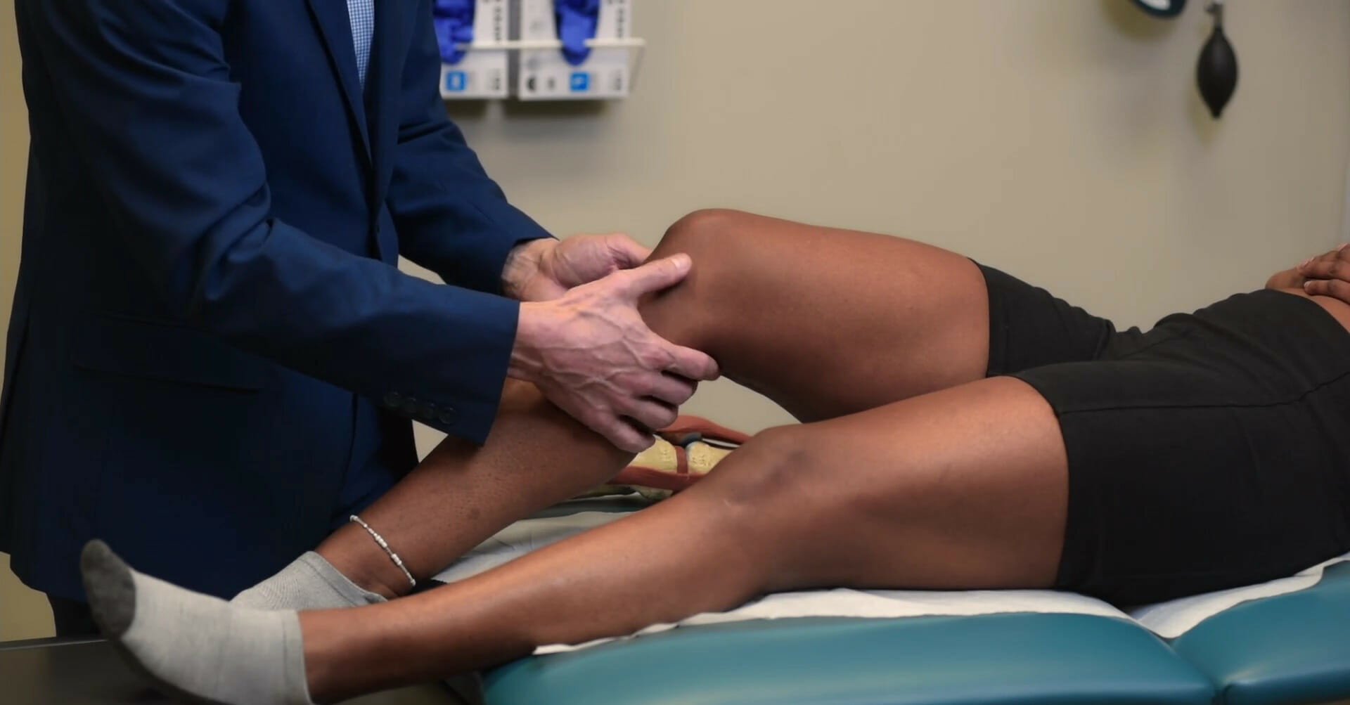
PalpationPalpationApplication of fingers with light pressure to the surface of the body to determine consistency of parts beneath in physical diagnosis; includes palpation for determining the outlines of organs.Dermatologic Examination and percussionPercussionAct of striking a part with short, sharp blows as an aid in diagnosing the condition beneath the sound obtained.Pulmonary Examination
Joint palpationPalpationApplication of fingers with light pressure to the surface of the body to determine consistency of parts beneath in physical diagnosis; includes palpation for determining the outlines of organs.Dermatologic Examination:
Best done with a neutral or flexed hip
Palpate the anterior joint line along the inguinal fold.
Palpate along the joint to feel for sponginess (synovitisSynovitisInflammation of the synovial membrane.Rheumatoid Arthritis) or bony growth (osteophytes).
Palpate for joint crepitusCrepitusOsteoarthritis (during active or passive range of motionRange of motionThe distance and direction to which a bone joint can be extended. Range of motion is a function of the condition of the joints, muscles, and connective tissues involved. Joint flexibility can be improved through appropriate muscle strength exercises.Examination of the Upper Limbs).
Feel for warmth of the skinSkinThe skin, also referred to as the integumentary system, is the largest organ of the body. The skin is primarily composed of the epidermis (outer layer) and dermis (deep layer). The epidermis is primarily composed of keratinocytes that undergo rapid turnover, while the dermis contains dense layers of connective tissue.Skin: Structure and Functions across the gradient (above and below the inguinal fold).
PercussionPercussionAct of striking a part with short, sharp blows as an aid in diagnosing the condition beneath the sound obtained.Pulmonary Examination:
Along the femoral neckNeckThe part of a human or animal body connecting the head to the rest of the body.Peritonsillar Abscess to detect femoral neckNeckThe part of a human or animal body connecting the head to the rest of the body.Peritonsillar Abscess (stress) fractureFractureA fracture is a disruption of the cortex of any bone and periosteum and is commonly due to mechanical stress after an injury or accident. Open fractures due to trauma can be a medical emergency. Fractures are frequently associated with automobile accidents, workplace injuries, and trauma.Overview of Bone Fractures
Assesses hip flexor strength against resistanceResistancePhysiologically, the opposition to flow of air caused by the forces of friction. As a part of pulmonary function testing, it is the ratio of driving pressure to the rate of air flow.Ventilation: Mechanics of Breathing
Assesses hamstring strength against resistanceResistancePhysiologically, the opposition to flow of air caused by the forces of friction. As a part of pulmonary function testing, it is the ratio of driving pressure to the rate of air flow.Ventilation: Mechanics of Breathing
Assesses hip abductor strength against resistanceResistancePhysiologically, the opposition to flow of air caused by the forces of friction. As a part of pulmonary function testing, it is the ratio of driving pressure to the rate of air flow.Ventilation: Mechanics of Breathing
Assesses hip adductor strength against resistanceResistancePhysiologically, the opposition to flow of air caused by the forces of friction. As a part of pulmonary function testing, it is the ratio of driving pressure to the rate of air flow.Ventilation: Mechanics of Breathing
Assesses hip internal rotator strength against resistanceResistancePhysiologically, the opposition to flow of air caused by the forces of friction. As a part of pulmonary function testing, it is the ratio of driving pressure to the rate of air flow.Ventilation: Mechanics of Breathing
Assesses hip internal rotator strength against resistanceResistancePhysiologically, the opposition to flow of air caused by the forces of friction. As a part of pulmonary function testing, it is the ratio of driving pressure to the rate of air flow.Ventilation: Mechanics of Breathing
Hip painPainAn unpleasant sensation induced by noxious stimuli which are detected by nerve endings of nociceptive neurons.Pain: Types and Pathways evaluation
“Hip painPainAn unpleasant sensation induced by noxious stimuli which are detected by nerve endings of nociceptive neurons.Pain: Types and Pathways,” as referred to by the layperson, is a common term that may be due to pathology in numerous structures. This painPainAn unpleasant sensation induced by noxious stimuli which are detected by nerve endings of nociceptive neurons.Pain: Types and Pathways should be further clarified by the examiner as to whether the painPainAn unpleasant sensation induced by noxious stimuli which are detected by nerve endings of nociceptive neurons.Pain: Types and Pathways is:
Posterior
Lateral
Anterior
Tests for posterior hip painPainAn unpleasant sensation induced by noxious stimuli which are detected by nerve endings of nociceptive neurons.Pain: Types and Pathways
Low lumbar spineSpineThe human spine, or vertebral column, is the most important anatomical and functional axis of the human body. It consists of 7 cervical vertebrae, 12 thoracic vertebrae, and 5 lumbar vertebrae and is limited cranially by the skull and caudally by the sacrum.Vertebral Column: Anatomy/lumbosacral spineSpineThe human spine, or vertebral column, is the most important anatomical and functional axis of the human body. It consists of 7 cervical vertebrae, 12 thoracic vertebrae, and 5 lumbar vertebrae and is limited cranially by the skull and caudally by the sacrum.Vertebral Column: Anatomy:
Muscular/tendinous/ligamentous elements related to these structures
Presents as localized painPainAn unpleasant sensation induced by noxious stimuli which are detected by nerve endings of nociceptive neurons.Pain: Types and Pathways at or just below the waistline that may be referred (or radiate) to the buttocks, proximal lower limb, and/or distal lower limb
Facet disease:
Generalized painPainAn unpleasant sensation induced by noxious stimuli which are detected by nerve endings of nociceptive neurons.Pain: Types and Pathways with extensionExtensionExamination of the Upper Limbs (backward bending) of the lumbar spineSpineThe human spine, or vertebral column, is the most important anatomical and functional axis of the human body. It consists of 7 cervical vertebrae, 12 thoracic vertebrae, and 5 lumbar vertebrae and is limited cranially by the skull and caudally by the sacrum.Vertebral Column: Anatomy
Positive Kemp test: localized painPainAn unpleasant sensation induced by noxious stimuli which are detected by nerve endings of nociceptive neurons.Pain: Types and Pathways (over the specific facet joint(s) affected) with extensionExtensionExamination of the Upper Limbs of the lumbar spineSpineThe human spine, or vertebral column, is the most important anatomical and functional axis of the human body. It consists of 7 cervical vertebrae, 12 thoracic vertebrae, and 5 lumbar vertebrae and is limited cranially by the skull and caudally by the sacrum.Vertebral Column: Anatomy
Tenderness to palpationPalpationApplication of fingers with light pressure to the surface of the body to determine consistency of parts beneath in physical diagnosis; includes palpation for determining the outlines of organs.Dermatologic Examination directly over the facet joint(s)
Disk disease:
Localized midline painPainAn unpleasant sensation induced by noxious stimuli which are detected by nerve endings of nociceptive neurons.Pain: Types and Pathways with flexionFlexionExamination of the Upper Limbs (forward bending) or lateral flexionFlexionExamination of the Upper Limbs (side bending) of the lumbar spineSpineThe human spine, or vertebral column, is the most important anatomical and functional axis of the human body. It consists of 7 cervical vertebrae, 12 thoracic vertebrae, and 5 lumbar vertebrae and is limited cranially by the skull and caudally by the sacrum.Vertebral Column: Anatomy
Localized midline painPainAn unpleasant sensation induced by noxious stimuli which are detected by nerve endings of nociceptive neurons.Pain: Types and Pathways with cough or sneeze (i.e., Valsalva)
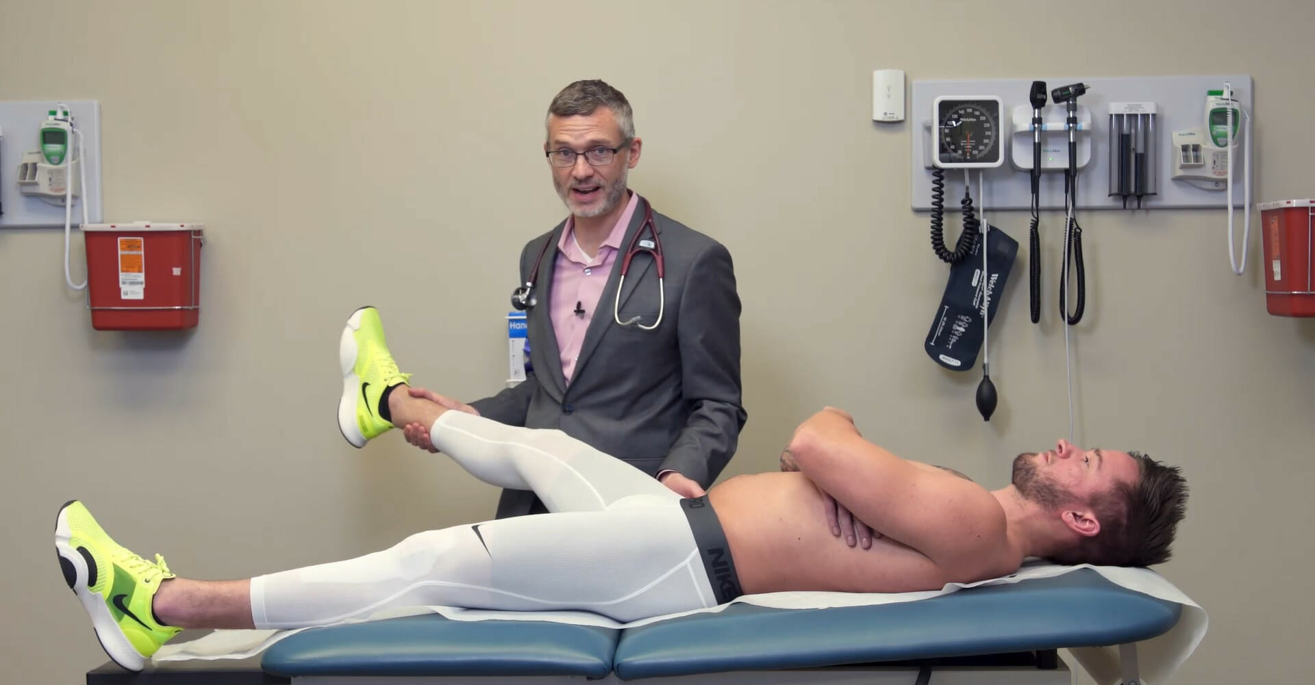
Positive straight leg raise testStraight Leg Raise TestSpinal Disk Herniation (Lasègue) test: With the individual in the supine position, the examiner passively raises a straight legLegThe lower leg, or just “leg” in anatomical terms, is the part of the lower limb between the knee and the ankle joint. The bony structure is composed of the tibia and fibula bones, and the muscles of the leg are grouped into the anterior, lateral, and posterior compartments by extensions of fascia.Leg: Anatomy (extended at the knee) into hip flexionFlexionExamination of the Upper Limbs. A positive test causes painPainAn unpleasant sensation induced by noxious stimuli which are detected by nerve endings of nociceptive neurons.Pain: Types and Pathways to radiate into the lower limb (typically in a dermatomalDermatomalDermatologic Examination pattern).
Significant (and commonly missed) cause of “low back” painPainAn unpleasant sensation induced by noxious stimuli which are detected by nerve endings of nociceptive neurons.Pain: Types and Pathways
Common in individuals with spondyloarthropathies (e.g., ankylosing spondylitisAnkylosing spondylitisAnkylosing spondylitis (also known as Bechterew’s disease or Marie-Strümpell disease) is a seronegative spondyloarthropathy characterized by chronic and indolent inflammation of the axial skeleton. Severe disease can lead to fusion and rigidity of the spine. Ankylosing Spondylitis, psoriatic arthritisPsoriatic ArthritisA type of inflammatory arthritis associated with psoriasis, often involving the axial joints and the peripheral terminal interphalangeal joints. It is characterized by the presence of hla-b27-associated spondyloarthropathy, and the absence of rheumatoid factor.Psoriasis)
Also common in the general population
Presents as localized painPainAn unpleasant sensation induced by noxious stimuli which are detected by nerve endings of nociceptive neurons.Pain: Types and Pathways at or just below the waistline that may be referred (or radiate) to the buttocks and/or proximal lower limb (rarely past the knee).
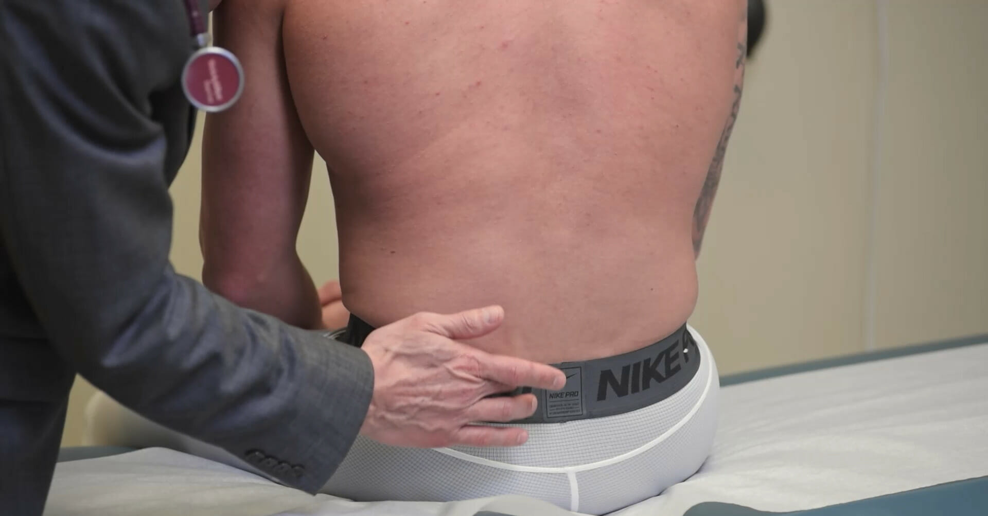
Positive Fortin finger point sign: Individual points within 2 cm of the posterior superior iliac spineSpineThe human spine, or vertebral column, is the most important anatomical and functional axis of the human body. It consists of 7 cervical vertebrae, 12 thoracic vertebrae, and 5 lumbar vertebrae and is limited cranially by the skull and caudally by the sacrum.Vertebral Column: Anatomy when asked to identify the source of their painPainAn unpleasant sensation induced by noxious stimuli which are detected by nerve endings of nociceptive neurons.Pain: Types and Pathways.
Tenderness to palpationPalpationApplication of fingers with light pressure to the surface of the body to determine consistency of parts beneath in physical diagnosis; includes palpation for determining the outlines of organs.Dermatologic Examination over the posterior superior iliac spineSpineThe human spine, or vertebral column, is the most important anatomical and functional axis of the human body. It consists of 7 cervical vertebrae, 12 thoracic vertebrae, and 5 lumbar vertebrae and is limited cranially by the skull and caudally by the sacrum.Vertebral Column: Anatomy
Provocative testing (designed to recreate the presenting painPainAn unpleasant sensation induced by noxious stimuli which are detected by nerve endings of nociceptive neurons.Pain: Types and Pathways):
Examiner compresses the innominates by applying a downward force through the anterior superior iliac spines.
Pelvic distraction test:
Individual is in the supine position.
Examiner separates (distracts) the innominates by applying a distractive force between the anterior superior iliac spines.
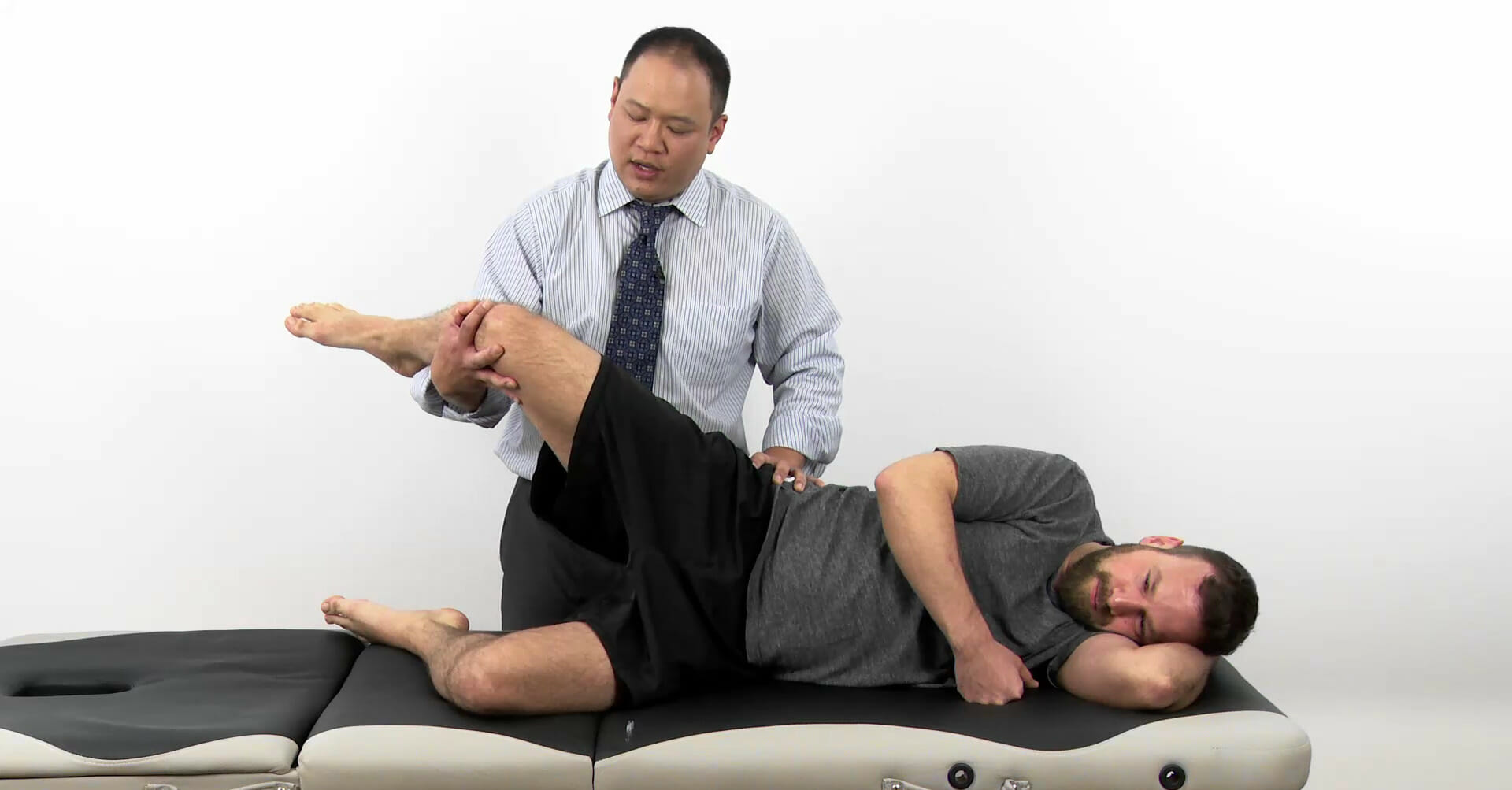
ThighThighThe thigh is the region of the lower limb found between the hip and the knee joint. There is a single bone in the thigh called the femur, which is surrounded by large muscles grouped into 3 fascial compartments. Thigh: Anatomy thrust test:
Individual is lying supine.
Affected hip is flexed to 90 degrees.
PelvisPelvisThe pelvis consists of the bony pelvic girdle, the muscular and ligamentous pelvic floor, and the pelvic cavity, which contains viscera, vessels, and multiple nerves and muscles. The pelvic girdle, composed of 2 “hip” bones and the sacrum, is a ring-like bony structure of the axial skeleton that links the vertebral column with the lower extremities.Pelvis: Anatomy is stabilized at the opposite anterior superior iliac spines by the examiner.
Examiner then applies downward pressure through the axis of the femur.
Gaenslen test:
Individual is lying supine.
Individual draws the nonaffected hip into full flexionFlexionExamination of the Upper Limbs and holds the position with their hands on a flexed knee while the affected limb stays lying on the table.
Examiner places one handHandThe hand constitutes the distal part of the upper limb and provides the fine, precise movements needed in activities of daily living. It consists of 5 metacarpal bones and 14 phalanges, as well as numerous muscles innervated by the median and ulnar nerves. Hand: Anatomy on the supine knee and the other over the hands of the individual on the flexed knee.
The examiner then applies a distractive force between the 2 knees, creating torsion stress at the sacroiliac (SI) joint.
Due to periosteal irritation associated with tight hamstrings (proximal attachment of the hamstrings is in the ischial tuberosityIschial TuberosityChronic Apophyseal Injury)
Pseuodosciatica: irritation of the sciatic nerveSciatic NerveA nerve which originates in the lumbar and sacral spinal cord (l4 to s3) and supplies motor and sensory innervation to the lower extremity. The sciatic nerve, which is the main continuation of the sacral plexus, is the largest nerve in the body. It has two major branches, the tibial nerve and the peroneal nerve.Gluteal Region: Anatomy as it passes under (or through, anatomic variant) the belly of the piriformisPiriformisVagina, Vulva, and Pelvic Floor: Anatomy
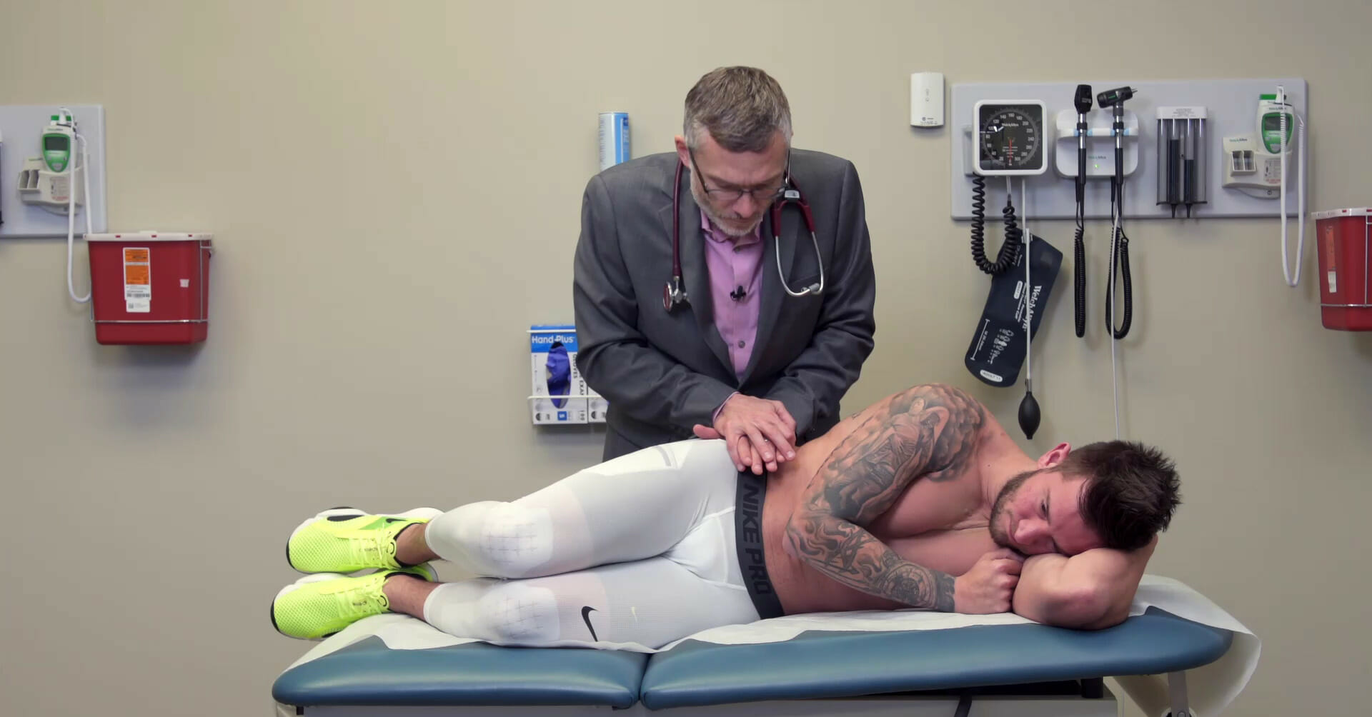
Individual is in the lateral decubitus position, with the hip and knee flexed.
Examiner applies a medial force against the lateral aspect of the knee to induce adductionAdductionExamination of the Upper Limbs of the hip against the individual’s active effort to abduct.
Palpation of the posterior superior iliac spine: This palpation is useful for identifying a bony landmark. In the Fortin finger point test, the individual is asked to point at the source of their pain. Localization of the individual’s fingertip near the posterior superior iliac spine is suggestive of SI joint disease. The posterior superior iliac spine is also commonly tender to palpation in SI joint disorders.
Image by Lecturio.
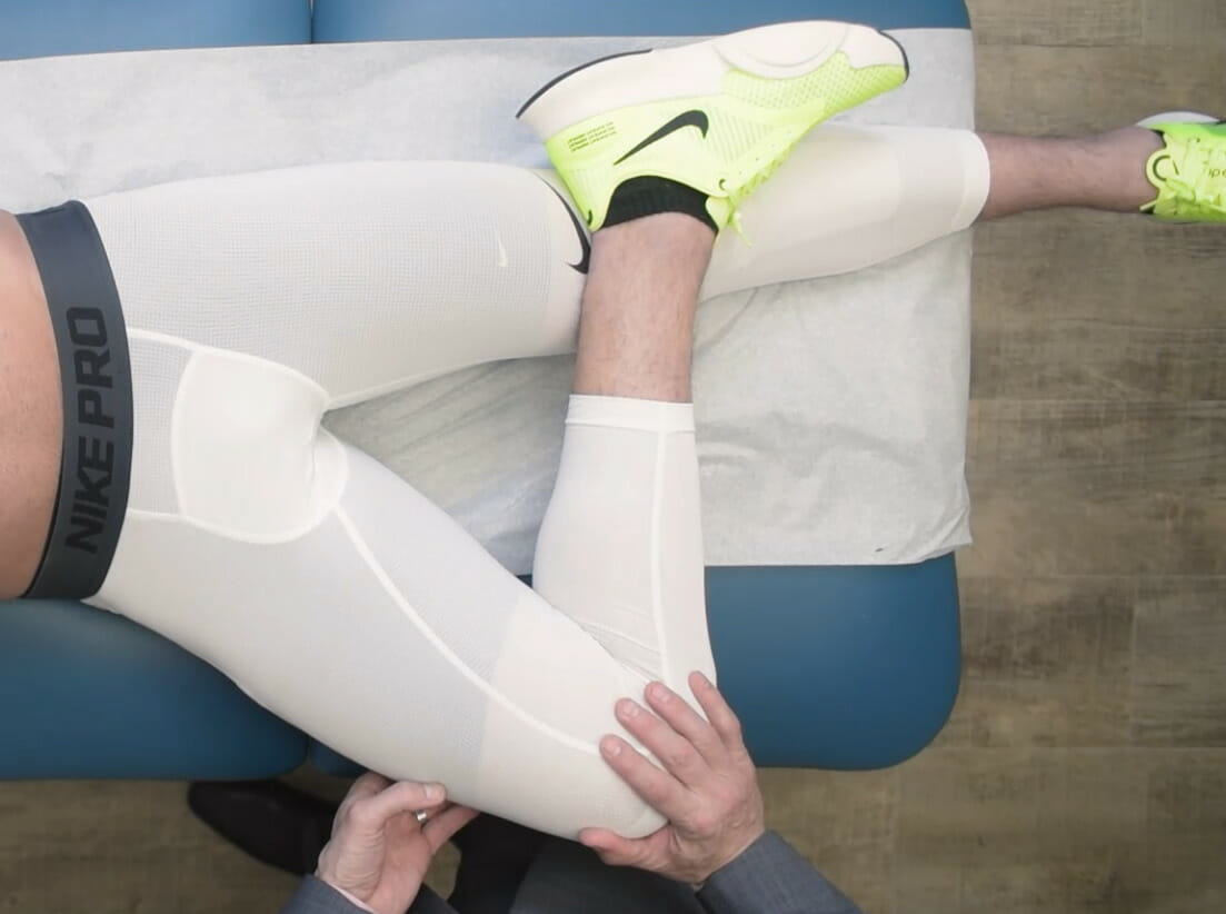
The figure 4, or FABER, test: With the individual in the supine position, the affected hip is carried into passive Flexion, ABduction, and External Rotation by the examiner, followed by a downward force applied by the examiner. This force causes stress against the SI joint(s). This provocative maneuver is designed to reproduce posterior hip pain and is associated with SI joint pathology.
Image by Lecturio.
The pelvic compression test: With the individual in the lateral decubitus position, the examiner compresses the innominates by applying a downward force through the anterior superior iliac spine. This force causes stress against the SI joint(s). This provocative maneuver is designed to reproduce posterior hip pain and is associated with SI joint pathology.
Image by Lecturio.
Active piriformis test: With the individual in the lateral decubitus position with the hip and knee flexed, the examiner applies a medial force against the lateral aspect of the knee to induce adduction of the hip against the individual’s active effort to abduct (the piriformis is an abductor and external rotator of the hip when the hip is flexed). This force causes the piriformis to contract. This provocative maneuver is designed to reproduce posterior hip pain and is associated with piriformis pathology or irritation of the sciatic nerve (pseudosciatica) as it passes under (or through) the piriformis.
Image by Lecturio.
Tests for lateral hip painPainAn unpleasant sensation induced by noxious stimuli which are detected by nerve endings of nociceptive neurons.Pain: Types and Pathways
May alternatively present as lateral knee painPainAn unpleasant sensation induced by noxious stimuli which are detected by nerve endings of nociceptive neurons.Pain: Types and Pathways and/or in conjunction with trochanteric bursitis
Exam findings:
Tenderness to palpationPalpationApplication of fingers with light pressure to the surface of the body to determine consistency of parts beneath in physical diagnosis; includes palpation for determining the outlines of organs.Dermatologic Examination over the tensor fascia lataFascia lataFemoral Region and Hernias: Anatomy and/or iliotibial bandIliotibial bandThigh: Anatomy (anywhere along its length, but especially over bony attachments)
Positive Ober test:
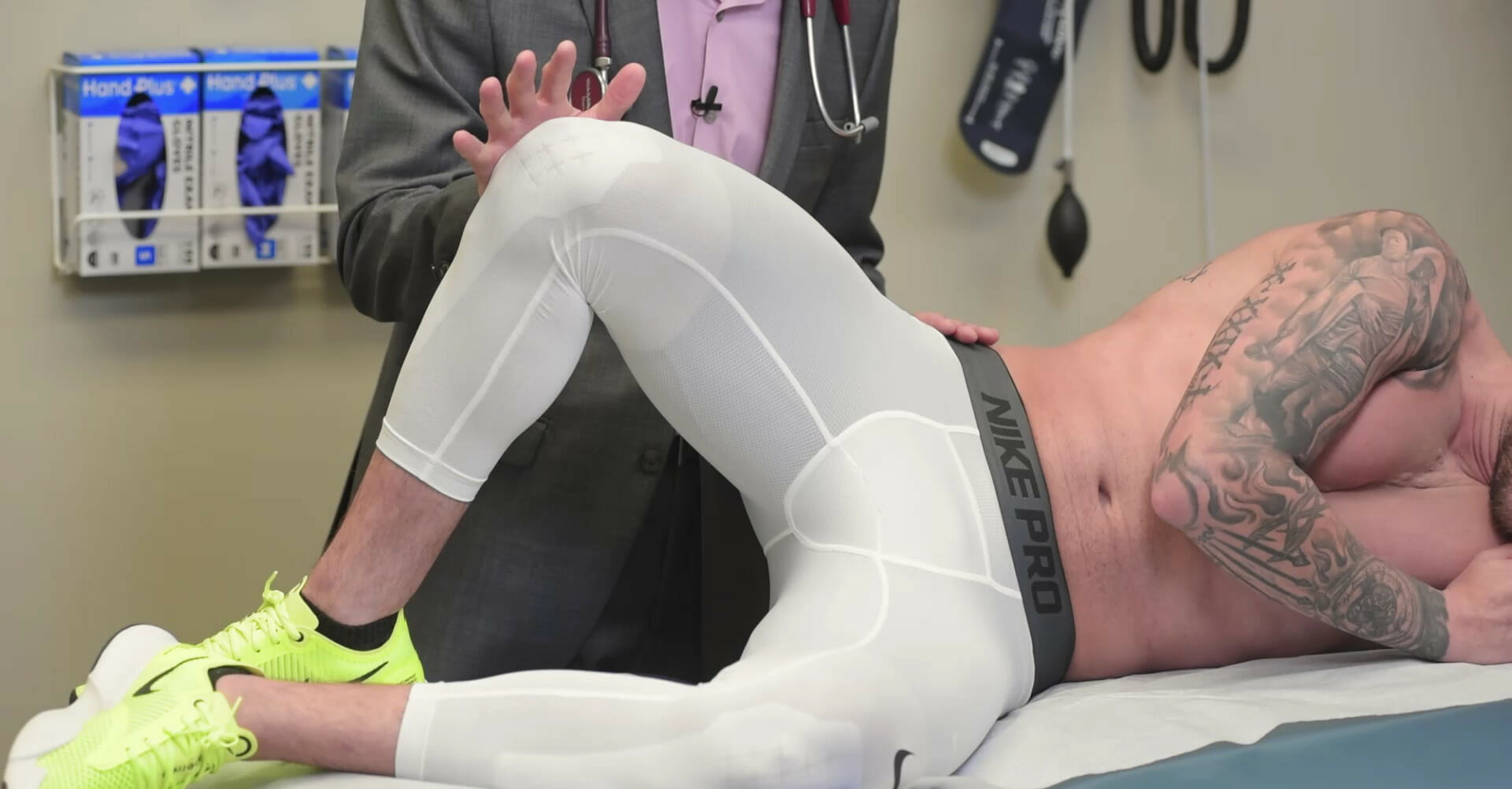
Individual is in the lateral decubitus position (affected side up) with the hip in the neutral position.
Examiner passively carries the hip into abductionAbductionExamination of the Upper Limbs (shortening the IT band) by lifting at the knee and then letting it fall.
In the absence of IT band (or tensor fascia lataFascia lataFemoral Region and Hernias: Anatomy) pathology, the knee will fall to its original position free of resistanceResistancePhysiologically, the opposition to flow of air caused by the forces of friction. As a part of pulmonary function testing, it is the ratio of driving pressure to the rate of air flow.Ventilation: Mechanics of Breathing.
If IT band pathology is present, the knee may fall slowly or may stop midway owing to contracture(s) of the IT band.
Exam findings: tenderness to palpationPalpationApplication of fingers with light pressure to the surface of the body to determine consistency of parts beneath in physical diagnosis; includes palpation for determining the outlines of organs.Dermatologic Examination directly over the greater trochanter/trochanteric bursaTrochanteric bursaHip Joint: Anatomy
The Ober test: With the individual in the lateral decubitus position (affected side up) and the hip in the neutral position, the examiner passively carries the hip into abduction (shortening the IT band) by lifting at the knee and then letting it fall. In the absence of IT band (or tensor fascia lata) pathology, the knee will fall to its original position free of resistance. If IT band pathology is present, the knee may fall slowly or may stop midway owing to contracture(s) of the IT band.
Image by Lecturio.
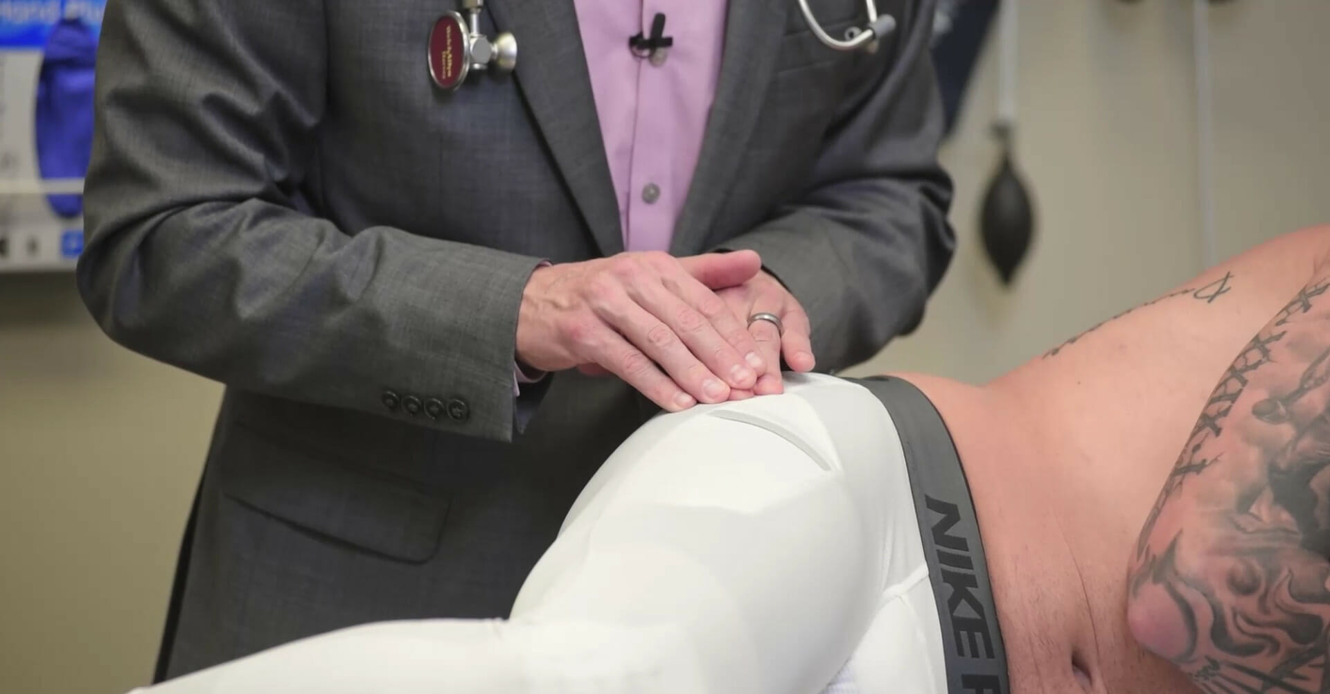
Palpation and provocation of the greater trochanter:
With the individual in the lateral decubitus position (affected side up), the examiner locates the greater trochanter and applies a downward provocative force. This force causes compression of the trochanteric bursa overlying the greater trochanter. This provocative maneuver is designed to reproduce lateral hip pain and is associated with pathology of the trochanteric bursa.
Image by Lecturio.
Tests for anterior hip painPainAn unpleasant sensation induced by noxious stimuli which are detected by nerve endings of nociceptive neurons.Pain: Types and Pathways
Femoroacetabular joint:
Most commonly due to arthritic conditions (e.g., osteoarthritisOsteoarthritisOsteoarthritis (OA) is the most common form of arthritis, and is due to cartilage destruction and changes of the subchondral bone. The risk of developing this disorder increases with age, obesity, and repetitive joint use or trauma. Patients develop gradual joint pain, stiffness lasting < 30 minutes, and decreased range of motion. Osteoarthritis (OAOAOsteoarthritis (OA) is the most common form of arthritis, and is due to cartilage destruction and changes of the subchondral bone. The risk of developing this disorder increases with age, obesity, and repetitive joint use or trauma. Patients develop gradual joint pain, stiffness lasting < 30 minutes, and decreased range of motion.Osteoarthritis), rheumatoid arthritisArthritisAcute or chronic inflammation of joints.Osteoarthritis (RA))
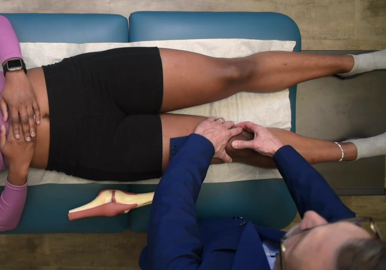
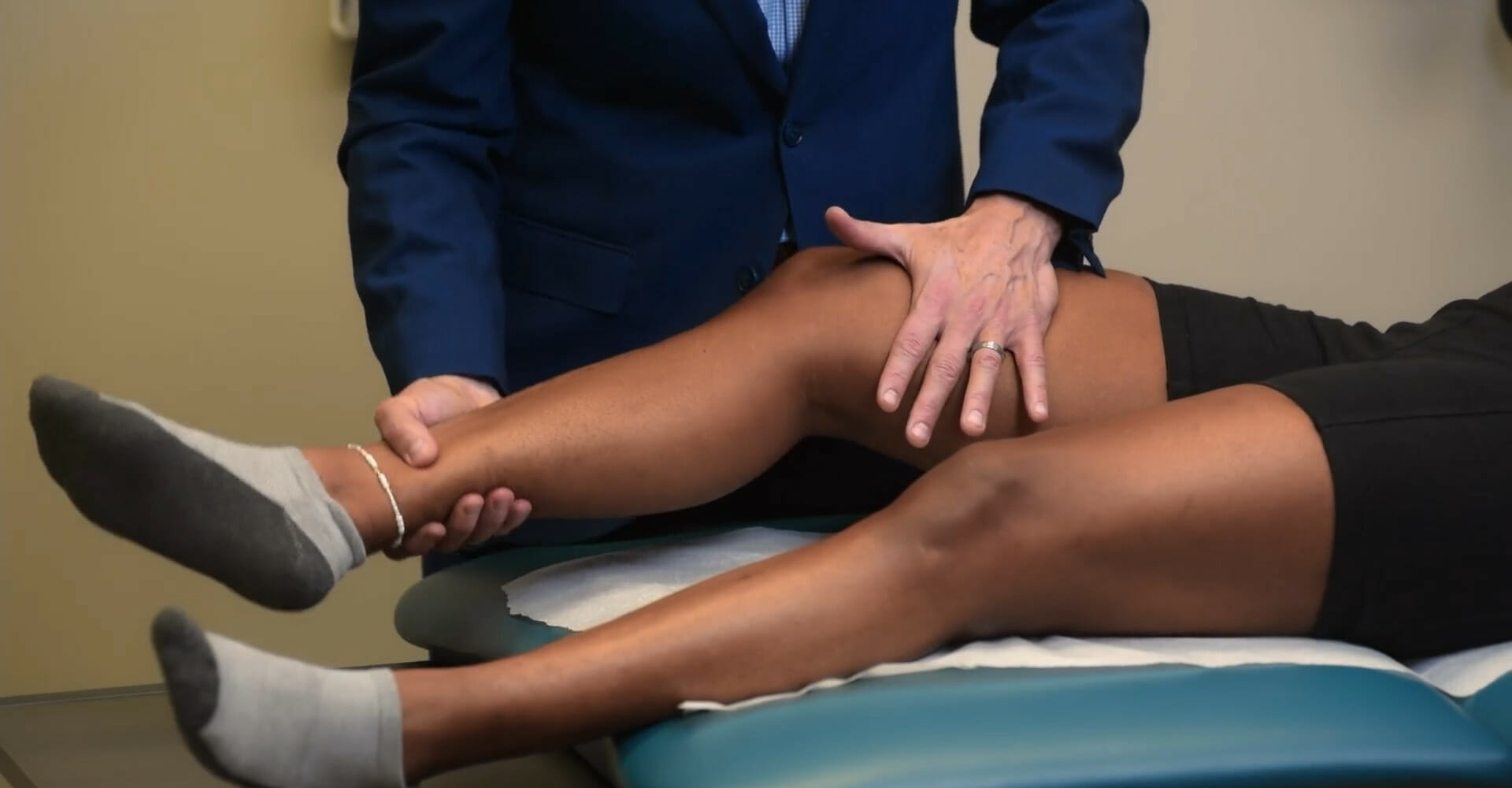
The femur is passively rotated internally and externally within the acetabulum.
This provocative maneuver is designed to reproduce painPainAn unpleasant sensation induced by noxious stimuli which are detected by nerve endings of nociceptive neurons.Pain: Types and Pathways in the anterior hip (generally with internal rotationInternal RotationExamination of the Upper Limbs) and is associated with femoroacetabular joint pathology.
This provocative maneuver is designed to reproduce painPainAn unpleasant sensation induced by noxious stimuli which are detected by nerve endings of nociceptive neurons.Pain: Types and Pathways in the anterior hip and is associated with femoroacetabular joint pathology.
Hip flexors/iliopsoas syndrome:
Common in individuals who stay seated for prolonged periods (office workers), during which the hip flexors stay in the shortened position
May occur with standing abruptly after a prolonged period of sitting
Exam findings:
Tenderness to palpationPalpationApplication of fingers with light pressure to the surface of the body to determine consistency of parts beneath in physical diagnosis; includes palpation for determining the outlines of organs.Dermatologic Examination and/or hypertonicityHypertonicityVolume Depletion and Dehydration of the iliopsoas muscle
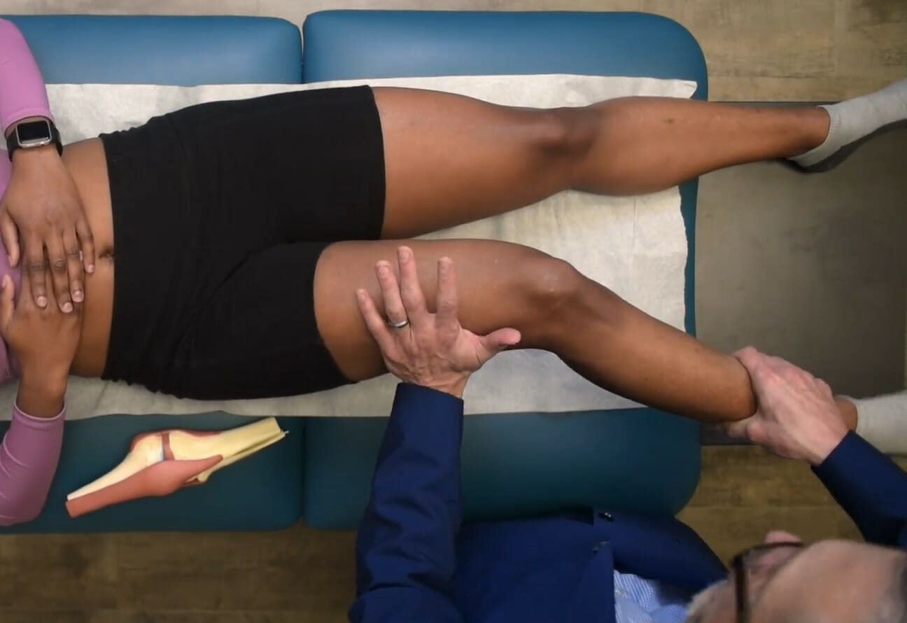
Examiner places a downward force against the individual’s distal femur while the individual resists by actively contracting the hip flexors.
This provocative maneuver is designed to reproduce painPainAn unpleasant sensation induced by noxious stimuli which are detected by nerve endings of nociceptive neurons.Pain: Types and Pathways in the anterior hip and is associated with pathology of the hip flexors.
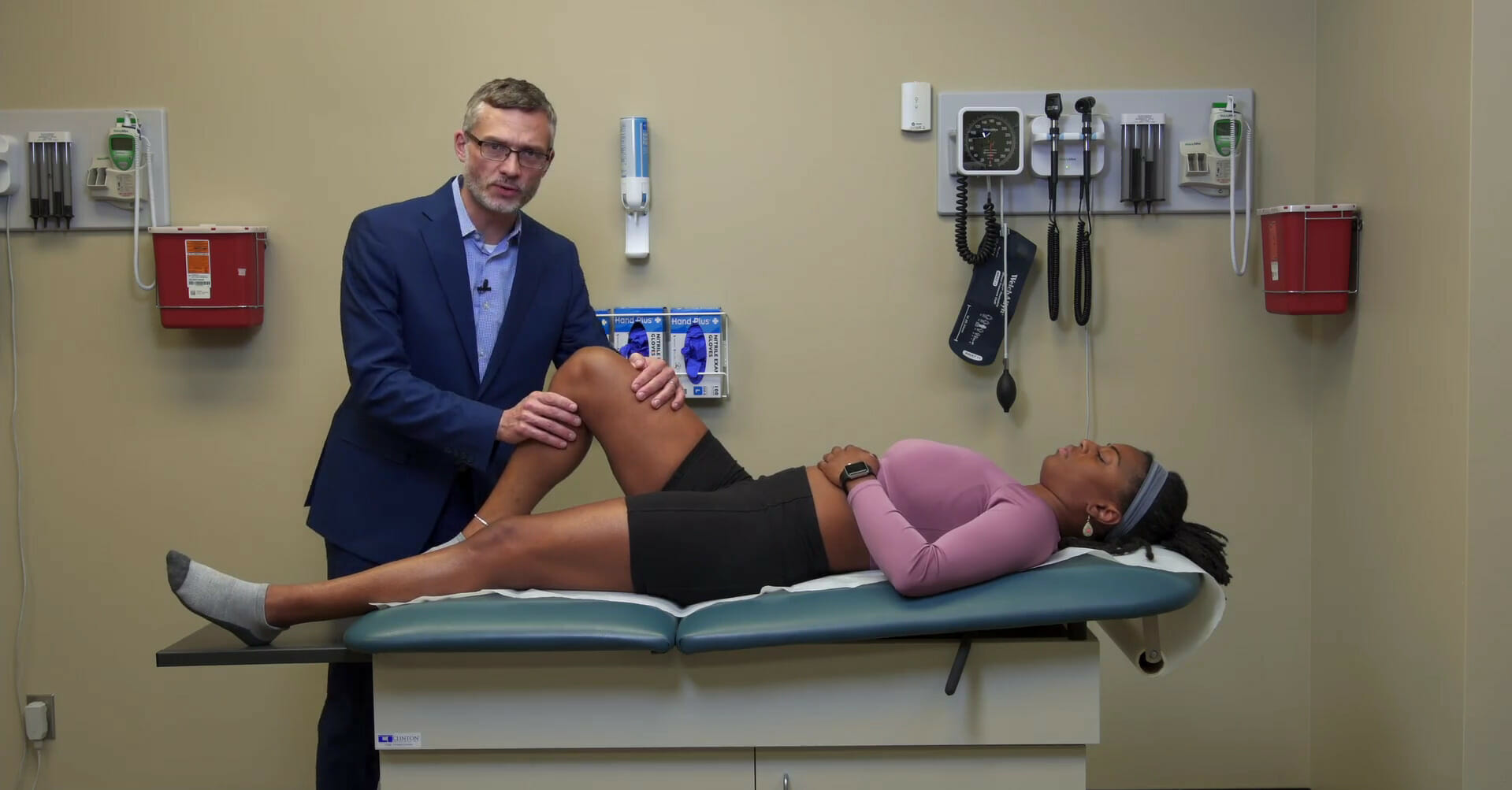
Positive Thomas test:
Individual is in the supine position
Unaffected hip is passively flexed to the chest.
If the iliopsoas is shortened (spasm/contracture), the affected hip will be unable to stay fully extended.
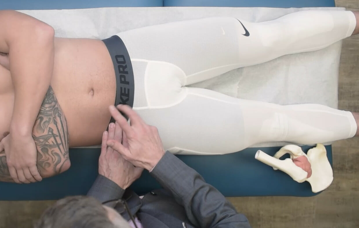
The lateral femoral cutaneous nerve exits the anterior abdominal wallAbdominal wallThe outer margins of the abdomen, extending from the osteocartilaginous thoracic cage to the pelvis. Though its major part is muscular, the abdominal wall consists of at least seven layers: the skin, subcutaneous fat, deep fascia; abdominal muscles, transversalis fascia, extraperitoneal fat, and the parietal peritoneum.Surgical Anatomy of the Abdomen just medial to the anterior superior iliac spineAnterior Superior Iliac SpineChronic Apophyseal Injury.
Entrapment/impingement/irritation may occur with:
Tight belt
Heavy belt (e.g., police belt, tool belt)
Abdominal pannusPannusA genus of cyanobacteria in the family synechococcaceae that is free-floating and occurs in various water environments attaching to algae and submersed plants.Septic Arthritis (i.e., obesityObesityObesity is a condition associated with excess body weight, specifically with the deposition of excessive adipose tissue. Obesity is considered a global epidemic. Major influences come from the western diet and sedentary lifestyles, but the exact mechanisms likely include a mixture of genetic and environmental factors. Obesity)
PregnancyPregnancyThe status during which female mammals carry their developing young (embryos or fetuses) in utero before birth, beginning from fertilization to birth.Pregnancy: Diagnosis, Physiology, and Care
Presents with painPainAn unpleasant sensation induced by noxious stimuli which are detected by nerve endings of nociceptive neurons.Pain: Types and Pathways/paresthesia in the distribution of the lateral femoral cutaneous nerve.
Exam findings:
SensorySensoryNeurons which conduct nerve impulses to the central nervous system.Nervous System: Histology disturbance (loss of sensation, altered sensation, allodyniaAllodyniaPain due to a stimulus that does not typically provoke pain.Pain Management) in the receptive field of the lateral femoral cutaneous nerve
Reproduces painPainAn unpleasant sensation induced by noxious stimuli which are detected by nerve endings of nociceptive neurons.Pain: Types and Pathways and/or paresthesia in the distribution of the nerve with percussionPercussionAct of striking a part with short, sharp blows as an aid in diagnosing the condition beneath the sound obtained.Pulmonary Examination medial to the anterior superior iliac spineAnterior Superior Iliac SpineChronic Apophyseal Injury
Log roll test: With the individual in the supine position, the femur is passively rotated internally and externally within the acetabulum. This provocative maneuver is designed to reproduce pain in the anterior hip (generally with internal rotation) and is associated with femoroacetabular joint pathology.
Image by Lecturio.
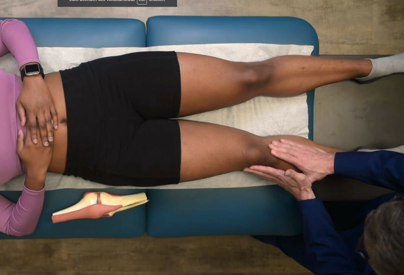
Hip abduction and adduction: With the individual in the supine position, the examiner passively carries the hip into abduction and adduction. This provocative maneuver is designed to reproduce pain in the anterior hip and is associated with femoroacetabular joint pathology.
Image by Lecturio.
Resisted flexion of the hip: With the individual in the supine position, the examiner places a downward force against the individual’s distal femur while the individual resists by actively contracting the hip flexors. This provocative maneuver is designed to reproduce pain in the anterior hip and is associated with pathology of the hip flexors.
Image by Lecturio.
Percussion over the lateral femoral cutaneous nerve at its exit point from the anterior abdominal wall just medial to the anterior superior iliac spine: This provocative maneuver is designed to reproduce pain and/or paresthesia in the distribution of the nerve and is associated with nerve entrapment.
Image by Lecturio.
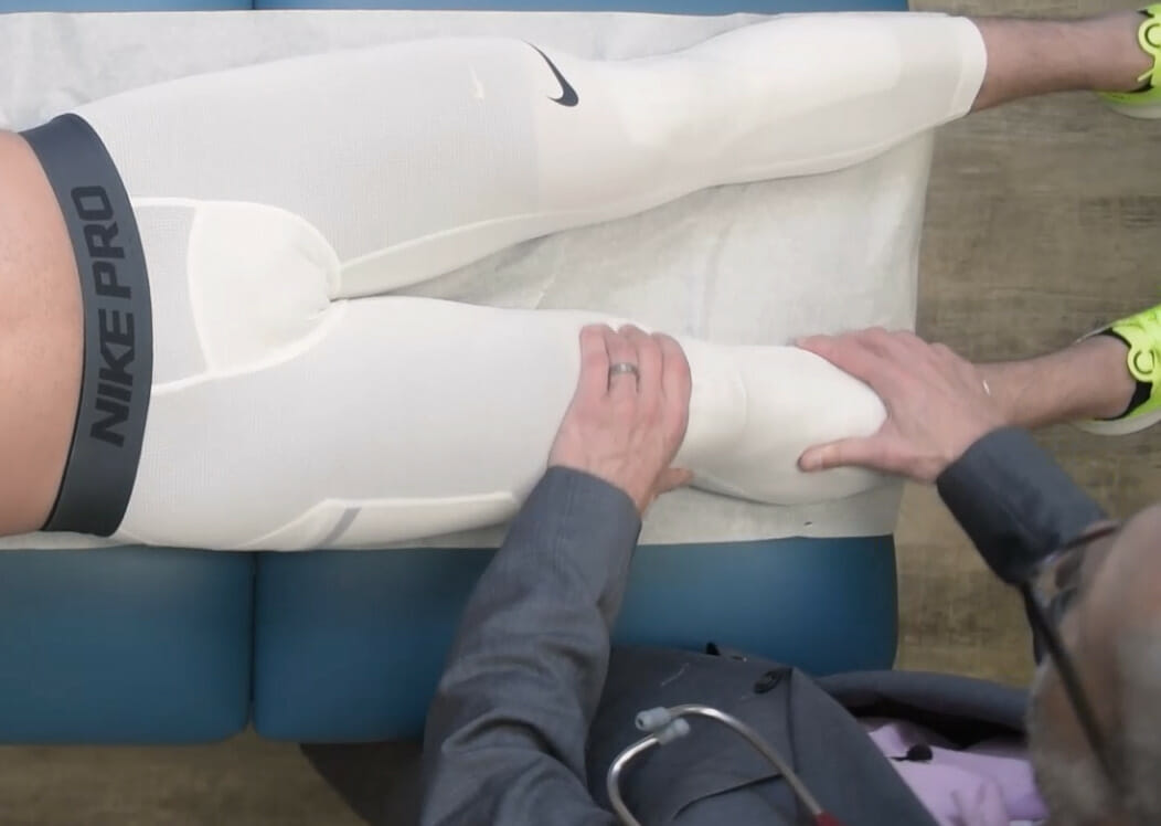
Vascular exam of the hip
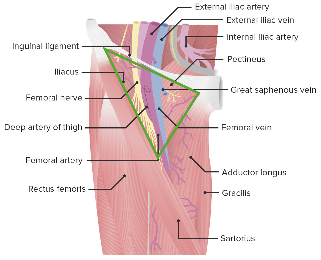
The femoral pulse can be palpated at the inguinal fold:
Normal alignment (valgus angulationAngulationBuckle or Torus Fracture) of the knee is at 8–14 degrees for men and 11–20 degrees degrees for women.
Angular deformities:
Genu varumGenu varumGenu varum is a deformation of the knee joint(s) that creates angulation of the lower limb(s) away from the midline in the coronal plane. Children ages 1-5 years are commonly affected. Genu Varum (bowlegged)
Genu valgumGenu valgumGenu valgum is a deformation of the knee joint(s) that creates angulation of the lower limb(s) toward the midline in the coronal plane. Children ages 1-5 years are commonly affected. Genu Valgum (knock-kneed)
SwellingSwellingInflammation or ecchymosisEcchymosisExtravasation of blood into the skin, resulting in a nonelevated, rounded or irregular, blue or purplish patch, larger than a petechia.Orbital Fractures
Muscle (quadriceps) wasting
Observe the popliteal fossaPopliteal fossaThe popliteal fossa or the “knee pit” is a diamond-shaped, fat-filled, shallow depression on the posterior aspect of the knee joint. The popliteal fossa is located at the dorsal aspect of the knee and contains an increased number of lymph nodes as well as structures of the neurovascular system that travel from the thigh to the lower leg.Popliteal Fossa: Anatomy for swellingSwellingInflammation.
Observe the knee capsuleCapsuleAn envelope of loose gel surrounding a bacterial cell which is associated with the virulence of pathogenic bacteria. Some capsules have a well-defined border, whereas others form a slime layer that trails off into the medium. Most capsules consist of relatively simple polysaccharides but there are some bacteria whose capsules are made of polypeptides.Bacteroides for effusion.
PalpationPalpationApplication of fingers with light pressure to the surface of the body to determine consistency of parts beneath in physical diagnosis; includes palpation for determining the outlines of organs.Dermatologic Examination and percussionPercussionAct of striking a part with short, sharp blows as an aid in diagnosing the condition beneath the sound obtained.Pulmonary Examination
Joint palpationPalpationApplication of fingers with light pressure to the surface of the body to determine consistency of parts beneath in physical diagnosis; includes palpation for determining the outlines of organs.Dermatologic Examination:
Palpate along the joint to feel for sponginess (synovitisSynovitisInflammation of the synovial membrane.Rheumatoid Arthritis) or bony growth (osteophytes).
Palpate for joint crepitusCrepitusOsteoarthritis (during active or passive range of motionRange of motionThe distance and direction to which a bone joint can be extended. Range of motion is a function of the condition of the joints, muscles, and connective tissues involved. Joint flexibility can be improved through appropriate muscle strength exercises.Examination of the Upper Limbs).
The posterior fossa must also be palpated for fullness, discomfort, or the presence of a cyst (Baker cyst, also known as popliteal cystPopliteal cystA synovial cyst most commonly at the back of the knee.Osteoarthritis).
Bursae:
PesPESRemoval of plasma and replacement with various fluids, e.g., fresh frozen plasma, plasma protein fractions (ppf), albumin preparations, dextran solutions, saline. Used in treatment of autoimmune diseases, immune complex diseases, diseases of excess plasma factors, and other conditions.Thrombotic Thrombocytopenic Purpura anserine (upper medial aspect of the tibiaTibiaThe second longest bone of the skeleton. It is located on the medial side of the lower leg, articulating with the fibula laterally, the talus distally, and the femur proximally.Knee Joint: Anatomy)
Feel for warmth of the skinSkinThe skin, also referred to as the integumentary system, is the largest organ of the body. The skin is primarily composed of the epidermis (outer layer) and dermis (deep layer). The epidermis is primarily composed of keratinocytes that undergo rapid turnover, while the dermis contains dense layers of connective tissue.Skin: Structure and Functions across the gradient (above the knee to the tibiaTibiaThe second longest bone of the skeleton. It is located on the medial side of the lower leg, articulating with the fibula laterally, the talus distally, and the femur proximally.Knee Joint: Anatomy)
Tests for effusion:
Patellar ballottement test/tap:
Extend the knee jointKnee jointThe knee joint is made up of the articulations between the femur, tibia, and patella bones, and is one of the largest and most complex joints of the human body. The knee is classified as a synovial hinge joint, which primarily allows for flexion and extension with a more limited degree of translation and rotation. Knee Joint: Anatomy.
Empty (milk) the suprapatellar pouch (by sliding handHandThe hand constitutes the distal part of the upper limb and provides the fine, precise movements needed in activities of daily living. It consists of 5 metacarpal bones and 14 phalanges, as well as numerous muscles innervated by the median and ulnar nerves. Hand: Anatomy down the thighThighThe thigh is the region of the lower limb found between the hip and the knee joint. There is a single bone in the thigh called the femur, which is surrounded by large muscles grouped into 3 fascial compartments. Thigh: Anatomy to the patellaPatellaThe flat, triangular bone situated at the anterior part of the knee.Knee Joint: Anatomy).
Tap the patellaPatellaThe flat, triangular bone situated at the anterior part of the knee.Knee Joint: Anatomy.
Note for a tapping sensation or fluid impulse on the milking handHandThe hand constitutes the distal part of the upper limb and provides the fine, precise movements needed in activities of daily living. It consists of 5 metacarpal bones and 14 phalanges, as well as numerous muscles innervated by the median and ulnar nerves. Hand: Anatomy.
Bulge sign/ripple test:
Extend the knee jointKnee jointThe knee joint is made up of the articulations between the femur, tibia, and patella bones, and is one of the largest and most complex joints of the human body. The knee is classified as a synovial hinge joint, which primarily allows for flexion and extension with a more limited degree of translation and rotation. Knee Joint: Anatomy.
Empty (milk) the suprapatellar pouch (by sliding handHandThe hand constitutes the distal part of the upper limb and provides the fine, precise movements needed in activities of daily living. It consists of 5 metacarpal bones and 14 phalanges, as well as numerous muscles innervated by the median and ulnar nerves. Hand: Anatomy down the thighThighThe thigh is the region of the lower limb found between the hip and the knee joint. There is a single bone in the thigh called the femur, which is surrounded by large muscles grouped into 3 fascial compartments. Thigh: Anatomy to the patellaPatellaThe flat, triangular bone situated at the anterior part of the knee.Knee Joint: Anatomy).
Stroke the lateral side of the joint.
Note any bulge or ripple on the medial side of the joint.
PercussionPercussionAct of striking a part with short, sharp blows as an aid in diagnosing the condition beneath the sound obtained.Pulmonary Examination:
Along the tibial plateauPlateauCardiac Physiology to detect tibial plateauPlateauCardiac Physiology (stress) fractureFractureA fracture is a disruption of the cortex of any bone and periosteum and is commonly due to mechanical stress after an injury or accident. Open fractures due to trauma can be a medical emergency. Fractures are frequently associated with automobile accidents, workplace injuries, and trauma.Overview of Bone Fractures
Along the anterior tibial shaft to detect tibial insufficiency (stress) fractureFractureA fracture is a disruption of the cortex of any bone and periosteum and is commonly due to mechanical stress after an injury or accident. Open fractures due to trauma can be a medical emergency. Fractures are frequently associated with automobile accidents, workplace injuries, and trauma.Overview of Bone Fractures
Palpation of the pes anserine bursa
Image by Lecturio.
Patellar ballottement test
Image by Lecturio.
Bulge sign (ripple test)
Image by Lecturio.
MotorMotorNeurons which send impulses peripherally to activate muscles or secretory cells.Nervous System: Histology and strength function
Assesses hamstring strength against resistanceResistancePhysiologically, the opposition to flow of air caused by the forces of friction. As a part of pulmonary function testing, it is the ratio of driving pressure to the rate of air flow.Ventilation: Mechanics of Breathing.
Straighten the legLegThe lower leg, or just “leg” in anatomical terms, is the part of the lower limb between the knee and the ankle joint. The bony structure is composed of the tibia and fibula bones, and the muscles of the leg are grouped into the anterior, lateral, and posterior compartments by extensions of fascia.Leg: Anatomy at the knee, bringing the knee into maximum extensionExtensionExamination of the Upper Limbs.
Normal range: 0–140 degrees
Assesses quadriceps strength against resistanceResistancePhysiologically, the opposition to flow of air caused by the forces of friction. As a part of pulmonary function testing, it is the ratio of driving pressure to the rate of air flow.Ventilation: Mechanics of Breathing
Patellar reflex: tests the reflex arc involving the L3 and L4 segments of the spinal cordSpinal cordThe spinal cord is the major conduction pathway connecting the brain to the body; it is part of the CNS. In cross section, the spinal cord is divided into an H-shaped area of gray matter (consisting of synapsing neuronal cell bodies) and a surrounding area of white matter (consisting of ascending and descending tracts of myelinated axons). Spinal Cord: Anatomy
Joint stability tests
Always test the unaffected knee 1st and compare the 2 knees.
Collateral ligament stability:
Start with the knee completely extended:
If stable
Then flex the knee to 30 degrees.
At 30 degrees, cruciate ligament stability is removed.
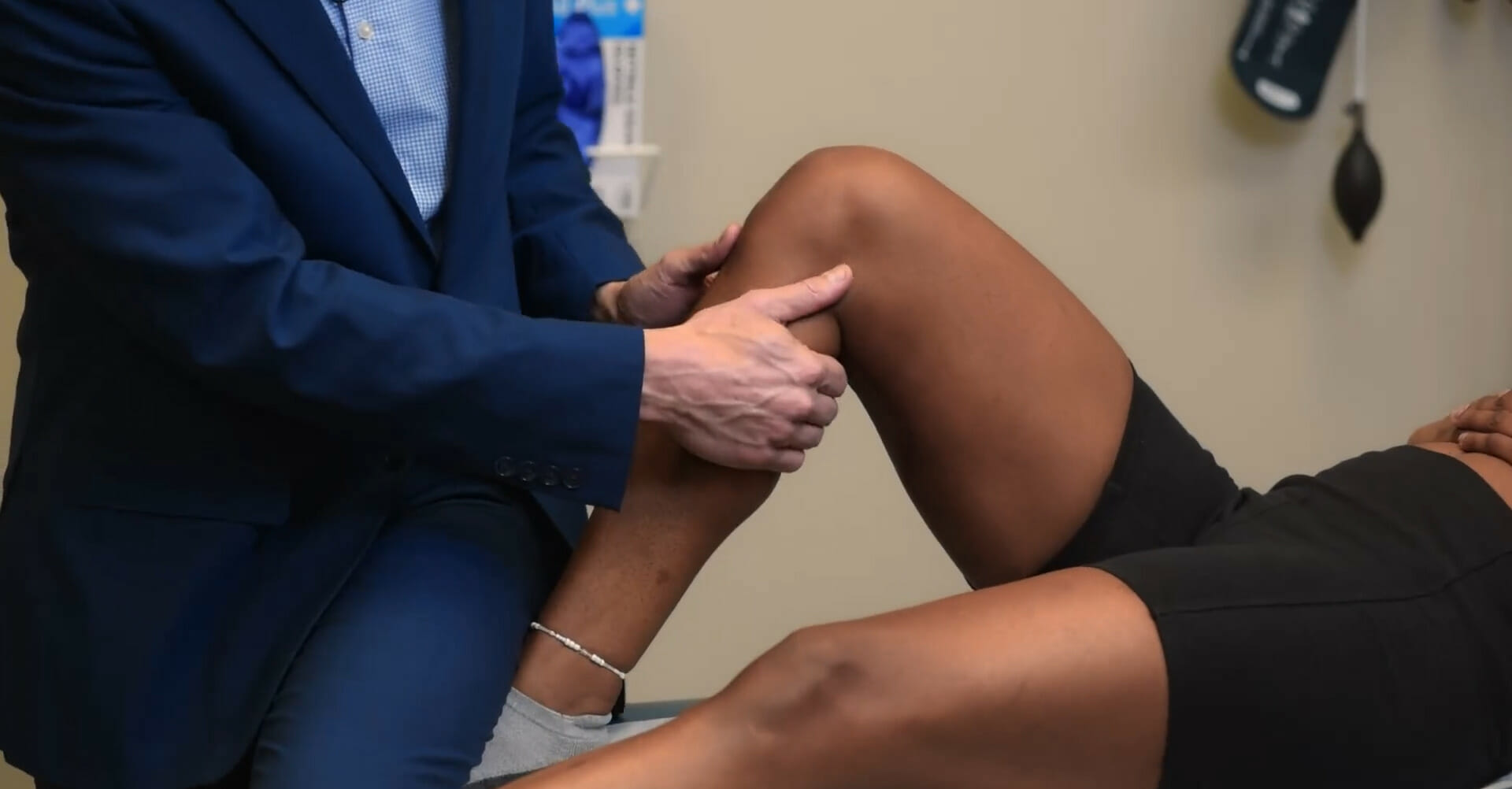
One handHandThe hand constitutes the distal part of the upper limb and provides the fine, precise movements needed in activities of daily living. It consists of 5 metacarpal bones and 14 phalanges, as well as numerous muscles innervated by the median and ulnar nerves. Hand: Anatomy is placed on the lateral aspect of the knee while the ankle is supported by the examiner’s other armArmThe arm, or “upper arm” in common usage, is the region of the upper limb that extends from the shoulder to the elbow joint and connects inferiorly to the forearm through the cubital fossa. It is divided into 2 fascial compartments (anterior and posterior).Arm: Anatomy.
Valgus force is applied to the knee while the thumb monitors the joint line.
Assess for excessive joint opening, ligamentous laxity, or painPainAn unpleasant sensation induced by noxious stimuli which are detected by nerve endings of nociceptive neurons.Pain: Types and Pathways.
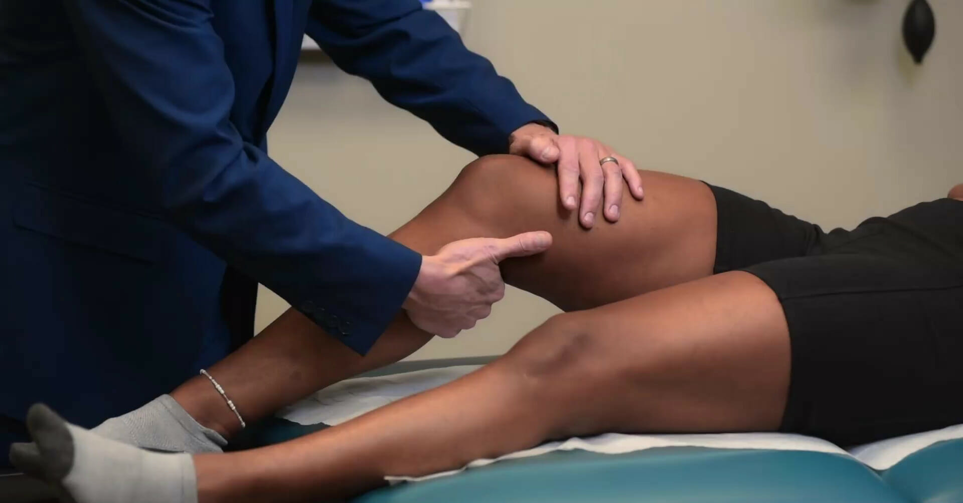
Examiner places one handHandThe hand constitutes the distal part of the upper limb and provides the fine, precise movements needed in activities of daily living. It consists of 5 metacarpal bones and 14 phalanges, as well as numerous muscles innervated by the median and ulnar nerves. Hand: Anatomy on the medial aspect of the knee while the ankle is supported by the examiner’s other armArmThe arm, or “upper arm” in common usage, is the region of the upper limb that extends from the shoulder to the elbow joint and connects inferiorly to the forearm through the cubital fossa. It is divided into 2 fascial compartments (anterior and posterior).Arm: Anatomy.
Apply varus force to the knee while holding the thumb in a position to monitor the joint line.
Assess for excessive joint opening, ligamentous laxity, or painPainAn unpleasant sensation induced by noxious stimuli which are detected by nerve endings of nociceptive neurons.Pain: Types and Pathways
Cruciate ligament stability:
Anterior cruciate ligamentAnterior Cruciate LigamentA strong ligament of the knee that originates from the posteromedial portion of the lateral condyle of the femur, passes anteriorly and inferiorly between the condyles, and attaches to the depression in front of the intercondylar eminence of the tibia.Knee Joint: Anatomy (ACLACLA strong ligament of the knee that originates from the posteromedial portion of the lateral condyle of the femur, passes anteriorly and inferiorly between the condyles, and attaches to the depression in front of the intercondylar eminence of the tibia.Knee Joint: Anatomy) injury is assessed by the anterior drawer, Lachman, and pivot shift tests.
Anterior drawer:
With the individual supine, the knee is bent at 90 degrees while the examiner stabilizes the legLegThe lower leg, or just “leg” in anatomical terms, is the part of the lower limb between the knee and the ankle joint. The bony structure is composed of the tibia and fibula bones, and the muscles of the leg are grouped into the anterior, lateral, and posterior compartments by extensions of fascia.Leg: Anatomy by placing their thighThighThe thigh is the region of the lower limb found between the hip and the knee joint. There is a single bone in the thigh called the femur, which is surrounded by large muscles grouped into 3 fascial compartments. Thigh: Anatomy on the footFootThe foot is the terminal portion of the lower limb, whose primary function is to bear weight and facilitate locomotion. The foot comprises 26 bones, including the tarsal bones, metatarsal bones, and phalanges. The bones of the foot form longitudinal and transverse arches and are supported by various muscles, ligaments, and tendons.Foot: Anatomy.
Place the hands behind the tibiaTibiaThe second longest bone of the skeleton. It is located on the medial side of the lower leg, articulating with the fibula laterally, the talus distally, and the femur proximally.Knee Joint: Anatomy and the thumbs over the tibial tuberosityTibial tuberosityLeg: Anatomy.
Pull the tibiaTibiaThe second longest bone of the skeleton. It is located on the medial side of the lower leg, articulating with the fibula laterally, the talus distally, and the femur proximally.Knee Joint: Anatomy anteriorly.
Excessive anterior translationTranslationTranslation is the process of synthesizing a protein from a messenger RNA (mRNA) transcript. This process is divided into three primary stages: initiation, elongation, and termination. Translation is catalyzed by structures known as ribosomes, which are large complexes of proteins and ribosomal RNA (rRNA). Stages and Regulation of Translation indicates rupture of the ACLACLA strong ligament of the knee that originates from the posteromedial portion of the lateral condyle of the femur, passes anteriorly and inferiorly between the condyles, and attaches to the depression in front of the intercondylar eminence of the tibia.Knee Joint: Anatomy.
With the individual supine, the knee is bent at 20–30 degrees.
One of the examiner’s hands stabilizes the distal femur while the other pulls the proximal tibiaTibiaThe second longest bone of the skeleton. It is located on the medial side of the lower leg, articulating with the fibula laterally, the talus distally, and the femur proximally.Knee Joint: Anatomy anteriorly.
Excessive anterior translationTranslationTranslation is the process of synthesizing a protein from a messenger RNA (mRNA) transcript. This process is divided into three primary stages: initiation, elongation, and termination. Translation is catalyzed by structures known as ribosomes, which are large complexes of proteins and ribosomal RNA (rRNA). Stages and Regulation of Translation indicates rupture of the ACLACLA strong ligament of the knee that originates from the posteromedial portion of the lateral condyle of the femur, passes anteriorly and inferiorly between the condyles, and attaches to the depression in front of the intercondylar eminence of the tibia.Knee Joint: Anatomy.
More sensitive and specific than the anterior drawer test.
Pivot shift:
Individual is positioned supine with the knee fully extended.
A “clunk” with flexionFlexionExamination of the Upper Limbs indicates rupture of the ACLACLA strong ligament of the knee that originates from the posteromedial portion of the lateral condyle of the femur, passes anteriorly and inferiorly between the condyles, and attaches to the depression in front of the intercondylar eminence of the tibia.Knee Joint: Anatomy.
Posterior cruciate ligamentPosterior Cruciate LigamentA strong ligament of the knee that originates from the anterolateral surface of the medial condyle of the femur, passes posteriorly and inferiorly between the condyles, and attaches to the posterior intercondylar area of the tibia.Knee Joint: Anatomy (PCLPCLA strong ligament of the knee that originates from the anterolateral surface of the medial condyle of the femur, passes posteriorly and inferiorly between the condyles, and attaches to the posterior intercondylar area of the tibia.Knee Joint: Anatomy) injury is assessed by the posterior drawer and quadriceps active tests.
Individual is positioned supine with the knee flexed at 90 degrees while the examiner stabilizes the legLegThe lower leg, or just “leg” in anatomical terms, is the part of the lower limb between the knee and the ankle joint. The bony structure is composed of the tibia and fibula bones, and the muscles of the leg are grouped into the anterior, lateral, and posterior compartments by extensions of fascia.Leg: Anatomy by placing their thighThighThe thigh is the region of the lower limb found between the hip and the knee joint. There is a single bone in the thigh called the femur, which is surrounded by large muscles grouped into 3 fascial compartments. Thigh: Anatomy on the footFootThe foot is the terminal portion of the lower limb, whose primary function is to bear weight and facilitate locomotion. The foot comprises 26 bones, including the tarsal bones, metatarsal bones, and phalanges. The bones of the foot form longitudinal and transverse arches and are supported by various muscles, ligaments, and tendons.Foot: Anatomy.
Both of the examiner’s hands are held at the individual’s proximal tibiaTibiaThe second longest bone of the skeleton. It is located on the medial side of the lower leg, articulating with the fibula laterally, the talus distally, and the femur proximally.Knee Joint: Anatomy while a posterior force is applied.
Excessive posterior translationTranslationTranslation is the process of synthesizing a protein from a messenger RNA (mRNA) transcript. This process is divided into three primary stages: initiation, elongation, and termination. Translation is catalyzed by structures known as ribosomes, which are large complexes of proteins and ribosomal RNA (rRNA). Stages and Regulation of Translation indicates rupture of the PCLPCLA strong ligament of the knee that originates from the anterolateral surface of the medial condyle of the femur, passes posteriorly and inferiorly between the condyles, and attaches to the posterior intercondylar area of the tibia.Knee Joint: Anatomy.
Individual is positioned supine and asked to raise the footFootThe foot is the terminal portion of the lower limb, whose primary function is to bear weight and facilitate locomotion. The foot comprises 26 bones, including the tarsal bones, metatarsal bones, and phalanges. The bones of the foot form longitudinal and transverse arches and are supported by various muscles, ligaments, and tendons.Foot: Anatomy off the table by actively flexing at the hip (this places the distal lower limb in a gravity-dependent position).
The individual is then asked to actively contract the quadriceps muscle.
The examiner assesses for anterior tibial movement (a tibiaTibiaThe second longest bone of the skeleton. It is located on the medial side of the lower leg, articulating with the fibula laterally, the talus distally, and the femur proximally.Knee Joint: Anatomy in posterior subluxationSubluxationRadial Head Subluxation (Nursemaid’s Elbow) will move anteriorly, indicating PCLPCLA strong ligament of the knee that originates from the anterolateral surface of the medial condyle of the femur, passes posteriorly and inferiorly between the condyles, and attaches to the posterior intercondylar area of the tibia.Knee Joint: Anatomy rupture).
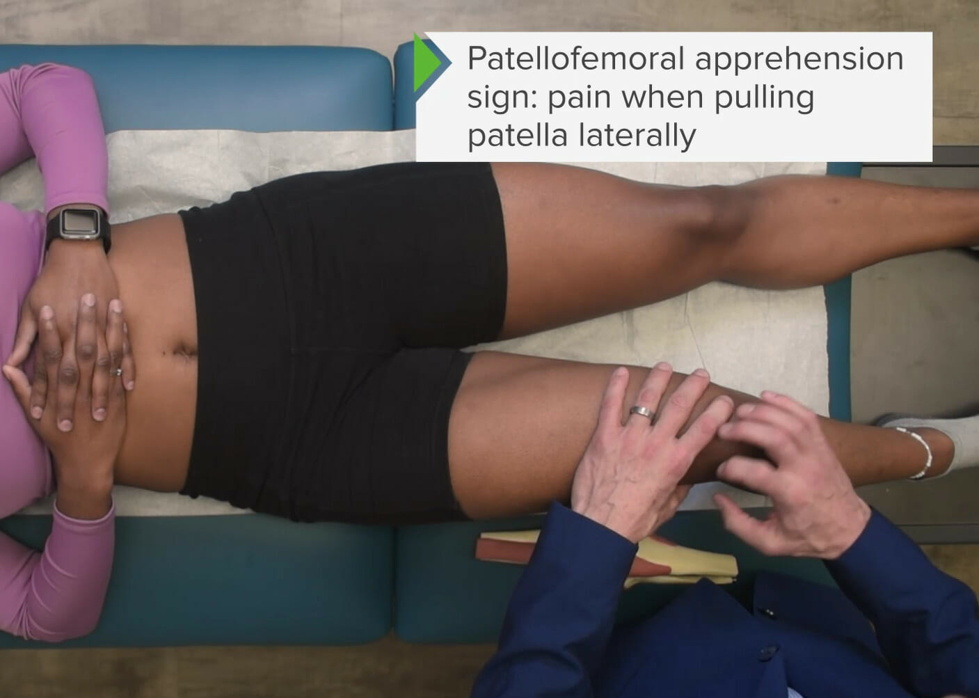
Patellar stability: patellar apprehension test
Tests for patellofemoral painPainAn unpleasant sensation induced by noxious stimuli which are detected by nerve endings of nociceptive neurons.Pain: Types and Pathways syndrome
With the individual supine, the knee is fully extended.
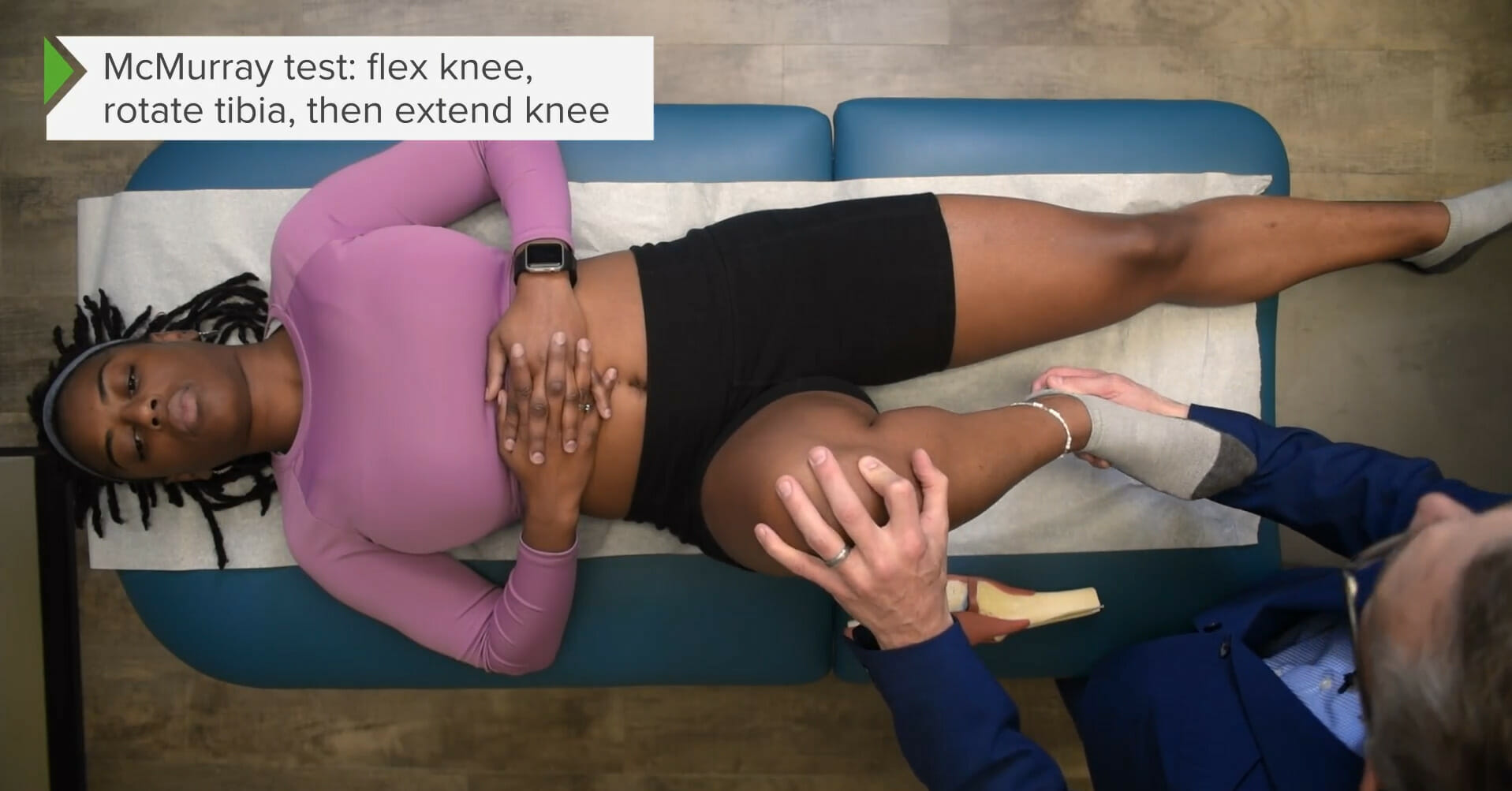
The examiner passively flexes the knee while applying varus stress and internally rotating the ankle.
The examiner then extends the knee smoothly while palpating the joint line.
Audible/palpable crepitusCrepitusOsteoarthritis (click or pop) or excessive discomfort indicates lateral meniscus injury.
Apley test:
Individual is placed in the prone position.
Knee is passively flexed to 90 degrees.
The examiner stabilizes the thighThighThe thigh is the region of the lower limb found between the hip and the knee joint. There is a single bone in the thigh called the femur, which is surrounded by large muscles grouped into 3 fascial compartments. Thigh: Anatomy with one handHandThe hand constitutes the distal part of the upper limb and provides the fine, precise movements needed in activities of daily living. It consists of 5 metacarpal bones and 14 phalanges, as well as numerous muscles innervated by the median and ulnar nerves. Hand: Anatomy and with the other handHandThe hand constitutes the distal part of the upper limb and provides the fine, precise movements needed in activities of daily living. It consists of 5 metacarpal bones and 14 phalanges, as well as numerous muscles innervated by the median and ulnar nerves. Hand: Anatomy applies compressionCompressionBlunt Chest Trauma through the tibiaTibiaThe second longest bone of the skeleton. It is located on the medial side of the lower leg, articulating with the fibula laterally, the talus distally, and the femur proximally.Knee Joint: Anatomy to the knee while passively carrying the ankle into internal and external rotationExternal RotationExamination of the Upper Limbs.
Excessive discomfort indicates meniscal injury.
Tests for iliotibial bandIliotibial bandThigh: AnatomyinflammationInflammationInflammation is a complex set of responses to infection and injury involving leukocytes as the principal cellular mediators in the body’s defense against pathogenic organisms. Inflammation is also seen as a response to tissue injury in the process of wound healing. The 5 cardinal signs of inflammation are pain, heat, redness, swelling, and loss of function. Inflammation:
The examiner supports the ankle with one handHandThe hand constitutes the distal part of the upper limb and provides the fine, precise movements needed in activities of daily living. It consists of 5 metacarpal bones and 14 phalanges, as well as numerous muscles innervated by the median and ulnar nerves. Hand: Anatomy and with the other handHandThe hand constitutes the distal part of the upper limb and provides the fine, precise movements needed in activities of daily living. It consists of 5 metacarpal bones and 14 phalanges, as well as numerous muscles innervated by the median and ulnar nerves. Hand: Anatomy palpates the lateral epicondyleLateral epicondyleArm: Anatomy of the femur while passively carrying the knee through flexionFlexionExamination of the Upper Limbs and extensionExtensionExamination of the Upper Limbs (0–90 degrees).
A test is positive when painPainAn unpleasant sensation induced by noxious stimuli which are detected by nerve endings of nociceptive neurons.Pain: Types and Pathways is produced at the lateral epicondyleLateral epicondyleArm: Anatomy as the IT band passes over the bony landmark with dynamic movement.
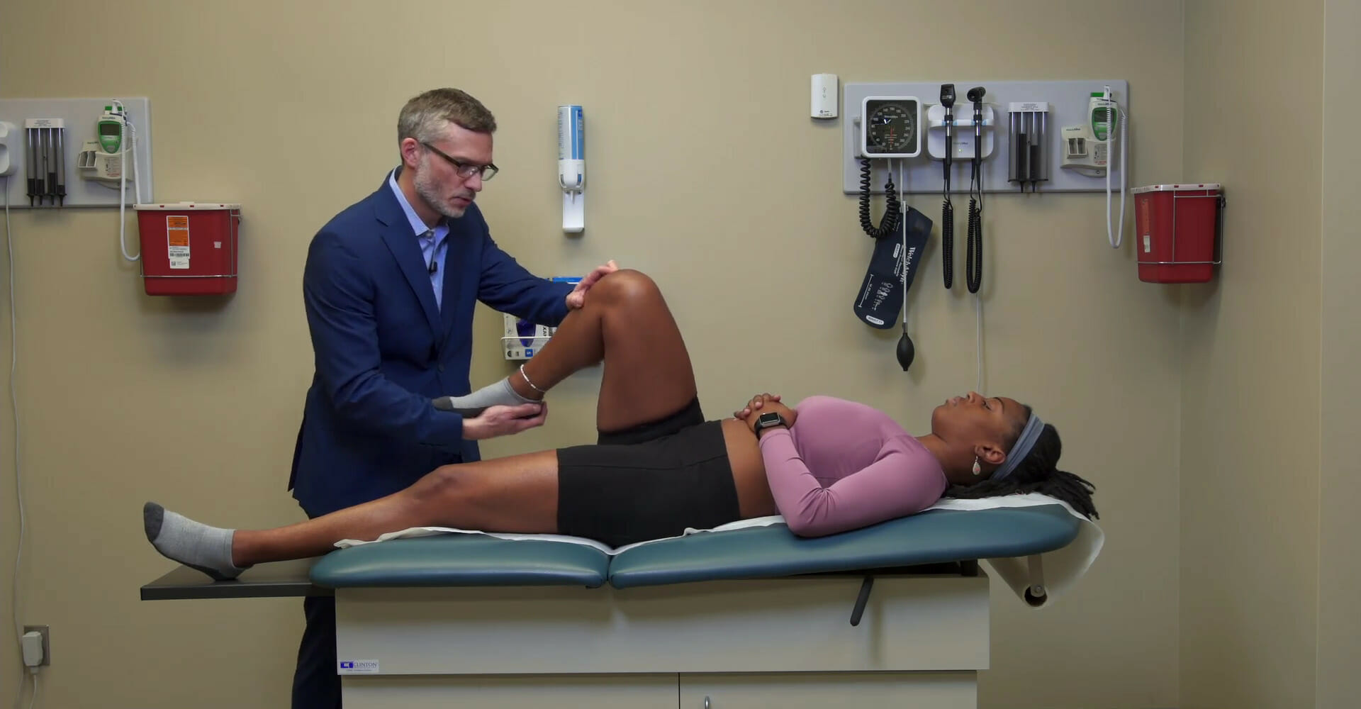
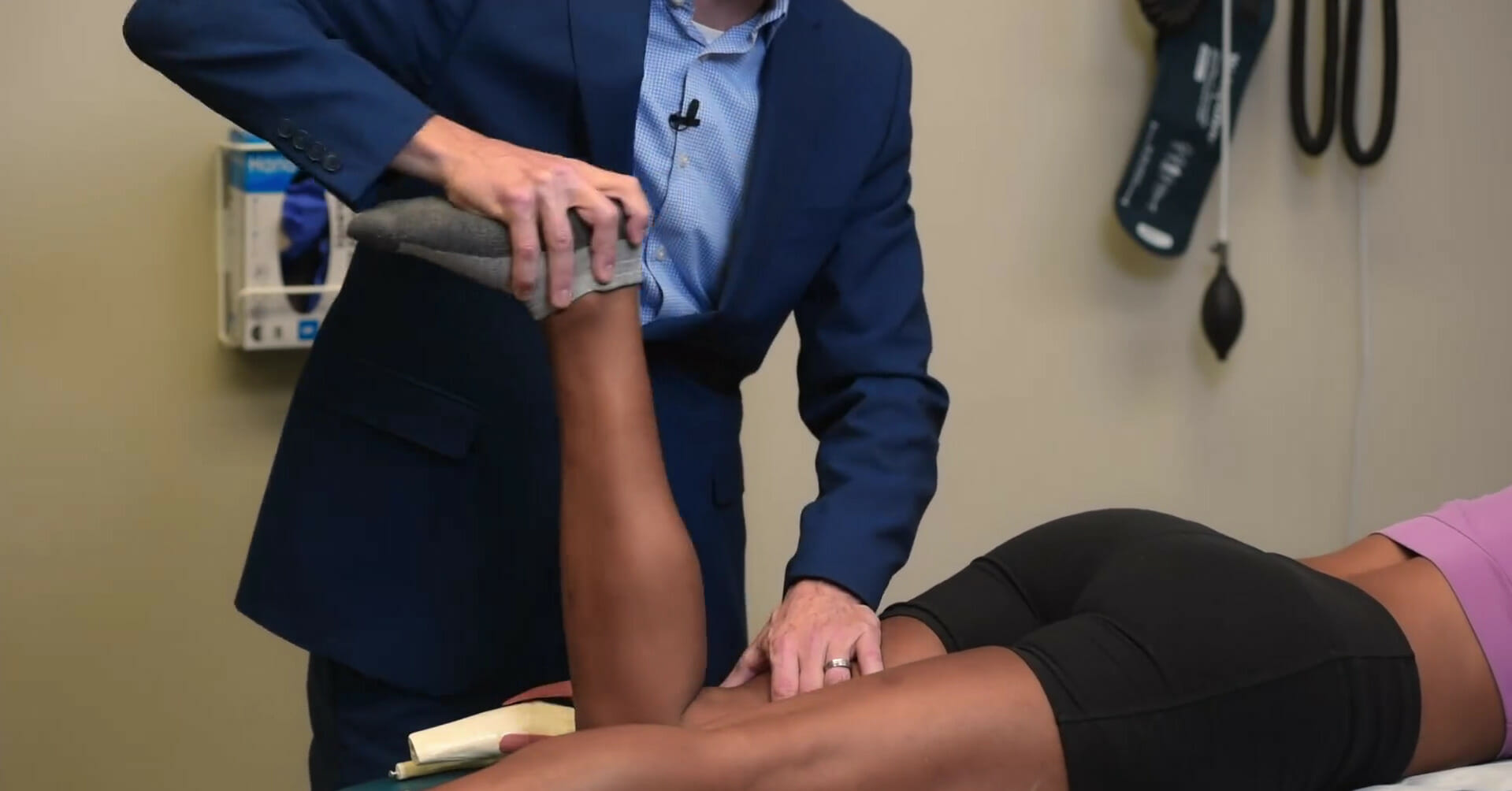
McMurray test: External rotation with varus stress to test for medial meniscus tear
Image by Lecturio.
McMurray test: Internal rotation with valgus stress to test for lateral meniscus tear
Image by Lecturio.
Apley test for meniscus tear
Image by Lecturio.
Noble test for iliotibial band syndrome
Image by Lecturio.
Vascular exam of the knee
The popliteal pulsePopliteal pulsePopliteal Fossa: Anatomy can be palpated at the popliteal fossaPopliteal fossaThe popliteal fossa or the “knee pit” is a diamond-shaped, fat-filled, shallow depression on the posterior aspect of the knee joint. The popliteal fossa is located at the dorsal aspect of the knee and contains an increased number of lymph nodes as well as structures of the neurovascular system that travel from the thigh to the lower leg.Popliteal Fossa: Anatomy:
Directly posterior to the joint line
Located between the tendons of the hamstrings muscle complex
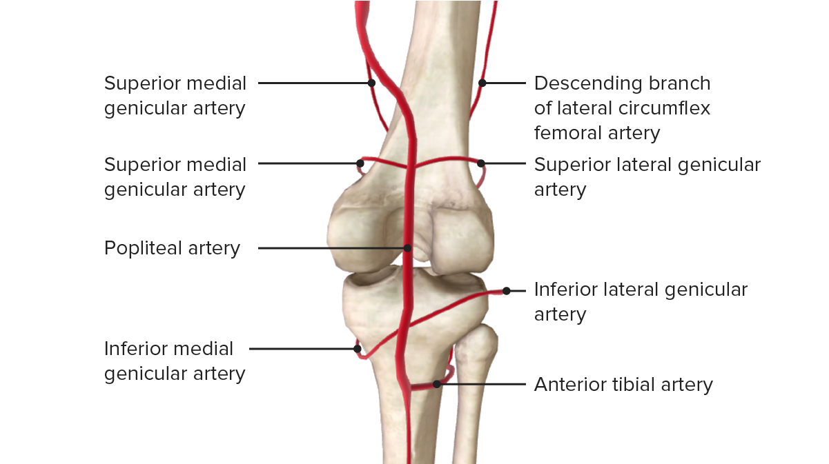
Posterior view of the vascular supply of the knee joint
Assess the arch of the footFootThe foot is the terminal portion of the lower limb, whose primary function is to bear weight and facilitate locomotion. The foot comprises 26 bones, including the tarsal bones, metatarsal bones, and phalanges. The bones of the foot form longitudinal and transverse arches and are supported by various muscles, ligaments, and tendons.Foot: Anatomy:
PesPESRemoval of plasma and replacement with various fluids, e.g., fresh frozen plasma, plasma protein fractions (ppf), albumin preparations, dextran solutions, saline. Used in treatment of autoimmune diseases, immune complex diseases, diseases of excess plasma factors, and other conditions.Thrombotic Thrombocytopenic Purpura cavus (exaggerated arch)
Pes planusPes PlanusEhlers-Danlos Syndrome (flat footFootThe foot is the terminal portion of the lower limb, whose primary function is to bear weight and facilitate locomotion. The foot comprises 26 bones, including the tarsal bones, metatarsal bones, and phalanges. The bones of the foot form longitudinal and transverse arches and are supported by various muscles, ligaments, and tendons.Foot: Anatomy)
SwellingSwellingInflammation of the 1st metatarsophalangeal jointMetatarsophalangeal JointFoot: Anatomy may indicate goutGoutGout is a heterogeneous metabolic disease associated with elevated serum uric acid levels (> 6.8 mg/dL) and abnormal deposits of monosodium urate in tissues. The condition is often familial and is initially characterized by painful, recurring, and usually monoarticular acute arthritis, or “gout flare,” followed later by chronic deforming arthritis. Gout.
EcchymosisEcchymosisExtravasation of blood into the skin, resulting in a nonelevated, rounded or irregular, blue or purplish patch, larger than a petechia.Orbital Fractures
Scars
Callous
Wounds/ulcers (especially in the diabetic footFootThe foot is the terminal portion of the lower limb, whose primary function is to bear weight and facilitate locomotion. The foot comprises 26 bones, including the tarsal bones, metatarsal bones, and phalanges. The bones of the foot form longitudinal and transverse arches and are supported by various muscles, ligaments, and tendons.Foot: Anatomy)
PalpationPalpationApplication of fingers with light pressure to the surface of the body to determine consistency of parts beneath in physical diagnosis; includes palpation for determining the outlines of organs.Dermatologic Examination and percussionPercussionAct of striking a part with short, sharp blows as an aid in diagnosing the condition beneath the sound obtained.Pulmonary Examination
Palpatory techniques of the following structures is performed, looking for tenderness, temperature, and swellingSwellingInflammation:
Proximal fibulaFibulaThe bone of the lower leg lateral to and smaller than the tibia. In proportion to its length, it is the most slender of the long bones.Leg: Anatomy (fibular head fractureFractureA fracture is a disruption of the cortex of any bone and periosteum and is commonly due to mechanical stress after an injury or accident. Open fractures due to trauma can be a medical emergency. Fractures are frequently associated with automobile accidents, workplace injuries, and trauma.Overview of Bone Fractures)
Tarsal bonesTarsal BonesThe seven bones which form the tarsus – namely, calcaneus; talus; cuboid, navicular, and the internal, middle, and external cuneiforms.Foot: Anatomy
Base of the 5th metatarsal (5th metatarsal fractures common)
1st metatarsophalangeal jointMetatarsophalangeal JointFoot: Anatomy (goutGoutGout is a heterogeneous metabolic disease associated with elevated serum uric acid levels (> 6.8 mg/dL) and abnormal deposits of monosodium urate in tissues. The condition is often familial and is initially characterized by painful, recurring, and usually monoarticular acute arthritis, or “gout flare,” followed later by chronic deforming arthritis. Gout, bunyon, arthritisArthritisAcute or chronic inflammation of joints.Osteoarthritis, capsulitis)
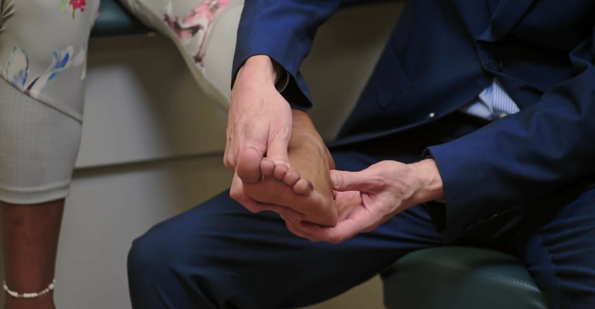
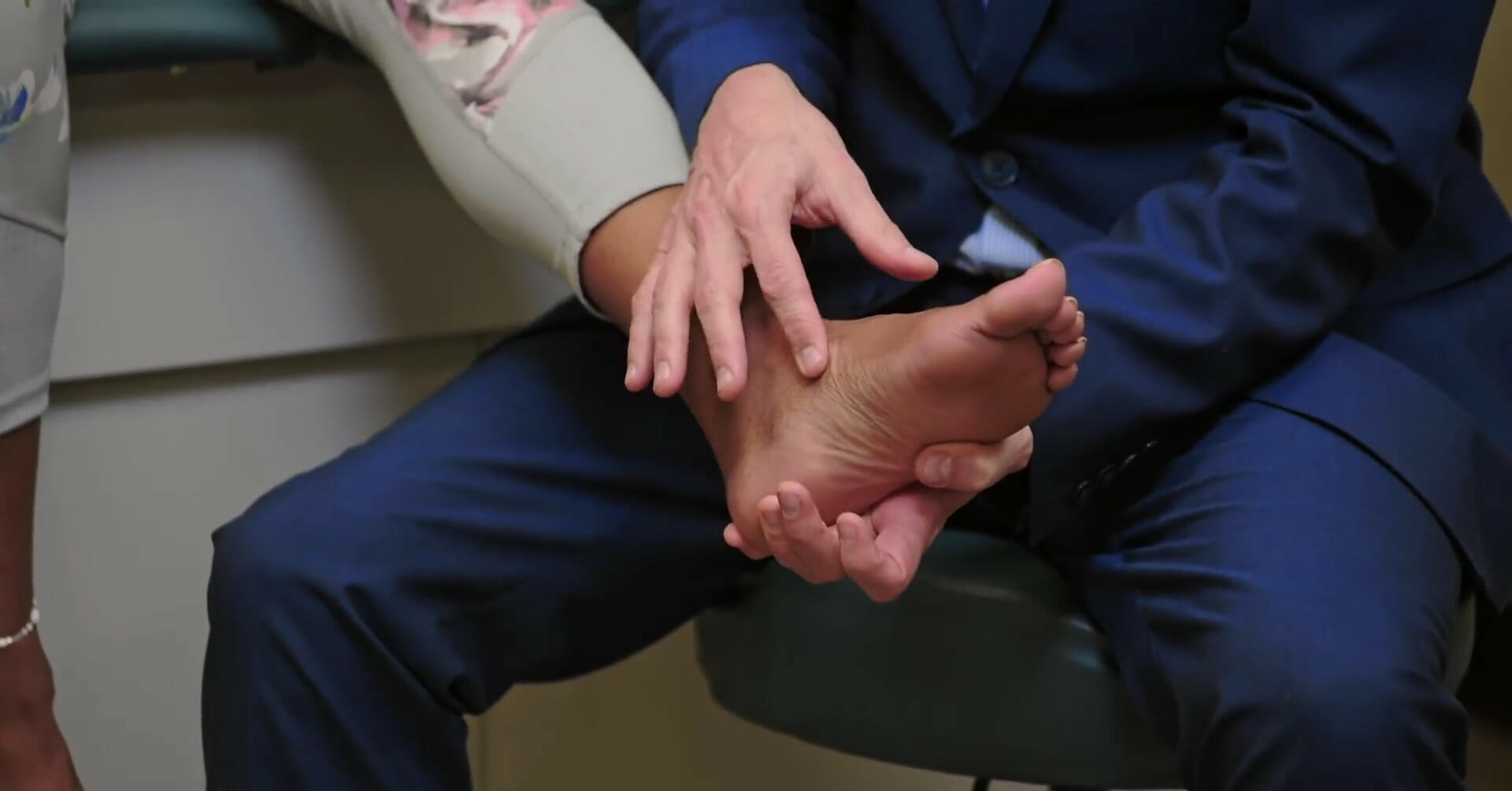
Anterior calcaneusCalcaneusThe largest of the tarsal bones which is situated at the lower and back part of the foot, forming the heel.Foot: Anatomy (tender at this point → plantar fasciitisPlantar fasciitisInflammation of the plantar fascia (aponeurosis) on the bottom of the foot causing heel pain. The etiology of plantar fasciitis remains controversial but is likely to involve a biomechanical imbalance. Though often presenting along with heel spur, they do not appear to be causally related.Ankle and Foot Pain)
Posterior calcaneusCalcaneusThe largest of the tarsal bones which is situated at the lower and back part of the foot, forming the heel.Foot: Anatomy/Achilles tendon (tender at this point → Achilles tendinitisTendinitisAnkylosing Spondylitis)
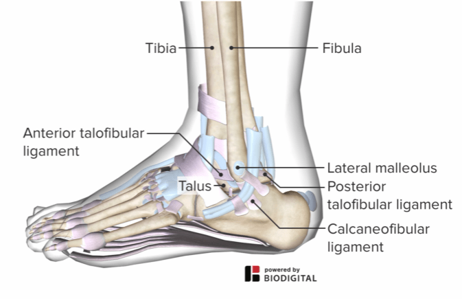
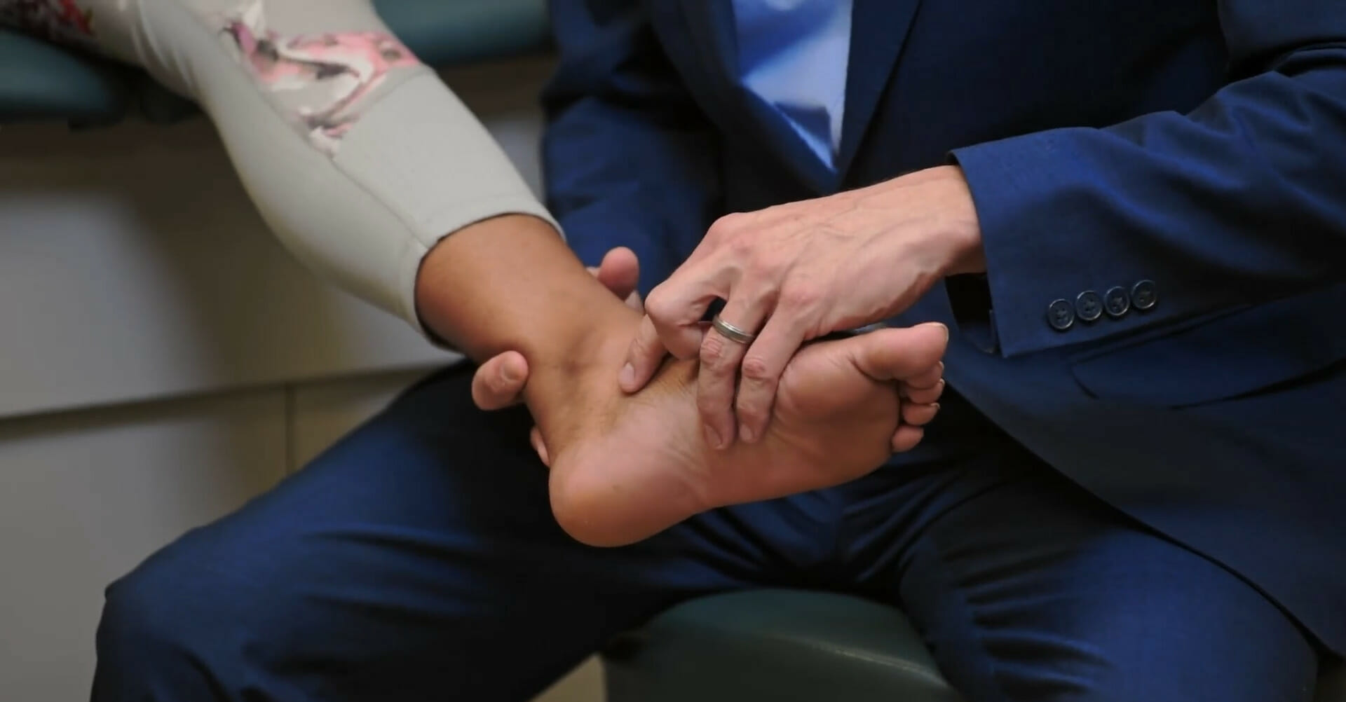
Palpation of the anterior talofibular ligament (ATFL) attachment
Image by Lecturio.
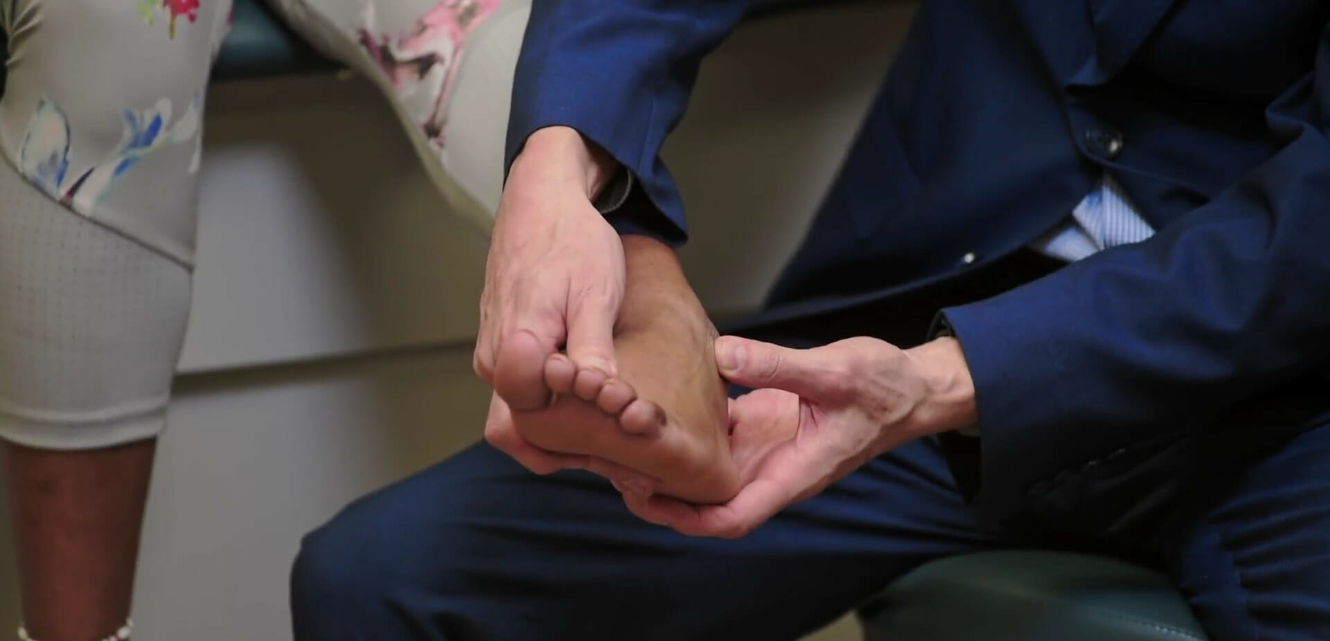
Palpation of the calcaneofibular ligament (CFL)
Image by Lecturio.
Palpation of the navicular bone
Image by Lecturio.
Palpation of the metatarsophalangeal joints
Image by Lecturio.
MotorMotorNeurons which send impulses peripherally to activate muscles or secretory cells.Nervous System: Histology strength
Dorsiflexion:
L4 nerve root
Assess strength in dorsiflexion against examiner’s resistanceResistancePhysiologically, the opposition to flow of air caused by the forces of friction. As a part of pulmonary function testing, it is the ratio of driving pressure to the rate of air flow.Ventilation: Mechanics of Breathing (weakness leads to foot dropFoot DropLeprosy).
Assess strength in plantar flexionFlexionExamination of the Upper Limbs against examiner’s resistanceResistancePhysiologically, the opposition to flow of air caused by the forces of friction. As a part of pulmonary function testing, it is the ratio of driving pressure to the rate of air flow.Ventilation: Mechanics of Breathing.
Normal range: 40–50 degrees
Inversion:
Resisted inversion: tests the integrity of the posterior tibial tendons
Achilles reflex: tests the reflex arc involving the L5 and S1S1Heart Sounds segments of the spinal cordSpinal cordThe spinal cord is the major conduction pathway connecting the brain to the body; it is part of the CNS. In cross section, the spinal cord is divided into an H-shaped area of gray matter (consisting of synapsing neuronal cell bodies) and a surrounding area of white matter (consisting of ascending and descending tracts of myelinated axons). Spinal Cord: Anatomy
Special tests
Calf-squeeze test (Thompson test):
Assesses for rupture of the Achilles tendon
May be carried out with the individual sitting on the edge of the examining table or lying prone with feet hanging over the edge
Observe for plantar flexionFlexionExamination of the Upper Limbs of the ipsilateral footFootThe foot is the terminal portion of the lower limb, whose primary function is to bear weight and facilitate locomotion. The foot comprises 26 bones, including the tarsal bones, metatarsal bones, and phalanges. The bones of the foot form longitudinal and transverse arches and are supported by various muscles, ligaments, and tendons.Foot: Anatomy.
If complete rupture → the footFootThe foot is the terminal portion of the lower limb, whose primary function is to bear weight and facilitate locomotion. The foot comprises 26 bones, including the tarsal bones, metatarsal bones, and phalanges. The bones of the foot form longitudinal and transverse arches and are supported by various muscles, ligaments, and tendons.Foot: Anatomy will remain neutral or dorsiflexed.
One of the examiner’s hands stabilizes the lower legLegThe lower leg, or just “leg” in anatomical terms, is the part of the lower limb between the knee and the ankle joint. The bony structure is composed of the tibia and fibula bones, and the muscles of the leg are grouped into the anterior, lateral, and posterior compartments by extensions of fascia.Leg: Anatomy and the other applies an anterior force at the heel.
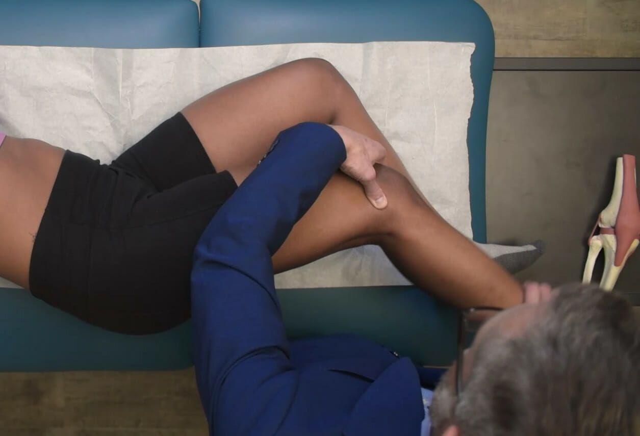
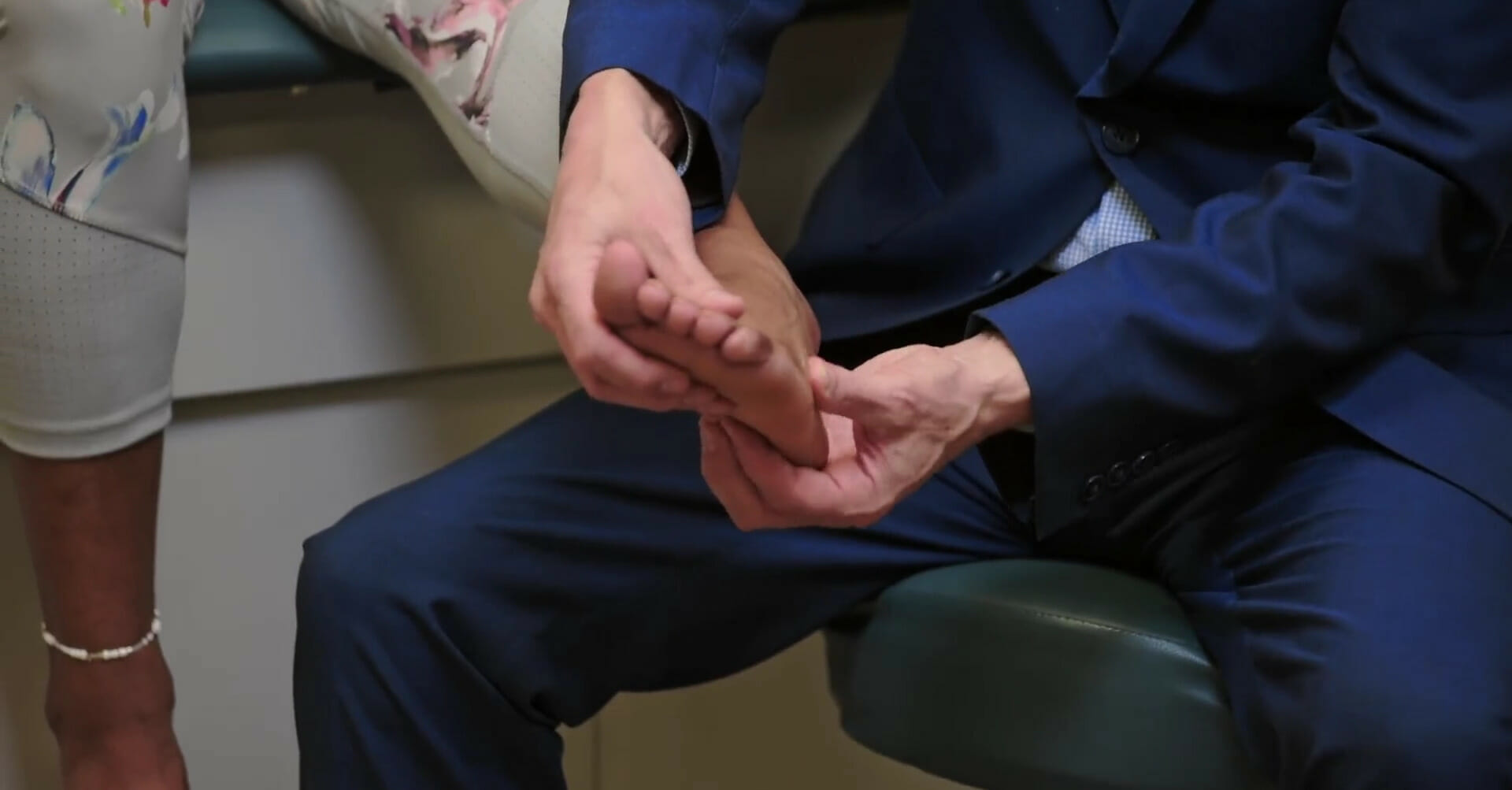
Heel painPainAn unpleasant sensation induced by noxious stimuli which are detected by nerve endings of nociceptive neurons.Pain: Types and Pathways with passive dorsiflexion of the toes indicates presence of plantar fasciitisPlantar fasciitisInflammation of the plantar fascia (aponeurosis) on the bottom of the foot causing heel pain. The etiology of plantar fasciitis remains controversial but is likely to involve a biomechanical imbalance. Though often presenting along with heel spur, they do not appear to be causally related.Ankle and Foot Pain.
Windlass test: Pain at the calcaneal attachment of the plantar aponeurosis with passive dorsiflexion of the toes indicates plantar fasciitis.
Image by Lecturio.
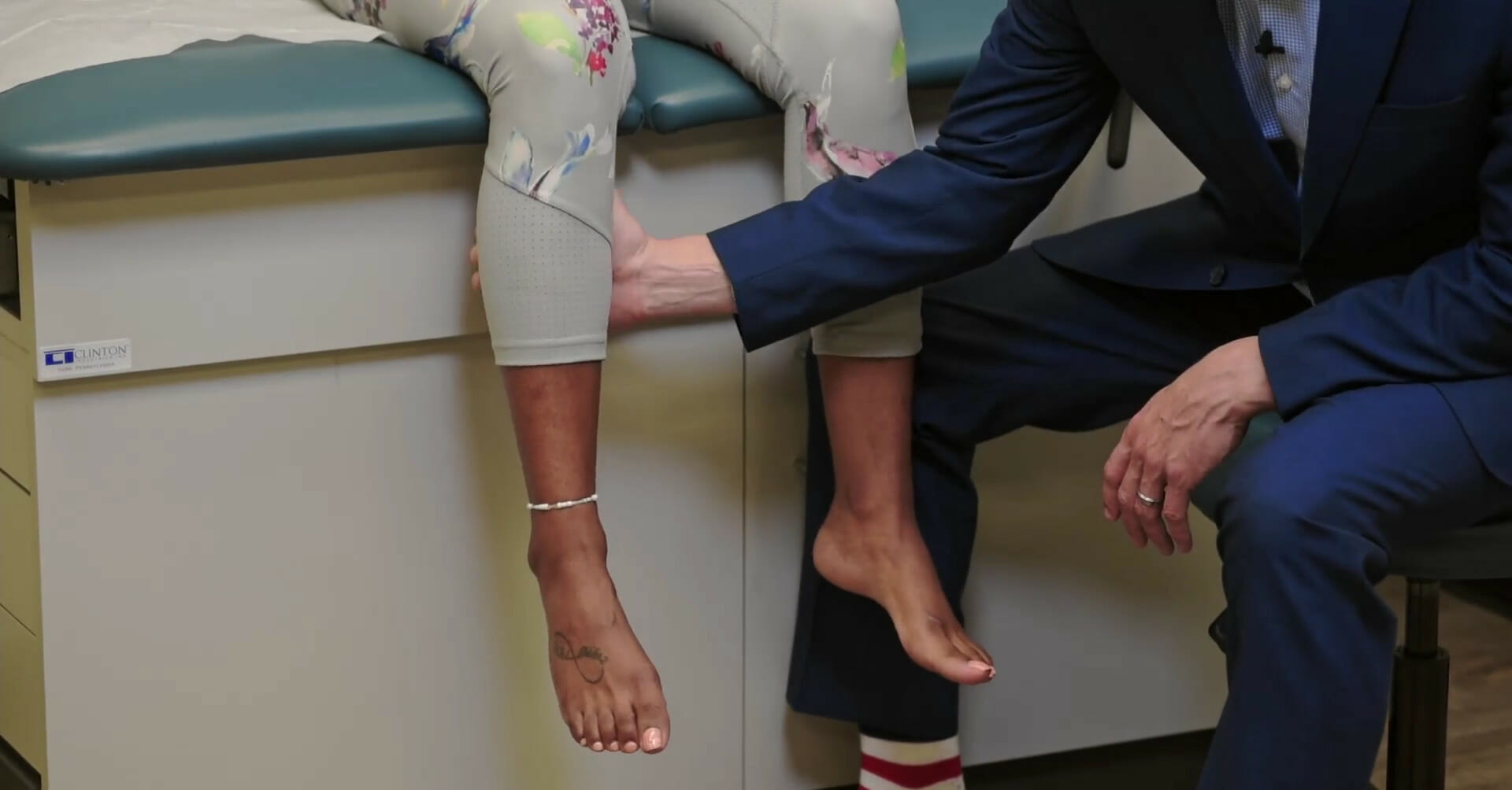
Talar tilt test
Image by Lecturio.
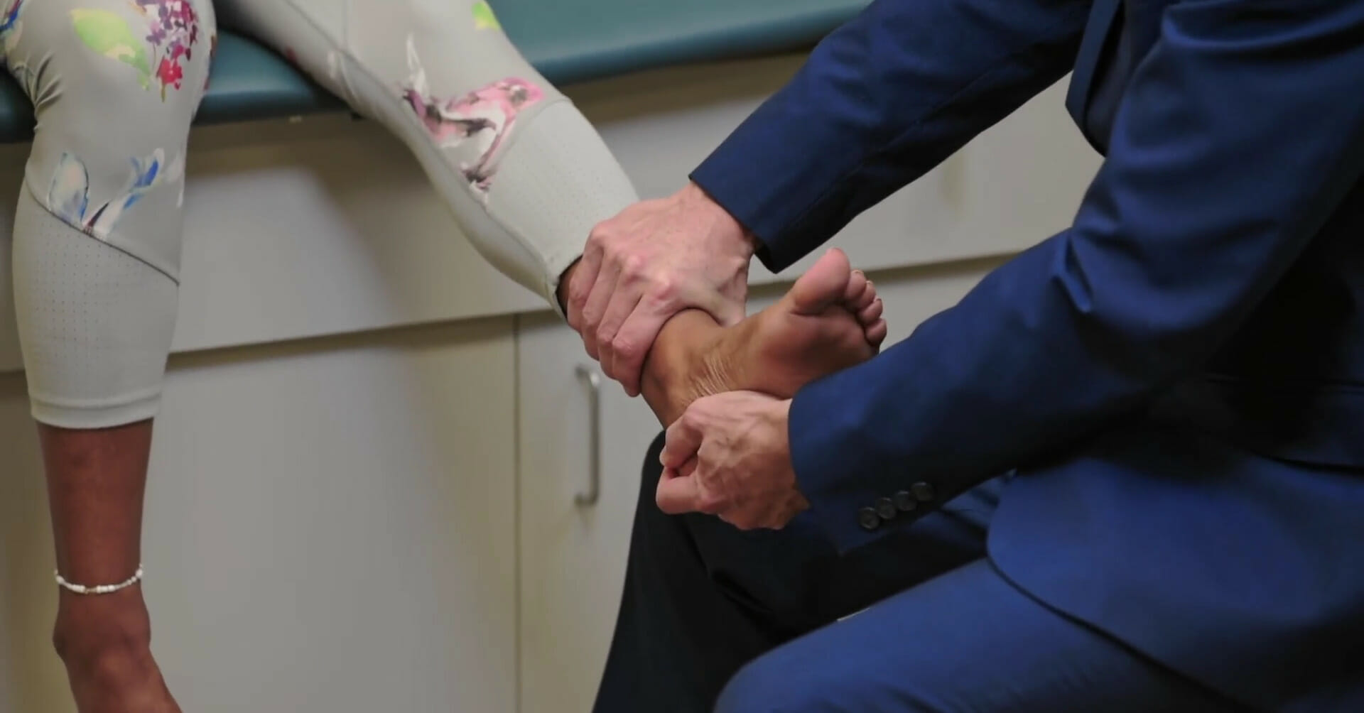
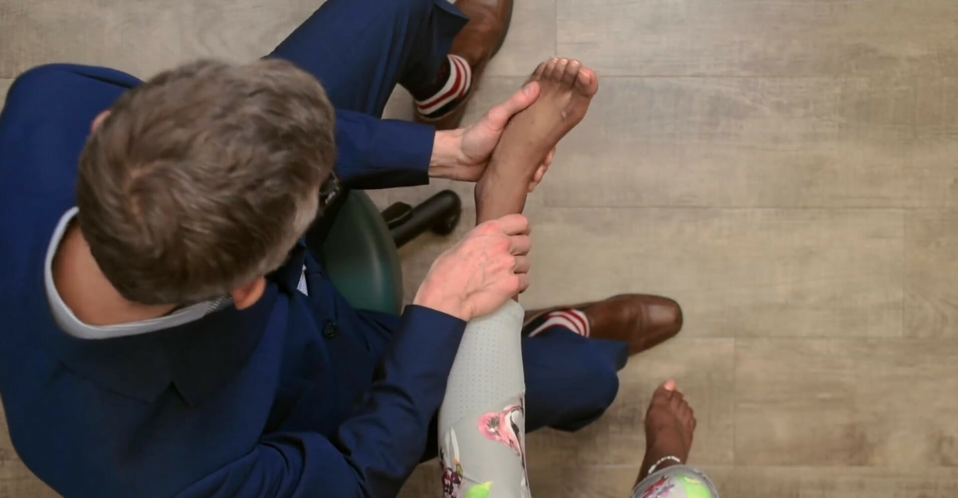
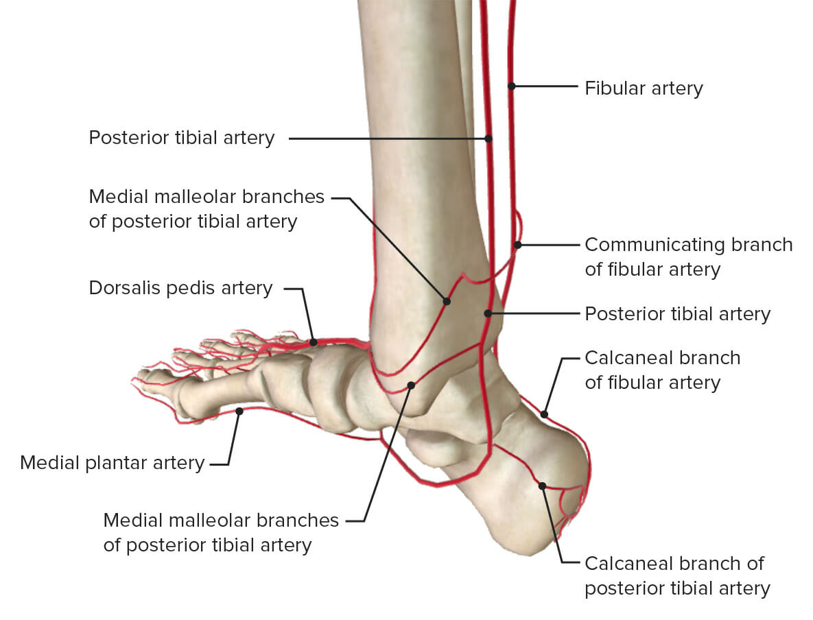
Vascular exam of the ankle
The dorsalis pedis pulse can be palpated at the mid-dorsal footFootThe foot is the terminal portion of the lower limb, whose primary function is to bear weight and facilitate locomotion. The foot comprises 26 bones, including the tarsal bones, metatarsal bones, and phalanges. The bones of the foot form longitudinal and transverse arches and are supported by various muscles, ligaments, and tendons.Foot: Anatomy.
Table: Overview of the most common conditions seen for knee painPainAn unpleasant sensation induced by noxious stimuli which are detected by nerve endings of nociceptive neurons.Pain: Types and Pathways
PalpationPalpationApplication of fingers with light pressure to the surface of the body to determine consistency of parts beneath in physical diagnosis; includes palpation for determining the outlines of organs.Dermatologic Examination
Effusion
Range of motionRange of motionThe distance and direction to which a bone joint can be extended. Range of motion is a function of the condition of the joints, muscles, and connective tissues involved. Joint flexibility can be improved through appropriate muscle strength exercises.Examination of the Upper Limbs
Special tests
OsteoarthritisOsteoarthritisOsteoarthritis (OA) is the most common form of arthritis, and is due to cartilage destruction and changes of the subchondral bone. The risk of developing this disorder increases with age, obesity, and repetitive joint use or trauma. Patients develop gradual joint pain, stiffness lasting < 30 minutes, and decreased range of motion. Osteoarthritis
Osteophytes and tender joint line
+/–
Limited by painPainAn unpleasant sensation induced by noxious stimuli which are detected by nerve endings of nociceptive neurons.Pain: Types and Pathways
Plain film radiography:
Joint space narrowing
Osteophytes
Meniscus tearMeniscus tearThe menisci are fibrocartilaginous wedge-shaped structures between the distal femur and proximal tibia that stabilize and dissipate weight-bearing forces at the knee joint. A meniscus tear is an injury to the meniscus caused by rotational or shearing forces across the tibiofemoral joint. Meniscus Tear
++ (ACLACLA strong ligament of the knee that originates from the posteromedial portion of the lateral condyle of the femur, passes anteriorly and inferiorly between the condyles, and attaches to the depression in front of the intercondylar eminence of the tibia.Knee Joint: Anatomy tears)
GoutGoutGout is a heterogeneous metabolic disease associated with elevated serum uric acid levels (> 6.8 mg/dL) and abnormal deposits of monosodium urate in tissues. The condition is often familial and is initially characterized by painful, recurring, and usually monoarticular acute arthritis, or “gout flare,” followed later by chronic deforming arthritis. Gout
Warm joint
++
Limited by painPainAn unpleasant sensation induced by noxious stimuli which are detected by nerve endings of nociceptive neurons.Pain: Types and Pathways
Joint aspiration and analysis
PesPESRemoval of plasma and replacement with various fluids, e.g., fresh frozen plasma, plasma protein fractions (ppf), albumin preparations, dextran solutions, saline. Used in treatment of autoimmune diseases, immune complex diseases, diseases of excess plasma factors, and other conditions.Thrombotic Thrombocytopenic Purpura anserine bursitis
Table: Most common conditions seen with ankle/footFootThe foot is the terminal portion of the lower limb, whose primary function is to bear weight and facilitate locomotion. The foot comprises 26 bones, including the tarsal bones, metatarsal bones, and phalanges. The bones of the foot form longitudinal and transverse arches and are supported by various muscles, ligaments, and tendons.Foot: AnatomypainPainAn unpleasant sensation induced by noxious stimuli which are detected by nerve endings of nociceptive neurons.Pain: Types and Pathways
PalpationPalpationApplication of fingers with light pressure to the surface of the body to determine consistency of parts beneath in physical diagnosis; includes palpation for determining the outlines of organs.Dermatologic Examination
Ottawa ankle rules: an ankle x-rayX-rayPenetrating electromagnetic radiation emitted when the inner orbital electrons of an atom are excited and release radiant energy. X-ray wavelengths range from 1 pm to 10 nm. Hard x-rays are the higher energy, shorter wavelength x-rays. Soft x-rays or grenz rays are less energetic and longer in wavelength. The short wavelength end of the x-ray spectrum overlaps the gamma rays wavelength range. The distinction between gamma rays and x-rays is based on their radiation source.Pulmonary Function Tests is indicated for any of the following:
PainPainAn unpleasant sensation induced by noxious stimuli which are detected by nerve endings of nociceptive neurons.Pain: Types and Pathways in the malleolar zone
Inability to bear weight for 4 steps
Bony tenderness along the distal 6 cm of the posterior edge of the fibulaFibulaThe bone of the lower leg lateral to and smaller than the tibia. In proportion to its length, it is the most slender of the long bones.Leg: Anatomy/tibiaTibiaThe second longest bone of the skeleton. It is located on the medial side of the lower leg, articulating with the fibula laterally, the talus distally, and the femur proximally.Knee Joint: Anatomy of the lateral/medial malleolusMedial malleolusAnkle Joint: Anatomy
Plantar fasciitisPlantar fasciitisInflammation of the plantar fascia (aponeurosis) on the bottom of the foot causing heel pain. The etiology of plantar fasciitis remains controversial but is likely to involve a biomechanical imbalance. Though often presenting along with heel spur, they do not appear to be causally related.Ankle and Foot Pain
Tenderness at the insertion point on the calcaneusCalcaneusThe largest of the tarsal bones which is situated at the lower and back part of the foot, forming the heel.Foot: Anatomy (plantar surface)
Windlass test
Loss of arch might be noted
Tarsal tunnel syndromeTarsal tunnel syndromeEntrapment of the distal branches of the posterior tibial nerve (which divides into the medial plantar, lateral plantar, and calcaneal nerves) in the tarsal tunnel, which lies posterior to the internal malleolus and beneath the retinaculum of the flexor muscles of the foot. Symptoms include ankle pain radiating into the foot which tends to be aggravated by walking. Examination may reveal tinel’s sign (radiating pain following nerve percussion) over the tibial nerve at the ankle, weakness and atrophy of the small foot muscles, or loss of sensation in the foot.Ankle and Foot Pain
PalpationPalpationApplication of fingers with light pressure to the surface of the body to determine consistency of parts beneath in physical diagnosis; includes palpation for determining the outlines of organs.Dermatologic Examination may reproduce symptoms of tinglingTinglingPosterior Cord Syndrome/burning radiating to the soles
Palpating along the tendon may show a gap (rupture) or tenderness at insertion (superior calcaneusCalcaneusThe largest of the tarsal bones which is situated at the lower and back part of the foot, forming the heel.Foot: Anatomy).
Thompson test
References
Macleod J, Munro JF, Edwards CRW, University of Edinburgh. (1990). Macleod’s Clinical Examination. Edinburgh: Churchill Livingstone.