An orbital fractureFractureA fracture is a disruption of the cortex of any bone and periosteum and is commonly due to mechanical stress after an injury or accident. Open fractures due to trauma can be a medical emergency. Fractures are frequently associated with automobile accidents, workplace injuries, and trauma.Overview of Bone Fractures is a break in the continuity of one or multiple bones of the eye socket, caused by direct or indirect trauma to the orbit. PatientsPatientsIndividuals participating in the health care system for the purpose of receiving therapeutic, diagnostic, or preventive procedures.Clinician–Patient Relationship frequently present with lacerations around the eye, orbital painPainAn unpleasant sensation induced by noxious stimuli which are detected by nerve endings of nociceptive neurons.Pain: Types and Pathways, edemaEdemaEdema is a condition in which excess serous fluid accumulates in the body cavity or interstitial space of connective tissues. Edema is a symptom observed in several medical conditions. It can be categorized into 2 types, namely, peripheral (in the extremities) and internal (in an organ or body cavity). Edema, ecchymosis, diplopiaDiplopiaA visual symptom in which a single object is perceived by the visual cortex as two objects rather than one. Disorders associated with this condition include refractive errors; strabismus; oculomotor nerve diseases; trochlear nerve diseases; abducens nerve diseases; and diseases of the brain stem and occipital lobe.Myasthenia Gravis on upward gaze, numbness around the eye, and signs of muscle entrapment. Diagnosis is based on clinical exam and imaging. The mainstay of management is to prevent further injury to the eye while determining whether surgery is needed. Complications include orbital compartment syndromeCompartment SyndromeCompartment syndrome is a surgical emergency usually occurring secondary to trauma. The condition is marked by increased pressure within a compartment that compromises the circulation and function of the tissues within that space.Compartment Syndrome, blindnessBlindnessThe inability to see or the loss or absence of perception of visual stimuli. This condition may be the result of eye diseases; optic nerve diseases; optic chiasm diseases; or brain diseases affecting the visual pathways or occipital lobe.Retinopathy of Prematurity, and persistent diplopiaDiplopiaA visual symptom in which a single object is perceived by the visual cortex as two objects rather than one. Disorders associated with this condition include refractive errors; strabismus; oculomotor nerve diseases; trochlear nerve diseases; abducens nerve diseases; and diseases of the brain stem and occipital lobe.Myasthenia Gravis.
An orbital fractureFractureA fracture is a disruption of the cortex of any bone and periosteum and is commonly due to mechanical stress after an injury or accident. Open fractures due to trauma can be a medical emergency. Fractures are frequently associated with automobile accidents, workplace injuries, and trauma.Overview of Bone Fractures is a broken boneBoneBone is a compact type of hardened connective tissue composed of bone cells, membranes, an extracellular mineralized matrix, and central bone marrow. The 2 primary types of bone are compact and spongy. Bones: Structure and Types involving the eye socket, either in the orbital rim, the orbital floor, or both.
Epidemiology
Orbital fractures account for approximately 3% of all patientsPatientsIndividuals participating in the health care system for the purpose of receiving therapeutic, diagnostic, or preventive procedures.Clinician–Patient Relationship seen in the ED.
More common in men than in women (4:1)
More common in adults than in children
Average age: 30 years
Etiology
The most common cause of orbital fractures is blunt force trauma to the eye.
About 85% of traumatic eye injuries (including orbital fractures) are caused by:
Auto accidents: most common cause of maxillofacial traumaMaxillofacial traumaGeneral or unspecified injuries involving the face and jaw (either upper, lower, or both).Basic Procedures
Contact sports
Occupational accidents
Home repair projects
About 15% are due to violent assaults.
For fractures in adult women, it is important to assess for domestic violence.
Anatomy and Pathophysiology
To understand the pathophysiology of orbital fractures, it is important to understand the anatomy of the orbit, the clinical presentation, and the potential consequences of fractures.
Anatomy
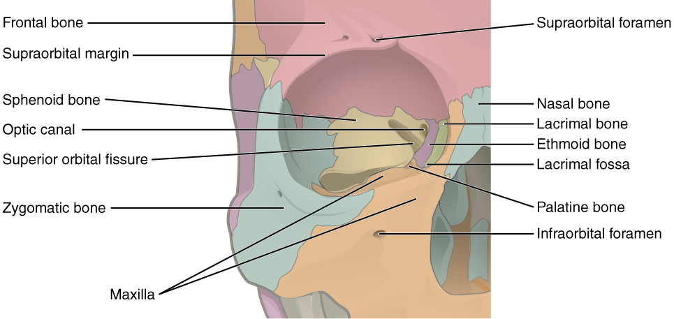
The 7 bones of the orbit are:
MaxillaMaxillaOne of a pair of irregularly shaped bones that form the upper jaw. A maxillary bone provides tooth sockets for the superior teeth, forms part of the orbit, and contains the maxillary sinus.Skull: Anatomy
ZygomaticZygomaticEither of a pair of bones that form the prominent part of the cheek and contribute to the orbit on each side of the skull.Skull: Anatomy
FrontalFrontalThe bone that forms the frontal aspect of the skull. Its flat part forms the forehead, articulating inferiorly with the nasal bone and the cheek bone on each side of the face.Skull: Anatomy
Ethmoid
Lacrimal
Sphenoid
Palatine
The walls of the orbit are:
Superior (roof):
Orbital part of the frontalFrontalThe bone that forms the frontal aspect of the skull. Its flat part forms the forehead, articulating inferiorly with the nasal bone and the cheek bone on each side of the face.Skull: AnatomyboneBoneBone is a compact type of hardened connective tissue composed of bone cells, membranes, an extracellular mineralized matrix, and central bone marrow. The 2 primary types of bone are compact and spongy. Bones: Structure and Types
Lesser wing of the sphenoid
Medial:
FrontalFrontalThe bone that forms the frontal aspect of the skull. Its flat part forms the forehead, articulating inferiorly with the nasal bone and the cheek bone on each side of the face.Skull: Anatomy process of the maxillaMaxillaOne of a pair of irregularly shaped bones that form the upper jaw. A maxillary bone provides tooth sockets for the superior teeth, forms part of the orbit, and contains the maxillary sinus.Skull: Anatomy
Orbital plate of the ethmoid boneEthmoid boneA light and spongy (pneumatized) bone that lies between the orbital part of frontal bone and the anterior of sphenoid bone. Ethmoid bone separates the orbit from the ethmoid sinus. It consists of a horizontal plate, a perpendicular plate, and two lateral labyrinths.Orbit and Extraocular Muscles: Anatomy
Sphenoid body
Inferior (floor):
Maxillary boneMaxillary boneOne of a pair of irregularly shaped bones that form the upper jaw. A maxillary bone provides tooth sockets for the superior teeth, forms part of the orbit, and contains the maxillary sinus.Skull: Anatomy
Orbital part of the frontalFrontalThe bone that forms the frontal aspect of the skull. Its flat part forms the forehead, articulating inferiorly with the nasal bone and the cheek bone on each side of the face.Skull: AnatomyboneBoneBone is a compact type of hardened connective tissue composed of bone cells, membranes, an extracellular mineralized matrix, and central bone marrow. The 2 primary types of bone are compact and spongy. Bones: Structure and Types
FrontalFrontalThe bone that forms the frontal aspect of the skull. Its flat part forms the forehead, articulating inferiorly with the nasal bone and the cheek bone on each side of the face.Skull: Anatomy process of the zygomatic boneZygomatic boneEither of a pair of bones that form the prominent part of the cheek and contribute to the orbit on each side of the skull.Orbit and Extraocular Muscles: Anatomy
The right orbit and the 7 bones that comprise its walls: frontal (red), maxilla (orange), lacrimal (green), ethmoid (purple), sphenoid (yellow), palatine (dark orange), and zygomatic (blue) bones
Image: “Illustration from Anatomy & Physiology” by OpenStax College. License: CC-BY-3.0
Pathophysiology
There are 4 main types of orbital fractures, which are classified on the basis of the anatomy involved.
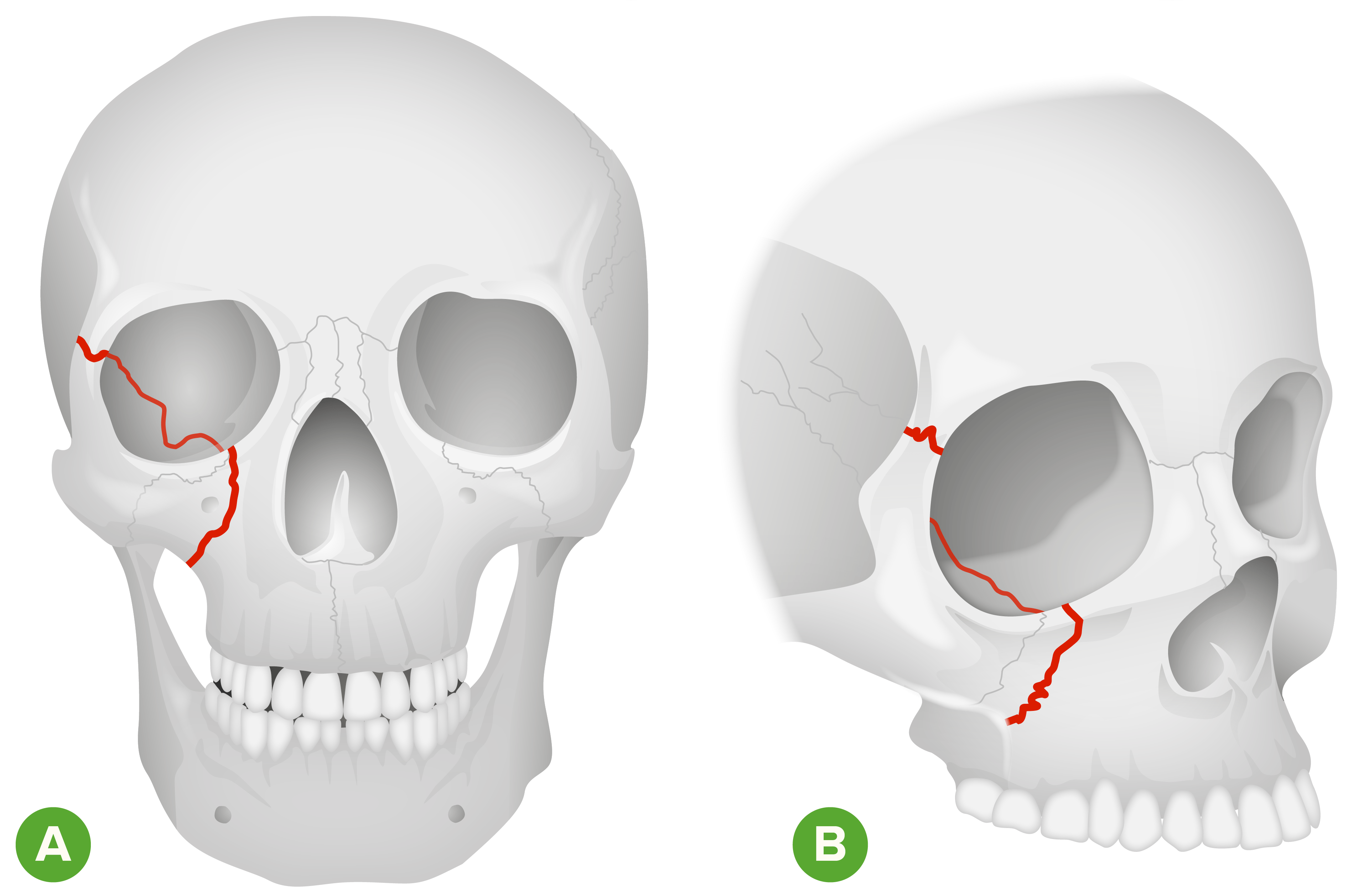
Orbital zygomaticZygomaticEither of a pair of bones that form the prominent part of the cheek and contribute to the orbit on each side of the skull.Skull: AnatomyfractureFractureA fracture is a disruption of the cortex of any bone and periosteum and is commonly due to mechanical stress after an injury or accident. Open fractures due to trauma can be a medical emergency. Fractures are frequently associated with automobile accidents, workplace injuries, and trauma.Overview of Bone Fractures:
ZygomaticZygomaticEither of a pair of bones that form the prominent part of the cheek and contribute to the orbit on each side of the skull.Skull: Anatomy region of the orbit is the most common location of an orbital rim fractureFractureA fracture is a disruption of the cortex of any bone and periosteum and is commonly due to mechanical stress after an injury or accident. Open fractures due to trauma can be a medical emergency. Fractures are frequently associated with automobile accidents, workplace injuries, and trauma.Overview of Bone Fractures.
Most common cause: a high-impact blow to the lateral orbit
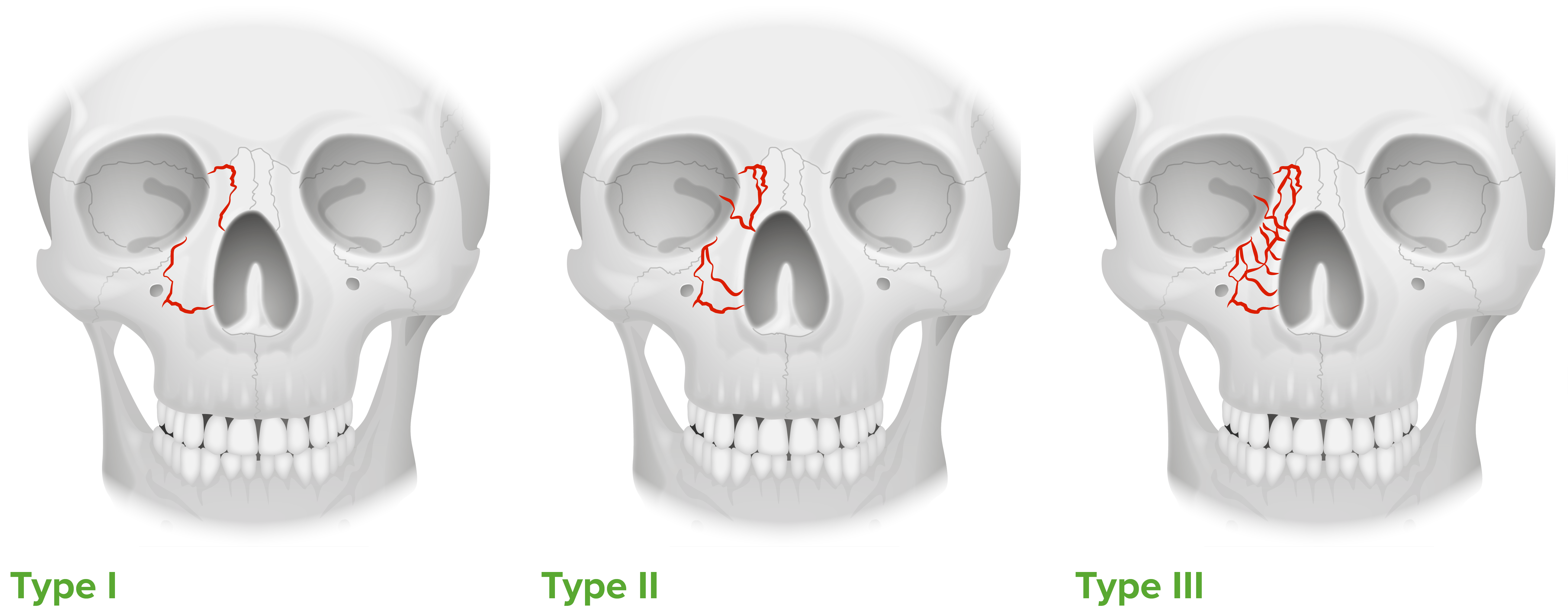
Nasoethmoid fractureFractureA fracture is a disruption of the cortex of any bone and periosteum and is commonly due to mechanical stress after an injury or accident. Open fractures due to trauma can be a medical emergency. Fractures are frequently associated with automobile accidents, workplace injuries, and trauma.Overview of Bone Fractures:
FractureFractureA fracture is a disruption of the cortex of any bone and periosteum and is commonly due to mechanical stress after an injury or accident. Open fractures due to trauma can be a medical emergency. Fractures are frequently associated with automobile accidents, workplace injuries, and trauma.Overview of Bone Fractures of the medial wall of the orbit
Often associated with fractures of the orbital floor
Due to direct force applied to the nasal boneBoneBone is a compact type of hardened connective tissue composed of bone cells, membranes, an extracellular mineralized matrix, and central bone marrow. The 2 primary types of bone are compact and spongy. Bones: Structure and Types
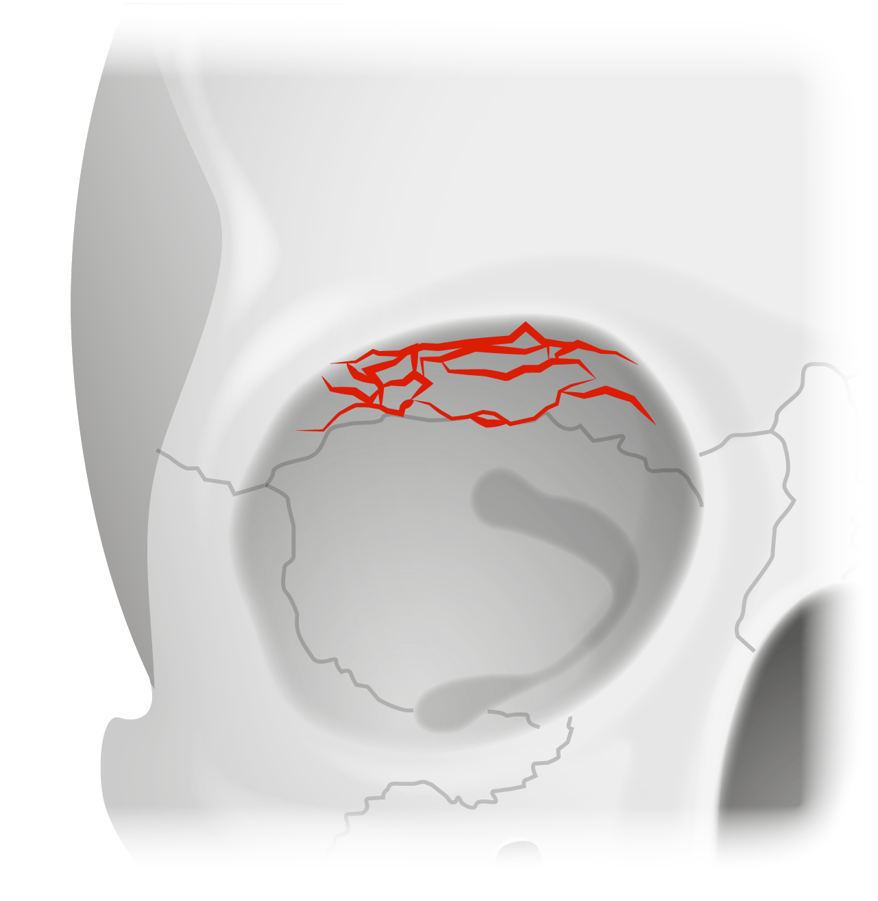
Orbital roof fractureFractureA fracture is a disruption of the cortex of any bone and periosteum and is commonly due to mechanical stress after an injury or accident. Open fractures due to trauma can be a medical emergency. Fractures are frequently associated with automobile accidents, workplace injuries, and trauma.Overview of Bone Fractures:
Location: frontalFrontalThe bone that forms the frontal aspect of the skull. Its flat part forms the forehead, articulating inferiorly with the nasal bone and the cheek bone on each side of the face.Skull: AnatomyboneBoneBone is a compact type of hardened connective tissue composed of bone cells, membranes, an extracellular mineralized matrix, and central bone marrow. The 2 primary types of bone are compact and spongy. Bones: Structure and Types (roof of the orbit)
Frequently associated with intracranial injuries
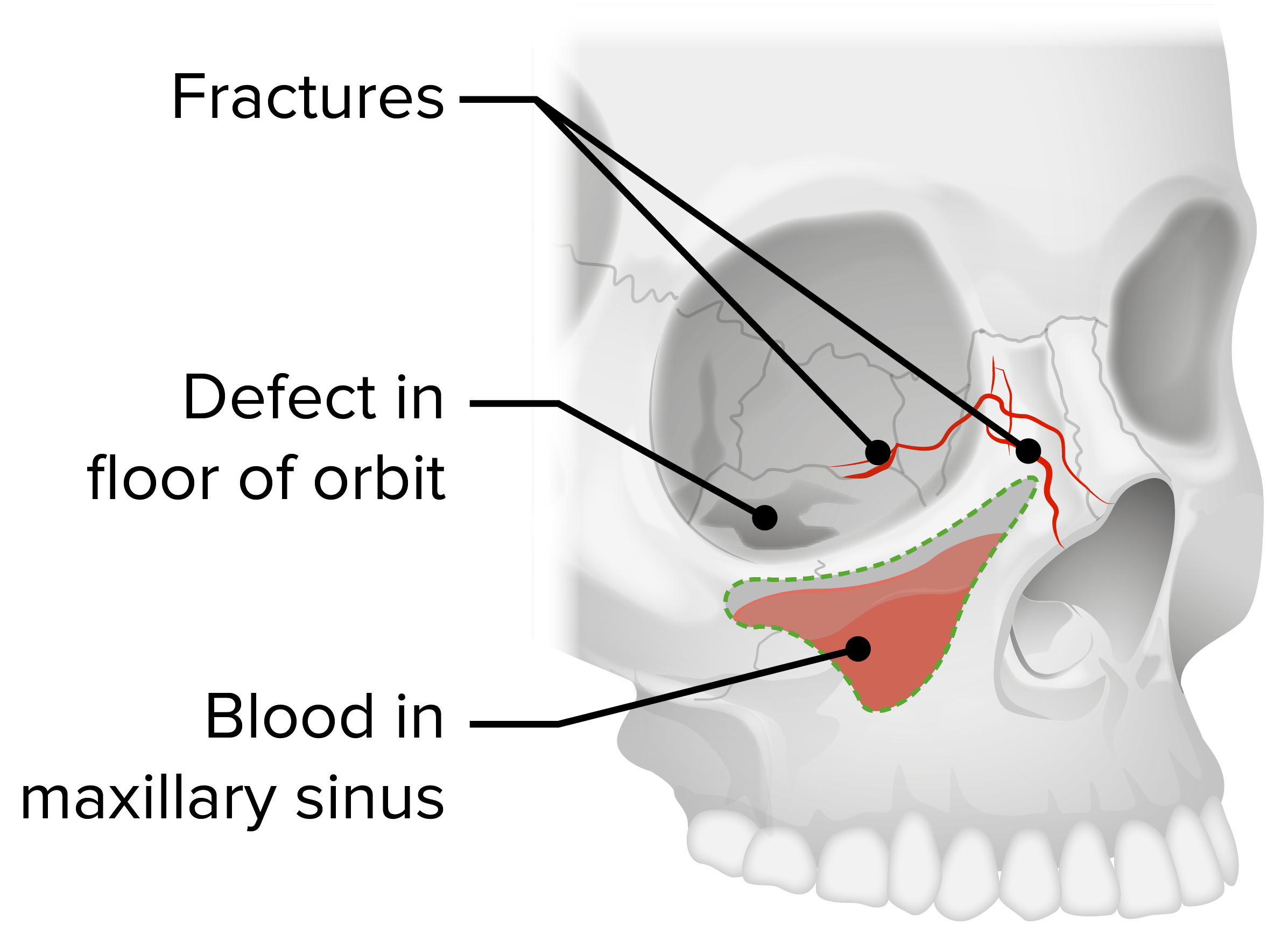
Orbital floor fractureFractureA fracture is a disruption of the cortex of any bone and periosteum and is commonly due to mechanical stress after an injury or accident. Open fractures due to trauma can be a medical emergency. Fractures are frequently associated with automobile accidents, workplace injuries, and trauma.Overview of Bone Fractures:
“Blowout fractureFractureA fracture is a disruption of the cortex of any bone and periosteum and is commonly due to mechanical stress after an injury or accident. Open fractures due to trauma can be a medical emergency. Fractures are frequently associated with automobile accidents, workplace injuries, and trauma.Overview of Bone Fractures”
Most common type of orbital fractureFractureA fracture is a disruption of the cortex of any bone and periosteum and is commonly due to mechanical stress after an injury or accident. Open fractures due to trauma can be a medical emergency. Fractures are frequently associated with automobile accidents, workplace injuries, and trauma.Overview of Bone Fractures
Location: maxillary, zygomaticZygomaticEither of a pair of bones that form the prominent part of the cheek and contribute to the orbit on each side of the skull.Skull: Anatomy, and palatine bones
Ocular motilityMotilityThe motor activity of the gastrointestinal tract.Gastrointestinal Motility may be disrupted, as orbital tissue is commonly involved.
Orbital zygomatic fracture
Image by Lecturio.
Nasoethmoid fracture
Image by Lecturio.
Orbital roof fracture
Image by Lecturio.
Orbital floor fracture
Image by Lecturio.
Clinical Presentation
In a patient presenting with possible orbital fractures, it is important to identify life-threatening and/or serious associated injuries, especially intracranial injury and cervical spineSpineThe human spine, or vertebral column, is the most important anatomical and functional axis of the human body. It consists of 7 cervical vertebrae, 12 thoracic vertebrae, and 5 lumbar vertebrae and is limited cranially by the skull and caudally by the sacrum.Vertebral Column: Anatomy fractures.
History and symptoms
History of blunt (more common) or penetrating trauma
Diffuse orbital painPainAn unpleasant sensation induced by noxious stimuli which are detected by nerve endings of nociceptive neurons.Pain: Types and Pathways
PainPainAn unpleasant sensation induced by noxious stimuli which are detected by nerve endings of nociceptive neurons.Pain: Types and Pathways on movement of the eyes suggests injury to the extraocular muscles.
DiplopiaDiplopiaA visual symptom in which a single object is perceived by the visual cortex as two objects rather than one. Disorders associated with this condition include refractive errors; strabismus; oculomotor nerve diseases; trochlear nerve diseases; abducens nerve diseases; and diseases of the brain stem and occipital lobe.Myasthenia Gravis on upward gaze suggests injury to the orbital floor.
Numbness of the foreheadForeheadThe part of the face above the eyes.Melasma suggests injury to the orbital roof.
NauseaNauseaAn unpleasant sensation in the stomach usually accompanied by the urge to vomit. Common causes are early pregnancy, sea and motion sickness, emotional stress, intense pain, food poisoning, and various enteroviruses.Antiemetics can be caused by entrapment of extraocular muscles.
Physical examination findings
PeriorbitalPeriorbitalOrbital and Preseptal CellulitisedemaEdemaEdema is a condition in which excess serous fluid accumulates in the body cavity or interstitial space of connective tissues. Edema is a symptom observed in several medical conditions. It can be categorized into 2 types, namely, peripheral (in the extremities) and internal (in an organ or body cavity). Edema
Ecchymosis
DiplopiaDiplopiaA visual symptom in which a single object is perceived by the visual cortex as two objects rather than one. Disorders associated with this condition include refractive errors; strabismus; oculomotor nerve diseases; trochlear nerve diseases; abducens nerve diseases; and diseases of the brain stem and occipital lobe.Myasthenia Gravis: due to mechanical entrapment of both inferior obliqueInferior obliqueOrbit and Extraocular Muscles: Anatomy and inferior rectusInferior rectusOrbit and Extraocular Muscles: Anatomy muscles
Absent pupillary light reflexPupillary Light ReflexConstriction of the pupil in response to light stimulation of the retina. It refers also to any reflex involving the iris, with resultant alteration of the diameter of the pupil.Pupil: Physiology and Abnormalities if damage to the afferentAfferentNeurons which conduct nerve impulses to the central nervous system.Nervous System: Histology nerves
EnophthalmosEnophthalmosRecession of the eyeball into the orbit.Marfan Syndrome: posterior displacementDisplacementThe process by which an emotional or behavioral response that is appropriate for one situation appears in another situation for which it is inappropriate.Defense Mechanisms of the eyeball within the orbit, seen in blowout fractures
ProptosisProptosisRetinoblastoma: protrusion of the eyeball, seen in orbital zygomaticZygomaticEither of a pair of bones that form the prominent part of the cheek and contribute to the orbit on each side of the skull.Skull: Anatomy fractures
Widened intercanthal distance due to disruption of the medial canthal ligament
BradycardiaBradycardiaBradyarrhythmia is a rhythm in which the heart rate is less than 60/min. Bradyarrhythmia can be physiologic, without symptoms or hemodynamic change. Pathologic bradyarrhythmia results in reduced cardiac output and hemodynamic instability causing syncope, dizziness, or dyspnea.Bradyarrhythmias: associated with the “oculocardiac reflex” (a reduction in pulse rate associated with pressure applied over extraocular muscles)
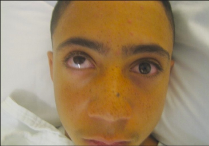
Restriction in left upward gaze due to entrapment of left extraocular muscles in a blowout fracture
Image: “Left orbital floor fracture” by Department of Ophthalmology, Division of Oculoplastic and Orbital Surgery, Rocky Mountain Lions Eye Institute, University of Colorado, Aurora, CO, USA. License: CC BY 2.0
Ocular injuries are present in up to 29% of patientsPatientsIndividuals participating in the health care system for the purpose of receiving therapeutic, diagnostic, or preventive procedures.Clinician–Patient Relationship with orbital fractures. It is imperative that an ocular exam be done as soon as possible to mitigate the risk of visionVisionOphthalmic Exam loss.
Ocular examination
Visual acuityVisual AcuityClarity or sharpness of ocular vision or the ability of the eye to see fine details. Visual acuity depends on the functions of retina, neuronal transmission, and the interpretative ability of the brain. Normal visual acuity is expressed as 20/20 indicating that one can see at 20 feet what should normally be seen at that distance. Visual acuity can also be influenced by brightness, color, and contrast.Ophthalmic Exam
X-rayX-rayPenetrating electromagnetic radiation emitted when the inner orbital electrons of an atom are excited and release radiant energy. X-ray wavelengths range from 1 pm to 10 nm. Hard x-rays are the higher energy, shorter wavelength x-rays. Soft x-rays or grenz rays are less energetic and longer in wavelength. The short wavelength end of the x-ray spectrum overlaps the gamma rays wavelength range. The distinction between gamma rays and x-rays is based on their radiation source.Pulmonary Function Tests:
Water’s view (also known as the occipitomental view): X-rayX-rayPenetrating electromagnetic radiation emitted when the inner orbital electrons of an atom are excited and release radiant energy. X-ray wavelengths range from 1 pm to 10 nm. Hard x-rays are the higher energy, shorter wavelength x-rays. Soft x-rays or grenz rays are less energetic and longer in wavelength. The short wavelength end of the x-ray spectrum overlaps the gamma rays wavelength range. The distinction between gamma rays and x-rays is based on their radiation source.Pulmonary Function Tests beam is angled at 45 degrees and shows “teardrop sign” in antrum.
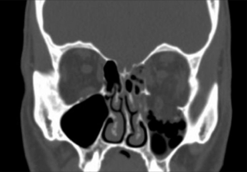
CT scan:
Gold standard for detection of orbital fractures
Thin slices < 2 mm are preferred.
CoronalCoronalComputed Tomography (CT) and sagittalSagittalComputed Tomography (CT) views differentiate between periorbitalPeriorbitalOrbital and Preseptal CellulitisedemaEdemaEdema is a condition in which excess serous fluid accumulates in the body cavity or interstitial space of connective tissues. Edema is a symptom observed in several medical conditions. It can be categorized into 2 types, namely, peripheral (in the extremities) and internal (in an organ or body cavity). Edema and entrapment of intraocular structures.
Image: “Left orbital floor fracture” by Department of Ophthalmology, Division of Oculoplastic and Orbital Surgery, Rocky Mountain Lions Eye Institute, University of Colorado, Aurora, CO, USA. License: CC BY 2.0
Management and Complications
Orbital fractures are facial injuries and should be managed emergently. Delay in diagnosis may lead to complications or postoperative complicationsPostoperative ComplicationsPathologic processes that affect patients after a surgical procedure. They may or may not be related to the disease for which the surgery was done, and they may or may not be direct results of the surgery.Postoperative Care.
IV fluidsIV fluidsIntravenous fluids are one of the most common interventions administered in medicine to approximate physiologic bodily fluids. Intravenous fluids are divided into 2 categories: crystalloid and colloid solutions. Intravenous fluids have a wide variety of indications, including intravascular volume expansion, electrolyte manipulation, and maintenance fluids. Intravenous Fluids
PainPainAn unpleasant sensation induced by noxious stimuli which are detected by nerve endings of nociceptive neurons.Pain: Types and Pathways and nauseaNauseaAn unpleasant sensation in the stomach usually accompanied by the urge to vomit. Common causes are early pregnancy, sea and motion sickness, emotional stress, intense pain, food poisoning, and various enteroviruses.Antiemetics medications
Elevate head.
Evaluate for:
Cervical spineSpineThe human spine, or vertebral column, is the most important anatomical and functional axis of the human body. It consists of 7 cervical vertebrae, 12 thoracic vertebrae, and 5 lumbar vertebrae and is limited cranially by the skull and caudally by the sacrum.Vertebral Column: Anatomy integrity
Possible head injuries
Soft tissueSoft TissueSoft Tissue Abscess and boneBoneBone is a compact type of hardened connective tissue composed of bone cells, membranes, an extracellular mineralized matrix, and central bone marrow. The 2 primary types of bone are compact and spongy. Bones: Structure and Types injuries in the head and neckNeckThe part of a human or animal body connecting the head to the rest of the body.Peritonsillar Abscess
Empiric management:
Antibiotics to cover sinus pathogens
Advise the patient not to blow the noseNoseThe nose is the human body’s primary organ of smell and functions as part of the upper respiratory system. The nose may be best known for inhaling oxygen and exhaling carbon dioxide, but it also contributes to other important functions, such as tasting. The anatomy of the nose can be divided into the external nose and the nasal cavity. Nose Anatomy (External & Internal), in order to avoid air in the orbital cavity.
Reduce periorbitalPeriorbitalOrbital and Preseptal CellulitisedemaEdemaEdema is a condition in which excess serous fluid accumulates in the body cavity or interstitial space of connective tissues. Edema is a symptom observed in several medical conditions. It can be categorized into 2 types, namely, peripheral (in the extremities) and internal (in an organ or body cavity). Edema:
Cold compresses
Oral steroidsSteroidsA group of polycyclic compounds closely related biochemically to terpenes. They include cholesterol, numerous hormones, precursors of certain vitamins, bile acids, alcohols (sterols), and certain natural drugs and poisons. Steroids have a common nucleus, a fused, reduced 17-carbon atom ring system, cyclopentanoperhydrophenanthrene. Most steroids also have two methyl groups and an aliphatic side-chain attached to the nucleus.Benign Liver Tumors
Urgent ophthalmology consult if:
Evidence of globe injury: seen in 30% of orbital fractures
Decreased visual acuityVisual AcuityClarity or sharpness of ocular vision or the ability of the eye to see fine details. Visual acuity depends on the functions of retina, neuronal transmission, and the interpretative ability of the brain. Normal visual acuity is expressed as 20/20 indicating that one can see at 20 feet what should normally be seen at that distance. Visual acuity can also be influenced by brightness, color, and contrast.Ophthalmic Exam
Widened intercanthal distance
Evidence of orbital compartment syndromeCompartment SyndromeCompartment syndrome is a surgical emergency usually occurring secondary to trauma. The condition is marked by increased pressure within a compartment that compromises the circulation and function of the tissues within that space.Compartment Syndrome:
Decreased retropulsion
“Rock hard” eyelidsEyelidsEach of the upper and lower folds of skin which cover the eye when closed.Blepharitis
Open globe
Severe vagal symptoms
Surgery to reduce the fractureFractureA fracture is a disruption of the cortex of any bone and periosteum and is commonly due to mechanical stress after an injury or accident. Open fractures due to trauma can be a medical emergency. Fractures are frequently associated with automobile accidents, workplace injuries, and trauma.Overview of Bone Fractures and prevent future deformities:
May be deferred up to 14 days if periorbitalPeriorbitalOrbital and Preseptal CellulitisedemaEdemaEdema is a condition in which excess serous fluid accumulates in the body cavity or interstitial space of connective tissues. Edema is a symptom observed in several medical conditions. It can be categorized into 2 types, namely, peripheral (in the extremities) and internal (in an organ or body cavity). Edema interferes with visualization and assessment
Is advised for patientsPatientsIndividuals participating in the health care system for the purpose of receiving therapeutic, diagnostic, or preventive procedures.Clinician–Patient Relationship with:
Orbital hematomaHematomaA collection of blood outside the blood vessels. Hematoma can be localized in an organ, space, or tissue.Intussusception
CSF leakage
Globe displacementDisplacementThe process by which an emotional or behavioral response that is appropriate for one situation appears in another situation for which it is inappropriate.Defense Mechanisms: exophthalmos or proptosisProptosisRetinoblastoma
Entrapment of infraorbital structures leading to the oculocardiac reflex
Commonly used implants for reconstruction are:
BoneBoneBone is a compact type of hardened connective tissue composed of bone cells, membranes, an extracellular mineralized matrix, and central bone marrow. The 2 primary types of bone are compact and spongy. Bones: Structure and TypesautograftAutograftTransplant comprised of an individual’s own tissue, transferred from one part of the body to another.Organ Transplantation:
Provides good strength for reconstruction
Commonly used in children < 7 years of age
CartilageCartilageCartilage is a type of connective tissue derived from embryonic mesenchyme that is responsible for structural support, resilience, and the smoothness of physical actions. Perichondrium (connective tissue membrane surrounding cartilage) compensates for the absence of vasculature in cartilage by providing nutrition and support. Cartilage: Histology:
Most biocompatible
Undergoes easy resorption
Most commonly used for small fractures
Alloplast:
Titanium mesh: used for large floor defects
Porous polyethylene: used for defects with well-defined edges
Resorbable sheeting: used in small gaps with well-defined edges
Complications
Complications arising from orbital fractures can be due to the injury itself or to surgery.
Injury-related complications:
Ruptured globe
Orbital hematomaHematomaA collection of blood outside the blood vessels. Hematoma can be localized in an organ, space, or tissue.Intussusception
Optic nerveOptic nerveThe 2nd cranial nerve which conveys visual information from the retina to the brain. The nerve carries the axons of the retinal ganglion cells which sort at the optic chiasm and continue via the optic tracts to the brain. The largest projection is to the lateral geniculate nuclei; other targets include the superior colliculi and the suprachiasmatic nuclei. Though known as the second cranial nerve, it is considered part of the central nervous system.The 12 Cranial Nerves: Overview and Functions sheath hematomaHematomaA collection of blood outside the blood vessels. Hematoma can be localized in an organ, space, or tissue.Intussusception
Retinal detachmentRetinal detachmentRetinal detachment is the separation of the neurosensory retina from the retinal pigmented epithelium and choroid. Rhegmatogenous retinal detachment, the most common type, stems from a break in the retina, allowing fluid to accumulate in the subretinal space. Retinal Detachment
Orbital compartment syndromeCompartment SyndromeCompartment syndrome is a surgical emergency usually occurring secondary to trauma. The condition is marked by increased pressure within a compartment that compromises the circulation and function of the tissues within that space.Compartment Syndrome
Surgical complicationsSurgical complicationsSurgical complications are conditions, disorders, or adverse events that occur following surgical procedures. The most common general surgical complications include bleeding, infections, injury to the surrounding organs, venous thromboembolic events, and complications from anesthesia.Surgical Complications:
Postoperative complicationsPostoperative ComplicationsPathologic processes that affect patients after a surgical procedure. They may or may not be related to the disease for which the surgery was done, and they may or may not be direct results of the surgery.Postoperative Care:
Persistent diplopiaDiplopiaA visual symptom in which a single object is perceived by the visual cortex as two objects rather than one. Disorders associated with this condition include refractive errors; strabismus; oculomotor nerve diseases; trochlear nerve diseases; abducens nerve diseases; and diseases of the brain stem and occipital lobe.Myasthenia Gravis
Boyette, J. R., et al. (2015). Management of orbital fractures: challenges and solutions. Clinical Ophthalmology 9:2127–2137. https://doi.org/10.2147/OPTH.S80463