A complex system of coordinated electrical circuitry within the heart governs cardiac muscleCardiac muscleThe muscle tissue of the heart. It is composed of striated, involuntary muscle cells connected to form the contractile pump to generate blood flow.Muscle Tissue: Histology activity. The heart generates its own electrical impulses within its pacemakerPacemakerA device designed to stimulate, by electric impulses, contraction of the heart muscles. It may be temporary (external) or permanent (internal or internal-external).Bradyarrhythmias cells. The signal then travels through specialized myocytesMyocytesMature contractile cells, commonly known as myocytes, that form one of three kinds of muscle. The three types of muscle cells are skeletal, cardiac, and smooth. They are derived from embryonic (precursor) muscle cells called myoblasts.Muscle Tissue: Histology, which act as electrical wiring, distributing the signal throughout the heart. Once the signal “leaves” the specialized conduction system, it passes to each myocyte through channelsChannelsThe Cell: Cell Membrane called gap junctionsGap JunctionsConnections between cells which allow passage of small molecules and electric current. Gap junctions were first described anatomically as regions of close apposition between cells with a narrow (1-2 nm) gap between cell membranes. The variety in the properties of gap junctions is reflected in the number of connexins, the family of proteins which form the junctions.The Cell: Cell Junctions (which connect myocytesMyocytesMature contractile cells, commonly known as myocytes, that form one of three kinds of muscle. The three types of muscle cells are skeletal, cardiac, and smooth. They are derived from embryonic (precursor) muscle cells called myoblasts.Muscle Tissue: Histology to each other) and causes them to contract. An electrical impulse is created by the opening and closing of ion channelsChannelsThe Cell: Cell Membrane, allowing the flowFlowBlood flows through the heart, arteries, capillaries, and veins in a closed, continuous circuit. Flow is the movement of volume per unit of time. Flow is affected by the pressure gradient and the resistance fluid encounters between 2 points. Vascular resistance is the opposition to flow, which is caused primarily by blood friction against vessel walls.Vascular Resistance, Flow, and Mean Arterial Pressure of charged particles across the myocardial cell membraneCell MembraneA cell membrane (also known as the plasma membrane or plasmalemma) is a biological membrane that separates the cell contents from the outside environment. A cell membrane is composed of a phospholipid bilayer and proteins that function to protect cellular DNA and mediate the exchange of ions and molecules. The Cell: Cell Membrane. The flowFlowBlood flows through the heart, arteries, capillaries, and veins in a closed, continuous circuit. Flow is the movement of volume per unit of time. Flow is affected by the pressure gradient and the resistance fluid encounters between 2 points. Vascular resistance is the opposition to flow, which is caused primarily by blood friction against vessel walls.Vascular Resistance, Flow, and Mean Arterial Pressure of charged particles changes the voltage across the membrane and opens up additional voltage-gated channelsChannelsThe Cell: Cell Membrane, allowing the signal to propagate throughout the heart.
A compact region of pacemakerPacemakerA device designed to stimulate, by electric impulses, contraction of the heart muscles. It may be temporary (external) or permanent (internal or internal-external).Bradyarrhythmias cells (modified myocytesMyocytesMature contractile cells, commonly known as myocytes, that form one of three kinds of muscle. The three types of muscle cells are skeletal, cardiac, and smooth. They are derived from embryonic (precursor) muscle cells called myoblasts.Muscle Tissue: Histology) that serves as the primary pacemakerPacemakerA device designed to stimulate, by electric impulses, contraction of the heart muscles. It may be temporary (external) or permanent (internal or internal-external).Bradyarrhythmias of the heart
Located within the subepicardial tissue at the junction of the right atrium and superior vena cavaSuperior vena cavaThe venous trunk which returns blood from the head, neck, upper extremities and chest.Mediastinum and Great Vessels: Anatomy
Depolarizes regularly:
Called the sinus rhythm: HR and rhythm are driven by the regularRegularInsulin firing of the SA node (60–100/min).
Signal originates in the SA node → atrial myocytesMyocytesMature contractile cells, commonly known as myocytes, that form one of three kinds of muscle. The three types of muscle cells are skeletal, cardiac, and smooth. They are derived from embryonic (precursor) muscle cells called myoblasts.Muscle Tissue: Histology → atrioventricular (AV) node
The AV node:
A compact region of specialized conducting cells that receives input from the SA node and propagates it toward the ventricles
Located within the interatrial septumInteratrial SeptumAtrial Septal Defect (ASD), near the coronary sinusCoronary SinusA short vein that collects about two thirds of the venous blood from the myocardium and drains into the right atrium. Coronary sinus, normally located between the left atrium and left ventricle on the posterior surface of the heart, can serve as an anatomical reference for cardiac procedures.Atrial Septal Defect (ASD) ostium and the septal leaflet of the tricuspid valveTricuspid valveThe valve consisting of three cusps situated between the right atrium and right ventricle of the heart.Heart: Anatomy (within the triangle of Koch)
Characterized by slow conduction and a long refractory period:
Causes a delay in propagation of electrical signal to the ventricles
Allows ventricles to fill with blood from the atrial contraction before the ventricles contract
If the SA node fails, the AV node has its own autorhythmicity and can serve as the primary pacemakerPacemakerA device designed to stimulate, by electric impulses, contraction of the heart muscles. It may be temporary (external) or permanent (internal or internal-external).Bradyarrhythmias with a slower rhythm (HR: 40–60/min).
Bundle of HisBundle of HisSmall band of specialized cardiac muscle fibers that originates in the atrioventricular node and extends into the membranous part of the interventricular septum. The bundle of his, consisting of the left and the right bundle branches, conducts the electrical impulses to the heart ventricles in generation of myocardial contraction.Heart: Anatomy and Purkinje fibersPurkinje fibersModified cardiac muscle fibers composing the terminal portion of the heart conduction system.Heart: Anatomy:
Purkinje fibersPurkinje fibersModified cardiac muscle fibers composing the terminal portion of the heart conduction system.Heart: Anatomy:
Arise from the bundle branches
Spread throughout the ventricular walls
Fastest conduction fibers
Have their own intrinsic rhythm of 30–40/min (although this is not fast enough to sustain life)
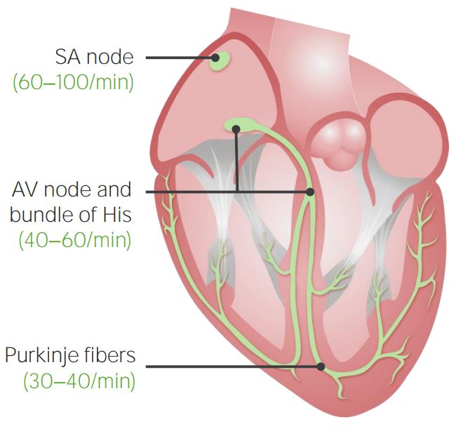
Cardiac conduction system and intrinsic rhythms: Location of pacemaker cells within the conduction system of the heart and their corresponding intrinsic rhythms
Image by Lecturio.
Nonpacemaker myocytesMyocytesMature contractile cells, commonly known as myocytes, that form one of three kinds of muscle. The three types of muscle cells are skeletal, cardiac, and smooth. They are derived from embryonic (precursor) muscle cells called myoblasts.Muscle Tissue: Histology:
Cardiac muscleCardiac muscleThe muscle tissue of the heart. It is composed of striated, involuntary muscle cells connected to form the contractile pump to generate blood flow.Muscle Tissue: Histology cells that are not part of the SA or AV nodes
Contract when they receive an electrical signal
Connected to each other via gap junctionsGap JunctionsConnections between cells which allow passage of small molecules and electric current. Gap junctions were first described anatomically as regions of close apposition between cells with a narrow (1-2 nm) gap between cell membranes. The variety in the properties of gap junctions is reflected in the number of connexins, the family of proteins which form the junctions.The Cell: Cell Junctions→ capable of conducting electrical signals from 1 cell to the next
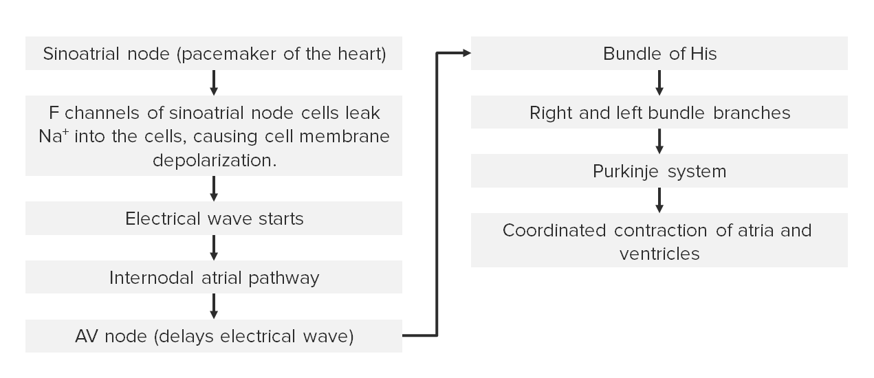
Summary of the electrical pathway
Clinically, the electrophysiological activity of the heart can be monitored using ECGECGAn electrocardiogram (ECG) is a graphic representation of the electrical activity of the heart plotted against time. Adhesive electrodes are affixed to the skin surface allowing measurement of cardiac impulses from many angles. The ECG provides 3-dimensional information about the conduction system of the heart, the myocardium, and other cardiac structures. Electrocardiogram (ECG).
The diagram below summarizes the formation and propagation of the electrical wave.
Diagram outlining the electrical pathway of the heart AV: atrioventricular
Image by Lecturio.
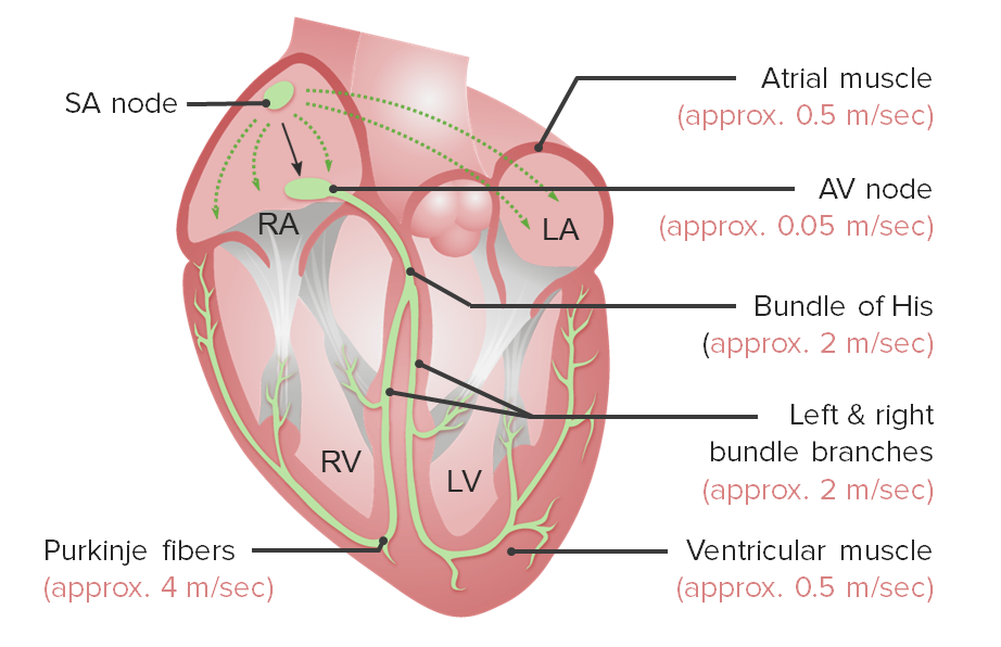
Conduction times
Action potentials travel at different speeds through different tissues and segments of the conduction system.
Atrial myocytesMyocytesMature contractile cells, commonly known as myocytes, that form one of three kinds of muscle. The three types of muscle cells are skeletal, cardiac, and smooth. They are derived from embryonic (precursor) muscle cells called myoblasts.Muscle Tissue: Histology: approximately 0.5‒1 m/sec
AV node: approximately 0.05 m/sec (slowest)
Bundle of HisBundle of HisSmall band of specialized cardiac muscle fibers that originates in the atrioventricular node and extends into the membranous part of the interventricular septum. The bundle of his, consisting of the left and the right bundle branches, conducts the electrical impulses to the heart ventricles in generation of myocardial contraction.Heart: Anatomy and the left and right bundle branches: approximately 2 m/sec
Purkinje fibersPurkinje fibersModified cardiac muscle fibers composing the terminal portion of the heart conduction system.Heart: Anatomy: approximately 4 m/sec (fastest)
Ventricular myocytesMyocytesMature contractile cells, commonly known as myocytes, that form one of three kinds of muscle. The three types of muscle cells are skeletal, cardiac, and smooth. They are derived from embryonic (precursor) muscle cells called myoblasts.Muscle Tissue: Histology: approximately 0.3–1 m/sec
Cardiac conduction system and conduction times of respective segments SA: sinoatrial AV: atrioventricular RV: right ventricle LV: left ventricle RA: right atrium LA: left atrium
Difference in the concentration of charged particles between 1 point and another (in physiology, usually across a cell membraneCell MembraneA cell membrane (also known as the plasma membrane or plasmalemma) is a biological membrane that separates the cell contents from the outside environment. A cell membrane is composed of a phospholipid bilayer and proteins that function to protect cellular DNA and mediate the exchange of ions and molecules. The Cell: Cell Membrane)
A form of potential energy
Capable of producing an electrical current
Unit: volts
Electrical current:
The flowFlowBlood flows through the heart, arteries, capillaries, and veins in a closed, continuous circuit. Flow is the movement of volume per unit of time. Flow is affected by the pressure gradient and the resistance fluid encounters between 2 points. Vascular resistance is the opposition to flow, which is caused primarily by blood friction against vessel walls.Vascular Resistance, Flow, and Mean Arterial Pressure of charged particles from 1 point to another (in physiology, usually across a cell membraneCell MembraneA cell membrane (also known as the plasma membrane or plasmalemma) is a biological membrane that separates the cell contents from the outside environment. A cell membrane is composed of a phospholipid bilayer and proteins that function to protect cellular DNA and mediate the exchange of ions and molecules. The Cell: Cell Membrane)
Written as “I” (e.g., flowFlowBlood flows through the heart, arteries, capillaries, and veins in a closed, continuous circuit. Flow is the movement of volume per unit of time. Flow is affected by the pressure gradient and the resistance fluid encounters between 2 points. Vascular resistance is the opposition to flow, which is caused primarily by blood friction against vessel walls.Vascular Resistance, Flow, and Mean Arterial Pressure of K+ ions: IK+)
Polarization:
Electrical potential exists across a membrane.
In cardiac physiology:
Hyperpolarized state: The cell is in a more negative state (i.e., a larger concentration gradient is present).
Depolarized state: The cell is in a less negative/slightly positive state (i.e., a smaller concentration gradient is present).
Nonpacemaker cardiac myocytesMyocytesMature contractile cells, commonly known as myocytes, that form one of three kinds of muscle. The three types of muscle cells are skeletal, cardiac, and smooth. They are derived from embryonic (precursor) muscle cells called myoblasts.Muscle Tissue: Histology depolarize only when they receive an electrical stimulus. When nonpacemaker cardiac myocytesMyocytesMature contractile cells, commonly known as myocytes, that form one of three kinds of muscle. The three types of muscle cells are skeletal, cardiac, and smooth. They are derived from embryonic (precursor) muscle cells called myoblasts.Muscle Tissue: Histology are not stimulated, they exist in a resting state and have an RMP.
Created by membrane permeability and the concentration differences of several key ions:
K+
Na+
CalciumCalciumA basic element found in nearly all tissues. It is a member of the alkaline earth family of metals with the atomic symbol ca, atomic number 20, and atomic weight 40. Calcium is the most abundant mineral in the body and combines with phosphorus to form calcium phosphate in the bones and teeth. It is essential for the normal functioning of nerves and muscles and plays a role in blood coagulation (as factor IV) and in many enzymatic processes.Electrolytes (CaCACondylomata acuminata are a clinical manifestation of genital HPV infection. Condylomata acuminata are described as raised, pearly, flesh-colored, papular, cauliflower-like lesions seen in the anogenital region that may cause itching, pain, or bleeding.Condylomata Acuminata (Genital Warts)2+)
Cl–
Can be calculated using the complex Goldman-Hodgkin-Katz equation
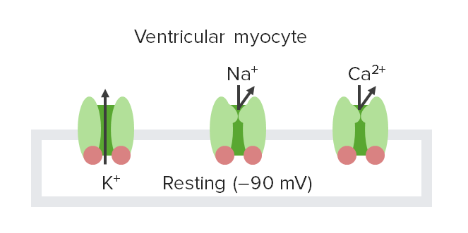
At rest, myocytesMyocytesMature contractile cells, commonly known as myocytes, that form one of three kinds of muscle. The three types of muscle cells are skeletal, cardiac, and smooth. They are derived from embryonic (precursor) muscle cells called myoblasts.Muscle Tissue: Histology are in a polarized state:
RMP = –90 mV
Depicted by an isoelectric (flat) line on graphs showing how the membrane potentialMembrane potentialThe membrane potential is the difference in electric charge between the interior and the exterior of a cell. All living cells maintain a potential difference across the membrane thanks to the insulating properties of their plasma membranes (PMs) and the selective transport of ions across this membrane by transporters.Membrane Potential changes over time
Ion conductances at resting potential: At the hyperpolarized resting potential, voltage-gated K+ channels are the only channels that are open; thus, K+ is the primary contributor to the resting membrane potential of cells.
Image by Lecturio.
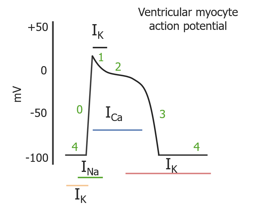
Action potentials
Action potentialAction PotentialAbrupt changes in the membrane potential that sweep along the cell membrane of excitable cells in response to excitation stimuli.Membrane Potential:
An electrical stimulus leads to the opening of voltage-gated ion channelsChannelsThe Cell: Cell Membrane, allowing ions to flowFlowBlood flows through the heart, arteries, capillaries, and veins in a closed, continuous circuit. Flow is the movement of volume per unit of time. Flow is affected by the pressure gradient and the resistance fluid encounters between 2 points. Vascular resistance is the opposition to flow, which is caused primarily by blood friction against vessel walls.Vascular Resistance, Flow, and Mean Arterial Pressure into and out of the cell down their concentration gradients (i.e., ion currents).
While the current is flowing, the membrane potentialMembrane potentialThe membrane potential is the difference in electric charge between the interior and the exterior of a cell. All living cells maintain a potential difference across the membrane thanks to the insulating properties of their plasma membranes (PMs) and the selective transport of ions across this membrane by transporters.Membrane Potential actively changes → action potentialAction PotentialAbrupt changes in the membrane potential that sweep along the cell membrane of excitable cells in response to excitation stimuli.Membrane Potential
An action potentialAction PotentialAbrupt changes in the membrane potential that sweep along the cell membrane of excitable cells in response to excitation stimuli.Membrane Potential can be divided into phases 0‒4 (usually described as beginning with 4).
Often depicted as a graph, showing the change in membrane potentialMembrane potentialThe membrane potential is the difference in electric charge between the interior and the exterior of a cell. All living cells maintain a potential difference across the membrane thanks to the insulating properties of their plasma membranes (PMs) and the selective transport of ions across this membrane by transporters.Membrane Potential over time
Induced by the voltage change from the action potentialAction PotentialAbrupt changes in the membrane potential that sweep along the cell membrane of excitable cells in response to excitation stimuli.Membrane Potential propagated from an adjacent myocyte via gap junctionsGap JunctionsConnections between cells which allow passage of small molecules and electric current. Gap junctions were first described anatomically as regions of close apposition between cells with a narrow (1-2 nm) gap between cell membranes. The variety in the properties of gap junctions is reflected in the number of connexins, the family of proteins which form the junctions.The Cell: Cell Junctions
Membrane potentialMembrane potentialThe membrane potential is the difference in electric charge between the interior and the exterior of a cell. All living cells maintain a potential difference across the membrane thanks to the insulating properties of their plasma membranes (PMs) and the selective transport of ions across this membrane by transporters.Membrane Potential has minimal overall change: plateau
The cell becomes more negative, returning to the baseline of ‒90 mV.
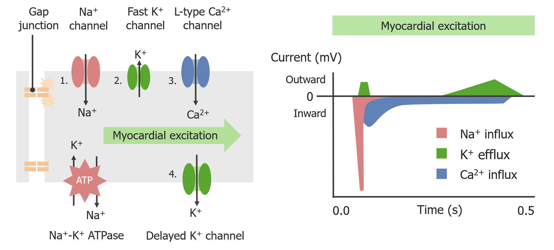
Depolarization of the cardiomyocyte: Propagation of action potentials is through gap junctions that connect cardiomyocytes, which influence voltage-gated Na+ and calcium (Ca2+) channels to open.
Image by Lecturio.
Phases of a cardiac myocyte action potential:
Phases 0, 1, 2, 3, and 4 occur in sequence. Colored lines depict the duration of the respective ion currents. IK: K+ current ICa: calcium (Ca2+) current INa: Na+ current
L-type CaCACondylomata acuminata are a clinical manifestation of genital HPV infection. Condylomata acuminata are described as raised, pearly, flesh-colored, papular, cauliflower-like lesions seen in the anogenital region that may cause itching, pain, or bleeding.Condylomata Acuminata (Genital Warts)2+channelsChannelsThe Cell: Cell Membrane
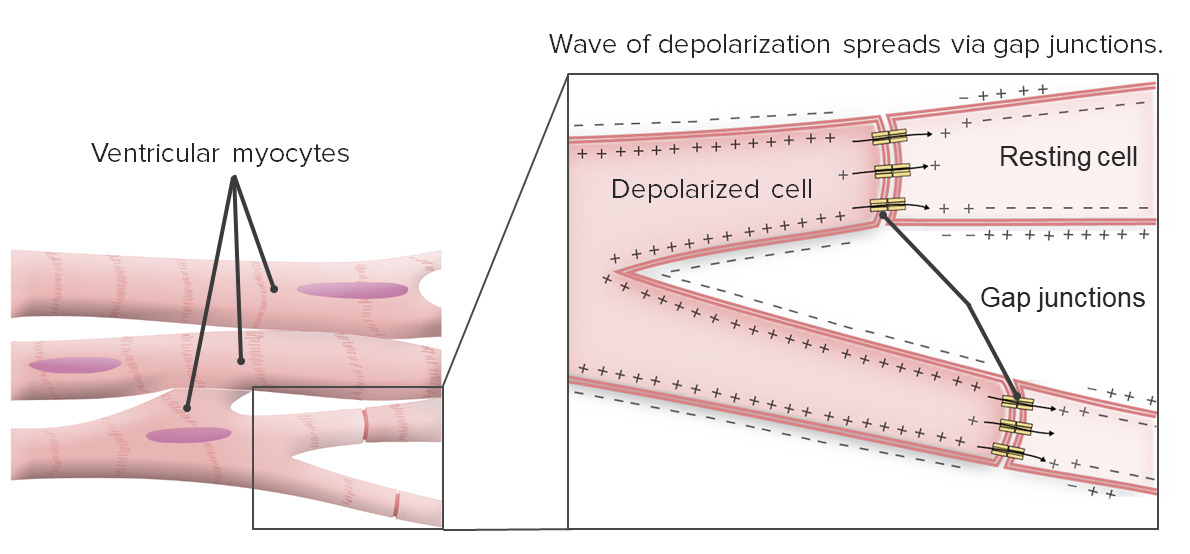
Propagation refers to how electrical signals spread to every myocyte in the heart.
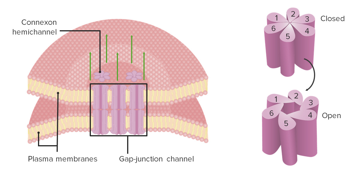
MyocytesMyocytesMature contractile cells, commonly known as myocytes, that form one of three kinds of muscle. The three types of muscle cells are skeletal, cardiac, and smooth. They are derived from embryonic (precursor) muscle cells called myoblasts.Muscle Tissue: Histology are connected to each other via gap junctionsGap JunctionsConnections between cells which allow passage of small molecules and electric current. Gap junctions were first described anatomically as regions of close apposition between cells with a narrow (1-2 nm) gap between cell membranes. The variety in the properties of gap junctions is reflected in the number of connexins, the family of proteins which form the junctions.The Cell: Cell Junctions.
Gap junctionsGap JunctionsConnections between cells which allow passage of small molecules and electric current. Gap junctions were first described anatomically as regions of close apposition between cells with a narrow (1-2 nm) gap between cell membranes. The variety in the properties of gap junctions is reflected in the number of connexins, the family of proteins which form the junctions.The Cell: Cell Junctions:
Action potentials (i.e., the flowFlowBlood flows through the heart, arteries, capillaries, and veins in a closed, continuous circuit. Flow is the movement of volume per unit of time. Flow is affected by the pressure gradient and the resistance fluid encounters between 2 points. Vascular resistance is the opposition to flow, which is caused primarily by blood friction against vessel walls.Vascular Resistance, Flow, and Mean Arterial Pressure of ions) pass through gap junctionsGap JunctionsConnections between cells which allow passage of small molecules and electric current. Gap junctions were first described anatomically as regions of close apposition between cells with a narrow (1-2 nm) gap between cell membranes. The variety in the properties of gap junctions is reflected in the number of connexins, the family of proteins which form the junctions.The Cell: Cell Junctions: propagation of action potentialAction PotentialAbrupt changes in the membrane potential that sweep along the cell membrane of excitable cells in response to excitation stimuli.Membrane Potential to the next myocyte
Ventricular myocytesMyocytesMature contractile cells, commonly known as myocytes, that form one of three kinds of muscle. The three types of muscle cells are skeletal, cardiac, and smooth. They are derived from embryonic (precursor) muscle cells called myoblasts.Muscle Tissue: Histology have an effective refractory period of approximately 200–250 msec (compared with 1–2 msec of skeletal muscle)
Cardiac myocytes are connected to each other via gap junctions. The wave of depolarization spreads through gap junctions.
PacemakerPacemakerA device designed to stimulate, by electric impulses, contraction of the heart muscles. It may be temporary (external) or permanent (internal or internal-external).Bradyarrhythmias cells located in the SA and AV nodes undergo continuous changes in action potentialAction PotentialAbrupt changes in the membrane potential that sweep along the cell membrane of excitable cells in response to excitation stimuli.Membrane Potential; thus, they do not have a true resting potential.
Phase 4 (pacemakerPacemakerA device designed to stimulate, by electric impulses, contraction of the heart muscles. It may be temporary (external) or permanent (internal or internal-external).Bradyarrhythmias potential):
Slow spontaneous depolarizationDepolarizationMembrane Potential during diastoleDiastolePost-systolic relaxation of the heart, especially the heart ventricles.Cardiac Cycle (relaxation of the heart muscle) from approximately ‒60 mV up to its thresholdThresholdMinimum voltage necessary to generate an action potential (an all-or-none response)Skeletal Muscle Contraction potential of ‒40 mV
Mediated primarily by the “funny” current (If) through hyperpolarization-activated cyclic nucleotide-gated (HCN) channelsChannelsThe Cell: Cell Membrane:
An outA mixed inward cation current (primarily Na+, with some K+)
Mediated partly by:
An inward current of CaCACondylomata acuminata are a clinical manifestation of genital HPV infection. Condylomata acuminata are described as raised, pearly, flesh-colored, papular, cauliflower-like lesions seen in the anogenital region that may cause itching, pain, or bleeding.Condylomata Acuminata (Genital Warts)2+ through transient or T-type (transient) CaCACondylomata acuminata are a clinical manifestation of genital HPV infection. Condylomata acuminata are described as raised, pearly, flesh-colored, papular, cauliflower-like lesions seen in the anogenital region that may cause itching, pain, or bleeding.Condylomata Acuminata (Genital Warts)2+channelsChannelsThe Cell: Cell Membrane
Occurs when the thresholdThresholdMinimum voltage necessary to generate an action potential (an all-or-none response)Skeletal Muscle Contraction potential (‒40 mV) is reached
Causes voltage-gated L-type CaCACondylomata acuminata are a clinical manifestation of genital HPV infection. Condylomata acuminata are described as raised, pearly, flesh-colored, papular, cauliflower-like lesions seen in the anogenital region that may cause itching, pain, or bleeding.Condylomata Acuminata (Genital Warts)2+channelsChannelsThe Cell: Cell Membrane to open:
Influx of CaCACondylomata acuminata are a clinical manifestation of genital HPV infection. Condylomata acuminata are described as raised, pearly, flesh-colored, papular, cauliflower-like lesions seen in the anogenital region that may cause itching, pain, or bleeding.Condylomata Acuminata (Genital Warts)2+
Voltage-gated L-type CaCACondylomata acuminata are a clinical manifestation of genital HPV infection. Condylomata acuminata are described as raised, pearly, flesh-colored, papular, cauliflower-like lesions seen in the anogenital region that may cause itching, pain, or bleeding.Condylomata Acuminata (Genital Warts)2+channelsChannelsThe Cell: Cell Membrane are inactivated → CaCACondylomata acuminata are a clinical manifestation of genital HPV infection. Condylomata acuminata are described as raised, pearly, flesh-colored, papular, cauliflower-like lesions seen in the anogenital region that may cause itching, pain, or bleeding.Condylomata Acuminata (Genital Warts)2+ influx stops
Delayed voltage-gated K+channelsChannelsThe Cell: Cell Membrane open → efflux of K+ → membrane potentialMembrane potentialThe membrane potential is the difference in electric charge between the interior and the exterior of a cell. All living cells maintain a potential difference across the membrane thanks to the insulating properties of their plasma membranes (PMs) and the selective transport of ions across this membrane by transporters.Membrane Potential becomes more negative again
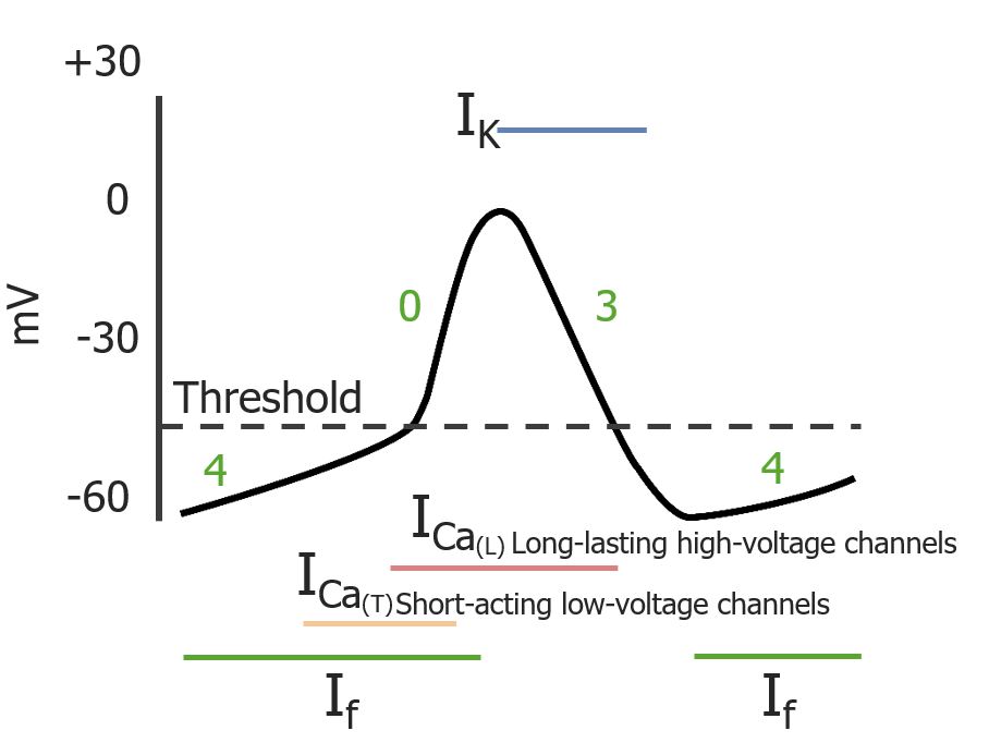
Phases of a cardiac pacemaker action potential: Phases 4, 0, 3, and 4 occur in sequence. Colored lines depict the duration of respective currents.
If: “funny” current
ICa(T): transient, short-acting calcium (Ca2+) current
ICa(L): long-lasting Ca2+ current
IK: K+ current
Image by Lecturio.
Table: Ion channelsChannelsThe Cell: Cell Membrane and their activity during pacemakerPacemakerA device designed to stimulate, by electric impulses, contraction of the heart muscles. It may be temporary (external) or permanent (internal or internal-external).Bradyarrhythmias action potentials
Transient or T-type CaCACondylomata acuminata are a clinical manifestation of genital HPV infection. Condylomata acuminata are described as raised, pearly, flesh-colored, papular, cauliflower-like lesions seen in the anogenital region that may cause itching, pain, or bleeding.Condylomata Acuminata (Genital Warts)2+channelsChannelsThe Cell: Cell Membrane
Active
Inactivated
–
L-type CaCACondylomata acuminata are a clinical manifestation of genital HPV infection. Condylomata acuminata are described as raised, pearly, flesh-colored, papular, cauliflower-like lesions seen in the anogenital region that may cause itching, pain, or bleeding.Condylomata Acuminata (Genital Warts)2+channelsChannelsThe Cell: Cell Membrane
*Primary current responsible for the membrane potential during the phase
HCN: hyperpolarization-activated cyclic nucleotide-gated Ca2+: calcium ions
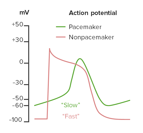
Comparison of pacemakerPacemakerA device designed to stimulate, by electric impulses, contraction of the heart muscles. It may be temporary (external) or permanent (internal or internal-external).Bradyarrhythmias and nonpacemaker action potentials
Compared with nonpacemaker action potentials, pacemakerPacemakerA device designed to stimulate, by electric impulses, contraction of the heart muscles. It may be temporary (external) or permanent (internal or internal-external).Bradyarrhythmias action potentials have the following characteristics:
Pacemaker (green) and nonpacemaker (red) action potentials: Nonpacemaker action potentials begin with quick depolarization followed by slow repolarization, whereas pacemaker action potentials have a longer depolarization phase. Nonpacemaker action potentials also start from an isoelectric (flat) line, whereas pacemaker action potentials have none because of their constant oscillation between repolarization and depolarization.
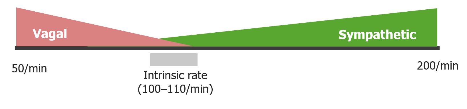
Chronotropy refers to the modulation of HR at the level of the pacemakerPacemakerA device designed to stimulate, by electric impulses, contraction of the heart muscles. It may be temporary (external) or permanent (internal or internal-external).Bradyarrhythmias cells. The SA node rate is primarily controlled by the ANSANSThe ans is a component of the peripheral nervous system that uses both afferent (sensory) and efferent (effector) neurons, which control the functioning of the internal organs and involuntary processes via connections with the CNS. The ans consists of the sympathetic and parasympathetic nervous systems.Autonomic Nervous System: Anatomy (sympathetic and parasympathetic nerves).
Normal resting HR: 60–100/min
TachycardiaTachycardiaAbnormally rapid heartbeat, usually with a heart rate above 100 beats per minute for adults. Tachycardia accompanied by disturbance in the cardiac depolarization (cardiac arrhythmia) is called tachyarrhythmia.Sepsis in Children: HR > 100/min
BradycardiaBradycardiaBradyarrhythmia is a rhythm in which the heart rate is less than 60/min. Bradyarrhythmia can be physiologic, without symptoms or hemodynamic change. Pathologic bradyarrhythmia results in reduced cardiac output and hemodynamic instability causing syncope, dizziness, or dyspnea.Bradyarrhythmias: HR < 60/min
Negative chronotropy:
Slowing down of the HR
Mediated by parasympathetic/vagal activation:
By acetylcholineAcetylcholineA neurotransmitter found at neuromuscular junctions, autonomic ganglia, parasympathetic effector junctions, a subset of sympathetic effector junctions, and at many sites in the central nervous system.Receptors and Neurotransmitters of the CNS
At the muscarinic (M2) receptorsReceptorsReceptors are proteins located either on the surface of or within a cell that can bind to signaling molecules known as ligands (e.g., hormones) and cause some type of response within the cell.Receptors
Positive chronotropy:
Increase in the HR
Mediated by sympathetic activation:
By norepinephrineNorepinephrinePrecursor of epinephrine that is secreted by the adrenal medulla and is a widespread central and autonomic neurotransmitter. Norepinephrine is the principal transmitter of most postganglionic sympathetic fibers, and of the diffuse projection system in the brain that arises from the locus ceruleus.Receptors and Neurotransmitters of the CNS
At the β1-adrenergic receptorsReceptorsReceptors are proteins located either on the surface of or within a cell that can bind to signaling molecules known as ligands (e.g., hormones) and cause some type of response within the cell.Receptors
There is a constant, low level of vagal tone slightly suppressing the intrinsic rate of the SA node.
Dromotropy is the modulation of conduction velocity through the AV node (also controlled by the ANSANSThe ans is a component of the peripheral nervous system that uses both afferent (sensory) and efferent (effector) neurons, which control the functioning of the internal organs and involuntary processes via connections with the CNS. The ans consists of the sympathetic and parasympathetic nervous systems.Autonomic Nervous System: Anatomy):
Sympathetic: speeds up conduction through the AV node
Parasympathetic: slows down conduction through the AV node
Autonomic control of the HR at the SA node: The sympathetic nervous system increases the HR (positive chronotropy) by acting on the β1-adrenergic receptors of the SA node. The parasympathetic nervous system decreases the HR (negative chronotropy) via the vagus by acting on the muscarinic (M2) receptors in the SA node.
Image by Lecturio.
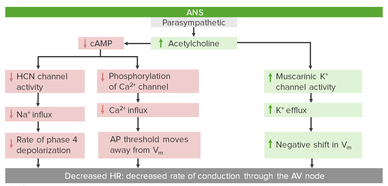
Parasympathetic control of HR
Cholinergic nerves release acetylcholineAcetylcholineA neurotransmitter found at neuromuscular junctions, autonomic ganglia, parasympathetic effector junctions, a subset of sympathetic effector junctions, and at many sites in the central nervous system.Receptors and Neurotransmitters of the CNS, which brings about 2 primary changes within myocytesMyocytesMature contractile cells, commonly known as myocytes, that form one of three kinds of muscle. The three types of muscle cells are skeletal, cardiac, and smooth. They are derived from embryonic (precursor) muscle cells called myoblasts.Muscle Tissue: Histology:
Decreases cAMPcAMPAn adenine nucleotide containing one phosphate group which is esterified to both the 3′- and 5′-positions of the sugar moiety. It is a second messenger and a key intracellular regulator, functioning as a mediator of activity for a number of hormones, including epinephrine, glucagon, and acth.Phosphodiesterase Inhibitors levels, which in turn:
Slows down depolarization by ↓ the “funny” current:
→ ↓ Na+flowFlowBlood flows through the heart, arteries, capillaries, and veins in a closed, continuous circuit. Flow is the movement of volume per unit of time. Flow is affected by the pressure gradient and the resistance fluid encounters between 2 points. Vascular resistance is the opposition to flow, which is caused primarily by blood friction against vessel walls.Vascular Resistance, Flow, and Mean Arterial Pressure into the cell during phase 4
→ Longer time for the membrane potentialMembrane potentialThe membrane potential is the difference in electric charge between the interior and the exterior of a cell. All living cells maintain a potential difference across the membrane thanks to the insulating properties of their plasma membranes (PMs) and the selective transport of ions across this membrane by transporters.Membrane Potential to reach its thresholdThresholdMinimum voltage necessary to generate an action potential (an all-or-none response)Skeletal Muscle Contraction (phase 4 line is flatter)
↓ PhosphorylationPhosphorylationThe introduction of a phosphoryl group into a compound through the formation of an ester bond between the compound and a phosphorus moiety.Post-translational Protein Processing of the CaCACondylomata acuminata are a clinical manifestation of genital HPV infection. Condylomata acuminata are described as raised, pearly, flesh-colored, papular, cauliflower-like lesions seen in the anogenital region that may cause itching, pain, or bleeding.Condylomata Acuminata (Genital Warts)2+ channel → ↓ CaCACondylomata acuminata are a clinical manifestation of genital HPV infection. Condylomata acuminata are described as raised, pearly, flesh-colored, papular, cauliflower-like lesions seen in the anogenital region that may cause itching, pain, or bleeding.Condylomata Acuminata (Genital Warts)2+ influx → moving the thresholdThresholdMinimum voltage necessary to generate an action potential (an all-or-none response)Skeletal Muscle Contraction potential farther from the current membrane potentialMembrane potentialThe membrane potential is the difference in electric charge between the interior and the exterior of a cell. All living cells maintain a potential difference across the membrane thanks to the insulating properties of their plasma membranes (PMs) and the selective transport of ions across this membrane by transporters.Membrane Potential
→ Longer time to reach the thresholdThresholdMinimum voltage necessary to generate an action potential (an all-or-none response)Skeletal Muscle Contraction potential
Parasympathetic control of the HR via the AV node AV: atrioventricular AP: action potential Vm: membrane potential HCN: hyperpolarization-activated cyclic nucleotide-gated
Image by Lecturio.
Sympathetic control of HR
NorepinephrineNorepinephrinePrecursor of epinephrine that is secreted by the adrenal medulla and is a widespread central and autonomic neurotransmitter. Norepinephrine is the principal transmitter of most postganglionic sympathetic fibers, and of the diffuse projection system in the brain that arises from the locus ceruleus.Receptors and Neurotransmitters of the CNS is released from sympathetic nerves, which binds to β1-adrenergic receptorsReceptorsReceptors are proteins located either on the surface of or within a cell that can bind to signaling molecules known as ligands (e.g., hormones) and cause some type of response within the cell.Receptors in the myocytesMyocytesMature contractile cells, commonly known as myocytes, that form one of three kinds of muscle. The three types of muscle cells are skeletal, cardiac, and smooth. They are derived from embryonic (precursor) muscle cells called myoblasts.Muscle Tissue: Histology and causes an intracellular increase in cAMPcAMPAn adenine nucleotide containing one phosphate group which is esterified to both the 3′- and 5′-positions of the sugar moiety. It is a second messenger and a key intracellular regulator, functioning as a mediator of activity for a number of hormones, including epinephrine, glucagon, and acth.Phosphodiesterase Inhibitors, thereby increasing the HR via 2 mechanisms:
↑ PhosphorylationPhosphorylationThe introduction of a phosphoryl group into a compound through the formation of an ester bond between the compound and a phosphorus moiety.Post-translational Protein Processing of CaCACondylomata acuminata are a clinical manifestation of genital HPV infection. Condylomata acuminata are described as raised, pearly, flesh-colored, papular, cauliflower-like lesions seen in the anogenital region that may cause itching, pain, or bleeding.Condylomata Acuminata (Genital Warts)2+channelsChannelsThe Cell: Cell Membrane → ↑ CaCACondylomata acuminata are a clinical manifestation of genital HPV infection. Condylomata acuminata are described as raised, pearly, flesh-colored, papular, cauliflower-like lesions seen in the anogenital region that may cause itching, pain, or bleeding.Condylomata Acuminata (Genital Warts)2+ influx → lowering of action potentialAction PotentialAbrupt changes in the membrane potential that sweep along the cell membrane of excitable cells in response to excitation stimuli.Membrane PotentialthresholdThresholdMinimum voltage necessary to generate an action potential (an all-or-none response)Skeletal Muscle Contraction (moves closer to the membrane potentialMembrane potentialThe membrane potential is the difference in electric charge between the interior and the exterior of a cell. All living cells maintain a potential difference across the membrane thanks to the insulating properties of their plasma membranes (PMs) and the selective transport of ions across this membrane by transporters.Membrane Potential)
Sympathetic control of HR via the AV node AV: atrioventricular AP: action potential Vm: membrane potential HCN: hyperpolarization-activated cyclic nucleotide-gated
Image by Lecturio.
Other factors influencing pacemakerPacemakerA device designed to stimulate, by electric impulses, contraction of the heart muscles. It may be temporary (external) or permanent (internal or internal-external).Bradyarrhythmias activity
↑ ThyroidThyroidThe thyroid gland is one of the largest endocrine glands in the human body. The thyroid gland is a highly vascular, brownish-red gland located in the visceral compartment of the anterior region of the neck.Thyroid Gland: AnatomyhormonesHormonesHormones are messenger molecules that are synthesized in one part of the body and move through the bloodstream to exert specific regulatory effects on another part of the body. Hormones play critical roles in coordinating cellular activities throughout the body in response to the constant changes in both the internal and external environments. Hormones: Overview and Types (i.e., T3T3A T3 thyroid hormone normally synthesized and secreted by the thyroid gland in much smaller quantities than thyroxine (T4). Most T3 is derived from peripheral monodeiodination of T4 at the 5′ position of the outer ring of the iodothyronine nucleus. The hormone finally delivered and used by the tissues is mainly t3.Thyroid Hormones and T4T4The major hormone derived from the thyroid gland. Thyroxine is synthesized via the iodination of tyrosines (monoiodotyrosine) and the coupling of iodotyrosines (diiodotyrosine) in the thyroglobulin. Thyroxine is released from thyroglobulin by proteolysis and secreted into the blood. Thyroxine is peripherally deiodinated to form triiodothyronine which exerts a broad spectrum of stimulatory effects on cell metabolism.Thyroid Hormones) → ↑ HR
↑ Circulating catecholaminesCatecholaminesA general class of ortho-dihydroxyphenylalkylamines derived from tyrosine.Adrenal Hormones (e.g., epinephrineEpinephrineThe active sympathomimetic hormone from the adrenal medulla. It stimulates both the alpha- and beta- adrenergic systems, causes systemic vasoconstriction and gastrointestinal relaxation, stimulates the heart, and dilates bronchi and cerebral vessels.Sympathomimetic Drugs and norepinephrineNorepinephrinePrecursor of epinephrine that is secreted by the adrenal medulla and is a widespread central and autonomic neurotransmitter. Norepinephrine is the principal transmitter of most postganglionic sympathetic fibers, and of the diffuse projection system in the brain that arises from the locus ceruleus.Receptors and Neurotransmitters of the CNS from the adrenal medullaAdrenal MedullaThe inner portion of the adrenal gland. Derived from ectoderm, adrenal medulla consists mainly of chromaffin cells that produces and stores a number of neurotransmitters, mainly adrenaline (epinephrine) and norepinephrine. The activity of the adrenal medulla is regulated by the sympathetic nervous system.Adrenal Glands: Anatomy) → ↑ HR
↑ K+ → ↓ HR (because K+ affects membrane potentialMembrane potentialThe membrane potential is the difference in electric charge between the interior and the exterior of a cell. All living cells maintain a potential difference across the membrane thanks to the insulating properties of their plasma membranes (PMs) and the selective transport of ions across this membrane by transporters.Membrane Potential)
IschemiaIschemiaA hypoperfusion of the blood through an organ or tissue caused by a pathologic constriction or obstruction of its blood vessels, or an absence of blood circulation.Ischemic Cell Damage (↓ in O2) → ↓ HR (works through a different K+ channel)
Drugs:
Antiarrhythmic agents
CaCACondylomata acuminata are a clinical manifestation of genital HPV infection. Condylomata acuminata are described as raised, pearly, flesh-colored, papular, cauliflower-like lesions seen in the anogenital region that may cause itching, pain, or bleeding.Condylomata Acuminata (Genital Warts)2+ channel blockers
β-adrenergic blockers
DigoxinDigoxinA cardiotonic glycoside obtained mainly from digitalis lanata; it consists of three sugars and the aglycone digoxigenin. Digoxin has positive inotropic and negative chronotropic activity. It is used to control ventricular rate in atrial fibrillation and in the management of congestive heart failure with atrial fibrillation. Its use in congestive heart failure and sinus rhythm is less certain. The margin between toxic and therapeutic doses is small.Cardiac Glycosides
Table: Major factors influencing the HR
Factor
Increased HR (positive chronotropy)
Decreased HR (negative chronotropy)
ANSANSThe ans is a component of the peripheral nervous system that uses both afferent (sensory) and efferent (effector) neurons, which control the functioning of the internal organs and involuntary processes via connections with the CNS. The ans consists of the sympathetic and parasympathetic nervous systems.Autonomic Nervous System: Anatomy*
Sympathetic nervous systemNervous systemThe nervous system is a small and complex system that consists of an intricate network of neural cells (or neurons) and even more glial cells (for support and insulation). It is divided according to its anatomical components as well as its functional characteristics. The brain and spinal cord are referred to as the central nervous system, and the branches of nerves from these structures are referred to as the peripheral nervous system.Nervous System: Anatomy, Structure, and Classification
Parasympathetic nervous systemNervous systemThe nervous system is a small and complex system that consists of an intricate network of neural cells (or neurons) and even more glial cells (for support and insulation). It is divided according to its anatomical components as well as its functional characteristics. The brain and spinal cord are referred to as the central nervous system, and the branches of nerves from these structures are referred to as the peripheral nervous system.Nervous System: Anatomy, Structure, and Classification
ThyroidThyroidThe thyroid gland is one of the largest endocrine glands in the human body. The thyroid gland is a highly vascular, brownish-red gland located in the visceral compartment of the anterior region of the neck.Thyroid Gland: AnatomyhormonesHormonesHormones are messenger molecules that are synthesized in one part of the body and move through the bloodstream to exert specific regulatory effects on another part of the body. Hormones play critical roles in coordinating cellular activities throughout the body in response to the constant changes in both the internal and external environments. Hormones: Overview and Types
HypothyroidismHypothyroidismHypothyroidism is a condition characterized by a deficiency of thyroid hormones. Iodine deficiency is the most common cause worldwide, but Hashimoto’s disease (autoimmune thyroiditis) is the leading cause in non-iodine-deficient regions. Hypothyroidism
K+
HypokalemiaHypokalemiaHypokalemia is defined as plasma potassium (K+) concentration < 3.5 mEq/L. Homeostatic mechanisms maintain plasma concentration between 3.5-5.2 mEq/L despite marked variation in dietary intake. Hypokalemia can be due to renal losses, GI losses, transcellular shifts, or poor dietary intake.Hypokalemia
HyperkalemiaHyperkalemiaHyperkalemia is defined as a serum potassium (K+) concentration >5.2 mEq/L. Homeostatic mechanisms maintain the serum K+ concentration between 3.5 and 5.2 mEq/L, despite marked variation in dietary intake. Hyperkalemia can be due to a variety of causes, which include transcellular shifts, tissue breakdown, inadequate renal excretion, and drugs. Hyperkalemia
Circulating catecholaminesCatecholaminesA general class of ortho-dihydroxyphenylalkylamines derived from tyrosine.Adrenal Hormones
↑ Serum epinephrineEpinephrineThe active sympathomimetic hormone from the adrenal medulla. It stimulates both the alpha- and beta- adrenergic systems, causes systemic vasoconstriction and gastrointestinal relaxation, stimulates the heart, and dilates bronchi and cerebral vessels.Sympathomimetic Drugs
↑ Serum norepinephrineNorepinephrinePrecursor of epinephrine that is secreted by the adrenal medulla and is a widespread central and autonomic neurotransmitter. Norepinephrine is the principal transmitter of most postganglionic sympathetic fibers, and of the diffuse projection system in the brain that arises from the locus ceruleus.Receptors and Neurotransmitters of the CNS
IschemiaIschemiaA hypoperfusion of the blood through an organ or tissue caused by a pathologic constriction or obstruction of its blood vessels, or an absence of blood circulation.Ischemic Cell Damage/hypoxiaHypoxiaSub-optimal oxygen levels in the ambient air of living organisms.Ischemic Cell Damage
Atrioventricular nodeAtrioventricular nodeA small nodular mass of specialized muscle fibers located in the interatrial septum near the opening of the coronary sinus. It gives rise to the atrioventricular bundle of the conduction system of the heart.Heart: Anatomy blocks
Atrioventricular nodeAtrioventricular nodeA small nodular mass of specialized muscle fibers located in the interatrial septum near the opening of the coronary sinus. It gives rise to the atrioventricular bundle of the conduction system of the heart.Heart: Anatomy blocks occur when an anatomical or functional impairment of the conduction system of the heart produces a delay or interruption in the transmission of action potentials from the atria to the ventricles through the AV node. Affected individuals may be asymptomatic or may present with syncopeSyncopeSyncope is a short-term loss of consciousness and loss of postural stability followed by spontaneous return of consciousness to the previous neurologic baseline without the need for resuscitation. The condition is caused by transient interruption of cerebral blood flow that may be benign or related to a underlying life-threatening condition. Syncope, chest painPainAn unpleasant sensation induced by noxious stimuli which are detected by nerve endings of nociceptive neurons.Pain: Types and Pathways, dyspneaDyspneaDyspnea is the subjective sensation of breathing discomfort. Dyspnea is a normal manifestation of heavy physical or psychological exertion, but also may be caused by underlying conditions (both pulmonary and extrapulmonary). Dyspnea, and bradycardiaBradycardiaBradyarrhythmia is a rhythm in which the heart rate is less than 60/min. Bradyarrhythmia can be physiologic, without symptoms or hemodynamic change. Pathologic bradyarrhythmia results in reduced cardiac output and hemodynamic instability causing syncope, dizziness, or dyspnea.Bradyarrhythmias depending on the severity of the block. Diagnosis is established based on ECGECGAn electrocardiogram (ECG) is a graphic representation of the electrical activity of the heart plotted against time. Adhesive electrodes are affixed to the skin surface allowing measurement of cardiac impulses from many angles. The ECG provides 3-dimensional information about the conduction system of the heart, the myocardium, and other cardiac structures. Electrocardiogram (ECG), and treatment is based on the type of block and hemodynamic stability of the affected individual.
1st-degree AV blockAV blockAtrioventricular (AV) block is a bradyarrhythmia caused by delay, or interruption, in the electrical conduction between the atria and the ventricles. Atrioventricular block occurs due to either anatomic or functional impairment, and is classified into 3 types. Atrioventricular block (AV block): delayed conduction through the AV node. Affected individuals have sinus rhythm; however, their overall HR is slower.
2nd-degree AV blockAV blockAtrioventricular (AV) block is a bradyarrhythmia caused by delay, or interruption, in the electrical conduction between the atria and the ventricles. Atrioventricular block occurs due to either anatomic or functional impairment, and is classified into 3 types. Atrioventricular block (AV block): delayed conduction through the AV node. Some atrial action potentials fail to make it through the AV node, resulting in ventricular bradycardiaBradycardiaBradyarrhythmia is a rhythm in which the heart rate is less than 60/min. Bradyarrhythmia can be physiologic, without symptoms or hemodynamic change. Pathologic bradyarrhythmia results in reduced cardiac output and hemodynamic instability causing syncope, dizziness, or dyspnea.Bradyarrhythmias.
Mobitz type I(WenckebachWenckebachAtrioventricular block (AV block)): progressive increase in conduction delay until a signal fails to make it through the AV node altogether, resulting in the signal (and thus a mechanical contraction) being “dropped.”
Mobitz type II: there is no progressive increase in delayed conduction; however, conduction through the AV node is intermittent with some “dropped” signals that do not make it through to the ventricles. Dropped signals often occur in a regularRegularInsulin pattern (e.g., 2:1 pattern). Mobitz type II block almost always results from conduction system disease below the level of the AV node.
3rd-degree AV blockAV blockAtrioventricular (AV) block is a bradyarrhythmia caused by delay, or interruption, in the electrical conduction between the atria and the ventricles. Atrioventricular block occurs due to either anatomic or functional impairment, and is classified into 3 types. Atrioventricular block (AV block): a complete block through the AV node resulting in atrial-ventricular dissociationDissociationDefense Mechanisms (activation and contraction are independent of each other as no atrial impulses reach the ventricles). Affected individuals have ventricular bradycardiaBradycardiaBradyarrhythmia is a rhythm in which the heart rate is less than 60/min. Bradyarrhythmia can be physiologic, without symptoms or hemodynamic change. Pathologic bradyarrhythmia results in reduced cardiac output and hemodynamic instability causing syncope, dizziness, or dyspnea.Bradyarrhythmias driven by an escape pacemakerPacemakerA device designed to stimulate, by electric impulses, contraction of the heart muscles. It may be temporary (external) or permanent (internal or internal-external).Bradyarrhythmias distal to the block.
Bundle branch and fascicular blocksFascicular BlocksBundle branch and fascicular blocks occur when the normal electrical activity in the His-Purkinje system is interrupted. These blocks can be due to many etiologies that may affect the structure of the heart or the conduction system directly. Bundle Branch and Fascicular Blocks
Bundle branch and fascicular blocksFascicular BlocksBundle branch and fascicular blocks occur when the normal electrical activity in the His-Purkinje system is interrupted. These blocks can be due to many etiologies that may affect the structure of the heart or the conduction system directly. Bundle Branch and Fascicular Blocks occur when normal electrical activity in the His-Purkinje system is interrupted. Bundle branch and fascicular blocksFascicular BlocksBundle branch and fascicular blocks occur when the normal electrical activity in the His-Purkinje system is interrupted. These blocks can be due to many etiologies that may affect the structure of the heart or the conduction system directly. Bundle Branch and Fascicular Blocks can occur due to many etiologies that may affect the structure of the heart or the conduction system directly (e.g., myocardial ischemiaMyocardial ischemiaA disorder of cardiac function caused by insufficient blood flow to the muscle tissue of the heart. The decreased blood flow may be due to narrowing of the coronary arteries (coronary artery disease), to obstruction by a thrombus (coronary thrombosis), or less commonly, to diffuse narrowing of arterioles and other small vessels within the heart.Coronary Heart Disease, myocarditisMyocarditisMyocarditis is an inflammatory disease of the myocardium, which may occur alone or in association with a systemic process. There are numerous etiologies of myocarditis, but all lead to inflammation and myocyte injury, most often leading to signs and symptoms of heart failure. Myocarditis, cardiomyopathyCardiomyopathyCardiomyopathy refers to a group of myocardial diseases associated with structural changes of the heart muscles (myocardium) and impaired systolic and/or diastolic function in the absence of other heart disorders (coronary artery disease, hypertension, valvular disease, and congenital heart disease). Cardiomyopathy: Overview and Types). Although usually asymptomatic, bundle branch and fascicular blocksFascicular BlocksBundle branch and fascicular blocks occur when the normal electrical activity in the His-Purkinje system is interrupted. These blocks can be due to many etiologies that may affect the structure of the heart or the conduction system directly. Bundle Branch and Fascicular Blocks may occasionally cause syncopeSyncopeSyncope is a short-term loss of consciousness and loss of postural stability followed by spontaneous return of consciousness to the previous neurologic baseline without the need for resuscitation. The condition is caused by transient interruption of cerebral blood flow that may be benign or related to a underlying life-threatening condition. Syncope.
Bundle branch blocksBundle Branch BlocksBundle branch and fascicular blocks occur when the normal electrical activity in the His-Purkinje system is interrupted. These blocks can be due to many etiologies that may affect the structure of the heart or the conduction system directly. Bundle Branch and Fascicular Blocks: a block in the downward progression of electrical impulses through 1 of the bundle branches in the interventricular septumInterventricular SeptumVentricular Septal Defect (VSD). The block forces electrical signals down the other bundle branch such that the ventricle depolarizes 1st. Electrical waves then travel through the myocytesMyocytesMature contractile cells, commonly known as myocytes, that form one of three kinds of muscle. The three types of muscle cells are skeletal, cardiac, and smooth. They are derived from embryonic (precursor) muscle cells called myoblasts.Muscle Tissue: Histology directly to cause depolarizationDepolarizationMembrane Potential on the affected side.
Fascicular blocksFascicular BlocksBundle branch and fascicular blocks occur when the normal electrical activity in the His-Purkinje system is interrupted. These blocks can be due to many etiologies that may affect the structure of the heart or the conduction system directly. Bundle Branch and Fascicular Blocks: a block in 1 of the more distal Purkinje fibersPurkinje fibersModified cardiac muscle fibers composing the terminal portion of the heart conduction system.Heart: Anatomy. The affected area will receive electrical signals more slowly from the surrounding myocytesMyocytesMature contractile cells, commonly known as myocytes, that form one of three kinds of muscle. The three types of muscle cells are skeletal, cardiac, and smooth. They are derived from embryonic (precursor) muscle cells called myoblasts.Muscle Tissue: Histology.
Antiarrhythmic medications
The following classes of drugs are used for the treatment of arrhythmias:
Class I antiarrhythmics are a group of medications that inhibit the Na+channelsChannelsThe Cell: Cell Membrane responsible for the depolarizationDepolarizationMembrane Potential of cardiomyocytes during phase 0 of the nonpacemaker action potentialAction PotentialAbrupt changes in the membrane potential that sweep along the cell membrane of excitable cells in response to excitation stimuli.Membrane Potential.
Class II antiarrhythmics are a group of medications that inhibit the β-adrenergic channelsChannelsThe Cell: Cell Membrane in cardiac muscleCardiac muscleThe muscle tissue of the heart. It is composed of striated, involuntary muscle cells connected to form the contractile pump to generate blood flow.Muscle Tissue: Histology. Drugs in this class are commonly known as beta-blockersBeta-blockersDrugs that bind to but do not activate beta-adrenergic receptors thereby blocking the actions of beta-adrenergic agonists. Adrenergic beta-antagonists are used for treatment of hypertension, cardiac arrhythmias, angina pectoris, glaucoma, migraine headaches, and anxiety.Class 2 Antiarrhythmic Drugs (Beta Blockers).
Class IV antiarrhythmics are a group of medications that inhibit the CaCACondylomata acuminata are a clinical manifestation of genital HPV infection. Condylomata acuminata are described as raised, pearly, flesh-colored, papular, cauliflower-like lesions seen in the anogenital region that may cause itching, pain, or bleeding.Condylomata Acuminata (Genital Warts)2+channelsChannelsThe Cell: Cell Membrane that are active during the repolarizationRepolarizationMembrane Potential of cardiomyocytes.
Baumann, B.M. (2016). Systemic hypertension. In Tintinalli, J.E., et al. (Eds.), Tintinalli’s emergency medicine: A comprehensive study guide, 8e. New York, NY: McGraw-Hill Education. Retrieved April 22, 2026, from https://accessmedicine.mhmedical.com/content.aspx?aid=1121496251