Ventricular septal defects (VSDs) are congenital Congenital Chorioretinitis cardiac Cardiac Total Anomalous Pulmonary Venous Return (TAPVR) malformations that feature an abnormal communication Communication The exchange or transmission of ideas, attitudes, or beliefs between individuals or groups. Decision-making Capacity and Legal Competence between the right and left ventricles. Presenting both in isolation or as part of a more complex disease, VSD is the most common congenital Congenital Chorioretinitis heart defect. While the degree of severity depends on the size of the defect, VSDs are classified on the basis of the anatomical location of the defect. Patients Patients Individuals participating in the health care system for the purpose of receiving therapeutic, diagnostic, or preventive procedures. Clinician–Patient Relationship may be asymptomatic with smaller defects, whereas larger defects can present with respiratory or heart failure Heart Failure A heterogeneous condition in which the heart is unable to pump out sufficient blood to meet the metabolic need of the body. Heart failure can be caused by structural defects, functional abnormalities (ventricular dysfunction), or a sudden overload beyond its capacity. Chronic heart failure is more common than acute heart failure which results from sudden insult to cardiac function, such as myocardial infarction. Total Anomalous Pulmonary Venous Return (TAPVR) during infancy or childhood. A common clinical sign is a holosystolic murmur Holosystolic Murmur Tricuspid Valve Atresia (TVA) audible at the left sternal border. Diagnosis, both pre- and post-natal, is confirmed by echocardiogram Echocardiogram Transposition of the Great Vessels. The majority of small VSDs close spontaneously, but those that are larger and symptomatic require medical stabilization followed by surgical repair.
Last updated: 19 Feb, 2021
A ventricular septal defect Ventricular Septal Defect Tetralogy of Fallot (VSD) is a malformation of the interventricular septum (IVS) resulting in an abnormal communication Communication The exchange or transmission of ideas, attitudes, or beliefs between individuals or groups. Decision-making Capacity and Legal Competence between the left ventricle (LV) and the right ventricle (RV). This defect may present in isolation, or be a part of another anomaly, such as tetralogy of Fallot Tetralogy of Fallot Tetralogy of Fallot is the most common cyanotic congenital heart disease. The disease is the confluence of 4 pathologic cardiac features: overriding aorta, ventricular septal defect, right ventricular outflow obstruction, and right ventricular hypertrophy. Tetralogy of Fallot.
There are 4 main subtypes of VSD based on location along IVS:
Exact mechanism is unknown. Genetic syndromes:
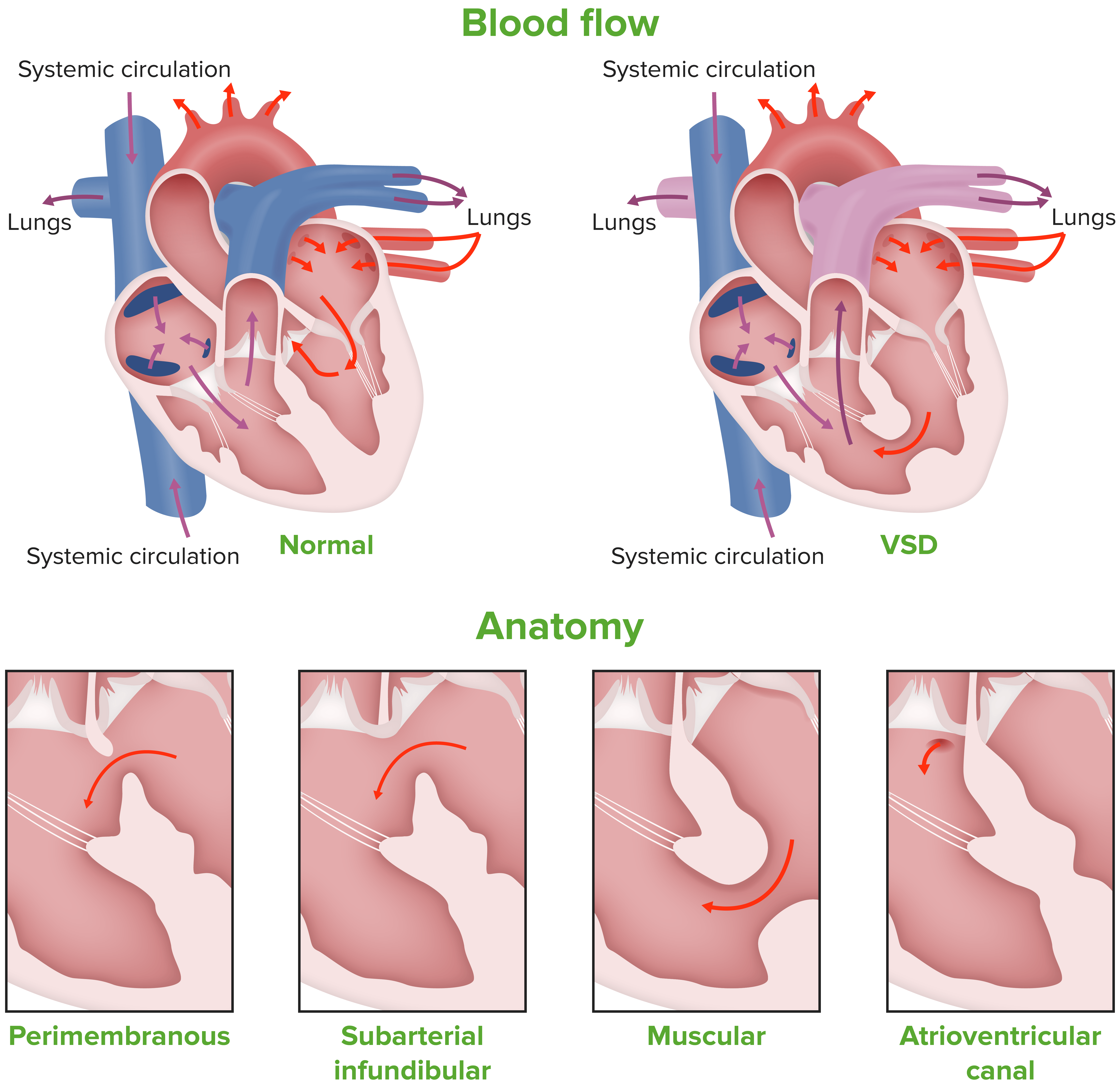
Schematic drawing depicting the various subtypes of the VSD along the interventricular septum:
The membranous subtypes are: (1) perimembranous with outlet
extension
Extension
Examination of the Upper Limbs; (2) perimembranous with trabecular
extension
Extension
Examination of the Upper Limbs; and (3) perimembranous with inlet
extension
Extension
Examination of the Upper Limbs
The muscular subtypes are: (4) inlet muscular; (5) central muscular; (6) apical muscular; and (7) outlet muscular. The defect known as (8) supracristal defect can also be seen.
Physiologic changes occur based on the size of the defect and the resistance Resistance Physiologically, the opposition to flow of air caused by the forces of friction. As a part of pulmonary function testing, it is the ratio of driving pressure to the rate of air flow. Ventilation: Mechanics of Breathing across which the blood has to flow Flow Blood flows through the heart, arteries, capillaries, and veins in a closed, continuous circuit. Flow is the movement of volume per unit of time. Flow is affected by the pressure gradient and the resistance fluid encounters between 2 points. Vascular resistance is the opposition to flow, which is caused primarily by blood friction against vessel walls. Vascular Resistance, Flow, and Mean Arterial Pressure.
Age of presentation Presentation The position or orientation of the fetus at near term or during obstetric labor, determined by its relation to the spine of the mother and the birth canal. The normal position is a vertical, cephalic presentation with the fetal vertex flexed on the neck. Normal and Abnormal Labor is usually around 2 months, unless a larger defect is present which becomes symptomatic within weeks of birth.
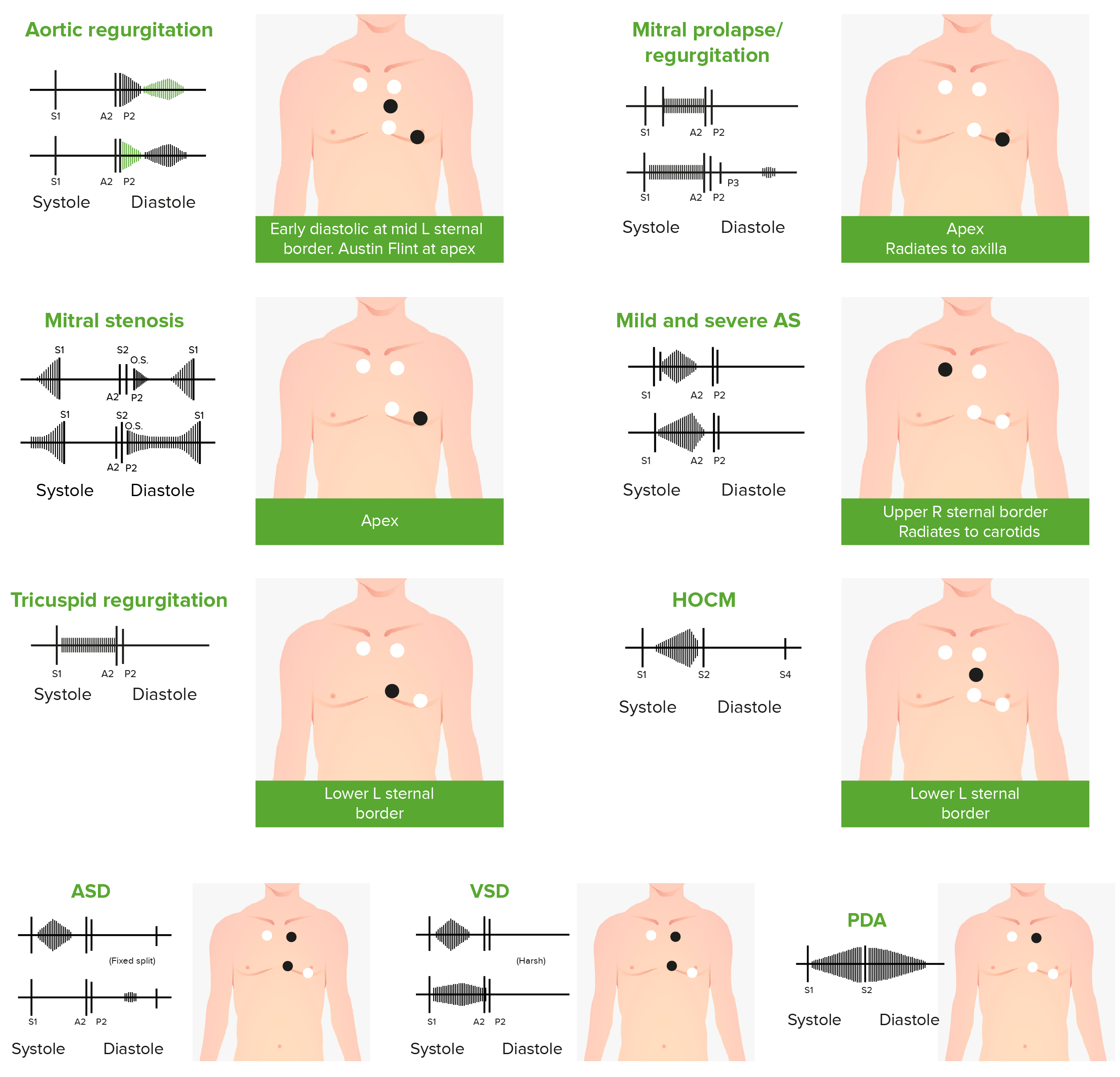
Phonocardiograms of abnormal
heart sounds
Heart sounds
Heart sounds are brief, transient sounds produced by valve opening and closure and by movement of blood in the heart. They are divided into systolic and diastolic sounds. In most cases, only the first (S1) and second (S2) heart sounds are heard. These are high-frequency sounds and arise from aortic and pulmonary valve closure (S1), as well as mitral and tricuspid valve closure (S2).
Heart Sounds caused by the following
cardiac
Cardiac
Total Anomalous Pulmonary Venous Return (TAPVR) defects:
Aortic regurgitation
Aortic regurgitation (AR) is a cardiac condition characterized by the backflow of blood from the aorta to the left ventricle during diastole. Aortic regurgitation is associated with an abnormal aortic valve and/or aortic root stemming from multiple causes, commonly rheumatic heart disease as well as congenital and degenerative valvular disorders.
Aortic Regurgitation,
mitral valve prolapse
Mitral valve prolapse
Abnormal protrusion or billowing of one or both of the leaflets of mitral valve into the left atrium during systole. This allows the backflow of blood into left atrium leading to mitral valve insufficiency, systolic murmurs, or cardiac arrhythmia.
Mitral Valve Prolapse, mitral
stenosis
Stenosis
Hypoplastic Left Heart Syndrome (HLHS) (
MS
MS
Multiple sclerosis (MS) is a chronic inflammatory autoimmune disease that leads to demyelination of the nerves in the CNS. Young women are more predominantly affected by this most common demyelinating condition.
Multiple Sclerosis), aortic
stenosis
Stenosis
Hypoplastic Left Heart Syndrome (HLHS) (AS),
tricuspid regurgitation
Tricuspid regurgitation
Tricuspid regurgitation (TR) is a valvular defect that allows backflow of blood from the right ventricle to the right atrium during systole. Tricuspid regurgitation can develop through a number of cardiac conditions that cause dilation of the right ventricle and tricuspid annulus. A blowing holosystolic murmur is best heard at the left lower sternal border.
Tricuspid Regurgitation, hypertrophic obstructive
cardiomyopathy
Cardiomyopathy
Cardiomyopathy refers to a group of myocardial diseases associated with structural changes of the heart muscles (myocardium) and impaired systolic and/or diastolic function in the absence of other heart disorders (coronary artery disease, hypertension, valvular disease, and congenital heart disease).
Cardiomyopathy: Overview and Types (HOCM),
atrial septal defect
Atrial Septal Defect
Atrial septal defects (ASDs) are benign acyanotic congenital heart defects characterized by an opening in the interatrial septum that causes blood to flow from the left atrium (LA) to the right atrium (RA) (left-to-right shunt).
Atrial Septal Defect (ASD) (
ASD
ASD
Autism spectrum disorder (ASD) is a neurodevelopmental disorder marked by poor social skills, restricted interests/social interactions, and repetitive/stereotyped behaviors. The condition is termed a “spectrum” because of the wide variability in the severity of symptoms exhibited.
Autism Spectrum Disorder),
ventricular septal defect
Ventricular Septal Defect
Tetralogy of Fallot (VSD), and
patent ductus arteriosus
Patent ductus arteriosus
The ductus arteriosus (DA) allows blood to bypass pulmonary circulation. After birth, the DA remains open for up to 72 hours and then constricts and involutes, becoming the ligamentum arteriosum. Failure of this process to occur results in patent ductus arteriosus (PDA), a condition that causes up to 10% of congenital heart defects.
Patent Ductus Arteriosus (PDA) (
PDA
PDA
The ductus arteriosus (DA) allows blood to bypass pulmonary circulation. After birth, the DA remains open for up to 72 hours and then constricts and involutes, becoming the ligamentum arteriosum. Failure of this process to occur results in patent ductus arteriosus (PDA), a condition that causes up to 10% of congenital heart defects.
Patent Ductus Arteriosus (PDA))
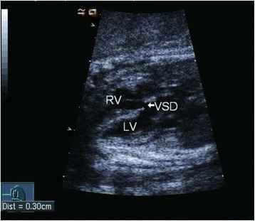
White arrow indicates a large VSD detected by fetal echocardiography Echocardiography Ultrasonic recording of the size, motion, and composition of the heart and surrounding tissues. The standard approach is transthoracic. Tricuspid Valve Atresia (TVA).
Image: “Prediction of spontaneous closure of isolated ventricular septal defects in utero and postnatal life” by BMC Pediatrics. License: CC BY 4.0The management of a VSD is based on the size of the defect and the clinical symptoms of the patient.
The following conditions are associated with VSD and can cause or modify the disease: