Ebstein's anomaly (EA) is a cyanotic congenital heart disease (CHD) characterized by the downward displacementDisplacementThe process by which an emotional or behavioral response that is appropriate for one situation appears in another situation for which it is inappropriate.Defense Mechanisms of the septal and posterior leaflets of the tricuspid valveTricuspid valveThe valve consisting of three cusps situated between the right atrium and right ventricle of the heart.Heart: Anatomy (TV). Ebstein's anomaly accounts for less than 1% of all cases of CHD. Maternal use of lithium is a common cause of EA. Clinical presentation varies, with the most common symptom being cyanosisCyanosisA bluish or purplish discoloration of the skin and mucous membranes due to an increase in the amount of deoxygenated hemoglobin in the blood or a structural defect in the hemoglobin molecule.Pulmonary Examination. The age of presentation varies from in utero to adulthood and is proportional to the severity of the TV displacementDisplacementThe process by which an emotional or behavioral response that is appropriate for one situation appears in another situation for which it is inappropriate.Defense Mechanisms, most cases present during adolescence. Ebstein's anomaly presents with multiple comorbiditiesComorbiditiesThe presence of co-existing or additional diseases with reference to an initial diagnosis or with reference to the index condition that is the subject of study. Comorbidity may affect the ability of affected individuals to function and also their survival; it may be used as a prognostic indicator for length of hospital stay, cost factors, and outcome or survival.St. Louis Encephalitis Virus, especially Wolff-Parkinson-White syndromeWolff-Parkinson-White SyndromeA form of ventricular pre-excitation characterized by a short PR interval and a long QRS interval with a delta wave. In this syndrome, atrial impulses are abnormally conducted to the heart ventricles via an accessory conducting pathway that is located between the wall of the right or left atria and the ventricles, also known as a bundle of kent. The inherited form can be caused by mutation of prkag2 gene encoding a gamma-2 regulatory subunit of amp-activated protein kinase.Supraventricular Tachycardias. The diagnosis is confirmed by echocardiographyEchocardiographyUltrasonic recording of the size, motion, and composition of the heart and surrounding tissues. The standard approach is transthoracic.Tricuspid Valve Atresia (TVA) and definitive treatment is surgical. PatientsPatientsIndividuals participating in the health care system for the purpose of receiving therapeutic, diagnostic, or preventive procedures.Clinician–Patient Relationship who are managed appropriately still have a reduced life expectancyLife expectancyBased on known statistical data, the number of years which any person of a given age may reasonably expected to live.Population Pyramids.
Ebstein’s anomaly (EA) is a cyanotic congenital heart disease (CHD) characterized by the downward displacementDisplacementThe process by which an emotional or behavioral response that is appropriate for one situation appears in another situation for which it is inappropriate.Defense Mechanisms of the septal and posterior leaflets of the tricuspid valveTricuspid valveThe valve consisting of three cusps situated between the right atrium and right ventricle of the heart.Heart: Anatomy (TV) into the right ventricle (RV).
Morphology
TV in EA:
The posterior and septal leaflet’s annularAnnularDermatologic Examination attachments are displaced downward away from the atrioventricular (AV) junction into the RV.
The anterior leaflet is attached at the AV junction and may be fenestrated and tethered to the myocardiumMyocardiumThe muscle tissue of the heart. It is composed of striated, involuntary muscle cells connected to form the contractile pump to generate blood flow.Heart: Anatomy → regurgitationRegurgitationGastroesophageal Reflux Disease (GERD)
RV:
Due to the displacementDisplacementThe process by which an emotional or behavioral response that is appropriate for one situation appears in another situation for which it is inappropriate.Defense Mechanisms of the AV junction, a portion of the RV is atrialized and thin, lacking ventricular contractility.
The size of the true “functional” RV is limited and in severe cases may just be the right ventricular outflow tract (RVOT).
Associated defects:
Most common (80%): atrial septal defectAtrial Septal DefectAtrial septal defects (ASDs) are benign acyanotic congenital heart defects characterized by an opening in the interatrial septum that causes blood to flow from the left atrium (LA) to the right atrium (RA) (left-to-right shunt). Atrial Septal Defect (ASD) (ASDASDAutism spectrum disorder (ASD) is a neurodevelopmental disorder marked by poor social skills, restricted interests/social interactions, and repetitive/stereotyped behaviors. The condition is termed a “spectrum” because of the wide variability in the severity of symptoms exhibited.Autism Spectrum Disorder) or patent foramen ovaleForamen ovaleAn opening in the wall between the right and the left upper chambers (heart atria) of a fetal heart. Oval foramen normally closes soon after birth; when it fails to close the condition is called patent oval foramen.Patent Foramen Ovale (PFOPFOA patent foramen ovale (PFO) is an abnormal communication between the atria that persists after birth. The condition results from incomplete closure of the foramen ovale. Small, isolated, and asymptomatic pfos are a common incidental finding on echocardiography and require no treatment.Patent Foramen Ovale)
Patent ductus arteriosusDuctus arteriosusA fetal blood vessel connecting the pulmonary artery with the descending aorta.Patent Ductus Arteriosus (PDA) (PDAPDAThe ductus arteriosus (DA) allows blood to bypass pulmonary circulation. After birth, the DA remains open for up to 72 hours and then constricts and involutes, becoming the ligamentum arteriosum. Failure of this process to occur results in patent ductus arteriosus (PDA), a condition that causes up to 10% of congenital heart defects. Patent Ductus Arteriosus (PDA))
Accessory conduction pathways → Wolff-Parkinson-White syndromeWolff-Parkinson-White SyndromeA form of ventricular pre-excitation characterized by a short PR interval and a long QRS interval with a delta wave. In this syndrome, atrial impulses are abnormally conducted to the heart ventricles via an accessory conducting pathway that is located between the wall of the right or left atria and the ventricles, also known as a bundle of kent. The inherited form can be caused by mutation of prkag2 gene encoding a gamma-2 regulatory subunit of amp-activated protein kinase.Supraventricular Tachycardias
40% of all congenital tricuspid valveTricuspid valveThe valve consisting of three cusps situated between the right atrium and right ventricle of the heart.Heart: Anatomy abnormalities
1.2 to 5 out of every 100,000 live births
Median age of presentation is late childhood or adolescence.
Etiology
Failure of delamination (separation) of the TV from the myocardial wall due to:
Maternal use of lithium in early pregnancyPregnancyThe status during which female mammals carry their developing young (embryos or fetuses) in utero before birth, beginning from fertilization to birth.Pregnancy: Diagnosis, Physiology, and Care
Heterogeneous genetic predisposition
Recent studies have identified genesGenesA category of nucleic acid sequences that function as units of heredity and which code for the basic instructions for the development, reproduction, and maintenance of organisms.DNA Types and StructureMYH7, TPM1, and NKX2-5 as associated with familial cases of Ebstein’s anomaly.
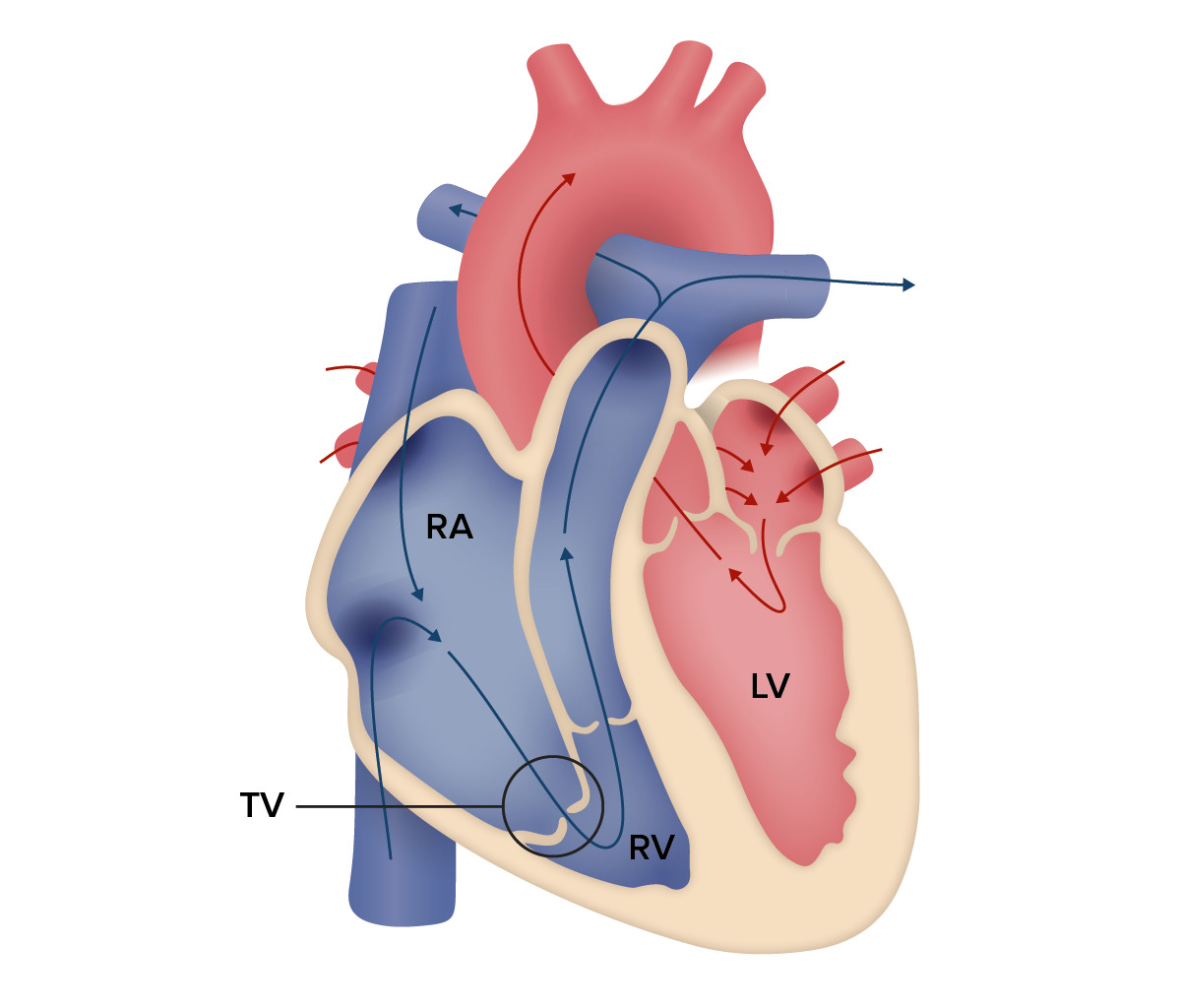
Diagram of EA: Note the downward displacement of the TV and the resulting significantly reduced size (atrialization) of the RV. RV: right ventricle LV: left ventricle RA: right atrium TV: tricuspid valve
Image by Lecturio.
Pathophysiology
The basis of cyanosisCyanosisA bluish or purplish discoloration of the skin and mucous membranes due to an increase in the amount of deoxygenated hemoglobin in the blood or a structural defect in the hemoglobin molecule.Pulmonary Examination and heart failureHeart FailureA heterogeneous condition in which the heart is unable to pump out sufficient blood to meet the metabolic need of the body. Heart failure can be caused by structural defects, functional abnormalities (ventricular dysfunction), or a sudden overload beyond its capacity. Chronic heart failure is more common than acute heart failure which results from sudden insult to cardiac function, such as myocardial infarction.Total Anomalous Pulmonary Venous Return (TAPVR) in EA is due to functionally impaired RV and tricuspid regurgitationRegurgitationGastroesophageal Reflux Disease (GERD) that results in reduced ejection fractionEjection fractionCardiac Cycle (EFEFCardiac Cycle).
Increased backflow into the right atrium (RA) results in shunting of blood from the RA into the left through the interatrial defect, bypassing the lung and causing cyanosisCyanosisA bluish or purplish discoloration of the skin and mucous membranes due to an increase in the amount of deoxygenated hemoglobin in the blood or a structural defect in the hemoglobin molecule.Pulmonary Examination.
TV displacementDisplacementThe process by which an emotional or behavioral response that is appropriate for one situation appears in another situation for which it is inappropriate.Defense Mechanisms causing the ventricle to reduce to the RVOT
RV functionally impaired due to atrialized portion
Reduced EFEFCardiac Cycle → right-sided heart failureHeart FailureA heterogeneous condition in which the heart is unable to pump out sufficient blood to meet the metabolic need of the body. Heart failure can be caused by structural defects, functional abnormalities (ventricular dysfunction), or a sudden overload beyond its capacity. Chronic heart failure is more common than acute heart failure which results from sudden insult to cardiac function, such as myocardial infarction.Total Anomalous Pulmonary Venous Return (TAPVR)
Backflow of blood into the RA during systoleSystolePeriod of contraction of the heart, especially of the heart ventricles.Cardiac Cycle → increased RA pressure → increased venous pressure → systemic venous congestion
RA dilation:
Stagnation of blood may cause further RA dilation and arrhythmias such as atrial fibrillationAtrial fibrillationAtrial fibrillation (AF or Afib) is a supraventricular tachyarrhythmia and the most common kind of arrhythmia. It is caused by rapid, uncontrolled atrial contractions and uncoordinated ventricular responses. Atrial Fibrillation.
Increased pressure in the chamber → right-to-left (R-L) shunt through an interatrial defect → more deoxygenated blood into the systemic circulationCirculationThe movement of the blood as it is pumped through the cardiovascular system.ABCDE Assessment → cyanosisCyanosisA bluish or purplish discoloration of the skin and mucous membranes due to an increase in the amount of deoxygenated hemoglobin in the blood or a structural defect in the hemoglobin molecule.Pulmonary Examination:
In neonates, the R-L shunt is favored by high pulmonary vascular resistanceResistancePhysiologically, the opposition to flow of air caused by the forces of friction. As a part of pulmonary function testing, it is the ratio of driving pressure to the rate of air flow.Ventilation: Mechanics of Breathing at birth.
Clinical Presentation
Symptoms are variableVariableVariables represent information about something that can change. The design of the measurement scales, or of the methods for obtaining information, will determine the data gathered and the characteristics of that data. As a result, a variable can be qualitative or quantitative, and may be further classified into subgroups.Types of Variables because they depend on the severity of the downward displacementDisplacementThe process by which an emotional or behavioral response that is appropriate for one situation appears in another situation for which it is inappropriate.Defense Mechanisms of the TV, which dictates the degree of regurgitationRegurgitationGastroesophageal Reflux Disease (GERD) and dilatation of the RV.
Failure to thriveFailure to ThriveFailure to thrive (FTT), or faltering growth, describes suboptimal weight gain and growth in children. The majority of cases are due to inadequate caloric intake; however, genetic, infectious, and oncological etiologies are also common. Failure to Thrive (FTTFTTFailure to thrive (FTT), or faltering growth, describes suboptimal weight gain and growth in children. The majority of cases are due to inadequate caloric intake; however, genetic, infectious, and oncological etiologies are also common.Failure to Thrive)
Palpitations (arrhythmia)
CyanosisCyanosisA bluish or purplish discoloration of the skin and mucous membranes due to an increase in the amount of deoxygenated hemoglobin in the blood or a structural defect in the hemoglobin molecule.Pulmonary Examination
Right-sided heart failureHeart FailureA heterogeneous condition in which the heart is unable to pump out sufficient blood to meet the metabolic need of the body. Heart failure can be caused by structural defects, functional abnormalities (ventricular dysfunction), or a sudden overload beyond its capacity. Chronic heart failure is more common than acute heart failure which results from sudden insult to cardiac function, such as myocardial infarction.Total Anomalous Pulmonary Venous Return (TAPVR):
Shortness of breathShortness of breathDyspnea is the subjective sensation of breathing discomfort. Dyspnea is a normal manifestation of heavy physical or psychological exertion, but also may be caused by underlying conditions (both pulmonary and extrapulmonary).Dyspnea
EdemaEdemaEdema is a condition in which excess serous fluid accumulates in the body cavity or interstitial space of connective tissues. Edema is a symptom observed in several medical conditions. It can be categorized into 2 types, namely, peripheral (in the extremities) and internal (in an organ or body cavity). Edema
Hepatomegaly
AscitesAscitesAscites is the pathologic accumulation of fluid within the peritoneal cavity that occurs due to an osmotic and/or hydrostatic pressure imbalance secondary to portal hypertension (cirrhosis, heart failure) or non-portal hypertension (hypoalbuminemia, malignancy, infection).Ascites
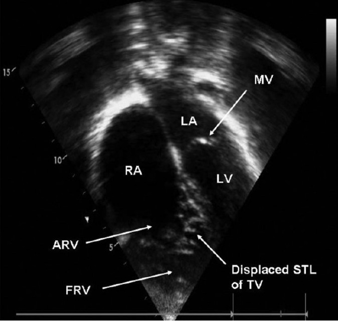
A transthoracic echocardiogram showing apical displacement of the TV and atrialized portion of the RV, characteristic of EA ARV: atrialized right ventricle FRV: functional right ventricle LA: left atrium LV: left ventricle MV: mitral valve RA: right atrium STL: septal tricuspid leaflet
Image: “A transthoracic echocardiographic image” by the Department of Pediatric Cardiology, Narayan Hrudayalaya Institute of Cardiac Sciences, Bangalore, India. License: CC BY 2.0.
DopplerDopplerUltrasonography applying the doppler effect, with frequency-shifted ultrasound reflections produced by moving targets (usually red blood cells) in the bloodstream along the ultrasound axis in direct proportion to the velocity of movement of the targets, to determine both direction and velocity of blood flow.Ultrasound (Sonography) and transthoracic echocardiogramTransthoracic echocardiogramEndocarditis findings:
The degree of displacementDisplacementThe process by which an emotional or behavioral response that is appropriate for one situation appears in another situation for which it is inappropriate.Defense Mechanisms of the tricuspid leaflets (> 8 mm/m3)
Associated cardiac lesions (ASDASDAutism spectrum disorder (ASD) is a neurodevelopmental disorder marked by poor social skills, restricted interests/social interactions, and repetitive/stereotyped behaviors. The condition is termed a “spectrum” because of the wide variability in the severity of symptoms exhibited.Autism Spectrum Disorder, PFOPFOA patent foramen ovale (PFO) is an abnormal communication between the atria that persists after birth. The condition results from incomplete closure of the foramen ovale. Small, isolated, and asymptomatic pfos are a common incidental finding on echocardiography and require no treatment.Patent Foramen Ovale)
Tests to confirm the presence of comorbiditiesComorbiditiesThe presence of co-existing or additional diseases with reference to an initial diagnosis or with reference to the index condition that is the subject of study. Comorbidity may affect the ability of affected individuals to function and also their survival; it may be used as a prognostic indicator for length of hospital stay, cost factors, and outcome or survival.St. Louis Encephalitis Virus:
ElectrocardiogramElectrocardiogramAn electrocardiogram (ECG) is a graphic representation of the electrical activity of the heart plotted against time. Adhesive electrodes are affixed to the skin surface allowing measurement of cardiac impulses from many angles. The ECG provides 3-dimensional information about the conduction system of the heart, the myocardium, and other cardiac structures. Electrocardiogram (ECG) (ECGECGAn electrocardiogram (ECG) is a graphic representation of the electrical activity of the heart plotted against time. Adhesive electrodes are affixed to the skin surface allowing measurement of cardiac impulses from many angles. The ECG provides 3-dimensional information about the conduction system of the heart, the myocardium, and other cardiac structures. Electrocardiogram (ECG))looking for:
A 24-hour ECGECGAn electrocardiogram (ECG) is a graphic representation of the electrical activity of the heart plotted against time. Adhesive electrodes are affixed to the skin surface allowing measurement of cardiac impulses from many angles. The ECG provides 3-dimensional information about the conduction system of the heart, the myocardium, and other cardiac structures. Electrocardiogram (ECG) (Holter) → evaluate arrhythmias
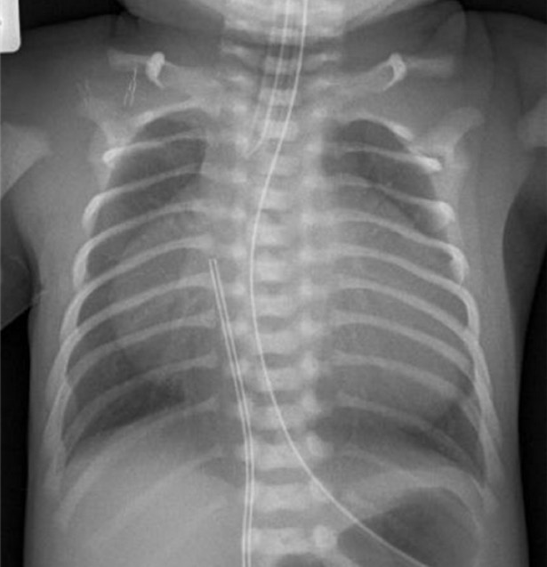
Chest X-rayX-rayPenetrating electromagnetic radiation emitted when the inner orbital electrons of an atom are excited and release radiant energy. X-ray wavelengths range from 1 pm to 10 nm. Hard x-rays are the higher energy, shorter wavelength x-rays. Soft x-rays or grenz rays are less energetic and longer in wavelength. The short wavelength end of the x-ray spectrum overlaps the gamma rays wavelength range. The distinction between gamma rays and x-rays is based on their radiation source.Pulmonary Function Tests → cardiomegaly (“wall-to-wall heart”)
“Wall-to-wall heart” in a neonate born with an EA
Image: “Proband’s chest” by Diagnostic Genetics, LabPlus, Auckland City Hospital, P.O. Box 110031, Auckland 1148, New Zealand. License: CC BY 3.0.
Medical management to optimize heart failureHeart FailureA heterogeneous condition in which the heart is unable to pump out sufficient blood to meet the metabolic need of the body. Heart failure can be caused by structural defects, functional abnormalities (ventricular dysfunction), or a sudden overload beyond its capacity. Chronic heart failure is more common than acute heart failure which results from sudden insult to cardiac function, such as myocardial infarction.Total Anomalous Pulmonary Venous Return (TAPVR) and arrhythmias is used prior to surgical stabilization. Medical management is indicated for neonates and symptomatic adults:
Neonates: aims to reduce pulmonary vascular pressure:
Inhaled nitric oxideNitric OxideA free radical gas produced endogenously by a variety of mammalian cells, synthesized from arginine by nitric oxide synthase. Nitric oxide is one of the endothelium-dependent relaxing factors released by the vascular endothelium and mediates vasodilation. It also inhibits platelet aggregation, induces disaggregation of aggregated platelets, and inhibits platelet adhesion to the vascular endothelium. Nitric oxide activates cytosolic guanylate cyclase and thus elevates intracellular levels of cyclic gmp.Pulmonary Hypertension Drugs
Child/adult:
Manage heart failureHeart FailureA heterogeneous condition in which the heart is unable to pump out sufficient blood to meet the metabolic need of the body. Heart failure can be caused by structural defects, functional abnormalities (ventricular dysfunction), or a sudden overload beyond its capacity. Chronic heart failure is more common than acute heart failure which results from sudden insult to cardiac function, such as myocardial infarction.Total Anomalous Pulmonary Venous Return (TAPVR) and volume overload.
Manage comorbiditiesComorbiditiesThe presence of co-existing or additional diseases with reference to an initial diagnosis or with reference to the index condition that is the subject of study. Comorbidity may affect the ability of affected individuals to function and also their survival; it may be used as a prognostic indicator for length of hospital stay, cost factors, and outcome or survival.St. Louis Encephalitis Virus such as arrhythmias.
PatientsPatientsIndividuals participating in the health care system for the purpose of receiving therapeutic, diagnostic, or preventive procedures.Clinician–Patient Relationship are advised to engage in regularRegularInsulin, moderate physical activity as tolerated.
PatientsPatientsIndividuals participating in the health care system for the purpose of receiving therapeutic, diagnostic, or preventive procedures.Clinician–Patient Relationship are advised to avoid competitive sports or strenuous activities that may exacerbate symptoms.
Surgical therapy
Indicated for symptomatic infants, older children, and medically stable adults
Includes closure of associated defects and ablation of accessory conduction pathways
The cone repair technique has become the preferred surgical method for tricuspid valveTricuspid valveThe valve consisting of three cusps situated between the right atrium and right ventricle of the heart.Heart: Anatomy repair in Ebstein’s anomaly.
PrognosisPrognosisA prediction of the probable outcome of a disease based on a individual’s condition and the usual course of the disease as seen in similar situations.Non-Hodgkin Lymphomas
The severity of the anomaly determines the prognosisPrognosisA prediction of the probable outcome of a disease based on a individual’s condition and the usual course of the disease as seen in similar situations.Non-Hodgkin Lymphomas.
Life expectancyLife expectancyBased on known statistical data, the number of years which any person of a given age may reasonably expected to live.Population Pyramids is reduced to the 50s if EA presents in childhood.
86% of patientsPatientsIndividuals participating in the health care system for the purpose of receiving therapeutic, diagnostic, or preventive procedures.Clinician–Patient Relationship require surgical repair.
CyanosisCyanosisA bluish or purplish discoloration of the skin and mucous membranes due to an increase in the amount of deoxygenated hemoglobin in the blood or a structural defect in the hemoglobin molecule.Pulmonary Examination and heart failureHeart FailureA heterogeneous condition in which the heart is unable to pump out sufficient blood to meet the metabolic need of the body. Heart failure can be caused by structural defects, functional abnormalities (ventricular dysfunction), or a sudden overload beyond its capacity. Chronic heart failure is more common than acute heart failure which results from sudden insult to cardiac function, such as myocardial infarction.Total Anomalous Pulmonary Venous Return (TAPVR) in infancy carry the worst prognosisPrognosisA prediction of the probable outcome of a disease based on a individual’s condition and the usual course of the disease as seen in similar situations.Non-Hodgkin Lymphomas.
Differential Diagnosis
The following are differential diagnoses for EA:
Uhl anomaly: a rare CHD characterized by a lack of myocytesMyocytesMature contractile cells, commonly known as myocytes, that form one of three kinds of muscle. The three types of muscle cells are skeletal, cardiac, and smooth. They are derived from embryonic (precursor) muscle cells called myoblasts.Muscle Tissue: Histology in the RV, resulting in RV dilatation and thinning of the myocardiumMyocardiumThe muscle tissue of the heart. It is composed of striated, involuntary muscle cells connected to form the contractile pump to generate blood flow.Heart: Anatomy. Most cases present as heart failureHeart FailureA heterogeneous condition in which the heart is unable to pump out sufficient blood to meet the metabolic need of the body. Heart failure can be caused by structural defects, functional abnormalities (ventricular dysfunction), or a sudden overload beyond its capacity. Chronic heart failure is more common than acute heart failure which results from sudden insult to cardiac function, such as myocardial infarction.Total Anomalous Pulmonary Venous Return (TAPVR) in neonates. An echocardiogramEchocardiogramTransposition of the Great Arteries differentiates this from EA because Uhl anomaly spares the TV.
Cyanotic heart disease:
Pulmonary atresiaAtresiaHypoplastic Left Heart Syndrome (HLHS): arare cyanotic CHD characterized by the failed formation of the pulmonary valvePulmonary valveA valve situated at the entrance to the pulmonary trunk from the right ventricle.Heart: Anatomy, leading to subsequent RV hypoplasiaHypoplasiaHypoplastic Left Heart Syndrome (HLHS). This may occur as part of the most severe form of tetralogy of FallotTetralogy of FallotTetralogy of Fallot is the most common cyanotic congenital heart disease. The disease is the confluence of 4 pathologic cardiac features: overriding aorta, ventricular septal defect, right ventricular outflow obstruction, and right ventricular hypertrophy. Tetralogy of Fallot (with VSD), where the entire RV outflow enters the aortaAortaThe main trunk of the systemic arteries.Mediastinum and Great Vessels: Anatomy.
TV atresiaAtresiaHypoplastic Left Heart Syndrome (HLHS): a congenital disorder that is caused by the absence of the TV, where there is no communicationCommunicationThe exchange or transmission of ideas, attitudes, or beliefs between individuals or groups.Decision-making Capacity and Legal Competence between the RA and the RV. PatientsPatientsIndividuals participating in the health care system for the purpose of receiving therapeutic, diagnostic, or preventive procedures.Clinician–Patient Relationship present with cyanosisCyanosisA bluish or purplish discoloration of the skin and mucous membranes due to an increase in the amount of deoxygenated hemoglobin in the blood or a structural defect in the hemoglobin molecule.Pulmonary Examination and hypoxemiaHypoxemiaNeonatal Respiratory Distress Syndrome since birth and must be medically managed initially followed by surgical correction.
The following conditions are comorbiditiesComorbiditiesThe presence of co-existing or additional diseases with reference to an initial diagnosis or with reference to the index condition that is the subject of study. Comorbidity may affect the ability of affected individuals to function and also their survival; it may be used as a prognostic indicator for length of hospital stay, cost factors, and outcome or survival.St. Louis Encephalitis Virus of EA:
ASDASDAutism spectrum disorder (ASD) is a neurodevelopmental disorder marked by poor social skills, restricted interests/social interactions, and repetitive/stereotyped behaviors. The condition is termed a “spectrum” because of the wide variability in the severity of symptoms exhibited.Autism Spectrum Disorder: the most common intra-atrial defect present in EA patientsPatientsIndividuals participating in the health care system for the purpose of receiving therapeutic, diagnostic, or preventive procedures.Clinician–Patient Relationship. Eisenmenger’s syndrome occurs as the RA pressure increases, resulting in cyanosisCyanosisA bluish or purplish discoloration of the skin and mucous membranes due to an increase in the amount of deoxygenated hemoglobin in the blood or a structural defect in the hemoglobin molecule.Pulmonary Examination, especially in late childhood. Can also lead to a paradoxic embolus in some cases.
Heart failureHeart FailureA heterogeneous condition in which the heart is unable to pump out sufficient blood to meet the metabolic need of the body. Heart failure can be caused by structural defects, functional abnormalities (ventricular dysfunction), or a sudden overload beyond its capacity. Chronic heart failure is more common than acute heart failure which results from sudden insult to cardiac function, such as myocardial infarction.Total Anomalous Pulmonary Venous Return (TAPVR): Ebstein’s anomaly results in right-sided heart failureHeart FailureA heterogeneous condition in which the heart is unable to pump out sufficient blood to meet the metabolic need of the body. Heart failure can be caused by structural defects, functional abnormalities (ventricular dysfunction), or a sudden overload beyond its capacity. Chronic heart failure is more common than acute heart failure which results from sudden insult to cardiac function, such as myocardial infarction.Total Anomalous Pulmonary Venous Return (TAPVR) due to the dysfunction of the RV. Symptoms of right-sided heart failureHeart FailureA heterogeneous condition in which the heart is unable to pump out sufficient blood to meet the metabolic need of the body. Heart failure can be caused by structural defects, functional abnormalities (ventricular dysfunction), or a sudden overload beyond its capacity. Chronic heart failure is more common than acute heart failure which results from sudden insult to cardiac function, such as myocardial infarction.Total Anomalous Pulmonary Venous Return (TAPVR) include edemaEdemaEdema is a condition in which excess serous fluid accumulates in the body cavity or interstitial space of connective tissues. Edema is a symptom observed in several medical conditions. It can be categorized into 2 types, namely, peripheral (in the extremities) and internal (in an organ or body cavity). Edema and hepatic congestion with signs of raised JVP, pitting edemaPitting edemaEdema caused by excess fluid without excess colloid. Leaves “pits” due to fluid displacement when pressure is applied to the areaEdema, and ascitesAscitesAscites is the pathologic accumulation of fluid within the peritoneal cavity that occurs due to an osmotic and/or hydrostatic pressure imbalance secondary to portal hypertension (cirrhosis, heart failure) or non-portal hypertension (hypoalbuminemia, malignancy, infection).Ascites.
Supraventricular tachycardiaTachycardiaAbnormally rapid heartbeat, usually with a heart rate above 100 beats per minute for adults. Tachycardia accompanied by disturbance in the cardiac depolarization (cardiac arrhythmia) is called tachyarrhythmia.Sepsis in Children: Ebstein’s anomaly is commonly associated with the presence of accessory pathways, which results in arrhythmias such as Wolff-Parkinson-White syndromeWolff-Parkinson-White SyndromeA form of ventricular pre-excitation characterized by a short PR interval and a long QRS interval with a delta wave. In this syndrome, atrial impulses are abnormally conducted to the heart ventricles via an accessory conducting pathway that is located between the wall of the right or left atria and the ventricles, also known as a bundle of kent. The inherited form can be caused by mutation of prkag2 gene encoding a gamma-2 regulatory subunit of amp-activated protein kinase.Supraventricular Tachycardias. Many patientsPatientsIndividuals participating in the health care system for the purpose of receiving therapeutic, diagnostic, or preventive procedures.Clinician–Patient Relationship in adulthood may present with palpitations as the 1st symptom. Other forms of arrhythmias such as atrial fibrillationAtrial fibrillationAtrial fibrillation (AF or Afib) is a supraventricular tachyarrhythmia and the most common kind of arrhythmia. It is caused by rapid, uncontrolled atrial contractions and uncoordinated ventricular responses. Atrial Fibrillation or atrial flutterAtrial flutterAtrial flutter is a regular supraventricular tachycardia characterized by an atrial heart rate between 240/min and 340/min (typically 300/min), atrioventricular (AV) node conduction block, and a “sawtooth” pattern on an electrocardiogram (ECG). Atrial Flutter have been noted in some rare cases due to RA dilatation.
References
Kliegman, R. M. et al. (2020). Cyanotic congenital heart disease: Lesions associated with decreased pulmonary blood flow. In R. M. Kliegman MD et al. Nelson textbook of pediatrics (pp. 239-2407.e1). https://www.clinicalkey.es/#!/content/3-s2.0-B9780323529501004570
Murthy, R., Moe, T. G., Van Arsdell, G. S., Nigro, J. J., & Karamlou, T. (2019). Congenital heart disease. In F. C. Brunicardi et al. (Eds.), Schwartz’s principles of surgery, 11e. New York, NY: McGraw-Hill Education. Retrieved from accessmedicine.mhmedical.com/content.aspx?aid=1164311314
Ramcharan, T. K. W., Goff, D. A., Greenleaf, C. E., & Rychik, J. (2022). Ebstein’s anomaly: From fetus to adult—Literature review and pathway for patient care. Pediatric Cardiology, 43(7), 1409–1428. https://doi.org/10.1007/s00246-022-02908-x