Achieve Mastery of Medical Concepts

Study for medical school and boards with Lecturio

Cardiac Glycosides

Cardiac glycosides are a class of drugs reversibly inhibiting the sodium-potassium ATPase pump Pump ACES and RUSH: Resuscitation Ultrasound Protocols in myocardial cells and increasing vagal tone, which results in increased cardiac contractility and slowed conduction through the atrioventricular node Atrioventricular node A small nodular mass of specialized muscle fibers located in the interatrial septum near the opening of the coronary sinus. It gives rise to the atrioventricular bundle of the conduction system of the heart. Heart: Anatomy. Digoxin is the only medically used drug in the cardiac glycoside class. Digoxin can be used for rate control in atrial fibrillation Atrial fibrillation Atrial fibrillation (AF or Afib) is a supraventricular tachyarrhythmia and the most common kind of arrhythmia. It is caused by rapid, uncontrolled atrial contractions and uncoordinated ventricular responses. Atrial Fibrillation/flutter and for systolic heart failure Heart Failure A heterogeneous condition in which the heart is unable to pump out sufficient blood to meet the metabolic need of the body. Heart failure can be caused by structural defects, functional abnormalities (ventricular dysfunction), or a sudden overload beyond its capacity. Chronic heart failure is more common than acute heart failure which results from sudden insult to cardiac function, such as myocardial infarction. Total Anomalous Pulmonary Venous Return (TAPVR). However, the medication needs to be used with caution due to a very narrow therapeutic window Therapeutic Window Dosage Calculation. Digoxin toxicity Toxicity Dosage Calculation can result in life-threatening arrhythmias as well as GI and neurologic symptoms; an antidote Antidote An antidote is a substance that counteracts poisoning or toxicity. Substances that can cause poisoning include heavy metals (from occupation, treatments, or diet), alcohols, environmental toxins, and medications. Antidotes of Common Poisonings is available.

Last updated: Jul 10, 2023

Editorial responsibility: Stanley Oiseth, Lindsay Jones, Evelin Maza

Chemistry and Pharmacodynamics

Chemical structure

Digoxin is the prototype drug of the cardiac glycoside class and the only drug in the class used for medicinal purposes.

  • Steroid nucleus Nucleus Within a eukaryotic cell, a membrane-limited body which contains chromosomes and one or more nucleoli (cell nucleolus). The nuclear membrane consists of a double unit-type membrane which is perforated by a number of pores; the outermost membrane is continuous with the endoplasmic reticulum. A cell may contain more than one nucleus. The Cell: Organelles with 4 fused rings
  • Lactone ring Lactone Ring Polyenes 
  • Glycoside attachment composed of 3 sugars
Chemical structure of digoxin cardiac glycosides

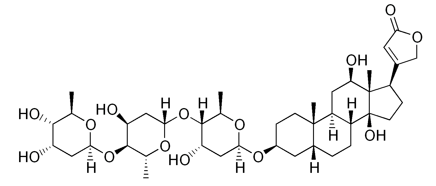
The chemical structure of digoxin:
Notice the steroid nucleus (4 fused rings) with an attached lactone ring (far right) and glycoside attachment (left).

Image: “The chemical structure of digoxin” by Edgar181. License: Public Domain

Mechanism of action

  • Digoxin reversibly inhibits the Na+-K+ ATPase of myocytes Myocytes Mature contractile cells, commonly known as myocytes, that form one of three kinds of muscle. The three types of muscle cells are skeletal, cardiac, and smooth. They are derived from embryonic (precursor) muscle cells called myoblasts. Muscle Tissue: Histology, resulting in:
    • ↑ Intracellular Na+ → ↓ Na+-calcium ( Ca CA Condylomata acuminata are a clinical manifestation of genital HPV infection. Condylomata acuminata are described as raised, pearly, flesh-colored, papular, cauliflower-like lesions seen in the anogenital region that may cause itching, pain, or bleeding. Condylomata Acuminata (Genital Warts)2+) antiporter Antiporter Membrane transporters that co-transport two or more dissimilar molecules in the opposite direction across a membrane. Usually the transport of one ion or molecule is against its electrochemical gradient and is ‘powered’ by the movement of another ion or molecule with its electrochemical gradient. The Cell: Cell Membrane exchange → ↓ Ca CA Condylomata acuminata are a clinical manifestation of genital HPV infection. Condylomata acuminata are described as raised, pearly, flesh-colored, papular, cauliflower-like lesions seen in the anogenital region that may cause itching, pain, or bleeding. Condylomata Acuminata (Genital Warts)2+ efflux 
    • ↑ Intracellular Ca CA Condylomata acuminata are a clinical manifestation of genital HPV infection. Condylomata acuminata are described as raised, pearly, flesh-colored, papular, cauliflower-like lesions seen in the anogenital region that may cause itching, pain, or bleeding. Condylomata Acuminata (Genital Warts)2+ → ↑ Ca CA Condylomata acuminata are a clinical manifestation of genital HPV infection. Condylomata acuminata are described as raised, pearly, flesh-colored, papular, cauliflower-like lesions seen in the anogenital region that may cause itching, pain, or bleeding. Condylomata Acuminata (Genital Warts)2+ binding to contractile proteins Proteins Linear polypeptides that are synthesized on ribosomes and may be further modified, crosslinked, cleaved, or assembled into complex proteins with several subunits. The specific sequence of amino acids determines the shape the polypeptide will take, during protein folding, and the function of the protein. Energy Homeostasis → ↑ cardiac contractility 
  • ↑ Vagal tone:
    • ↑ Refractory period → ↓ conduction velocity in the atrioventricular (AV) node
    • ↓ Sinoatrial (SA) node automaticity
Mechanism of action of digoxin

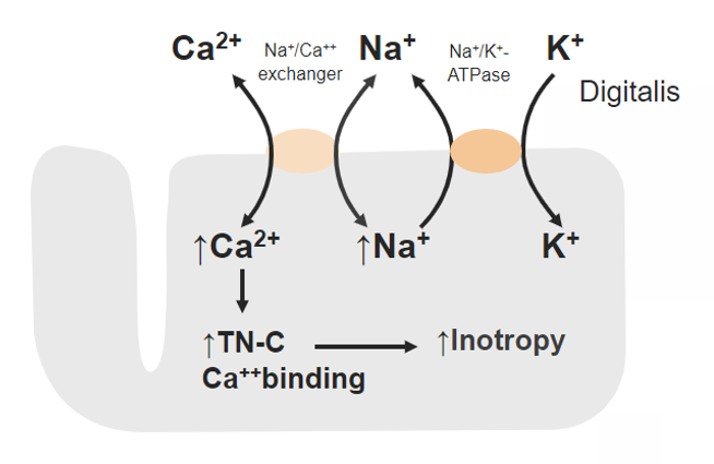
Mechanism of action of digoxin:
Inhibition of sodium (Na+)-potassium (K+) ATPase leads to increased intracellular Na+, which lowers the exchange of the Na+-calcium (Ca2+) antiporter and inhibits the efflux of Ca2+.
More intracellular Ca2+ can bind to contractile proteins (such as troponin (TN-C)), resulting in increased cardiac contractility.

Image by Lecturio. License: CC BY-NC-SA 4.0

Physiologic effects

  • ↑ Cardiac contractility (positive inotropy) → ↑ cardiac output Cardiac output The volume of blood passing through the heart per unit of time. It is usually expressed as liters (volume) per minute so as not to be confused with stroke volume (volume per beat). Cardiac Mechanics
  • AV and SA node slowing → ↓ heart rate Heart rate The number of times the heart ventricles contract per unit of time, usually per minute. Cardiac Physiology
  • Blood pressure is not significantly impacted.
  • May cause characteristic changes to a resting ECG ECG An electrocardiogram (ECG) is a graphic representation of the electrical activity of the heart plotted against time. Adhesive electrodes are affixed to the skin surface allowing measurement of cardiac impulses from many angles. The ECG provides 3-dimensional information about the conduction system of the heart, the myocardium, and other cardiac structures. Electrocardiogram (ECG) (“ digitalis effect Digitalis Effect Class 5 Antiarrhythmic Drugs”):
Ecg scooping st segment depressionin

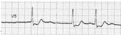
Typical “scooped” ST-depression resulting from digoxin use.

Image: “F15: An ECG with characteristic scooping ST segment depressionin a patient taking Digoxin.” by Christopher Yates and Alex F Manini. License: CC BY 2.5

Pharmacokinetics

Absorption Absorption Absorption involves the uptake of nutrient molecules and their transfer from the lumen of the GI tract across the enterocytes and into the interstitial space, where they can be taken up in the venous or lymphatic circulation. Digestion and Absorption

  • Oral and IV forms are available.
  • Oral absorption Absorption Absorption involves the uptake of nutrient molecules and their transfer from the lumen of the GI tract across the enterocytes and into the interstitial space, where they can be taken up in the venous or lymphatic circulation. Digestion and Absorption:
    • Passive, nonsaturable diffusion Diffusion The tendency of a gas or solute to pass from a point of higher pressure or concentration to a point of lower pressure or concentration and to distribute itself throughout the available space. Diffusion, especially facilitated diffusion, is a major mechanism of biological transport. Peritoneal Dialysis and Hemodialysis in the proximal small intestine Small intestine The small intestine is the longest part of the GI tract, extending from the pyloric orifice of the stomach to the ileocecal junction. The small intestine is the major organ responsible for chemical digestion and absorption of nutrients. It is divided into 3 segments: the duodenum, the jejunum, and the ileum. Small Intestine: Anatomy
    • Food may delay, but not impact, the extent of absorption Absorption Absorption involves the uptake of nutrient molecules and their transfer from the lumen of the GI tract across the enterocytes and into the interstitial space, where they can be taken up in the venous or lymphatic circulation. Digestion and Absorption.

Distribution

  • Extensive in peripheral tissues:
    • Distribution phase: 6–8 hours
    • Higher concentrations in heart, liver Liver The liver is the largest gland in the human body. The liver is found in the superior right quadrant of the abdomen and weighs approximately 1.5 kilograms. Its main functions are detoxification, metabolism, nutrient storage (e.g., iron and vitamins), synthesis of coagulation factors, formation of bile, filtration, and storage of blood. Liver: Anatomy, kidney, and skeletal muscle
  • Protein binding:
    • Approximately 25% is protein bound.
    • Uremic patients Patients Individuals participating in the health care system for the purpose of receiving therapeutic, diagnostic, or preventive procedures. Clinician–Patient Relationship: Digoxin is displaced from plasma Plasma The residual portion of blood that is left after removal of blood cells by centrifugation without prior blood coagulation. Transfusion Products protein binding sites.

Metabolism and excretion

  • Minimal hepatic metabolism:
    • Approximately 16% of an absorbed dose is metabolized to active metabolites.
    • Does not interact with the cytochrome P450 Cytochrome P450 A superfamily of hundreds of closely related hemeproteins found throughout the phylogenetic spectrum, from animals, plants, fungi, to bacteria. They include numerous complex monooxygenases (mixed function oxygenases). In animals, these p450 enzymes serve two major functions: (1) biosynthesis of steroids, fatty acids, and bile acids; (2) metabolism of endogenous and a wide variety of exogenous substrates, such as toxins and drugs (biotransformation). They are classified, according to their sequence similarities rather than functions, into cyp gene families (>40% homology) and subfamilies (>59% homology). For example, enzymes from the cyp1, cyp2, and cyp3 gene families are responsible for most drug metabolism. Drug-Induced Liver Injury system
  • Predominantly excreted in the urine (50%–70% as an unchanged drug)

Indications

Heart failure Heart Failure A heterogeneous condition in which the heart is unable to pump out sufficient blood to meet the metabolic need of the body. Heart failure can be caused by structural defects, functional abnormalities (ventricular dysfunction), or a sudden overload beyond its capacity. Chronic heart failure is more common than acute heart failure which results from sudden insult to cardiac function, such as myocardial infarction. Total Anomalous Pulmonary Venous Return (TAPVR)

  • 2nd-line therapy for heart failure Heart Failure A heterogeneous condition in which the heart is unable to pump out sufficient blood to meet the metabolic need of the body. Heart failure can be caused by structural defects, functional abnormalities (ventricular dysfunction), or a sudden overload beyond its capacity. Chronic heart failure is more common than acute heart failure which results from sudden insult to cardiac function, such as myocardial infarction. Total Anomalous Pulmonary Venous Return (TAPVR) with reduced ejection fraction Ejection fraction Cardiac Cycle:
    • Provides a positive inotropic effect
    • ↓ Symptoms of heart failure Heart Failure A heterogeneous condition in which the heart is unable to pump out sufficient blood to meet the metabolic need of the body. Heart failure can be caused by structural defects, functional abnormalities (ventricular dysfunction), or a sudden overload beyond its capacity. Chronic heart failure is more common than acute heart failure which results from sudden insult to cardiac function, such as myocardial infarction. Total Anomalous Pulmonary Venous Return (TAPVR) and the need for hospitalization Hospitalization The confinement of a patient in a hospital. Delirium
    • Not shown to improve mortality Mortality All deaths reported in a given population. Measures of Health Status
  • 1st-line choice in patients Patients Individuals participating in the health care system for the purpose of receiving therapeutic, diagnostic, or preventive procedures. Clinician–Patient Relationship with heart failure Heart Failure A heterogeneous condition in which the heart is unable to pump out sufficient blood to meet the metabolic need of the body. Heart failure can be caused by structural defects, functional abnormalities (ventricular dysfunction), or a sudden overload beyond its capacity. Chronic heart failure is more common than acute heart failure which results from sudden insult to cardiac function, such as myocardial infarction. Total Anomalous Pulmonary Venous Return (TAPVR) with reduced ejection fraction Ejection fraction Cardiac Cycle complicated by atrial fibrillation Atrial fibrillation Atrial fibrillation (AF or Afib) is a supraventricular tachyarrhythmia and the most common kind of arrhythmia. It is caused by rapid, uncontrolled atrial contractions and uncoordinated ventricular responses. Atrial Fibrillation.

Arrhythmia

Digoxin is indicated for rate control when other therapies are ineffective or contraindicated:

  • Atrial fibrillation Atrial fibrillation Atrial fibrillation (AF or Afib) is a supraventricular tachyarrhythmia and the most common kind of arrhythmia. It is caused by rapid, uncontrolled atrial contractions and uncoordinated ventricular responses. Atrial Fibrillation
  • Atrial flutter Atrial flutter Atrial flutter is a regular supraventricular tachycardia characterized by an atrial heart rate between 240/min and 340/min (typically 300/min), atrioventricular (AV) node conduction block, and a “sawtooth” pattern on an electrocardiogram (ECG). Atrial Flutter
  • Supraventricular tachycardia Tachycardia Abnormally rapid heartbeat, usually with a heart rate above 100 beats per minute for adults. Tachycardia accompanied by disturbance in the cardiac depolarization (cardiac arrhythmia) is called tachyarrhythmia. Sepsis in Children

Adverse Effects

Adverse effects

Digoxin has a very narrow therapeutic window Therapeutic Window Dosage Calculation and several signs of toxicity Toxicity Dosage Calculation:

  • Arrhythmias can occur through multiple mechanisms:
    • ↑ Intracellular Ca CA Condylomata acuminata are a clinical manifestation of genital HPV infection. Condylomata acuminata are described as raised, pearly, flesh-colored, papular, cauliflower-like lesions seen in the anogenital region that may cause itching, pain, or bleeding. Condylomata Acuminata (Genital Warts)2+ → delayed afterdepolarizations and ↑ automaticity
    • Slowed conduction
  • GI symptoms:
    • Nausea Nausea An unpleasant sensation in the stomach usually accompanied by the urge to vomit. Common causes are early pregnancy, sea and motion sickness, emotional stress, intense pain, food poisoning, and various enteroviruses. Antiemetics
    • Vomiting Vomiting The forcible expulsion of the contents of the stomach through the mouth. Hypokalemia
    • Diarrhea Diarrhea Diarrhea is defined as ≥ 3 watery or loose stools in a 24-hour period. There are a multitude of etiologies, which can be classified based on the underlying mechanism of disease. The duration of symptoms (acute or chronic) and characteristics of the stools (e.g., watery, bloody, steatorrheic, mucoid) can help guide further diagnostic evaluation. Diarrhea
    • Anorexia Anorexia The lack or loss of appetite accompanied by an aversion to food and the inability to eat. It is the defining characteristic of the disorder anorexia nervosa. Anorexia Nervosa
  • Neurologic symptoms:

Warnings and precautions

  • As with all AV node-blocking agents, digoxin should not be used in supraventricular tachyarrhythmias caused by an accessory pathway (e.g., Wolff-Parkinson-White syndrome Wolff-Parkinson-White Syndrome A form of ventricular pre-excitation characterized by a short PR interval and a long QRS interval with a delta wave. In this syndrome, atrial impulses are abnormally conducted to the heart ventricles via an accessory conducting pathway that is located between the wall of the right or left atria and the ventricles, also known as a bundle of kent. The inherited form can be caused by mutation of prkag2 gene encoding a gamma-2 regulatory subunit of amp-activated protein kinase. Supraventricular Tachycardias).
  • Avoid in sinus node disease and AV block AV block Atrioventricular (AV) block is a bradyarrhythmia caused by delay, or interruption, in the electrical conduction between the atria and the ventricles. Atrioventricular block occurs due to either anatomic or functional impairment, and is classified into 3 types. Atrioventricular block (AV block)
  • Acute coronary syndrome:
    • Use caution in patients Patients Individuals participating in the health care system for the purpose of receiving therapeutic, diagnostic, or preventive procedures. Clinician–Patient Relationship with an acute MI MI MI is ischemia and death of an area of myocardial tissue due to insufficient blood flow and oxygenation, usually from thrombus formation on a ruptured atherosclerotic plaque in the epicardial arteries. Clinical presentation is most commonly with chest pain, but women and patients with diabetes may have atypical symptoms. Myocardial Infarction.
    • May ↑ myocardial oxygen demand Myocardial oxygen demand Stable and Unstable Angina ischemia Ischemia A hypoperfusion of the blood through an organ or tissue caused by a pathologic constriction or obstruction of its blood vessels, or an absence of blood circulation. Ischemic Cell Damage
  • Hypertrophic cardiomyopathy Hypertrophic Cardiomyopathy Hypertrophic cardiomyopathy (HCM) is the most commonly inherited cardiomyopathy, which is characterized by an asymmetric increase in thickness (hypertrophy) of the left ventricular wall, diastolic dysfunction, and often left ventricular outflow tract obstruction. Hypertrophic Cardiomyopathy with left ventricular outflow tract obstruction Left ventricular outflow tract obstruction Hypertrophic Cardiomyopathy:
    • Outflow obstruction may worsen.
    • Due to digoxin’s positive inotropic effects
  • Thyroid Thyroid The thyroid gland is one of the largest endocrine glands in the human body. The thyroid gland is a highly vascular, brownish-red gland located in the visceral compartment of the anterior region of the neck. Thyroid Gland: Anatomy disease:
    • Use caution in patients Patients Individuals participating in the health care system for the purpose of receiving therapeutic, diagnostic, or preventive procedures. Clinician–Patient Relationship with hypothyroidism Hypothyroidism Hypothyroidism is a condition characterized by a deficiency of thyroid hormones. Iodine deficiency is the most common cause worldwide, but Hashimoto’s disease (autoimmune thyroiditis) is the leading cause in non-iodine-deficient regions. Hypothyroidism or hyperthyroidism Hyperthyroidism Hypersecretion of thyroid hormones from the thyroid gland. Elevated levels of thyroid hormones increase basal metabolic rate. Thyrotoxicosis and Hyperthyroidism.
    • May cause significant changes in digoxin clearance

Drug interactions

Drug interactions may lead to:

  • ↑ AV blocking/bradycardic effect:
    • Calcium Calcium A basic element found in nearly all tissues. It is a member of the alkaline earth family of metals with the atomic symbol ca, atomic number 20, and atomic weight 40. Calcium is the most abundant mineral in the body and combines with phosphorus to form calcium phosphate in the bones and teeth. It is essential for the normal functioning of nerves and muscles and plays a role in blood coagulation (as factor IV) and in many enzymatic processes. Electrolytes channel blockers
    • Beta blockers
    • Dronedarone Dronedarone A non-iodinated derivative of amiodarone that is used for the treatment of arrhythmia. Class 3 Antiarrhythmic Drugs (Potassium Channel Blockers)
    • Lacosamide Lacosamide An acetamide derivative that acts as a blocker of voltage-gated sodium channels. It is used as an anticonvulsant, for adjunctive or monotherapy, in the treatment of partial seizures. Second-Generation Anticonvulsant Drugs
  • ↑ Risk of toxicity Toxicity Dosage Calculation due to:
    • ↑ Digoxin concentration:
      • Amiodarone Amiodarone An antianginal and class III antiarrhythmic drug. It increases the duration of ventricular and atrial muscle action by inhibiting potassium channels and voltage-gated sodium channels. There is a resulting decrease in heart rate and in vascular resistance. Pulmonary Fibrosis
      • Quinidine Quinidine An optical isomer of quinine, extracted from the bark of the cinchona tree and similar plant species. This alkaloid dampens the excitability of cardiac and skeletal muscles by blocking sodium and potassium currents across cellular membranes. It prolongs cellular action potentials, and decreases automaticity. Quinidine also blocks muscarinic and alpha-adrenergic neurotransmission. Class 1 Antiarrhythmic Drugs (Sodium Channel Blockers)
      • Spironolactone Spironolactone A potassium sparing diuretic that acts by antagonism of aldosterone in the distal renal tubules. It is used mainly in the treatment of refractory edema in patients with congestive heart failure, nephrotic syndrome, or hepatic cirrhosis. Its effects on the endocrine system are utilized in the treatments of hirsutism and acne but they can lead to adverse effects. Potassium-sparing Diuretics
    • Hypokalemia Hypokalemia Hypokalemia is defined as plasma potassium (K+) concentration < 3.5 mEq/L. Homeostatic mechanisms maintain plasma concentration between 3.5-5.2 mEq/L despite marked variation in dietary intake. Hypokalemia can be due to renal losses, GI losses, transcellular shifts, or poor dietary intake. Hypokalemia and/or hypomagnesemia Hypomagnesemia A nutritional condition produced by a deficiency of magnesium in the diet, characterized by anorexia, nausea, vomiting, lethargy, and weakness. Symptoms are paresthesias, muscle cramps, irritability, decreased attention span, and mental confusion, possibly requiring months to appear. Deficiency of body magnesium can exist even when serum values are normal. In addition, magnesium deficiency may be organ-selective, since certain tissues become deficient before others. Electrolytes:

Overdose

Risk factors

  • Factors affecting digoxin levels:
  • Potential triggers for toxicity Toxicity Dosage Calculation:
    • Hypokalemia Hypokalemia Hypokalemia is defined as plasma potassium (K+) concentration < 3.5 mEq/L. Homeostatic mechanisms maintain plasma concentration between 3.5-5.2 mEq/L despite marked variation in dietary intake. Hypokalemia can be due to renal losses, GI losses, transcellular shifts, or poor dietary intake. Hypokalemia
    • Hypomagnesemia Hypomagnesemia A nutritional condition produced by a deficiency of magnesium in the diet, characterized by anorexia, nausea, vomiting, lethargy, and weakness. Symptoms are paresthesias, muscle cramps, irritability, decreased attention span, and mental confusion, possibly requiring months to appear. Deficiency of body magnesium can exist even when serum values are normal. In addition, magnesium deficiency may be organ-selective, since certain tissues become deficient before others. Electrolytes
    • Hypercalcemia Hypercalcemia Hypercalcemia (serum calcium > 10.5 mg/dL) can result from various conditions, the majority of which are due to hyperparathyroidism and malignancy. Other causes include disorders leading to vitamin D elevation, granulomatous diseases, and the use of certain pharmacological agents. Symptoms vary depending on calcium levels and the onset of hypercalcemia. Hypercalcemia

Clinical presentation

  • Arrhythmia:
    • The most serious manifestation of digoxin overdose
    • May be any type of arrhythmia (except rapidly-conducted atrial arrhythmias)
    • May be life-threatening
  • GI symptoms:
    • Anorexia Anorexia The lack or loss of appetite accompanied by an aversion to food and the inability to eat. It is the defining characteristic of the disorder anorexia nervosa. Anorexia Nervosa
    • Nausea Nausea An unpleasant sensation in the stomach usually accompanied by the urge to vomit. Common causes are early pregnancy, sea and motion sickness, emotional stress, intense pain, food poisoning, and various enteroviruses. Antiemetics
    • Vomiting Vomiting The forcible expulsion of the contents of the stomach through the mouth. Hypokalemia
    • Abdominal pain Abdominal Pain Acute Abdomen
  • Neurologic symptoms:

Laboratory evaluation

  • Serum digoxin concentration:
  • ↑ Serum K+ level:
    • Due to Na+-K+ ATPase inhibition
    • Degree of elevation correlates with mortality Mortality All deaths reported in a given population. Measures of Health Status risk
    • Note: Hypokalemia Hypokalemia Hypokalemia is defined as plasma potassium (K+) concentration < 3.5 mEq/L. Homeostatic mechanisms maintain plasma concentration between 3.5-5.2 mEq/L despite marked variation in dietary intake. Hypokalemia can be due to renal losses, GI losses, transcellular shifts, or poor dietary intake. Hypokalemia is a potential trigger Trigger The type of signal that initiates the inspiratory phase by the ventilator Invasive Mechanical Ventilation for digoxin toxicity Toxicity Dosage Calculation.
  • BUN and creatinine → renal dysfunction may be a precipitating factor
  • ECG ECG An electrocardiogram (ECG) is a graphic representation of the electrical activity of the heart plotted against time. Adhesive electrodes are affixed to the skin surface allowing measurement of cardiac impulses from many angles. The ECG provides 3-dimensional information about the conduction system of the heart, the myocardium, and other cardiac structures. Electrocardiogram (ECG):

Management

  • Antidote Antidote An antidote is a substance that counteracts poisoning or toxicity. Substances that can cause poisoning include heavy metals (from occupation, treatments, or diet), alcohols, environmental toxins, and medications. Antidotes of Common Poisonings: digoxin-specific antibody ( Fab Fab Univalent antigen-binding fragments composed of one entire immunoglobulin light chain and the amino terminal end of one of the immunoglobulin heavy chains from the hinge region, linked to each other by disulfide bonds. Fab contains the immunoglobulin variable regions, which are part of the antigen-binding site, and the first immunoglobulin constant regions. This fragment can be obtained by digestion of immunoglobulins with the proteolytic enzyme papain. Immunoglobulins: Types and Functions) fragments
  • Supportive treatment:
    • Bradyarrhythmias Bradyarrhythmias Bradyarrhythmia is a rhythm in which the heart rate is less than 60/min. Bradyarrhythmia can be physiologic, without symptoms or hemodynamic change. Pathologic bradyarrhythmia results in reduced cardiac output and hemodynamic instability causing syncope, dizziness, or dyspnea. Bradyarrhythmias: atropine Atropine An alkaloid, originally from atropa belladonna, but found in other plants, mainly solanaceae. Hyoscyamine is the 3(s)-endo isomer of atropine. Anticholinergic Drugs or temporary pacemaker Pacemaker A device designed to stimulate, by electric impulses, contraction of the heart muscles. It may be temporary (external) or permanent (internal or internal-external). Bradyarrhythmias
    • Hypotension Hypotension Hypotension is defined as low blood pressure, specifically < 90/60 mm Hg, and is most commonly a physiologic response. Hypotension may be mild, serious, or life threatening, depending on the cause. Hypotension: bolus IV fluids IV fluids Intravenous fluids are one of the most common interventions administered in medicine to approximate physiologic bodily fluids. Intravenous fluids are divided into 2 categories: crystalloid and colloid solutions. Intravenous fluids have a wide variety of indications, including intravascular volume expansion, electrolyte manipulation, and maintenance fluids. Intravenous Fluids
    • Correct electrolyte abnormalities.
    • Life-threatening arrhythmia treatment
  • Activated charcoal Charcoal An amorphous form of carbon prepared from the incomplete combustion of animal or vegetable matter, e.g., wood. The activated form of charcoal is used in the treatment of poisoning. Antidotes of Common Poisonings can be given for acute digoxin intoxication within 1–2 hours.

References

  1. Katzung, B.G. (2012). Drugs used in heart failure. In Katzung, B.G., Masters, S.B., and Trevor, A.J. (Eds.), Basic & Clinical Pharmacology (12th edition, pp. 211-225). https://pharmacomedicale.org/images/cnpm/CNPM_2016/katzung-pharmacology.pdf
  2. Kumar, K., and Zimetbaum, P. (2021). Antiarrhythmic drugs to maintain sinus rhythm in patients with atrial fibrillation: Clinical trials. In Knight, B. (Ed.), UpToDate. Retrieved July 6, 2021, from https://www.uptodate.com/contents/antiarrhythmic-drugs-to-maintain-sinus-rhythm-in-patients-with-atrial-fibrillation-clinical-trials
  3. Makielski, J., and Eckhardt, L. (2021). Cardiac excitability, mechanisms of arrhythmia, and action of antiarrhythmic drugs. In Levy, S. (Ed.), UpToDate. Retrieved July 7, 2021, from https://www.uptodate.com/contents/cardiac-excitability-mechanisms-of-arrhythmia-and-action-of-antiarrhythmic-drugs
  4. Levine, M., and O’Connor, A. (2021). Digitalis (cardiac glycoside) poisoning. In Traub, S. and Burns, M. (Ed.), UpToDate. Retrieved July 7, 2021, from https://www.uptodate.com/contents/digitalis-cardiac-glycoside-poisoning
  5. Giardina, E., and Sylvia, L. (2021). Treatment with digoxin: Initial dosing, monitoring, and dose modification. In Dardas, T. (Ed.), UpToDate. Retrieved July 7, 2021, from https://www.uptodate.com/contents/treatment-with-digoxin-initial-dosing-monitoring-and-dose-modification
  6. Wyse D.G., Waldo A.L., DiMarco J.P., et al. (2002). A comparison of rate control and rhythm control in patients with atrial fibrillation. N Engl J Med; 347:1825. https://www.uptodate.com/contents/antiarrhythmic-drugs-to-maintain-sinus-rhythm-in-patients-with-atrial-fibrillation-clinical-trials/abstract/1
  7. Falk R.H. (2001). Atrial fibrillation. N Engl J Med; 344:1067. https://www.uptodate.com/contents/antiarrhythmic-drugs-to-maintain-sinus-rhythm-in-patients-with-atrial-fibrillation-clinical-trials/abstract/3
  8. Dan G.A., Martinez-Rubio A., Agewall S., et al. (2018). Antiarrhythmic drugs-clinical use and clinical decision making: a consensus document from the European Heart Rhythm Association (EHRA) and European Society of Cardiology (ESC) Working Group. https://www.uptodate.com/contents/cardiac-excitability-mechanisms-of-arrhythmia-and-action-of-antiarrhythmic-drugs/abstract/4 
  9. Busti, A.J. (2015). The mechanism of digoxin’s increase in inotropy (force of contraction of the heart). In Evidence-Based Medicine Consult. Retrieved July 22, 2021, from https://www.ebmconsult.com/articles/mechanism-of-action-digoxin-inotropy-force-contraction-heart

Create your free account or log in to continue reading!

Sign up now and get free access to Lecturio with concept pages, medical videos, and questions for your medical education.

User Reviews

Details