Digestion refers to the process of the mechanical and chemical breakdown of food into smaller constituent molecules, which can then be absorbed and utilized by the body. Absorption involves the uptake of nutrient molecules and their transfer from the lumen of the GI tract across the enterocytes and into the interstitial space, where they can be taken up in the venous or lymphatic circulationCirculationThe movement of the blood as it is pumped through the cardiovascular system.ABCDE Assessment. CarbohydratesCarbohydratesA class of organic compounds composed of carbon, hydrogen, and oxygen in a ratio of cn(H2O)n. The largest class of organic compounds, including starch; glycogen; cellulose; polysaccharides; and simple monosaccharides.Basics of Carbohydrates, proteinsProteinsLinear polypeptides that are synthesized on ribosomes and may be further modified, crosslinked, cleaved, or assembled into complex proteins with several subunits. The specific sequence of amino acids determines the shape the polypeptide will take, during protein folding, and the function of the protein.Energy Homeostasis, lipidsLipidsLipids are a diverse group of hydrophobic organic molecules, which include fats, oils, sterols, and waxes.Fatty Acids and Lipids, and micronutrients are digested and absorbed differently and require several enzymesEnzymesEnzymes are complex protein biocatalysts that accelerate chemical reactions without being consumed by them. Due to the body's constant metabolic needs, the absence of enzymes would make life unsustainable, as reactions would occur too slowly without these molecules. Basics of Enzymes and transport proteinsTransport proteinsProteins and Peptides to complete the process.
Digestion and absorption are complex processes that begin in the mouth but occur primarily in the stomachStomachThe stomach is a muscular sac in the upper left portion of the abdomen that plays a critical role in digestion. The stomach develops from the foregut and connects the esophagus with the duodenum. Structurally, the stomach is C-shaped and forms a greater and lesser curvature and is divided grossly into regions: the cardia, fundus, body, and pylorus. Stomach: Anatomy and small intestineSmall intestineThe small intestine is the longest part of the GI tract, extending from the pyloric orifice of the stomach to the ileocecal junction. The small intestine is the major organ responsible for chemical digestion and absorption of nutrients. It is divided into 3 segments: the duodenum, the jejunum, and the ileum. Small Intestine: Anatomy.
CarbohydratesCarbohydratesA class of organic compounds composed of carbon, hydrogen, and oxygen in a ratio of cn(H2O)n. The largest class of organic compounds, including starch; glycogen; cellulose; polysaccharides; and simple monosaccharides.Basics of Carbohydrates:
Must be broken down into monosaccharidesMonosaccharidesSingle chain carbohydrates that are the most basic units of carbohydrates. They are typically colorless crystalline substances with a sweet taste and have the same general formula CNH2NON.Basics of Carbohydrates to be absorbed
Primary digestive enzymesEnzymesEnzymes are complex protein biocatalysts that accelerate chemical reactions without being consumed by them. Due to the body’s constant metabolic needs, the absence of enzymes would make life unsustainable, as reactions would occur too slowly without these molecules. Basics of Enzymes:
Amylase
Brush borderBrush borderTubular SystemenzymesEnzymesEnzymes are complex protein biocatalysts that accelerate chemical reactions without being consumed by them. Due to the body’s constant metabolic needs, the absence of enzymes would make life unsustainable, as reactions would occur too slowly without these molecules. Basics of Enzymes
ProteinsProteinsLinear polypeptides that are synthesized on ribosomes and may be further modified, crosslinked, cleaved, or assembled into complex proteins with several subunits. The specific sequence of amino acids determines the shape the polypeptide will take, during protein folding, and the function of the protein.Energy Homeostasis:
Broken down into peptides and individual amino acidsAmino acidsOrganic compounds that generally contain an amino (-NH2) and a carboxyl (-COOH) group. Twenty alpha-amino acids are the subunits which are polymerized to form proteins.Basics of Amino Acids (AAs)
Primary digestive enzymesEnzymesEnzymes are complex protein biocatalysts that accelerate chemical reactions without being consumed by them. Due to the body’s constant metabolic needs, the absence of enzymes would make life unsustainable, as reactions would occur too slowly without these molecules. Basics of Enzymes:
PepsinPepsinPepsin breaks down proteins into proteoses, peptones, and large polypeptides.Proteins and Peptides
TrypsinTrypsinA serine endopeptidase that is formed from trypsinogen in the pancreas. It is converted into its active form by enteropeptidase in the small intestine. It catalyzes hydrolysis of the carboxyl group of either arginine or lysine.Proteins and Peptides
ChymotrypsinChymotrypsinA serine endopeptidase secreted by the pancreas as its zymogen, chymotrypsinogen and carried in the pancreatic juice to the duodenum where it is activated by trypsin. It selectively cleaves aromatic amino acids on the carboxyl side.Pancreatic Parameters
CarboxypeptidaseCarboxypeptidaseEnzymes that act at a free c-terminus of a polypeptide to liberate a single amino acid residue.Pancreatic Parameters
ElastaseElastaseA protease of broad specificity, obtained from dried pancreas. Molecular weight is approximately 25, 000. The enzyme breaks down elastin, the specific protein of elastic fibers, and digests other proteins such as fibrin, hemoglobin, and albumin.Proteins and Peptides
Absorbed by specialized cotransporters
LipidsLipidsLipids are a diverse group of hydrophobic organic molecules, which include fats, oils, sterols, and waxes.Fatty Acids and Lipids:
Broken down into its constituents (e.g., triacylglycerides (TAGs) → glycerol + free fatty acidsAcidsChemical compounds which yield hydrogen ions or protons when dissolved in water, whose hydrogen can be replaced by metals or basic radicals, or which react with bases to form salts and water (neutralization). An extension of the term includes substances dissolved in media other than water.Acid-Base Balance)
Primary digestive enzymesEnzymesEnzymes are complex protein biocatalysts that accelerate chemical reactions without being consumed by them. Due to the body’s constant metabolic needs, the absence of enzymes would make life unsustainable, as reactions would occur too slowly without these molecules. Basics of Enzymes:
LipasesLipasesAn enzyme of the hydrolase class that catalyzes the reaction of triacylglycerol and water to yield diacylglycerol and a fatty acid anion. It is produced by glands on the tongue and by the pancreas and initiates the digestion of dietary fats.Lipid Metabolism
CholesterolCholesterolThe principal sterol of all higher animals, distributed in body tissues, especially the brain and spinal cord, and in animal fats and oils.Cholesterol Metabolism esterase
Phospholipase A2Phospholipase A2Phospholipases that hydrolyze the Acyl group attached to the 2-position of phosphoglycerides.Nephrotic Syndrome
LipidsLipidsLipids are a diverse group of hydrophobic organic molecules, which include fats, oils, sterols, and waxes.Fatty Acids and Lipids are reassembled in enterocytes before being released into the interstitial space.
Absorbed into the lymphatic circulationCirculationThe movement of the blood as it is pumped through the cardiovascular system.ABCDE Assessment
Vitamin and mineral absorption varies and depends on the nutrient.
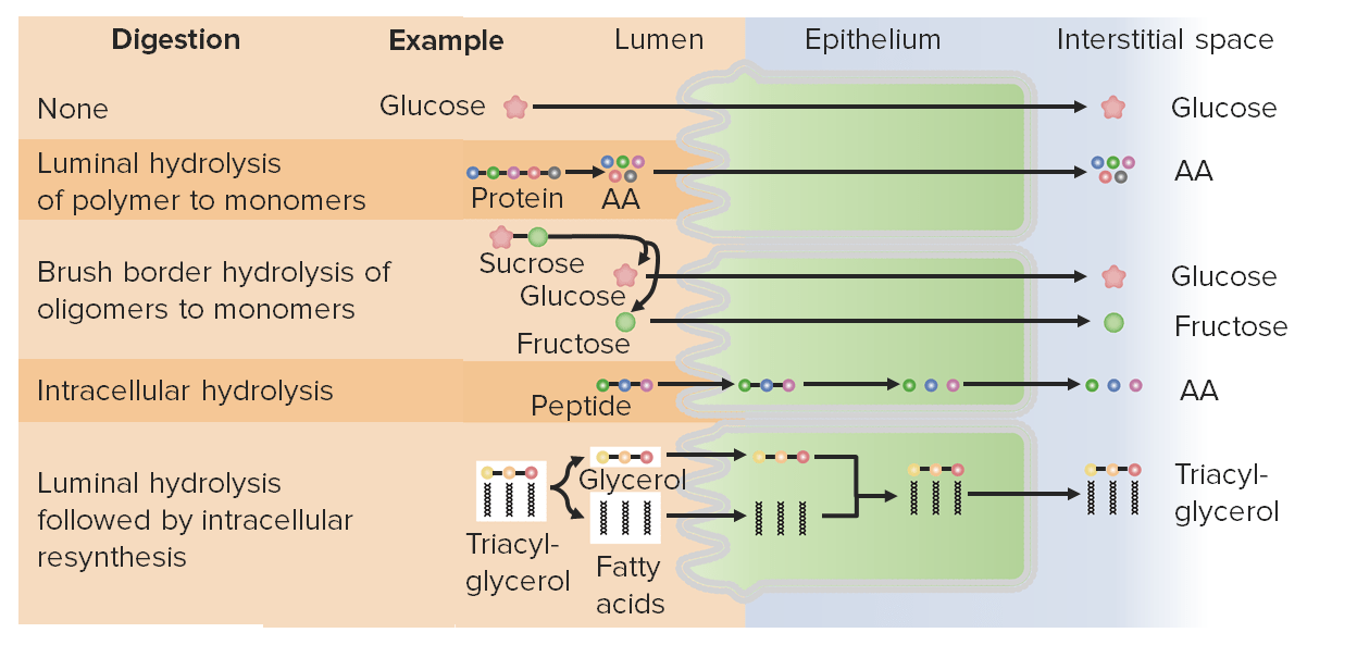
Overview of macromolecule digestion and absorption AA: amino acid
Digested primarily by amylasesAmylasesA group of amylolytic enzymes that cleave starch, glycogen, and related alpha-1, 4-glucans.Digestion and Absorption of Carbohydrates and brush borderBrush borderTubular SystemenzymesEnzymesEnzymes are complex protein biocatalysts that accelerate chemical reactions without being consumed by them. Due to the body’s constant metabolic needs, the absence of enzymes would make life unsustainable, as reactions would occur too slowly without these molecules. Basics of Enzymes
EnzymesEnzymesEnzymes are complex protein biocatalysts that accelerate chemical reactions without being consumed by them. Due to the body’s constant metabolic needs, the absence of enzymes would make life unsustainable, as reactions would occur too slowly without these molecules. Basics of Enzymes hydrolyze large starch molecules to monosaccharidesMonosaccharidesSingle chain carbohydrates that are the most basic units of carbohydrates. They are typically colorless crystalline substances with a sweet taste and have the same general formula CNH2NON.Basics of Carbohydrates
Cleavage of α-1,4-glycosidic bonds in sugar molecules
Creation of increasingly smaller polysaccharide chains until most α-1,4-glycosidic bonds are broken, leaving behind:
MonosaccharidesMonosaccharidesSingle chain carbohydrates that are the most basic units of carbohydrates. They are typically colorless crystalline substances with a sweet taste and have the same general formula CNH2NON.Basics of Carbohydrates: individual sugar molecules
DisaccharidesDisaccharidesOligosaccharides containing two monosaccharide units linked by a glycosidic bond.Basics of Carbohydrates: starches with 2 sugar molecules
OligosaccharidesOligosaccharidesCarbohydrates consisting of between two (disaccharides) and ten monosaccharides connected by either an alpha- or beta-glycosidic link. They are found throughout nature in both the free and bound form.Basics of Carbohydrates: starches with 3‒10 sugar molecules
Indigestible starches: sugars joined by other types of bonds
Active at higher pHpHThe quantitative measurement of the acidity or basicity of a solution.Acid-Base Balance:
Active in the mouth and small intestines
Inactivated in the stomachStomachThe stomach is a muscular sac in the upper left portion of the abdomen that plays a critical role in digestion. The stomach develops from the foregut and connects the esophagus with the duodenum. Structurally, the stomach is C-shaped and forms a greater and lesser curvature and is divided grossly into regions: the cardia, fundus, body, and pylorus. Stomach: Anatomy
Types and location of amylase:
Salivary amylaseSalivary amylaseA subclass of alpha-amylase isoenzymes that are secreted into saliva.Digestion and Absorption of Carbohydrates: secreted in the mouth by the salivary glandsSalivary glandsThe salivary glands are exocrine glands positioned in and around the oral cavity. These glands are responsible for secreting saliva into the mouth, which aids in digestion. There are 3 major paired salivary glands: the sublingual, submandibular, and parotid glands.Salivary Glands: Anatomy
Pancreatic amylasePancreatic AmylaseA group of amylolytic enzymes that cleave starch, glycogen, and related alpha-1, 4-glucans.Pancreatic Parameters: secreted into the duodenumDuodenumThe shortest and widest portion of the small intestine adjacent to the pylorus of the stomach. It is named for having the length equal to about the width of 12 fingers.Small Intestine: Anatomy by the exocrine pancreasExocrine pancreasThe major component (about 80%) of the pancreas composed of acinar functional units of tubular and spherical cells. The acinar cells synthesize and secrete several digestive enzymes such as trypsinogen; lipase; amylase; and ribonuclease. Secretion from the exocrine pancreas drains into the pancreatic ductal system and empties into the duodenum.Pancreas: Anatomy
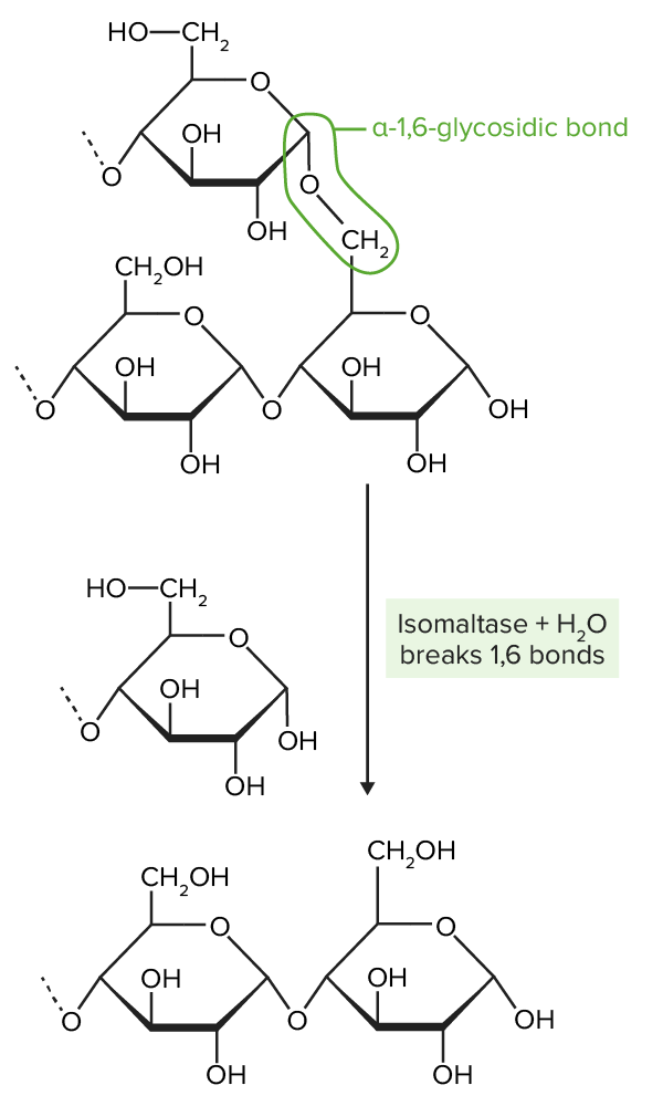
Amylopectin is partially digested by amylase. Amylopectin molecules are chains of glucose, joined together by α-1,4-glycosidic bonds (creation of a straight chain of glucose molecules) and α-1,6-glycosidic bonds (creation of a branch from the straight chain). Amylase breaks the α-1,4-glycosidic bonds.
Image by Lecturio.
Brush borderBrush borderTubular SystemenzymesEnzymesEnzymes are complex protein biocatalysts that accelerate chemical reactions without being consumed by them. Due to the body’s constant metabolic needs, the absence of enzymes would make life unsustainable, as reactions would occur too slowly without these molecules. Basics of Enzymes:
Brush borderBrush borderTubular SystemenzymesEnzymesEnzymes are complex protein biocatalysts that accelerate chemical reactions without being consumed by them. Due to the body’s constant metabolic needs, the absence of enzymes would make life unsustainable, as reactions would occur too slowly without these molecules. Basics of Enzymes are membrane-bound proteinsProteinsLinear polypeptides that are synthesized on ribosomes and may be further modified, crosslinked, cleaved, or assembled into complex proteins with several subunits. The specific sequence of amino acids determines the shape the polypeptide will take, during protein folding, and the function of the protein.Energy Homeostasis on the luminal surface of enterocytes in the small intestineSmall intestineThe small intestine is the longest part of the GI tract, extending from the pyloric orifice of the stomach to the ileocecal junction. The small intestine is the major organ responsible for chemical digestion and absorption of nutrients. It is divided into 3 segments: the duodenum, the jejunum, and the ileum. Small Intestine: Anatomy. There are 4 major brush borderBrush borderTubular SystemenzymesEnzymesEnzymes are complex protein biocatalysts that accelerate chemical reactions without being consumed by them. Due to the body’s constant metabolic needs, the absence of enzymes would make life unsustainable, as reactions would occur too slowly without these molecules. Basics of Enzymes involved in carbohydrate digestion.
IsomaltaseIsomaltaseAn enzyme that catalyzes the endohydrolysis of 1, 6-alpha-glycosidic linkages in isomaltose and dextrins produced from starch and glycogen by alpha-amylases.Digestion and Absorption of Carbohydrates: cleaves the α-1,6-glycosidic bonds
Hydrolyzes maltose → glucoseGlucoseA primary source of energy for living organisms. It is naturally occurring and is found in fruits and other parts of plants in its free state. It is used therapeutically in fluid and nutrient replacement.Lactose Intolerance + glucoseGlucoseA primary source of energy for living organisms. It is naturally occurring and is found in fruits and other parts of plants in its free state. It is used therapeutically in fluid and nutrient replacement.Lactose Intolerance
Hydrolyzes maltotriose → glucoseGlucoseA primary source of energy for living organisms. It is naturally occurring and is found in fruits and other parts of plants in its free state. It is used therapeutically in fluid and nutrient replacement.Lactose Intolerance + glucoseGlucoseA primary source of energy for living organisms. It is naturally occurring and is found in fruits and other parts of plants in its free state. It is used therapeutically in fluid and nutrient replacement.Lactose Intolerance + glucoseGlucoseA primary source of energy for living organisms. It is naturally occurring and is found in fruits and other parts of plants in its free state. It is used therapeutically in fluid and nutrient replacement.Lactose Intolerance
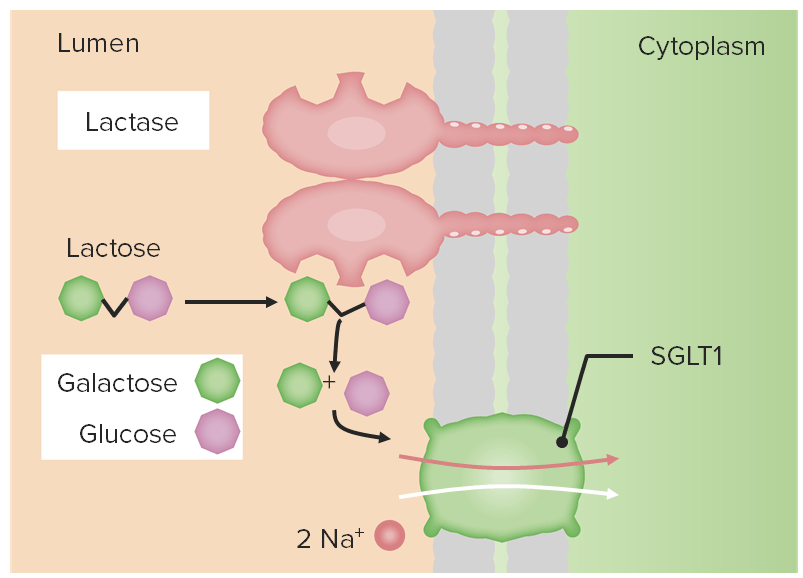
LactaseLactaseAn enzyme which catalyzes the hydrolysis of lactose to d-galactose and d-glucose. Defects in the enzyme cause lactose intolerance.Digestion and Absorption of Carbohydrates: hydrolyzes lactose → glucoseGlucoseA primary source of energy for living organisms. It is naturally occurring and is found in fruits and other parts of plants in its free state. It is used therapeutically in fluid and nutrient replacement.Lactose Intolerance + galactoseGalactoseAn aldohexose that occurs naturally in the d-form in lactose, cerebrosides, gangliosides, and mucoproteins. Deficiency of galactosyl-1-phosphate uridyltransferase causes an error in galactose metabolism called galactosemia, resulting in elevations of galactose in the blood.Lactose Intolerance
Hydrolyzes sucrose → glucoseGlucoseA primary source of energy for living organisms. It is naturally occurring and is found in fruits and other parts of plants in its free state. It is used therapeutically in fluid and nutrient replacement.Lactose Intolerance + fructose
Hydrolyzes other small oligosaccharidesOligosaccharidesCarbohydrates consisting of between two (disaccharides) and ten monosaccharides connected by either an alpha- or beta-glycosidic link. They are found throughout nature in both the free and bound form.Basics of Carbohydrates
Isomaltase breaks the α-1,6-glycosidic bonds present in amylopectin. The α-1,6-glycosidic bonds create branches off the “straight chains” of glucose by joining the 1st carbon on 1 chain (top ring structure) to the 6th carbon on another chain (bottom 2-ring structure). Isomaltase hydrolyzes these bonds.
Image by Lecturio.
Brush border digestion: Diagram of the disaccharide lactose being hydrolyzed by lactase into constituent monosaccharides, galactose and glucose. Both glucose and galactose can be absorbed into the enterocytes (along with 2 Na+ atoms moving down their concentration gradient) through the sodium–glucose-linked transporter (SGLT) 1 cotransporter.
Image by Lecturio.
Absorption
CarbohydratesCarbohydratesA class of organic compounds composed of carbon, hydrogen, and oxygen in a ratio of cn(H2O)n. The largest class of organic compounds, including starch; glycogen; cellulose; polysaccharides; and simple monosaccharides.Basics of Carbohydrates are absorbed as monosaccharidesMonosaccharidesSingle chain carbohydrates that are the most basic units of carbohydrates. They are typically colorless crystalline substances with a sweet taste and have the same general formula CNH2NON.Basics of Carbohydrates by the enterocytes in the small intestines and transported via blood to the portal circulationCirculationThe movement of the blood as it is pumped through the cardiovascular system.ABCDE Assessment. CarbohydratesCarbohydratesA class of organic compounds composed of carbon, hydrogen, and oxygen in a ratio of cn(H2O)n. The largest class of organic compounds, including starch; glycogen; cellulose; polysaccharides; and simple monosaccharides.Basics of Carbohydrates that cannot be broken down into monosaccharidesMonosaccharidesSingle chain carbohydrates that are the most basic units of carbohydrates. They are typically colorless crystalline substances with a sweet taste and have the same general formula CNH2NON.Basics of Carbohydrates are not absorbed (e.g., fibers).
MonosaccharidesMonosaccharidesSingle chain carbohydrates that are the most basic units of carbohydrates. They are typically colorless crystalline substances with a sweet taste and have the same general formula CNH2NON.Basics of Carbohydrates are:
Found in the small intestineSmall intestineThe small intestine is the longest part of the GI tract, extending from the pyloric orifice of the stomach to the ileocecal junction. The small intestine is the major organ responsible for chemical digestion and absorption of nutrients. It is divided into 3 segments: the duodenum, the jejunum, and the ileum. Small Intestine: Anatomy
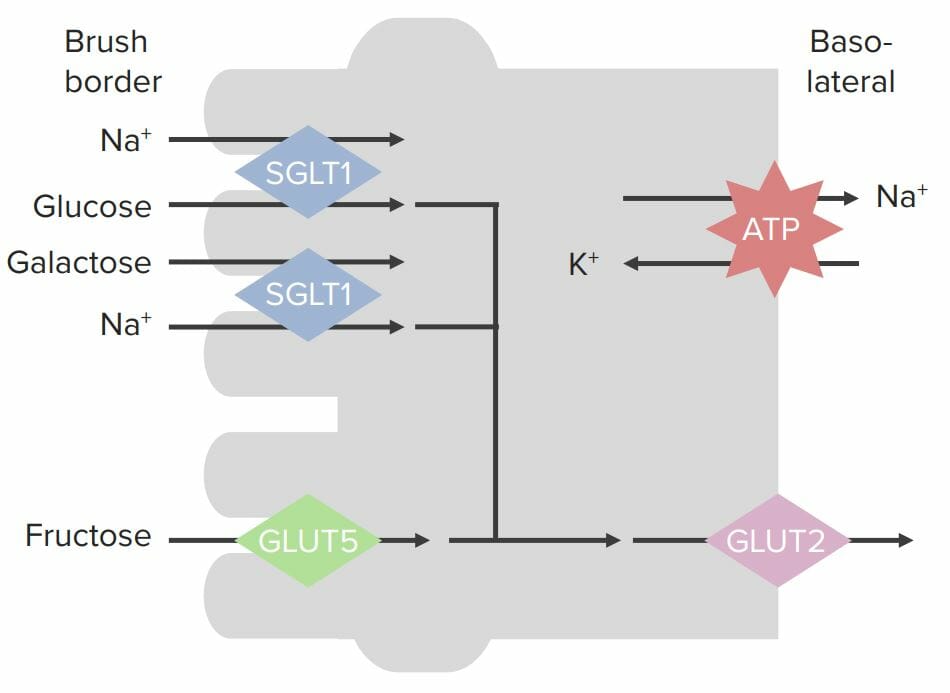
Transports 2 Na+, 1 glucoseGlucoseA primary source of energy for living organisms. It is naturally occurring and is found in fruits and other parts of plants in its free state. It is used therapeutically in fluid and nutrient replacement.Lactose Intolerance or galactoseGalactoseAn aldohexose that occurs naturally in the d-form in lactose, cerebrosides, gangliosides, and mucoproteins. Deficiency of galactosyl-1-phosphate uridyltransferase causes an error in galactose metabolism called galactosemia, resulting in elevations of galactose in the blood.Lactose Intolerance, and water
Leads to secondary active transportActive transportThe movement of materials across cell membranes and epithelial layers against an electrochemical gradient, requiring the expenditure of metabolic energy.The Cell: Cell Membrane
GLUT5: transports fructose into the cell down its concentration gradient via facilitated diffusionDiffusionThe tendency of a gas or solute to pass from a point of higher pressure or concentration to a point of lower pressure or concentration and to distribute itself throughout the available space. Diffusion, especially facilitated diffusion, is a major mechanism of biological transport.Peritoneal Dialysis and Hemodialysis
Moved out into the interstitial space by a different transport protein located in the basolateral membrane:
GLUT2:
Can move all 3 primary monosaccharidesMonosaccharidesSingle chain carbohydrates that are the most basic units of carbohydrates. They are typically colorless crystalline substances with a sweet taste and have the same general formula CNH2NON.Basics of Carbohydrates: glucoseGlucoseA primary source of energy for living organisms. It is naturally occurring and is found in fruits and other parts of plants in its free state. It is used therapeutically in fluid and nutrient replacement.Lactose Intolerance, galactoseGalactoseAn aldohexose that occurs naturally in the d-form in lactose, cerebrosides, gangliosides, and mucoproteins. Deficiency of galactosyl-1-phosphate uridyltransferase causes an error in galactose metabolism called galactosemia, resulting in elevations of galactose in the blood.Lactose Intolerance, and fructose
Works via facilitated diffusionDiffusionThe tendency of a gas or solute to pass from a point of higher pressure or concentration to a point of lower pressure or concentration and to distribute itself throughout the available space. Diffusion, especially facilitated diffusion, is a major mechanism of biological transport.Peritoneal Dialysis and Hemodialysis
Absorbed into the capillariesCapillariesCapillaries are the primary structures in the circulatory system that allow the exchange of gas, nutrients, and other materials between the blood and the extracellular fluid (ECF). Capillaries are the smallest of the blood vessels. Because a capillary diameter is so small, only 1 RBC may pass through at a time.Capillaries: Histology from the interstitial space
CapillariesCapillariesCapillaries are the primary structures in the circulatory system that allow the exchange of gas, nutrients, and other materials between the blood and the extracellular fluid (ECF). Capillaries are the smallest of the blood vessels. Because a capillary diameter is so small, only 1 RBC may pass through at a time.Capillaries: Histology drain into veinsVeinsVeins are tubular collections of cells, which transport deoxygenated blood and waste from the capillary beds back to the heart. Veins are classified into 3 types: small veins/venules, medium veins, and large veins. Each type contains 3 primary layers: tunica intima, tunica media, and tunica adventitia. Veins: Histology → portal veinPortal veinA short thick vein formed by union of the superior mesenteric vein and the splenic vein.Liver: Anatomy → liverLiverThe liver is the largest gland in the human body. The liver is found in the superior right quadrant of the abdomen and weighs approximately 1.5 kilograms. Its main functions are detoxification, metabolism, nutrient storage (e.g., iron and vitamins), synthesis of coagulation factors, formation of bile, filtration, and storage of blood. Liver: Anatomy for metabolism
Absorption of monosaccharides across enterocytes SGLT: sodium–glucose-linked transporter GLUT: glucose transporter
Image by Lecturio.
Digestion and Absorption of Proteins
Digestion
Protein digestionProtein digestionThe digestive system breaks down proteins into individual amino acids that are absorbed by the cells to build other proteins and macromolecules.Proteins and Peptides mainly occurs in the stomachStomachThe stomach is a muscular sac in the upper left portion of the abdomen that plays a critical role in digestion. The stomach develops from the foregut and connects the esophagus with the duodenum. Structurally, the stomach is C-shaped and forms a greater and lesser curvature and is divided grossly into regions: the cardia, fundus, body, and pylorus. Stomach: Anatomy and duodenumDuodenumThe shortest and widest portion of the small intestine adjacent to the pylorus of the stomach. It is named for having the length equal to about the width of 12 fingers.Small Intestine: Anatomy.
Recall: Peptide bonds join the amino terminus of AAAAAmyloidosis to the carboxy terminus of the next AAAAAmyloidosis.
Protein digestionProtein digestionThe digestive system breaks down proteins into individual amino acids that are absorbed by the cells to build other proteins and macromolecules.Proteins and Peptides occurs via enzymatic hydrolysisHydrolysisThe process of cleaving a chemical compound by the addition of a molecule of water.Proteins and Peptides of peptide bonds, breaking down proteinsProteinsLinear polypeptides that are synthesized on ribosomes and may be further modified, crosslinked, cleaved, or assembled into complex proteins with several subunits. The specific sequence of amino acids determines the shape the polypeptide will take, during protein folding, and the function of the protein.Energy Homeostasis into:
Small peptides composed of short AAAAAmyloidosis chains
Individual AAs
EnzymesEnzymesEnzymes are complex protein biocatalysts that accelerate chemical reactions without being consumed by them. Due to the body’s constant metabolic needs, the absence of enzymes would make life unsustainable, as reactions would occur too slowly without these molecules. Basics of Enzymes involved are:
Secreted by the stomachStomachThe stomach is a muscular sac in the upper left portion of the abdomen that plays a critical role in digestion. The stomach develops from the foregut and connects the esophagus with the duodenum. Structurally, the stomach is C-shaped and forms a greater and lesser curvature and is divided grossly into regions: the cardia, fundus, body, and pylorus. Stomach: Anatomy and pancreasPancreasThe pancreas lies mostly posterior to the stomach and extends across the posterior abdominal wall from the duodenum on the right to the spleen on the left. This organ has both exocrine and endocrine tissue. Pancreas: Anatomy (see table)
AminopeptidasesAminopeptidasesA subclass of exopeptidases that act on the free n terminus end of a polypeptide liberating a single amino acid residue.Proteins and Peptidesbreak down small peptides from their amino end (i.e., N-terminus).
DipeptidasesDipeptidasesExopeptidases that specifically act on dipeptides.Proteins and Peptides break peptide bonds between 2 AAs → 2 single AAs
Table: Secreted enzymesEnzymesEnzymes are complex protein biocatalysts that accelerate chemical reactions without being consumed by them. Due to the body’s constant metabolic needs, the absence of enzymes would make life unsustainable, as reactions would occur too slowly without these molecules. Basics of Enzymes involved in protein digestionProtein digestionThe digestive system breaks down proteins into individual amino acids that are absorbed by the cells to build other proteins and macromolecules.Proteins and Peptides
Enzyme
Zymogen (precursor)
Activated by
Notes on activity
Gastric enzymesEnzymesEnzymes are complex protein biocatalysts that accelerate chemical reactions without being consumed by them. Due to the body’s constant metabolic needs, the absence of enzymes would make life unsustainable, as reactions would occur too slowly without these molecules. Basics of Enzymes secreted into the stomachStomachThe stomach is a muscular sac in the upper left portion of the abdomen that plays a critical role in digestion. The stomach develops from the foregut and connects the esophagus with the duodenum. Structurally, the stomach is C-shaped and forms a greater and lesser curvature and is divided grossly into regions: the cardia, fundus, body, and pylorus. Stomach: Anatomy
PepsinPepsinPepsin breaks down proteins into proteoses, peptones, and large polypeptides.Proteins and Peptides
Pepsinogen
Hydrochloric acidHydrochloric acidA strong corrosive acid that is commonly used as a laboratory reagent. It is formed by dissolving hydrogen chloride in water. Gastric acid is the hydrochloric acid component of gastric juice.Caustic Ingestion (Cleaning Products)
Most efficient between hydrophobic AAs
Pancreatic enzymesEnzymesEnzymes are complex protein biocatalysts that accelerate chemical reactions without being consumed by them. Due to the body’s constant metabolic needs, the absence of enzymes would make life unsustainable, as reactions would occur too slowly without these molecules. Basics of Enzymes secreted into the duodenumDuodenumThe shortest and widest portion of the small intestine adjacent to the pylorus of the stomach. It is named for having the length equal to about the width of 12 fingers.Small Intestine: Anatomy
TrypsinTrypsinA serine endopeptidase that is formed from trypsinogen in the pancreas. It is converted into its active form by enteropeptidase in the small intestine. It catalyzes hydrolysis of the carboxyl group of either arginine or lysine.Proteins and Peptides
TrypsinogenTrypsinogenThe inactive proenzyme of trypsin secreted by the pancreas, activated in the duodenum via cleavage by enteropeptidase.Pancreatic Parameters
Enteropeptidase
Able to activate:
More trypsinogenTrypsinogenThe inactive proenzyme of trypsin secreted by the pancreas, activated in the duodenum via cleavage by enteropeptidase.Pancreatic Parameters → trypsinTrypsinA serine endopeptidase that is formed from trypsinogen in the pancreas. It is converted into its active form by enteropeptidase in the small intestine. It catalyzes hydrolysis of the carboxyl group of either arginine or lysine.Proteins and Peptides
All other pancreatic zymogens
Most efficient between lysine and arginineArginineAn essential amino acid that is physiologically active in the l-form.Urea Cycle
ChymotrypsinChymotrypsinA serine endopeptidase secreted by the pancreas as its zymogen, chymotrypsinogen and carried in the pancreatic juice to the duodenum where it is activated by trypsin. It selectively cleaves aromatic amino acids on the carboxyl side.Pancreatic Parameters
Chymotrypsinogen
TrypsinTrypsinA serine endopeptidase that is formed from trypsinogen in the pancreas. It is converted into its active form by enteropeptidase in the small intestine. It catalyzes hydrolysis of the carboxyl group of either arginine or lysine.Proteins and Peptides
Most efficient between hydrophobic AAs
CarboxypeptidaseCarboxypeptidaseEnzymes that act at a free c-terminus of a polypeptide to liberate a single amino acid residue.Pancreatic Parameters
Procarboxypeptidase
TrypsinTrypsinA serine endopeptidase that is formed from trypsinogen in the pancreas. It is converted into its active form by enteropeptidase in the small intestine. It catalyzes hydrolysis of the carboxyl group of either arginine or lysine.Proteins and Peptides
Attacks the carboxy end of peptide chains
Generates individual AAs or very short peptide chains
ElastaseElastaseA protease of broad specificity, obtained from dried pancreas. Molecular weight is approximately 25, 000. The enzyme breaks down elastin, the specific protein of elastic fibers, and digests other proteins such as fibrin, hemoglobin, and albumin.Proteins and Peptides
Proelastase
TrypsinTrypsinA serine endopeptidase that is formed from trypsinogen in the pancreas. It is converted into its active form by enteropeptidase in the small intestine. It catalyzes hydrolysis of the carboxyl group of either arginine or lysine.Proteins and Peptides
Same as carboxypeptidaseCarboxypeptidaseEnzymes that act at a free c-terminus of a polypeptide to liberate a single amino acid residue.Pancreatic Parameters
AA: amino acid
Absorption
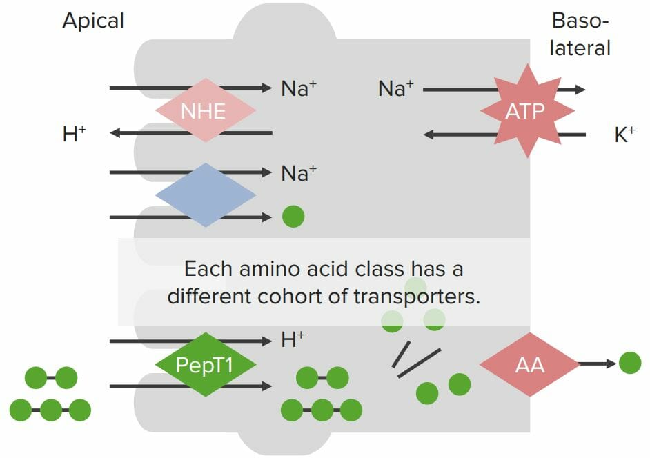
Absorption occurs in the small intestineSmall intestineThe small intestine is the longest part of the GI tract, extending from the pyloric orifice of the stomach to the ileocecal junction. The small intestine is the major organ responsible for chemical digestion and absorption of nutrients. It is divided into 3 segments: the duodenum, the jejunum, and the ileum. Small Intestine: Anatomy.
Only AAs, dipeptides, and tripeptides can be absorbed across the apical membrane into the enterocytes.
Only individual AAs can be absorbed across the basolateral membrane into the interstitial space.
Individual AAs:
Absorbed into the enterocytes across the apical membrane via specialized Na+/AAAAAmyloidosis cotransporters:
[Na+] is high in the lumen but low in the enterocytes → Na+ moves down its concentration gradient into the cell, transporting an AAAAAmyloidosis with it
Absorbed across the basolateral membrane by specialized transporters (different types of transporters for different types of AAs)
Dipeptides and tripeptides:
Absorbed by the enterocytes across the apical membrane via specialized H+/PepT cotransporters
Uses the H+ gradient created by the H+/Na+ exchanger on the apical membrane (which pumps 1 H+ ion into the lumen and brings 1 Na+ into the enterocytes)
Peptides are broken down into individual AAs by peptidases within the enterocytes.
Absorbed across the basolateral membrane in the same manner as AAs
Once in the interstitial space, AAs are absorbed into the venous circulationCirculationThe movement of the blood as it is pumped through the cardiovascular system.ABCDE Assessment → transported through portal circulationCirculationThe movement of the blood as it is pumped through the cardiovascular system.ABCDE Assessment to the liverLiverThe liver is the largest gland in the human body. The liver is found in the superior right quadrant of the abdomen and weighs approximately 1.5 kilograms. Its main functions are detoxification, metabolism, nutrient storage (e.g., iron and vitamins), synthesis of coagulation factors, formation of bile, filtration, and storage of blood. Liver: Anatomy
Transport proteins on enterocyte membranes involved in protein absorption: The Na+/K+ ATPase on the basolateral membrane generates a Na+ gradient within the cell. A Na+/H+ exchanger (NHE) on the apical membrane also generates the H+ gradient. Individual amino acids (AAs; green balls) are absorbed via a Na+/AA cotransporter, where Na+ flows across the apical membrane into the enterocytes down its concentration gradient, bringing the AA with it (despite moving against the chemical AA gradient). Small peptides are absorbed via the H+/PepT cotransporter with H+ flowing down its concentration gradient into the cell, bringing the small peptides with it. Peptides are broken down into individual AAs by peptidases within the enterocytes. All AAs are then absorbed through specialized transporters on the basolateral membrane.
Image by Lecturio.
Digestion and Absorption of Fats
Digestion
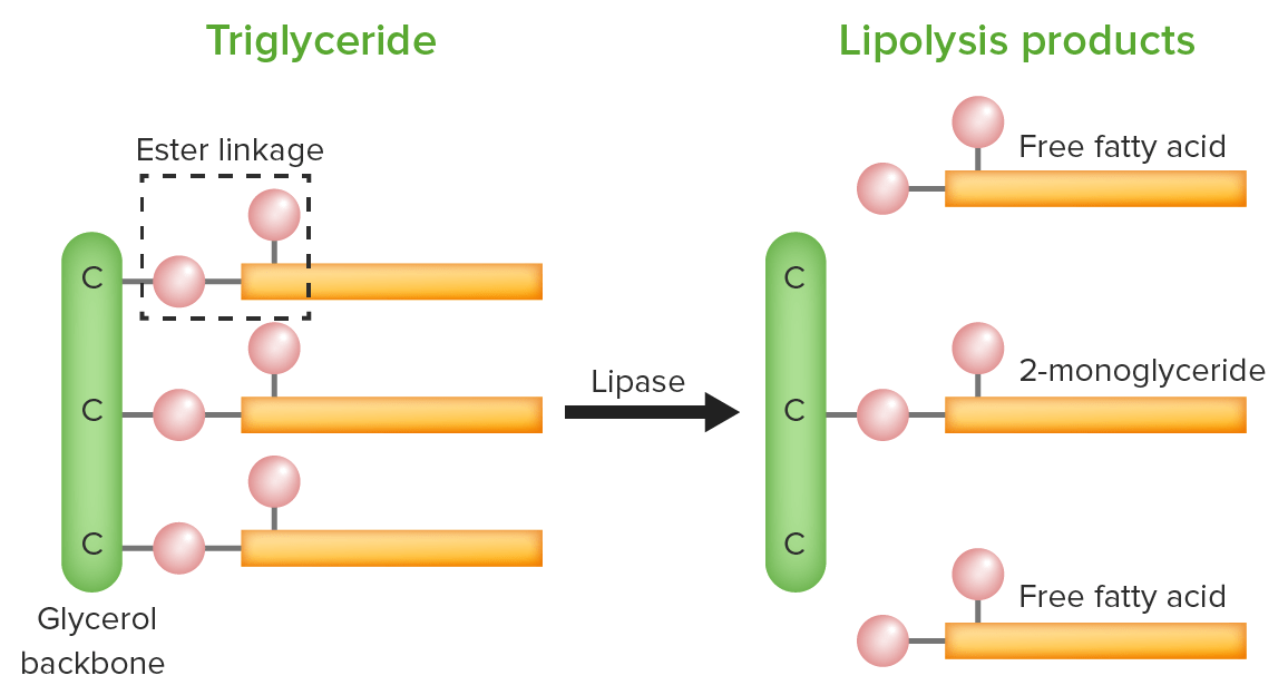
There are 3 primary types of fatsFatsThe glyceryl esters of a fatty acid, or of a mixture of fatty acids. They are generally odorless, colorless, and tasteless if pure, but they may be flavored according to origin. Fats are insoluble in water, soluble in most organic solvents. They occur in animal and vegetable tissue and are generally obtained by boiling or by extraction under pressure. They are important in the diet (dietary fats) as a source of energy.Energy Homeostasis that are digested and absorbed: triglyceridesTriglyceridesFatty Acids and Lipids (triacylglycerolsTriacylglycerolsFatty Acids and Lipids, or TAGs), phospholipidsPhospholipidsLipids containing one or more phosphate groups, particularly those derived from either glycerol (phosphoglycerides) or sphingosine (sphingolipids). They are polar lipids that are of great importance for the structure and function of cell membranes and are the most abundant of membrane lipids, although not stored in large amounts in the system.Lipid Metabolism, and cholesterolCholesterolThe principal sterol of all higher animals, distributed in body tissues, especially the brain and spinal cord, and in animal fats and oils.Cholesterol Metabolism esters. All 3 types contain ester bonds (R1‒(C=O)‒O ‒R2) that are broken during digestion.
TAGs:
Structure:
Glycerol backbone: 3-carbon chain with each carbon attached to an alcohol group
Fatty acidsAcidsChemical compounds which yield hydrogen ions or protons when dissolved in water, whose hydrogen can be replaced by metals or basic radicals, or which react with bases to form salts and water (neutralization). An extension of the term includes substances dissolved in media other than water.Acid-Base Balance: hydrocarbon chain with a carboxyl group at 1 end
Each carbon on the glycerol backbone is bound to the carboxy end of a fatty-acid chain by an ester bond.
Ester bonds are hydrolyzed by lipasesLipasesAn enzyme of the hydrolase class that catalyzes the reaction of triacylglycerol and water to yield diacylglycerol and a fatty acid anion. It is produced by glands on the tongue and by the pancreas and initiates the digestion of dietary fats.Lipid Metabolism:
Lingual lipaseLipaseAn enzyme of the hydrolase class that catalyzes the reaction of triacylglycerol and water to yield diacylglycerol and a fatty acid anion. It is produced by glands on the tongue and by the pancreas and initiates the digestion of dietary fats.Malabsorption and Maldigestion (from salivary glandsSalivary glandsThe salivary glands are exocrine glands positioned in and around the oral cavity. These glands are responsible for secreting saliva into the mouth, which aids in digestion. There are 3 major paired salivary glands: the sublingual, submandibular, and parotid glands.Salivary Glands: Anatomy)
Gastric lipaseLipaseAn enzyme of the hydrolase class that catalyzes the reaction of triacylglycerol and water to yield diacylglycerol and a fatty acid anion. It is produced by glands on the tongue and by the pancreas and initiates the digestion of dietary fats.Malabsorption and Maldigestion (from chief cellsChief cellsEpithelial cells that line the basal half of the gastric glands. Chief cells synthesize and export an inactive enzyme pepsinogen which is converted into the highly proteolytic enzyme pepsin in the acid environment of the stomach.Stomach: Anatomy)
Pancreatic lipaseLipaseAn enzyme of the hydrolase class that catalyzes the reaction of triacylglycerol and water to yield diacylglycerol and a fatty acid anion. It is produced by glands on the tongue and by the pancreas and initiates the digestion of dietary fats.Malabsorption and Maldigestion (from exocrine pancreasExocrine pancreasThe major component (about 80%) of the pancreas composed of acinar functional units of tubular and spherical cells. The acinar cells synthesize and secrete several digestive enzymes such as trypsinogen; lipase; amylase; and ribonuclease. Secretion from the exocrine pancreas drains into the pancreatic ductal system and empties into the duodenum.Pancreas: Anatomy, the most important)
Lipase catalyzes the hydrolysis of ester bonds, resulting in 2 free fatty acids and a monoglyceride. Red balls represent oxygen molecules.
Image by Lecturio.
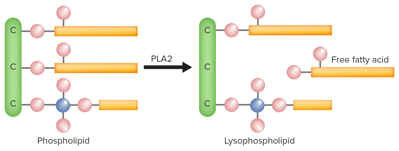
PhospholipidsPhospholipidsLipids containing one or more phosphate groups, particularly those derived from either glycerol (phosphoglycerides) or sphingosine (sphingolipids). They are polar lipids that are of great importance for the structure and function of cell membranes and are the most abundant of membrane lipids, although not stored in large amounts in the system.Lipid Metabolism:
Phospholipase A2 (PLA2) hydrolyzes the bond between the 2nd fatty acid of a phospholipid and the glycerol backbone, resulting in a lysophospholipid and free fatty acid.
Image by Lecturio.
CholesterolCholesterolThe principal sterol of all higher animals, distributed in body tissues, especially the brain and spinal cord, and in animal fats and oils.Cholesterol Metabolism esters:
CholesterolCholesterolThe principal sterol of all higher animals, distributed in body tissues, especially the brain and spinal cord, and in animal fats and oils.Cholesterol Metabolism bound to a fatty acid by an ester bond
Cholesteryl ester hydrolase catalyzes the hydrolysis of cholesteryl esters into cholesterol and a fatty acid.
Image by Lecturio.
Table: LipidsLipidsLipids are a diverse group of hydrophobic organic molecules, which include fats, oils, sterols, and waxes.Fatty Acids and Lipids and their digestive enzymesEnzymesEnzymes are complex protein biocatalysts that accelerate chemical reactions without being consumed by them. Due to the body’s constant metabolic needs, the absence of enzymes would make life unsustainable, as reactions would occur too slowly without these molecules. Basics of Enzymes
LipasesLipasesAn enzyme of the hydrolase class that catalyzes the reaction of triacylglycerol and water to yield diacylglycerol and a fatty acid anion. It is produced by glands on the tongue and by the pancreas and initiates the digestion of dietary fats.Lipid Metabolism
Monoglyceride and 2 fatty acidsAcidsChemical compounds which yield hydrogen ions or protons when dissolved in water, whose hydrogen can be replaced by metals or basic radicals, or which react with bases to form salts and water (neutralization). An extension of the term includes substances dissolved in media other than water.Acid-Base Balance
CholesterolCholesterolThe principal sterol of all higher animals, distributed in body tissues, especially the brain and spinal cord, and in animal fats and oils.Cholesterol Metabolism esters
CholesterolCholesterolThe principal sterol of all higher animals, distributed in body tissues, especially the brain and spinal cord, and in animal fats and oils.Cholesterol Metabolism and fatty acid
PhospholipidsPhospholipidsLipids containing one or more phosphate groups, particularly those derived from either glycerol (phosphoglycerides) or sphingosine (sphingolipids). They are polar lipids that are of great importance for the structure and function of cell membranes and are the most abundant of membrane lipids, although not stored in large amounts in the system.Lipid Metabolism
Phospholipase A2Phospholipase A2Phospholipases that hydrolyze the Acyl group attached to the 2-position of phosphoglycerides.Nephrotic Syndrome
LysolecithinLysolecithinDerivatives of phosphatidylcholines obtained by their partial hydrolysis which removes one of the fatty acid moieties.Cholecystitis and a fatty acid
Substances required for lipaseLipaseAn enzyme of the hydrolase class that catalyzes the reaction of triacylglycerol and water to yield diacylglycerol and a fatty acid anion. It is produced by glands on the tongue and by the pancreas and initiates the digestion of dietary fats.Malabsorption and Maldigestion activity/digestion of lipidsLipidsLipids are a diverse group of hydrophobic organic molecules, which include fats, oils, sterols, and waxes.Fatty Acids and Lipids:
BileBileAn emulsifying agent produced in the liver and secreted into the duodenum. Its composition includes bile acids and salts; cholesterol; and electrolytes. It aids digestion of fats in the duodenum.Gallbladder and Biliary Tract: Anatomy:
Emulsifier produced by the liverLiverThe liver is the largest gland in the human body. The liver is found in the superior right quadrant of the abdomen and weighs approximately 1.5 kilograms. Its main functions are detoxification, metabolism, nutrient storage (e.g., iron and vitamins), synthesis of coagulation factors, formation of bile, filtration, and storage of blood. Liver: Anatomy and stored and secreted by the gallbladderGallbladderThe gallbladder is a pear-shaped sac, located directly beneath the liver, that sits on top of the superior part of the duodenum. The primary functions of the gallbladder include concentrating and storing up to 50 mL of bile. Gallbladder and Biliary Tract: Anatomy
Contains lecithinLecithinA complex mixture of phospholipids; glycolipids; and triglycerides; with substantial amounts of phosphatidylcholines; phosphatidylethanolamines; and phosphatidylinositols, which are sometimes loosely termed as 1, 2-diacyl-3-phosphocholines. Lecithin is a component of the cell membrane and commercially extracted from soybeans and egg yolk. The emulsifying and surfactant properties are useful in food additives and for forming organogels (gels).Fatty Acids and Lipids (a phospholipid) and bileBileAn emulsifying agent produced in the liver and secreted into the duodenum. Its composition includes bile acids and salts; cholesterol; and electrolytes. It aids digestion of fats in the duodenum.Gallbladder and Biliary Tract: Anatomy salts
Forms smaller fat dropletsDropletsVaricella-Zoster Virus/Chickenpox, providing the water-soluble lipasesLipasesAn enzyme of the hydrolase class that catalyzes the reaction of triacylglycerol and water to yield diacylglycerol and a fatty acid anion. It is produced by glands on the tongue and by the pancreas and initiates the digestion of dietary fats.Lipid Metabolism more surface area to digest lipidsLipidsLipids are a diverse group of hydrophobic organic molecules, which include fats, oils, sterols, and waxes.Fatty Acids and Lipids
Colipase: helps lipasesLipasesAn enzyme of the hydrolase class that catalyzes the reaction of triacylglycerol and water to yield diacylglycerol and a fatty acid anion. It is produced by glands on the tongue and by the pancreas and initiates the digestion of dietary fats.Lipid MetabolismbindBINDHyperbilirubinemia of the Newborn to (and digest) the emulsified fat dropletsDropletsVaricella-Zoster Virus/Chickenpox
MicellesMicellesParticles consisting of aggregates of molecules held loosely together by secondary bonds. The surface of micelles are usually comprised of amphiphatic compounds that are oriented in a way that minimizes the energy of interaction between the micelle and its environment. Liquids that contain large numbers of suspended micelles are referred to as emulsions.Malabsorption and Maldigestion:
As lipidsLipidsLipids are a diverse group of hydrophobic organic molecules, which include fats, oils, sterols, and waxes.Fatty Acids and Lipids are broken down, they (along with components from the bileBileAn emulsifying agent produced in the liver and secreted into the duodenum. Its composition includes bile acids and salts; cholesterol; and electrolytes. It aids digestion of fats in the duodenum.Gallbladder and Biliary Tract: Anatomy) arrange themselves in structures called micellesMicellesParticles consisting of aggregates of molecules held loosely together by secondary bonds. The surface of micelles are usually comprised of amphiphatic compounds that are oriented in a way that minimizes the energy of interaction between the micelle and its environment. Liquids that contain large numbers of suspended micelles are referred to as emulsions.Malabsorption and Maldigestion:
Surrounded by phospholipidsPhospholipidsLipids containing one or more phosphate groups, particularly those derived from either glycerol (phosphoglycerides) or sphingosine (sphingolipids). They are polar lipids that are of great importance for the structure and function of cell membranes and are the most abundant of membrane lipids, although not stored in large amounts in the system.Lipid Metabolism from bileBileAn emulsifying agent produced in the liver and secreted into the duodenum. Its composition includes bile acids and salts; cholesterol; and electrolytes. It aids digestion of fats in the duodenum.Gallbladder and Biliary Tract: Anatomy
Contain all fat-soluble components to be absorbed:
Free fatty acidsAcidsChemical compounds which yield hydrogen ions or protons when dissolved in water, whose hydrogen can be replaced by metals or basic radicals, or which react with bases to form salts and water (neutralization). An extension of the term includes substances dissolved in media other than water.Acid-Base Balance
Monoacylglycerides
CholesterolCholesterolThe principal sterol of all higher animals, distributed in body tissues, especially the brain and spinal cord, and in animal fats and oils.Cholesterol Metabolism
PhospholipidsPhospholipidsLipids containing one or more phosphate groups, particularly those derived from either glycerol (phosphoglycerides) or sphingosine (sphingolipids). They are polar lipids that are of great importance for the structure and function of cell membranes and are the most abundant of membrane lipids, although not stored in large amounts in the system.Lipid Metabolism
Fat-soluble vitamins: A, D, E, and K
MicellesMicellesParticles consisting of aggregates of molecules held loosely together by secondary bonds. The surface of micelles are usually comprised of amphiphatic compounds that are oriented in a way that minimizes the energy of interaction between the micelle and its environment. Liquids that contain large numbers of suspended micelles are referred to as emulsions.Malabsorption and Maldigestion carry the lipid components to the enterocyte walls for absorption.
Absorption
While the majority of absorption occurs in the small intestineSmall intestineThe small intestine is the longest part of the GI tract, extending from the pyloric orifice of the stomach to the ileocecal junction. The small intestine is the major organ responsible for chemical digestion and absorption of nutrients. It is divided into 3 segments: the duodenum, the jejunum, and the ileum. Small Intestine: Anatomy, some absorption may begin in the stomachStomachThe stomach is a muscular sac in the upper left portion of the abdomen that plays a critical role in digestion. The stomach develops from the foregut and connects the esophagus with the duodenum. Structurally, the stomach is C-shaped and forms a greater and lesser curvature and is divided grossly into regions: the cardia, fundus, body, and pylorus. Stomach: Anatomy.
Long-chain fatty acidsAcidsChemical compounds which yield hydrogen ions or protons when dissolved in water, whose hydrogen can be replaced by metals or basic radicals, or which react with bases to form salts and water (neutralization). An extension of the term includes substances dissolved in media other than water.Acid-Base Balance (LCFAs):
Mixed micellesMicellesParticles consisting of aggregates of molecules held loosely together by secondary bonds. The surface of micelles are usually comprised of amphiphatic compounds that are oriented in a way that minimizes the energy of interaction between the micelle and its environment. Liquids that contain large numbers of suspended micelles are referred to as emulsions.Malabsorption and Maldigestion package LCFAs and bring them to the enterocyte border.
pHpHThe quantitative measurement of the acidity or basicity of a solution.Acid-Base Balance change near the brush borderBrush borderTubular System breaks open the micellesMicellesParticles consisting of aggregates of molecules held loosely together by secondary bonds. The surface of micelles are usually comprised of amphiphatic compounds that are oriented in a way that minimizes the energy of interaction between the micelle and its environment. Liquids that contain large numbers of suspended micelles are referred to as emulsions.Malabsorption and Maldigestion.
Lipid components (e.g., fatty acidsAcidsChemical compounds which yield hydrogen ions or protons when dissolved in water, whose hydrogen can be replaced by metals or basic radicals, or which react with bases to form salts and water (neutralization). An extension of the term includes substances dissolved in media other than water.Acid-Base Balance and monoglyceridesMonoglyceridesGlycerol esterified with a single Acyl (fatty acids) chain.Lipid Metabolism) travel across the membrane to enter the cytosolCytosolA cell’s cytoskeleton is a network of intracellular protein fibers that provides structural support, anchors organelles, and aids intra- and extracellular movement.The Cell: Cytosol and Cytoskeleton of enterocytes.
Lipid components are:
Lipid solubleLipid SolubleChloramphenicol → can cross the phospholipid membrane without specialized transport molecules
Resynthesized via esterificationEsterificationThe process of converting an acid into an alkyl or aryl derivative. Most frequently the process consists of the reaction of an acid with an alcohol in the presence of a trace of mineral acid as catalyst or the reaction of an Acyl chloride with an alcohol. Esterification can also be accomplished by enzymatic processes.Lipid Metabolism in the ER
Repackaged as chylomicrons in the golgi apparatus
Chylomicrons exit the enterocytes on their basolateral side → enter lymphatic circulationCirculationThe movement of the blood as it is pumped through the cardiovascular system.ABCDE Assessment
Short-chain fatty acidsAcidsChemical compounds which yield hydrogen ions or protons when dissolved in water, whose hydrogen can be replaced by metals or basic radicals, or which react with bases to form salts and water (neutralization). An extension of the term includes substances dissolved in media other than water.Acid-Base Balance (SCFAs) and medium-chain fatty acidsAcidsChemical compounds which yield hydrogen ions or protons when dissolved in water, whose hydrogen can be replaced by metals or basic radicals, or which react with bases to form salts and water (neutralization). An extension of the term includes substances dissolved in media other than water.Acid-Base Balance (MCFAs):
In the small intestineSmall intestineThe small intestine is the longest part of the GI tract, extending from the pyloric orifice of the stomach to the ileocecal junction. The small intestine is the major organ responsible for chemical digestion and absorption of nutrients. It is divided into 3 segments: the duodenum, the jejunum, and the ileum. Small Intestine: Anatomy:
SCFAs and MCFAs travel across the enterocytes without assistance.
SCFAs and MCFAs are absorbed into venous circulationCirculationThe movement of the blood as it is pumped through the cardiovascular system.ABCDE Assessment → hepatic portalHepatic portalLiver: Anatomy vein → liverLiverThe liver is the largest gland in the human body. The liver is found in the superior right quadrant of the abdomen and weighs approximately 1.5 kilograms. Its main functions are detoxification, metabolism, nutrient storage (e.g., iron and vitamins), synthesis of coagulation factors, formation of bile, filtration, and storage of blood. Liver: Anatomy
In the large intestineLarge intestineThe large intestines constitute the last portion of the digestive system. The large intestine consists of the cecum, appendix, colon (with ascending, transverse, descending, and sigmoid segments), rectum, and anal canal. The primary function of the colon is to remove water and compact the stool prior to expulsion from the body via the rectum and anal canal. Colon, Cecum, and Appendix: Anatomy, SCFAs use the sodiumSodiumA member of the alkali group of metals. It has the atomic symbol na, atomic number 11, and atomic weight 23.Hyponatremia monocarboxylate transporter (SMCT) 1:
The pumpPumpACES and RUSH: Resuscitation Ultrasound Protocols also assists in the absorption of water in the large intestineLarge intestineThe large intestines constitute the last portion of the digestive system. The large intestine consists of the cecum, appendix, colon (with ascending, transverse, descending, and sigmoid segments), rectum, and anal canal. The primary function of the colon is to remove water and compact the stool prior to expulsion from the body via the rectum and anal canal. Colon, Cecum, and Appendix: Anatomy.
Digestion and Absorption of Micronutrients
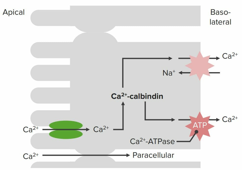
CalciumCalciumA basic element found in nearly all tissues. It is a member of the alkaline earth family of metals with the atomic symbol ca, atomic number 20, and atomic weight 40. Calcium is the most abundant mineral in the body and combines with phosphorus to form calcium phosphate in the bones and teeth. It is essential for the normal functioning of nerves and muscles and plays a role in blood coagulation (as factor IV) and in many enzymatic processes.Electrolytes (CaCACondylomata acuminata are a clinical manifestation of genital HPV infection. Condylomata acuminata are described as raised, pearly, flesh-colored, papular, cauliflower-like lesions seen in the anogenital region that may cause itching, pain, or bleeding.Condylomata Acuminata (Genital Warts)2+) absorption
CaCACondylomata acuminata are a clinical manifestation of genital HPV infection. Condylomata acuminata are described as raised, pearly, flesh-colored, papular, cauliflower-like lesions seen in the anogenital region that may cause itching, pain, or bleeding.Condylomata Acuminata (Genital Warts)2+ is absorbed across the apical membrane via CaCACondylomata acuminata are a clinical manifestation of genital HPV infection. Condylomata acuminata are described as raised, pearly, flesh-colored, papular, cauliflower-like lesions seen in the anogenital region that may cause itching, pain, or bleeding.Condylomata Acuminata (Genital Warts)2+ transporters (primarily TRPV6).
CalbindinCalbindinCalcium-binding proteins that are found in distal kidney tubules, intestines, brain, and other tissues where they bind, buffer and transport cytoplasmic calcium. Calbindins possess a variable number of ef-hand motifs which contain calcium-binding sites. Some isoforms are regulated by vitamin d.Calcium Hemostasis and Bone Metabolism: an intracellular CaCACondylomata acuminata are a clinical manifestation of genital HPV infection. Condylomata acuminata are described as raised, pearly, flesh-colored, papular, cauliflower-like lesions seen in the anogenital region that may cause itching, pain, or bleeding.Condylomata Acuminata (Genital Warts)2+-binding protein that immediately binds all absorbed CaCACondylomata acuminata are a clinical manifestation of genital HPV infection. Condylomata acuminata are described as raised, pearly, flesh-colored, papular, cauliflower-like lesions seen in the anogenital region that may cause itching, pain, or bleeding.Condylomata Acuminata (Genital Warts)2+and transports it to the basolateral membrane
Purpose:
Free CaCACondylomata acuminata are a clinical manifestation of genital HPV infection. Condylomata acuminata are described as raised, pearly, flesh-colored, papular, cauliflower-like lesions seen in the anogenital region that may cause itching, pain, or bleeding.Condylomata Acuminata (Genital Warts)2+ could act as an intracellular signaling molecule.
High levels of free CaCACondylomata acuminata are a clinical manifestation of genital HPV infection. Condylomata acuminata are described as raised, pearly, flesh-colored, papular, cauliflower-like lesions seen in the anogenital region that may cause itching, pain, or bleeding.Condylomata Acuminata (Genital Warts)2+ can be toxic.
CalbindinCalbindinCalcium-binding proteins that are found in distal kidney tubules, intestines, brain, and other tissues where they bind, buffer and transport cytoplasmic calcium. Calbindins possess a variable number of ef-hand motifs which contain calcium-binding sites. Some isoforms are regulated by vitamin d.Calcium Hemostasis and Bone Metabolism level in the cell determines how much CaCACondylomata acuminata are a clinical manifestation of genital HPV infection. Condylomata acuminata are described as raised, pearly, flesh-colored, papular, cauliflower-like lesions seen in the anogenital region that may cause itching, pain, or bleeding.Condylomata Acuminata (Genital Warts)2+ can be absorbed.
CaCACondylomata acuminata are a clinical manifestation of genital HPV infection. Condylomata acuminata are described as raised, pearly, flesh-colored, papular, cauliflower-like lesions seen in the anogenital region that may cause itching, pain, or bleeding.Condylomata Acuminata (Genital Warts)2+ is absorbed across the basolateral membrane by:
CaCACondylomata acuminata are a clinical manifestation of genital HPV infection. Condylomata acuminata are described as raised, pearly, flesh-colored, papular, cauliflower-like lesions seen in the anogenital region that may cause itching, pain, or bleeding.Condylomata Acuminata (Genital Warts)2+ ATPase
CaCACondylomata acuminata are a clinical manifestation of genital HPV infection. Condylomata acuminata are described as raised, pearly, flesh-colored, papular, cauliflower-like lesions seen in the anogenital region that may cause itching, pain, or bleeding.Condylomata Acuminata (Genital Warts)2+/Na+ exchanger
Note: A small amount of CaCACondylomata acuminata are a clinical manifestation of genital HPV infection. Condylomata acuminata are described as raised, pearly, flesh-colored, papular, cauliflower-like lesions seen in the anogenital region that may cause itching, pain, or bleeding.Condylomata Acuminata (Genital Warts)2+ can also be absorbed paracellularly.
Regulation:
Production of the apical CaCACondylomata acuminata are a clinical manifestation of genital HPV infection. Condylomata acuminata are described as raised, pearly, flesh-colored, papular, cauliflower-like lesions seen in the anogenital region that may cause itching, pain, or bleeding.Condylomata Acuminata (Genital Warts)2+ transporter is induced by:
Vitamin DVitamin DA vitamin that includes both cholecalciferols and ergocalciferols, which have the common effect of preventing or curing rickets in animals. It can also be viewed as a hormone since it can be formed in skin by action of ultraviolet rays upon the precursors, 7-dehydrocholesterol and ergosterol, and acts on vitamin D receptors to regulate calcium in opposition to parathyroid hormone.Fat-soluble Vitamins and their Deficiencies
Estrogens
CalbindinCalbindinCalcium-binding proteins that are found in distal kidney tubules, intestines, brain, and other tissues where they bind, buffer and transport cytoplasmic calcium. Calbindins possess a variable number of ef-hand motifs which contain calcium-binding sites. Some isoforms are regulated by vitamin d.Calcium Hemostasis and Bone MetabolismsynthesisSynthesisPolymerase Chain Reaction (PCR) is induced by vitamin DVitamin DA vitamin that includes both cholecalciferols and ergocalciferols, which have the common effect of preventing or curing rickets in animals. It can also be viewed as a hormone since it can be formed in skin by action of ultraviolet rays upon the precursors, 7-dehydrocholesterol and ergosterol, and acts on vitamin D receptors to regulate calcium in opposition to parathyroid hormone.Fat-soluble Vitamins and their Deficiencies.
Schematic diagram depicting calcium (Ca2+) absorption: Calcium is absorbed across the apical membrane by a specialized Ca2+-transport protein and then immediately bound to a Ca2+-binding protein called calbindin. Calbindin transports Ca2+ to the basolateral membrane where it is absorbed by Ca2+ ATPase and/or a Ca2+/Na+ exchanger.
Image by Lecturio.
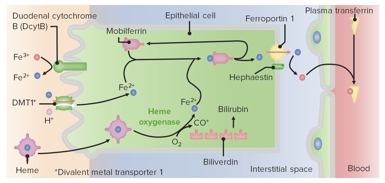
IronIronA metallic element with atomic symbol fe, atomic number 26, and atomic weight 55. 85. It is an essential constituent of hemoglobins; cytochromes; and iron-binding proteins. It plays a role in cellular redox reactions and in the transport of oxygen.Trace Elements absorption
Primarily absorbed in the duodenumDuodenumThe shortest and widest portion of the small intestine adjacent to the pylorus of the stomach. It is named for having the length equal to about the width of 12 fingers.Small Intestine: Anatomy
Fe3+ (the primary form of ironIronA metallic element with atomic symbol fe, atomic number 26, and atomic weight 55. 85. It is an essential constituent of hemoglobins; cytochromes; and iron-binding proteins. It plays a role in cellular redox reactions and in the transport of oxygen.Trace Elements found in food) must be reduced to Fe2+ for absorption via duodenal cytochrome B (DcytB) located on the brush borderBrush borderTubular System of enterocytes.
Absorption across the apical membrane:
DMT (divalent metal transporterDivalent metal transporterTransports Fe²⁺ (not Fe³⁺) from the apical surface of enterocytes to the interior of the cell.Heme Metabolism) 1: a specialized Fe2+/H+ cotransporter absorbing most nonheme ironIronA metallic element with atomic symbol fe, atomic number 26, and atomic weight 55. 85. It is an essential constituent of hemoglobins; cytochromes; and iron-binding proteins. It plays a role in cellular redox reactions and in the transport of oxygen.Trace Elements
Within heme molecules (e.g., from meat)
Within the cell:
Mobilferrin: an iron-binding protein that binds Fe2+ and transports it across the enterocyte to the basolateral membrane
Heme oxygenaseHeme oxygenaseA mixed function oxidase enzyme which during hemoglobin catabolism catalyzes the degradation of heme to ferrous iron, carbon monoxide and biliverdin in the presence of molecular oxygen and reduced NADPH.Heme Metabolism: releases Fe2+ from heme → Fe2+ is bound and transported by mobilferrin
Release across the basolateral membrane:
FerroportinFerroportinHelps export iron from the intestinal cell.Heme Metabolism 1: a membrane-bound transport protein that releases Fe2+ into the interstitial space
HephaestinHephaestinA copper-containing membrane protein which has a ferroxidase activity.Heme Metabolism:
A copper-dependent membrane-bound ferroxidase
Oxidizes Fe2+ to Fe3+, which is necessary for ironIronA metallic element with atomic symbol fe, atomic number 26, and atomic weight 55. 85. It is an essential constituent of hemoglobins; cytochromes; and iron-binding proteins. It plays a role in cellular redox reactions and in the transport of oxygen.Trace Elements to move into capillariesCapillariesCapillaries are the primary structures in the circulatory system that allow the exchange of gas, nutrients, and other materials between the blood and the extracellular fluid (ECF). Capillaries are the smallest of the blood vessels. Because a capillary diameter is so small, only 1 RBC may pass through at a time.Capillaries: Histology and bindBINDHyperbilirubinemia of the NewborntransferrinTransferrinAn iron-binding beta1-globulin that is synthesized in the liver and secreted into the blood. It plays a central role in the transport of iron throughout the circulation.Heme Metabolism(plasmaPlasmaThe residual portion of blood that is left after removal of blood cells by centrifugation without prior blood coagulation.Transfusion ProductsironIronA metallic element with atomic symbol fe, atomic number 26, and atomic weight 55. 85. It is an essential constituent of hemoglobins; cytochromes; and iron-binding proteins. It plays a role in cellular redox reactions and in the transport of oxygen.Trace Elements transport protein)
Regulation:
HepcidinHepcidinForms of hepcidin, a cationic amphipathic peptide synthesized in the liver as a prepropeptide which is first processed into prohepcidin and then into the biologically active hepcidin forms, including in human the 20-, 22-, and 25-amino acid residue peptide forms. Hepcidin acts as a homeostatic regulators of iron metabolism and also possesses antimicrobial activity.Hereditary Hemochromatosis:
Inactivates ferroportinFerroportinHelps export iron from the intestinal cell.Heme Metabolism 1 (the primary negative regulatory outcome of intestinal ironIronA metallic element with atomic symbol fe, atomic number 26, and atomic weight 55. 85. It is an essential constituent of hemoglobins; cytochromes; and iron-binding proteins. It plays a role in cellular redox reactions and in the transport of oxygen.Trace Elements absorption)
Clinical relevance: MutationMutationGenetic mutations are errors in DNA that can cause protein misfolding and dysfunction. There are various types of mutations, including chromosomal, point, frameshift, and expansion mutations. Types of Mutations/deficiency of hepcidinHepcidinForms of hepcidin, a cationic amphipathic peptide synthesized in the liver as a prepropeptide which is first processed into prohepcidin and then into the biologically active hepcidin forms, including in human the 20-, 22-, and 25-amino acid residue peptide forms. Hepcidin acts as a homeostatic regulators of iron metabolism and also possesses antimicrobial activity.Hereditary Hemochromatosis results in hereditary hemochromatosisHemochromatosisA disorder of iron metabolism characterized by a triad of hemosiderosis; liver cirrhosis; and diabetes mellitus. It is caused by massive iron deposits in parenchymal cells that may develop after a prolonged increase of iron absorption.Hereditary Hemochromatosis.
Hypoxia-inducible factorHypoxia-inducible factorHypoxia-inducible factor 1, alpha subunit is a basic helix-loop-helix transcription factor that is regulated by oxygen availability and is targeted for degradation by VHL tumor suppressor protein.Von Hippel-Lindau Disease (HIFHIFHypoxia-inducible factor 1, alpha subunit is a basic helix-loop-helix transcription factor that is regulated by oxygen availability and is targeted for degradation by VHL tumor suppressor protein.Von Hippel-Lindau Disease)-2α induces synthesisSynthesisPolymerase Chain Reaction (PCR) of DMT1DMT1Transports Fe²⁺ (not Fe³⁺) from the apical surface of enterocytes to the interior of the cell.Heme Metabolism and DcytB → ↑ absorption
HypoxiaHypoxiaSub-optimal oxygen levels in the ambient air of living organisms.Ischemic Cell Damage and anemiaAnemiaAnemia is a condition in which individuals have low Hb levels, which can arise from various causes. Anemia is accompanied by a reduced number of RBCs and may manifest with fatigue, shortness of breath, pallor, and weakness. Subtypes are classified by the size of RBCs, chronicity, and etiology. Anemia: Overview and Types → ↑ ironIronA metallic element with atomic symbol fe, atomic number 26, and atomic weight 55. 85. It is an essential constituent of hemoglobins; cytochromes; and iron-binding proteins. It plays a role in cellular redox reactions and in the transport of oxygen.Trace Elements absorption via:
↓ HepcidinHepcidinForms of hepcidin, a cationic amphipathic peptide synthesized in the liver as a prepropeptide which is first processed into prohepcidin and then into the biologically active hepcidin forms, including in human the 20-, 22-, and 25-amino acid residue peptide forms. Hepcidin acts as a homeostatic regulators of iron metabolism and also possesses antimicrobial activity.Hereditary Hemochromatosis → ↑ activity of ferroportinFerroportinHelps export iron from the intestinal cell.Heme Metabolism 1
↑ HIF-2α
Ascorbic acid (vitamin CVitamin CA six carbon compound related to glucose. It is found naturally in citrus fruits and many vegetables. Ascorbic acid is an essential nutrient in human diets, and necessary to maintain connective tissue and bone. Its biologically active form, vitamin C, functions as a reducing agent and coenzyme in several metabolic pathways. Vitamin C is considered an antioxidant.Water-soluble Vitamins and their Deficiencies) enhances absorption.
Phosphates (present in teas, bran) inhibit absorption.
Schematic diagram depicting iron absorption
Image by Lecturio.
Digestion and absorption of other micronutrients
Fat-soluble vitamins:
A, D, E, and K
Absorbed with lipidsLipidsLipids are a diverse group of hydrophobic organic molecules, which include fats, oils, sterols, and waxes.Fatty Acids and Lipids → packaged in micellesMicellesParticles consisting of aggregates of molecules held loosely together by secondary bonds. The surface of micelles are usually comprised of amphiphatic compounds that are oriented in a way that minimizes the energy of interaction between the micelle and its environment. Liquids that contain large numbers of suspended micelles are referred to as emulsions.Malabsorption and Maldigestion
Absorbed across the apical membrane → repackaged into chylomicrons
Includes all B vitamins and vitamin CVitamin CA six carbon compound related to glucose. It is found naturally in citrus fruits and many vegetables. Ascorbic acid is an essential nutrient in human diets, and necessary to maintain connective tissue and bone. Its biologically active form, vitamin C, functions as a reducing agent and coenzyme in several metabolic pathways. Vitamin C is considered an antioxidant.Water-soluble Vitamins and their Deficiencies
Primarily absorbed in the small intestineSmall intestineThe small intestine is the longest part of the GI tract, extending from the pyloric orifice of the stomach to the ileocecal junction. The small intestine is the major organ responsible for chemical digestion and absorption of nutrients. It is divided into 3 segments: the duodenum, the jejunum, and the ileum. Small Intestine: Anatomy:
Via active transportActive transportThe movement of materials across cell membranes and epithelial layers against an electrochemical gradient, requiring the expenditure of metabolic energy.The Cell: Cell Membrane:
Vitamin CVitamin CA six carbon compound related to glucose. It is found naturally in citrus fruits and many vegetables. Ascorbic acid is an essential nutrient in human diets, and necessary to maintain connective tissue and bone. Its biologically active form, vitamin C, functions as a reducing agent and coenzyme in several metabolic pathways. Vitamin C is considered an antioxidant.Water-soluble Vitamins and their Deficiencies
ThiamineThiamineAlso known as thiamine or thiamin, it is a vitamin C12H17N4OSCl of the vitamin B complex that is essential to normal metabolism and nerve function and is widespread in plants and animalsWater-soluble Vitamins and their Deficiencies (B1)
Pantothenic acid (B5)
FolateFolateFolate and vitamin B12 are 2 of the most clinically important water-soluble vitamins. Deficiencies can present with megaloblastic anemia, GI symptoms, neuropsychiatric symptoms, and adverse pregnancy complications, including neural tube defects. Folate and Vitamin B12 (B9)
Cobalamine (B12)
Via passive transportPassive transportThe passive movement of molecules exceeding the rate expected by simple diffusion. No energy is expended in the process. It is achieved by the introduction of passively diffusing molecules to an environment or path that is more favorable to the movement of those molecules. Examples of facilitated diffusion are passive transport of hydrophilic substances across a lipid membrane through hydrophilic pores that traverse the membrane, and the sliding of a DNA binding protein along a strand of DNA.The Cell: Cell Membrane and/or facilitated diffusionDiffusionThe tendency of a gas or solute to pass from a point of higher pressure or concentration to a point of lower pressure or concentration and to distribute itself throughout the available space. Diffusion, especially facilitated diffusion, is a major mechanism of biological transport.Peritoneal Dialysis and Hemodialysis:
Riboflavin (B2)
NiacinNiacinA water-soluble vitamin of the B complex occurring in various animal and plant tissues. It is required by the body for the formation of coenzymes nad and NADP. It has pellagra-curative, vasodilating, and antilipemic properties.Lipid Control Drugs (B3)
Pyridoxine (B6)
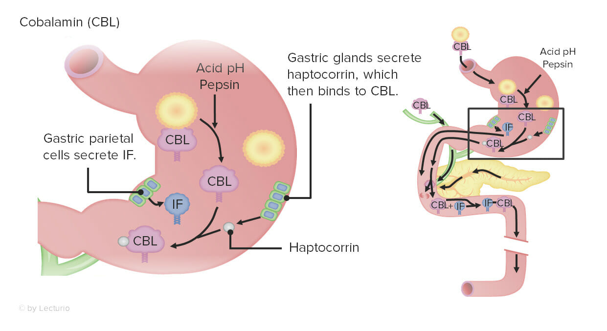
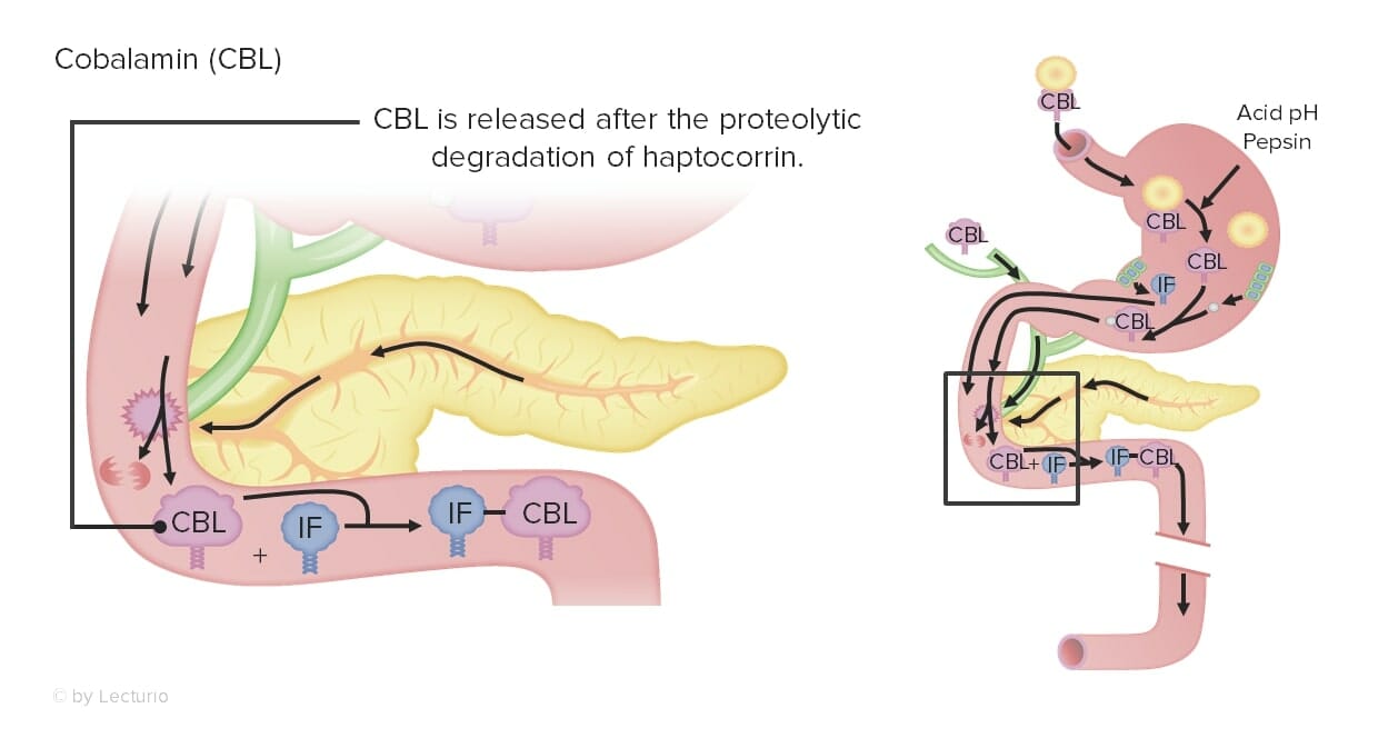
Absorption of B12:
Vitamin B12Vitamin B12A cobalt-containing coordination compound produced by intestinal microorganisms and found also in soil and water. Higher plants do not concentrate vitamin B 12 from the soil and so are a poor source of the substance as compared with animal tissues. Intrinsic factor is important for the assimilation of vitamin B 12.Folate and Vitamin B12 is bound to proteinsProteinsLinear polypeptides that are synthesized on ribosomes and may be further modified, crosslinked, cleaved, or assembled into complex proteins with several subunits. The specific sequence of amino acids determines the shape the polypeptide will take, during protein folding, and the function of the protein.Energy Homeostasis in food.
In the stomachStomachThe stomach is a muscular sac in the upper left portion of the abdomen that plays a critical role in digestion. The stomach develops from the foregut and connects the esophagus with the duodenum. Structurally, the stomach is C-shaped and forms a greater and lesser curvature and is divided grossly into regions: the cardia, fundus, body, and pylorus. Stomach: Anatomy:
Acid and pepsinPepsinPepsin breaks down proteins into proteoses, peptones, and large polypeptides.Proteins and Peptides release vitamin B12Vitamin B12A cobalt-containing coordination compound produced by intestinal microorganisms and found also in soil and water. Higher plants do not concentrate vitamin B 12 from the soil and so are a poor source of the substance as compared with animal tissues. Intrinsic factor is important for the assimilation of vitamin B 12.Folate and Vitamin B12 from dietary proteinsProteinsLinear polypeptides that are synthesized on ribosomes and may be further modified, crosslinked, cleaved, or assembled into complex proteins with several subunits. The specific sequence of amino acids determines the shape the polypeptide will take, during protein folding, and the function of the protein.Energy Homeostasis.
Vitamin B12Vitamin B12A cobalt-containing coordination compound produced by intestinal microorganisms and found also in soil and water. Higher plants do not concentrate vitamin B 12 from the soil and so are a poor source of the substance as compared with animal tissues. Intrinsic factor is important for the assimilation of vitamin B 12.Folate and Vitamin B12 binds haptocorrin.
Parietal cellsParietal cellsRounded or pyramidal cells of the gastric glands. They secrete hydrochloric acid and produce gastric intrinsic factor, a glycoprotein that binds vitamin B12.Stomach: Anatomy release intrinsic factorIntrinsic factorA glycoprotein secreted by the cells of the gastric glands that is required for the absorption of vitamin B 12 (cyanocobalamin). Deficiency of intrinsic factor leads to vitamin B12 deficiency and anemia, pernicious.Gastritis(IF).
In the duodenumDuodenumThe shortest and widest portion of the small intestine adjacent to the pylorus of the stomach. It is named for having the length equal to about the width of 12 fingers.Small Intestine: Anatomy:
The B12-IF complex is absorbed by receptor-mediated endocytosisEndocytosisCellular uptake of extracellular materials within membrane-limited vacuoles or microvesicles. Endosomes play a central role in endocytosis.The Cell: Cell Membrane in the terminal ileumIleumThe distal and narrowest portion of the small intestine, between the jejunum and the ileocecal valve of the large intestine.Small Intestine: Anatomy.
Vitamin B12Vitamin B12A cobalt-containing coordination compound produced by intestinal microorganisms and found also in soil and water. Higher plants do not concentrate vitamin B 12 from the soil and so are a poor source of the substance as compared with animal tissues. Intrinsic factor is important for the assimilation of vitamin B 12.Folate and Vitamin B12 is secreted into the blood by active transportActive transportThe movement of materials across cell membranes and epithelial layers against an electrochemical gradient, requiring the expenditure of metabolic energy.The Cell: Cell Membrane, where it binds to transcobalamin.
Clinical relevance:
↓ Vitamin CVitamin CA six carbon compound related to glucose. It is found naturally in citrus fruits and many vegetables. Ascorbic acid is an essential nutrient in human diets, and necessary to maintain connective tissue and bone. Its biologically active form, vitamin C, functions as a reducing agent and coenzyme in several metabolic pathways. Vitamin C is considered an antioxidant.Water-soluble Vitamins and their Deficiencies → scurvyScurvyAn acquired blood vessel disorder caused by severe deficiency of vitamin C (ascorbic acid) in the diet leading to defective collagen formation in small blood vessels. Scurvy is characterized by bleeding in any tissue, weakness, anemia, spongy gums, and a brawny induration of the muscles of the calves and legs.Water-soluble Vitamins and their Deficiencies
↓ Vitamin B1Vitamin B1Also known as thiamine or thiamin, it is a vitamin C12H17N4OSCl of the vitamin B complex that is essential to normal metabolism and nerve function and is widespread in plants and animalsWater-soluble Vitamins and their Deficiencies → beriberiBeriberiA disease caused by a deficiency of thiamine (vitamin B1) and characterized by polyneuritis, cardiac pathology, and edema. The epidemic form is found primarily in areas in which white (polished) rice is the staple food, as in japan, china, the philippines, india, and other countries of southeast Asia.Water-soluble Vitamins and their Deficiencies; Wernicke-Korsakoff syndromeWernicke-Korsakoff syndrome2 different syndromes that are different stages of the same disease including Wernicke’s encephalopathy and Korsakoff psychosis.Wernicke-Korsakoff syndrome is seen in patients with alcohol use disorder 8–10 times more than in the general population.Wernicke Encephalopathy and Korsakoff Syndrome
↓ Vitamin B3Vitamin B3A water-soluble vitamin of the B complex occurring in various animal and plant tissues. It is required by the body for the formation of coenzymes nad and NADP. It has pellagra-curative, vasodilating, and antilipemic properties.Water-soluble Vitamins and their Deficiencies → pellagraPellagraA disease due to deficiency of niacin, a b-complex vitamin, or its precursor tryptophan. It is characterized by scaly dermatitis which is often associated with diarrhea and dementia (the three d’s).Water-soluble Vitamins and their Deficiencies
↓ FolateFolateFolate and vitamin B12 are 2 of the most clinically important water-soluble vitamins. Deficiencies can present with megaloblastic anemia, GI symptoms, neuropsychiatric symptoms, and adverse pregnancy complications, including neural tube defects. Folate and Vitamin B12 and/or vitamin B12Vitamin B12A cobalt-containing coordination compound produced by intestinal microorganisms and found also in soil and water. Higher plants do not concentrate vitamin B 12 from the soil and so are a poor source of the substance as compared with animal tissues. Intrinsic factor is important for the assimilation of vitamin B 12.Folate and Vitamin B12 → megaloblastic anemiaMegaloblastic anemiaMegaloblastic anemia is a subset of macrocytic anemias that arises because of impaired nucleic acid synthesis in erythroid precursors. This impairment leads to ineffective RBC production and intramedullary hemolysis that is characterized by large cells with arrested nuclear maturation. The most common causes are vitamin B12 and folic acid deficiencies.Megaloblastic Anemia
↓ FolateFolateFolate and vitamin B12 are 2 of the most clinically important water-soluble vitamins. Deficiencies can present with megaloblastic anemia, GI symptoms, neuropsychiatric symptoms, and adverse pregnancy complications, including neural tube defects. Folate and Vitamin B12 in early pregnancyPregnancyThe status during which female mammals carry their developing young (embryos or fetuses) in utero before birth, beginning from fertilization to birth.Pregnancy: Diagnosis, Physiology, and Care → neural-tube defects in infants
Gastric bypassGastric bypassSurgical procedure in which the stomach is transected high on the body. The resulting small proximal gastric pouch is joined to any parts of the small intestine by an end-to-side surgical anastomosis, depending on the amounts of intestinal surface being bypasses. This procedure is used frequently in the treatment of morbid obesity by limiting the size of functional stomach, food intake, and food absorption.Gastroesophageal Reflux Disease (GERD) → may remove a majority of parietal cellsParietal cellsRounded or pyramidal cells of the gastric glands. They secrete hydrochloric acid and produce gastric intrinsic factor, a glycoprotein that binds vitamin B12.Stomach: Anatomy → ↓ IF → ↓ vitamin B12Vitamin B12A cobalt-containing coordination compound produced by intestinal microorganisms and found also in soil and water. Higher plants do not concentrate vitamin B 12 from the soil and so are a poor source of the substance as compared with animal tissues. Intrinsic factor is important for the assimilation of vitamin B 12.Folate and Vitamin B12 absorption
Surgical removal of the terminal ileumIleumThe distal and narrowest portion of the small intestine, between the jejunum and the ileocecal valve of the large intestine.Small Intestine: Anatomy → ↓ vitamin B12Vitamin B12A cobalt-containing coordination compound produced by intestinal microorganisms and found also in soil and water. Higher plants do not concentrate vitamin B 12 from the soil and so are a poor source of the substance as compared with animal tissues. Intrinsic factor is important for the assimilation of vitamin B 12.Folate and Vitamin B12 absorption
Process of CBL (vitamin B12) absorption: Dietary CBL is highly protein bound. In the stomach, acid and pepsin release CBL from these dietary proteins. Cobalamin is bound by haptocorrin; and separately, parietal cells release intrinsic factor (IF).
Image by Lecturio.
Process of CBL (vitamin B12) absorption: In the duodenum, pancreatic proteases degrade haptocorrin and the CBL is now free to bind the intrinsic factor (IF).
Image by Lecturio.
Process of CBL (vitamin B12) absorption: The IF-CBL complex is absorbed by endocytosis in the terminal ileum.
Image by Lecturio.
Clinical Relevance of Malabsorption and Maldigestion
MalabsorptionMalabsorptionGeneral term for a group of malnutrition syndromes caused by failure of normal intestinal absorption of nutrients.Malabsorption and Maldigestion involves many disorders wherein the gut cannot absorb nutrients including water and/or electrolytesElectrolytesElectrolytes are mineral salts that dissolve in water and dissociate into charged particles called ions, which can be either be positively (cations) or negatively (anions) charged. Electrolytes are distributed in the extracellular and intracellular compartments in different concentrations. Electrolytes are essential for various basic life-sustaining functions.Electrolytes from the diet. MaldigestionMaldigestionMalabsorption involves many disorders in which there is an inability of the gut to absorb nutrients from dietary intake, potentially including water and/or electrolytes. A closely related term, maldigestion is the inability to break down large molecules of food into their smaller constituents. Malabsorption and maldigestion can affect macronutrients (fats, proteins, and carbohydrates), micronutrients (vitamins and minerals), or both. Malabsorption and Maldigestion is a closely related term, which is the inability of the gut to break down large food molecules into their smaller constituents. MaldigestionMaldigestionMalabsorption involves many disorders in which there is an inability of the gut to absorb nutrients from dietary intake, potentially including water and/or electrolytes. A closely related term, maldigestion is the inability to break down large molecules of food into their smaller constituents. Malabsorption and maldigestion can affect macronutrients (fats, proteins, and carbohydrates), micronutrients (vitamins and minerals), or both. Malabsorption and Maldigestion can affect macronutrients (fatsFatsThe glyceryl esters of a fatty acid, or of a mixture of fatty acids. They are generally odorless, colorless, and tasteless if pure, but they may be flavored according to origin. Fats are insoluble in water, soluble in most organic solvents. They occur in animal and vegetable tissue and are generally obtained by boiling or by extraction under pressure. They are important in the diet (dietary fats) as a source of energy.Energy Homeostasis, proteinsProteinsLinear polypeptides that are synthesized on ribosomes and may be further modified, crosslinked, cleaved, or assembled into complex proteins with several subunits. The specific sequence of amino acids determines the shape the polypeptide will take, during protein folding, and the function of the protein.Energy Homeostasis, and carbohydratesCarbohydratesA class of organic compounds composed of carbon, hydrogen, and oxygen in a ratio of cn(H2O)n. The largest class of organic compounds, including starch; glycogen; cellulose; polysaccharides; and simple monosaccharides.Basics of Carbohydrates), micronutrients (vitamins, mineralsMineralsElectrolytes), or both. MalabsorptionMalabsorptionGeneral term for a group of malnutrition syndromes caused by failure of normal intestinal absorption of nutrients.Malabsorption and Maldigestion and maldigestionMaldigestionMalabsorption involves many disorders in which there is an inability of the gut to absorb nutrients from dietary intake, potentially including water and/or electrolytes. A closely related term, maldigestion is the inability to break down large molecules of food into their smaller constituents. Malabsorption and maldigestion can affect macronutrients (fats, proteins, and carbohydrates), micronutrients (vitamins and minerals), or both. Malabsorption and Maldigestion present with weight lossWeight lossDecrease in existing body weight.Bariatric Surgery, diarrheaDiarrheaDiarrhea is defined as ≥ 3 watery or loose stools in a 24-hour period. There are a multitude of etiologies, which can be classified based on the underlying mechanism of disease. The duration of symptoms (acute or chronic) and characteristics of the stools (e.g., watery, bloody, steatorrheic, mucoid) can help guide further diagnostic evaluation. Diarrhea, weakness, and fatigueFatigueThe state of weariness following a period of exertion, mental or physical, characterized by a decreased capacity for work and reduced efficiency to respond to stimuli.Fibromyalgia.
Etiologies of maldigestionMaldigestionMalabsorption involves many disorders in which there is an inability of the gut to absorb nutrients from dietary intake, potentially including water and/or electrolytes. A closely related term, maldigestion is the inability to break down large molecules of food into their smaller constituents. Malabsorption and maldigestion can affect macronutrients (fats, proteins, and carbohydrates), micronutrients (vitamins and minerals), or both. Malabsorption and Maldigestion:
Zollinger-Ellison syndromeZollinger-ellison syndromeA syndrome that is characterized by the triad of severe peptic ulcer, hypersecretion of gastric acid, and gastrin-producing tumors of the pancreas or other tissue (gastrinoma). This syndrome may be sporadic or be associated with multiple endocrine neoplasia type 1.Esophagitis (hypersecretion of gastric acidGastric acidHydrochloric acid present in gastric juice.Gastroesophageal Reflux Disease (GERD) resulting in low pHpHThe quantitative measurement of the acidity or basicity of a solution.Acid-Base Balance)
Diabetic gastroparesisGastroparesisChronic delayed gastric emptying. Gastroparesis may be caused by motor dysfunction or paralysis of stomach muscles or may be associated with other systemic diseases such as diabetes mellitus.Malabsorption and Maldigestion
Systemic sclerosisSclerosisA pathological process consisting of hardening or fibrosis of an anatomical structure, often a vessel or a nerve.Wilms Tumor
ThyroidThyroidThe thyroid gland is one of the largest endocrine glands in the human body. The thyroid gland is a highly vascular, brownish-red gland located in the visceral compartment of the anterior region of the neck.Thyroid Gland: Anatomy dysfunction
Biliary obstruction and/or cholestasis
BileBileAn emulsifying agent produced in the liver and secreted into the duodenum. Its composition includes bile acids and salts; cholesterol; and electrolytes. It aids digestion of fats in the duodenum.Gallbladder and Biliary Tract: Anatomy salt deficiency due to:
Hepatic cirrhosisCirrhosisCirrhosis is a late stage of hepatic parenchymal necrosis and scarring (fibrosis) most commonly due to hepatitis C infection and alcoholic liver disease. Patients may present with jaundice, ascites, and hepatosplenomegaly. Cirrhosis can also cause complications such as hepatic encephalopathy, portal hypertension, portal vein thrombosis, and hepatorenal syndrome. Cirrhosis
Primary biliary cirrhosisBiliary cirrhosisFibrosis of the hepatic parenchyma due to obstruction of bile flow (cholestasis) in the intrahepatic or extrahepatic bile ducts. Primary biliary cholangitis involves the destruction of small intrahepatic bile ducts and decreased bile secretion. Secondary biliary cholangitis is produced by prolonged obstruction of large intrahepatic or extrahepatic bile ducts from a variety of causes.Cystic Fibrosis
Pancreatic diseases:
Exocrine pancreatic insufficiencyExocrine pancreatic insufficiencyA malabsorption condition resulting from greater than 10% reduction in the secretion of pancreatic digestive enzymes (lipase; proteases; and amylase) by the exocrine pancreas into the duodenum. This condition is often associated with cystic fibrosis and with chronic pancreatitis.Malabsorption and Maldigestion
Chronic pancreatitisPancreatitisInflammation of the pancreas. Pancreatitis is classified as acute unless there are computed tomographic or endoscopic retrograde cholangiopancreatographic findings of chronic pancreatitis. The two most common forms of acute pancreatitis are alcoholic pancreatitis and gallstone pancreatitis.Acute Pancreatitis
Pancreatic cancer
DisaccharidaseDisaccharidaseEnzymes belonging to the class of glycoside hydrolases which break down disaccharides into simpler sugars, monosaccharides.Rotavirus deficiencies:
Lactase deficiencyLactase deficiencyLactose Intolerance (resulting in lactose intoleranceLactose intoleranceLactose intolerance (LI) describes a constellation of symptoms due to lactase deficiency (LD), the enzyme located in the brush border of the absorptive cells in the small intestine. Lactose is the disaccharide present in milk and requires hydrolysis by lactase to break it down into its 2 absorbable constituents, glucose and galactose. Lactose intolerance typically presents with bloating, abdominal cramping, diarrhea, and flatulence. Lactose Intolerance)
Etiologies of malabsorptionMalabsorptionGeneral term for a group of malnutrition syndromes caused by failure of normal intestinal absorption of nutrients.Malabsorption and Maldigestion:
Acutely abnormal epitheliumEpitheliumThe epithelium is a complex of specialized cellular organizations arranged into sheets and lining cavities and covering the surfaces of the body. The cells exhibit polarity, having an apical and a basal pole. Structures important for the epithelial integrity and function involve the basement membrane, the semipermeable sheet on which the cells rest, and interdigitations, as well as cellular junctions. Surface Epithelium: Histology:
Acute intestinal infectionsInfectionsInvasion of the host organism by microorganisms or their toxins or by parasites that can cause pathological conditions or diseases.Chronic Granulomatous Disease such as giardiasisGiardiasisAn infection of the small intestine caused by the flagellated protozoan giardia. It is spread via contaminated food and water and by direct person-to-person contact.Giardia/Giardiasis
Alcohol ingestion
Chronically abnormal epitheliumEpitheliumThe epithelium is a complex of specialized cellular organizations arranged into sheets and lining cavities and covering the surfaces of the body. The cells exhibit polarity, having an apical and a basal pole. Structures important for the epithelial integrity and function involve the basement membrane, the semipermeable sheet on which the cells rest, and interdigitations, as well as cellular junctions. Surface Epithelium: Histology:
Celiac diseaseCeliac diseaseCeliac disease (also known as celiac sprue or gluten enteropathy) is an autoimmune reaction to gliadin, which is a component of gluten. Celiac disease is closely associated with HLA-DQ2 and HLA-DQ8. The immune response is localized to the proximal small intestine and causes the characteristic histologic findings of villous atrophy, crypt hyperplasia, and intraepithelial lymphocytosis. Celiac Disease
Inflammatory bowel disease (IBD): Crohn disease and ulcerative colitisColitisInflammation of the colon section of the large intestine, usually with symptoms such as diarrhea (often with blood and mucus), abdominal pain, and fever.Pseudomembranous Colitis
Intestinal ischemiaIschemiaA hypoperfusion of the blood through an organ or tissue caused by a pathologic constriction or obstruction of its blood vessels, or an absence of blood circulation.Ischemic Cell Damage
RadiationRadiationEmission or propagation of acoustic waves (sound), electromagnetic energy waves (such as light; radio waves; gamma rays; or x-rays), or a stream of subatomic particles (such as electrons; neutrons; protons; or alpha particles).OsteosarcomaenteritisEnteritisInflammation of any segment of the small intestine.Lactose Intolerance
Whipple disease
Infiltrative disease: can affect both motilityMotilityThe motor activity of the gastrointestinal tract.Gastrointestinal Motility and absorptive ability
SarcoidosisSarcoidosisSarcoidosis is a multisystem inflammatory disease that causes noncaseating granulomas. The exact etiology is unknown. Sarcoidosis usually affects the lungs and thoracic lymph nodes, but it can also affect almost every system in the body, including the skin, heart, and eyes, most commonly. Sarcoidosis
AmyloidosisAmyloidosisAmyloidosis is a disease caused by abnormal extracellular tissue deposition of fibrils composed of various misfolded low-molecular-weight protein subunits. These proteins are frequently byproducts of other pathological processes (e.g., multiple myeloma). Amyloidosis
Systemic sclerosisSclerosisA pathological process consisting of hardening or fibrosis of an anatomical structure, often a vessel or a nerve.Wilms Tumor
MalabsorptionMalabsorptionGeneral term for a group of malnutrition syndromes caused by failure of normal intestinal absorption of nutrients.Malabsorption and Maldigestion of carbohydratesCarbohydratesA class of organic compounds composed of carbon, hydrogen, and oxygen in a ratio of cn(H2O)n. The largest class of organic compounds, including starch; glycogen; cellulose; polysaccharides; and simple monosaccharides.Basics of Carbohydrates:
Most commonly due to deficiency of disaccharidases (e.g., lactaseLactaseAn enzyme which catalyzes the hydrolysis of lactose to d-galactose and d-glucose. Defects in the enzyme cause lactose intolerance.Digestion and Absorption of Carbohydrates)
Colonic bacteriaBacteriaBacteria are prokaryotic single-celled microorganisms that are metabolically active and divide by binary fission. Some of these organisms play a significant role in the pathogenesis of diseases. Bacteriology ferment unabsorbed carbohydratesCarbohydratesA class of organic compounds composed of carbon, hydrogen, and oxygen in a ratio of cn(H2O)n. The largest class of organic compounds, including starch; glycogen; cellulose; polysaccharides; and simple monosaccharides.Basics of Carbohydrates into gases and fatty acidsAcidsChemical compounds which yield hydrogen ions or protons when dissolved in water, whose hydrogen can be replaced by metals or basic radicals, or which react with bases to form salts and water (neutralization). An extension of the term includes substances dissolved in media other than water.Acid-Base Balance.
Results in flatulence, bloatingBloatingConstipation, and diarrheaDiarrheaDiarrhea is defined as ≥ 3 watery or loose stools in a 24-hour period. There are a multitude of etiologies, which can be classified based on the underlying mechanism of disease. The duration of symptoms (acute or chronic) and characteristics of the stools (e.g., watery, bloody, steatorrheic, mucoid) can help guide further diagnostic evaluation. Diarrhea
MalabsorptionMalabsorptionGeneral term for a group of malnutrition syndromes caused by failure of normal intestinal absorption of nutrients.Malabsorption and Maldigestion of proteinsProteinsLinear polypeptides that are synthesized on ribosomes and may be further modified, crosslinked, cleaved, or assembled into complex proteins with several subunits. The specific sequence of amino acids determines the shape the polypeptide will take, during protein folding, and the function of the protein.Energy Homeostasis: Deficiency can result in muscle atrophyAtrophyDecrease in the size of a cell, tissue, organ, or multiple organs, associated with a variety of pathological conditions such as abnormal cellular changes, ischemia, malnutrition, or hormonal changes.Cellular Adaptation and edemaEdemaEdema is a condition in which excess serous fluid accumulates in the body cavity or interstitial space of connective tissues. Edema is a symptom observed in several medical conditions. It can be categorized into 2 types, namely, peripheral (in the extremities) and internal (in an organ or body cavity). Edema.
MalabsorptionMalabsorptionGeneral term for a group of malnutrition syndromes caused by failure of normal intestinal absorption of nutrients.Malabsorption and Maldigestion of lipidsLipidsLipids are a diverse group of hydrophobic organic molecules, which include fats, oils, sterols, and waxes.Fatty Acids and Lipids:
Deficiency in lipasesLipasesAn enzyme of the hydrolase class that catalyzes the reaction of triacylglycerol and water to yield diacylglycerol and a fatty acid anion. It is produced by glands on the tongue and by the pancreas and initiates the digestion of dietary fats.Lipid Metabolism prevents the effective breakdown of fatsFatsThe glyceryl esters of a fatty acid, or of a mixture of fatty acids. They are generally odorless, colorless, and tasteless if pure, but they may be flavored according to origin. Fats are insoluble in water, soluble in most organic solvents. They occur in animal and vegetable tissue and are generally obtained by boiling or by extraction under pressure. They are important in the diet (dietary fats) as a source of energy.Energy Homeostasis.
Most commonly due to pancreatic insufficiency
Results in:
SteatorrheaSteatorrheaA condition that is characterized by chronic fatty diarrhea, a result of abnormal digestion and/or intestinal absorption of fats.Diarrhea
Deficiencies of fat-soluble vitamins
DiarrheaDiarrheaDiarrhea is defined as ≥ 3 watery or loose stools in a 24-hour period. There are a multitude of etiologies, which can be classified based on the underlying mechanism of disease. The duration of symptoms (acute or chronic) and characteristics of the stools (e.g., watery, bloody, steatorrheic, mucoid) can help guide further diagnostic evaluation. Diarrhea (stimulation of water secretionSecretionCoagulation Studies in the colonColonThe large intestines constitute the last portion of the digestive system. The large intestine consists of the cecum, appendix, colon (with ascending, transverse, descending, and sigmoid segments), rectum, and anal canal. The primary function of the colon is to remove water and compact the stool prior to expulsion from the body via the rectum and anal canal. Colon, Cecum, and Appendix: Anatomy by unabsorbed bileBileAn emulsifying agent produced in the liver and secreted into the duodenum. Its composition includes bile acids and salts; cholesterol; and electrolytes. It aids digestion of fats in the duodenum.Gallbladder and Biliary Tract: Anatomy salts)
References
Boland, M. (2016). Human digestion–a processing perspective. J Sci Food Agric. 96(7), 2275-2283. doi: 10.1002/jsfa.7601. Epub 2016 Feb 5. PMID: 26711173.