Achieve Mastery of Medical Concepts

Study for medical school and boards with Lecturio

Cardiomyopathy: Overview and Types

Cardiomyopathy refers to a group of myocardial diseases associated with structural changes of the heart muscles ( myocardium Myocardium The muscle tissue of the heart. It is composed of striated, involuntary muscle cells connected to form the contractile pump to generate blood flow. Heart: Anatomy) and impaired systolic and/or diastolic function in the absence of other heart disorders ( coronary artery Coronary Artery Truncus Arteriosus disease, hypertension Hypertension Hypertension, or high blood pressure, is a common disease that manifests as elevated systemic arterial pressures. Hypertension is most often asymptomatic and is found incidentally as part of a routine physical examination or during triage for an unrelated medical encounter. Hypertension, valvular disease, and congenital Congenital Chorioretinitis heart disease). The list of causes is extensive, ranging from familial disorders to underlying diseases and infections Infections Invasion of the host organism by microorganisms or their toxins or by parasites that can cause pathological conditions or diseases. Chronic Granulomatous Disease. Patients Patients Individuals participating in the health care system for the purpose of receiving therapeutic, diagnostic, or preventive procedures. Clinician–Patient Relationship often present with chest pain Pain An unpleasant sensation induced by noxious stimuli which are detected by nerve endings of nociceptive neurons. Pain: Types and Pathways, dyspnea Dyspnea Dyspnea is the subjective sensation of breathing discomfort. Dyspnea is a normal manifestation of heavy physical or psychological exertion, but also may be caused by underlying conditions (both pulmonary and extrapulmonary). Dyspnea, palpitations Palpitations Ebstein’s Anomaly, and/or syncope Syncope Syncope is a short-term loss of consciousness and loss of postural stability followed by spontaneous return of consciousness to the previous neurologic baseline without the need for resuscitation. The condition is caused by transient interruption of cerebral blood flow that may be benign or related to a underlying life-threatening condition. Syncope. Some patients Patients Individuals participating in the health care system for the purpose of receiving therapeutic, diagnostic, or preventive procedures. Clinician–Patient Relationship may be completely asymptomatic, while others may present with sudden cardiac death Sudden cardiac death Cardiac arrest is the sudden, complete cessation of cardiac output with hemodynamic collapse. Patients present as pulseless, unresponsive, and apneic. Rhythms associated with cardiac arrest are ventricular fibrillation/tachycardia, asystole, or pulseless electrical activity. Cardiac Arrest as the first sign of an underlying condition. Diagnosis is made through the use of ECG ECG An electrocardiogram (ECG) is a graphic representation of the electrical activity of the heart plotted against time. Adhesive electrodes are affixed to the skin surface allowing measurement of cardiac impulses from many angles. The ECG provides 3-dimensional information about the conduction system of the heart, the myocardium, and other cardiac structures. Electrocardiogram (ECG) and cardiac imaging such as echocardiography Echocardiography Ultrasonic recording of the size, motion, and composition of the heart and surrounding tissues. The standard approach is transthoracic. Tricuspid Valve Atresia (TVA) and cardiac MRI Cardiac MRI Imaging of the Heart and Great Vessels. Management involves medications typically used to treat heart failure Heart Failure A heterogeneous condition in which the heart is unable to pump out sufficient blood to meet the metabolic need of the body. Heart failure can be caused by structural defects, functional abnormalities (ventricular dysfunction), or a sudden overload beyond its capacity. Chronic heart failure is more common than acute heart failure which results from sudden insult to cardiac function, such as myocardial infarction. Total Anomalous Pulmonary Venous Return (TAPVR), as well as implantable devices. In severe cases, heart transplantation Heart Transplantation The transference of a heart from one human or animal to another. Organ Transplantation may be necessary.

Last updated: Mar 29, 2023

Editorial responsibility: Stanley Oiseth, Lindsay Jones, Evelin Maza

Overview

Definition

Cardiomyopathies are diseases that affect the structure and function of the heart muscle ( myocardium Myocardium The muscle tissue of the heart. It is composed of striated, involuntary muscle cells connected to form the contractile pump to generate blood flow. Heart: Anatomy) in the absence of secondary causes (e.g., coronary artery Coronary Artery Truncus Arteriosus disease, hypertension Hypertension Hypertension, or high blood pressure, is a common disease that manifests as elevated systemic arterial pressures. Hypertension is most often asymptomatic and is found incidentally as part of a routine physical examination or during triage for an unrelated medical encounter. Hypertension, valvular disease, and congenital Congenital Chorioretinitis heart disease).

Epidemiology

  • Incidence Incidence The number of new cases of a given disease during a given period in a specified population. It also is used for the rate at which new events occur in a defined population. It is differentiated from prevalence, which refers to all cases in the population at a given time. Measures of Disease Frequency and prevalence Prevalence The total number of cases of a given disease in a specified population at a designated time. It is differentiated from incidence, which refers to the number of new cases in the population at a given time. Measures of Disease Frequency difficult to estimate due to inconsistent reporting
  • Second most common cause of sudden death after ischemic heart disease Ischemic heart disease Coronary heart disease (CHD), or ischemic heart disease, describes a situation in which an inadequate supply of blood to the myocardium exists due to a stenosis of the coronary arteries, typically from atherosclerosis. Coronary Heart Disease
  • Both sporadic Sporadic Selective IgA Deficiency and familial forms exist.
  • No clear gender Gender Gender Dysphoria or racial predominance

Etiology

Causes vary:

  • Infectious diseases
  • Inflammatory/autoimmune conditions
  • Endocrinologic disorders
  • Toxins
  • Medications
  • Nutritional deficiencies
  • Deposition diseases
  • Familial storage diseases
  • Genetic neurologic and neuromuscular disorders
  • Hematologic/oncologic disorders
  • Cardiovascular disorders
  • Endomyocardial diseases
  • Genetic ( gene Gene A category of nucleic acid sequences that function as units of heredity and which code for the basic instructions for the development, reproduction, and maintenance of organisms. Basic Terms of Genetics mutations)

Approximately ⅓ of cases have a genetic cause.

Classification

By morphology:

  • Dilated cardiomyopathy Dilated Cardiomyopathy Dilated cardiomyopathy (DCM) is the most common type of non-ischemic cardiomyopathy and a common cause of heart failure (HF). The cause may be idiopathic, familial, or secondary to a variety of underlying conditions. The disease is characterized by the enlargement of 1 or both ventricles and reduced systolic function. Dilated Cardiomyopathy ( DCM DCM Dilated cardiomyopathy (DCM) is the most common type of non-ischemic cardiomyopathy and a common cause of heart failure (HF). The cause may be idiopathic, familial, or secondary to a variety of underlying conditions. The disease is characterized by the enlargement of 1 or both ventricles and reduced systolic function. Dilated Cardiomyopathy):
    • Heart appears enlarged.
    • Muscle is thinned.
  • Restrictive cardiomyopathy Restrictive Cardiomyopathy Restrictive cardiomyopathy (RCM) is a fairly uncommon condition characterized by progressive stiffening of the cardiac muscle, which causes impaired relaxation and refilling of the heart during diastole, resulting in diastolic dysfunction and eventual heart failure. Restrictive Cardiomyopathy ( RCM RCM Restrictive cardiomyopathy (RCM) is a fairly uncommon condition characterized by progressive stiffening of the cardiac muscle, which causes impaired relaxation and refilling of the heart during diastole, resulting in diastolic dysfunction and eventual heart failure. Restrictive Cardiomyopathy):
    • Heart is normal size (or smaller).
    • Muscle is stiffened.
  • Hypertrophic cardiomyopathy Hypertrophic Cardiomyopathy Hypertrophic cardiomyopathy (HCM) is the most commonly inherited cardiomyopathy, which is characterized by an asymmetric increase in thickness (hypertrophy) of the left ventricular wall, diastolic dysfunction, and often left ventricular outflow tract obstruction. Hypertrophic Cardiomyopathy ( HCM HCM Hypertrophic cardiomyopathy (HCM) is the most commonly inherited cardiomyopathy, which is characterized by an asymmetric increase in thickness (hypertrophy) of the left ventricular wall, diastolic dysfunction, and often left ventricular outflow tract obstruction. Hypertrophic Cardiomyopathy)
    • Heart is enlarged.
    • Muscle is thickened.
  • Arrhythmogenic right ventricular cardiomyopathy Arrhythmogenic Right Ventricular Cardiomyopathy Arrhythmogenic right ventricular cardiomyopathy is an inherited disorder of the heart muscle that affects the right ventricle (RV); it can cause rhythm disturbances and sudden cardiac death (SCD). The disorder results from mutations in the genes that encode desmosomal proteins involved in cell-to-cell adhesion. Arrhythmogenic Right Ventricular Cardiomyopathy/dysplasia
  • Unclassified cardiomyopathy

By etiology:

  • Primary: arises from an exclusively cardiac issue
    • Genetic
    • Mixed (nongenetic/genetic)
    • Acquired
  • Secondary: caused by or associated with a known underlying systemic condition
    • Disorders of connective tissue Connective tissue Connective tissues originate from embryonic mesenchyme and are present throughout the body except inside the brain and spinal cord. The main function of connective tissues is to provide structural support to organs. Connective tissues consist of cells and an extracellular matrix. Connective Tissue: Histology 
    • Muscular dystrophies
    • Autoimmune disease
    • Infections Infections Invasion of the host organism by microorganisms or their toxins or by parasites that can cause pathological conditions or diseases. Chronic Granulomatous Disease of the heart muscle
    • Infiltrative diseases ( hemochromatosis Hemochromatosis A disorder of iron metabolism characterized by a triad of hemosiderosis; liver cirrhosis; and diabetes mellitus. It is caused by massive iron deposits in parenchymal cells that may develop after a prolonged increase of iron absorption. Hereditary Hemochromatosis, sarcoidosis Sarcoidosis Sarcoidosis is a multisystem inflammatory disease that causes noncaseating granulomas. The exact etiology is unknown. Sarcoidosis usually affects the lungs and thoracic lymph nodes, but it can also affect almost every system in the body, including the skin, heart, and eyes, most commonly. Sarcoidosis, or amyloidosis Amyloidosis Amyloidosis is a disease caused by abnormal extracellular tissue deposition of fibrils composed of various misfolded low-molecular-weight protein subunits. These proteins are frequently byproducts of other pathological processes (e.g., multiple myeloma). Amyloidosis)
    • Endocrine disorders ( thyroid Thyroid The thyroid gland is one of the largest endocrine glands in the human body. The thyroid gland is a highly vascular, brownish-red gland located in the visceral compartment of the anterior region of the neck. Thyroid Gland: Anatomy conditions, diabetes Diabetes Diabetes mellitus (DM) is a metabolic disease characterized by hyperglycemia and dysfunction of the regulation of glucose metabolism by insulin. Type 1 DM is diagnosed mostly in children and young adults as the result of autoimmune destruction of β cells in the pancreas and the resulting lack of insulin. Type 2 DM has a significant association with obesity and is characterized by insulin resistance. Diabetes Mellitus)
    • Chronic substance use disorder (especially alcohol or cocaine Cocaine An alkaloid ester extracted from the leaves of plants including coca. It is a local anesthetic and vasoconstrictor and is clinically used for that purpose, particularly in the eye, ear, nose, and throat. It also has powerful central nervous system effects similar to the amphetamines and is a drug of abuse. Cocaine, like amphetamines, acts by multiple mechanisms on brain catecholaminergic neurons; the mechanism of its reinforcing effects is thought to involve inhibition of dopamine uptake. Local Anesthetics)
    • Pregnancy Pregnancy The status during which female mammals carry their developing young (embryos or fetuses) in utero before birth, beginning from fertilization to birth. Pregnancy: Diagnosis, Physiology, and Care
Overview of cardiomyopathies

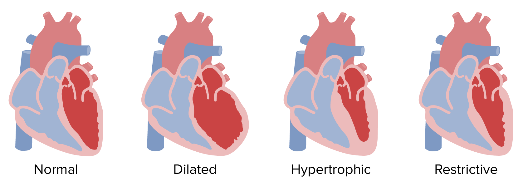
Morphological classification of cardiomyopathies

Image by Lecturio.

Pathophysiology

  • Myocardium Myocardium The muscle tissue of the heart. It is composed of striated, involuntary muscle cells connected to form the contractile pump to generate blood flow. Heart: Anatomy: thickest layer of the heart
    • Specialized form of striated muscle Striated muscle One of two types of muscle in the body, characterized by the array of bands observed under microscope. Striated muscles can be divided into two subtypes: the cardiac muscle and the skeletal muscle. Muscle Tissue: Histology
    • Composed of cardiomyocytes connected by intercalated discs Intercalated discs Irregular, transverse, thick parts of the sarcolemma at the terminal ends of the cell branches. Muscle Tissue: Histology
    • Structure, size, and arrangement of muscle fibers and heart chambers optimized for pumping force
  • End effect of cardiomyopathies: structural change in heart muscle
    • May cause hypertrophy Hypertrophy General increase in bulk of a part or organ due to cell enlargement and accumulation of fluids and secretions, not due to tumor formation, nor to an increase in the number of cells (hyperplasia). Cellular Adaptation or dilation of one or both of the heart ventricles Heart ventricles The lower right and left chambers of the heart. The right ventricle pumps venous blood into the lungs and the left ventricle pumps oxygenated blood into the systemic arterial circulation. Heart: Anatomy
    • Changes shape and capacity of ventricles
  • Cause systolic or diastolic dysfunction Diastolic dysfunction Restrictive Cardiomyopathy, depending on the disease process
  • Net end effect is inability to pump Pump ACES and RUSH: Resuscitation Ultrasound Protocols blood efficiently:
    • Heart failure Heart Failure A heterogeneous condition in which the heart is unable to pump out sufficient blood to meet the metabolic need of the body. Heart failure can be caused by structural defects, functional abnormalities (ventricular dysfunction), or a sudden overload beyond its capacity. Chronic heart failure is more common than acute heart failure which results from sudden insult to cardiac function, such as myocardial infarction. Total Anomalous Pulmonary Venous Return (TAPVR)
    • Volume overload

Diagnosis

Diagnostic approach varies based on the type of cardiomyopathy. Physicians Physicians Individuals licensed to practice medicine. Clinician–Patient Relationship must have a high index of suspicion for disorders of the myocardium Myocardium The muscle tissue of the heart. It is composed of striated, involuntary muscle cells connected to form the contractile pump to generate blood flow. Heart: Anatomy in patients Patients Individuals participating in the health care system for the purpose of receiving therapeutic, diagnostic, or preventive procedures. Clinician–Patient Relationship with heart failure Heart Failure A heterogeneous condition in which the heart is unable to pump out sufficient blood to meet the metabolic need of the body. Heart failure can be caused by structural defects, functional abnormalities (ventricular dysfunction), or a sudden overload beyond its capacity. Chronic heart failure is more common than acute heart failure which results from sudden insult to cardiac function, such as myocardial infarction. Total Anomalous Pulmonary Venous Return (TAPVR).

  • Echocardiography Echocardiography Ultrasonic recording of the size, motion, and composition of the heart and surrounding tissues. The standard approach is transthoracic. Tricuspid Valve Atresia (TVA) with Doppler Doppler Ultrasonography applying the doppler effect, with frequency-shifted ultrasound reflections produced by moving targets (usually red blood cells) in the bloodstream along the ultrasound axis in direct proportion to the velocity of movement of the targets, to determine both direction and velocity of blood flow. Ultrasound (Sonography):
    • Used in evaluation of all cases
    • Assesses cardiac size, anatomy, structure, and function 
  • Cardiac MRI Cardiac MRI Imaging of the Heart and Great Vessels:
    • Advanced imaging technique
    • Allows greater characterization of cardiac structure and function 
    • Used to determine extent of myocardial damage, infiltration, and fibrosis Fibrosis Any pathological condition where fibrous connective tissue invades any organ, usually as a consequence of inflammation or other injury. Bronchiolitis Obliterans
  • ECG ECG An electrocardiogram (ECG) is a graphic representation of the electrical activity of the heart plotted against time. Adhesive electrodes are affixed to the skin surface allowing measurement of cardiac impulses from many angles. The ECG provides 3-dimensional information about the conduction system of the heart, the myocardium, and other cardiac structures. Electrocardiogram (ECG):
    • Low sensitivity for diagnosis of cardiomyopathies
    • Used to identify abnormal heart rhythms, such as associated arrhythmias or conduction delays

Management

Goals:

  • Optimize cardiac output Cardiac output The volume of blood passing through the heart per unit of time. It is usually expressed as liters (volume) per minute so as not to be confused with stroke volume (volume per beat). Cardiac Mechanics:
  • Treat or slow underlying disease progression.
  • Manage symptoms of heart failure Heart Failure A heterogeneous condition in which the heart is unable to pump out sufficient blood to meet the metabolic need of the body. Heart failure can be caused by structural defects, functional abnormalities (ventricular dysfunction), or a sudden overload beyond its capacity. Chronic heart failure is more common than acute heart failure which results from sudden insult to cardiac function, such as myocardial infarction. Total Anomalous Pulmonary Venous Return (TAPVR):
    • Volume overload
    • Blood pressure
  • Manage symptoms caused by distortion Distortion Defense Mechanisms of heart muscle:
    • Arrhythmias: medication, pacemaker Pacemaker A device designed to stimulate, by electric impulses, contraction of the heart muscles. It may be temporary (external) or permanent (internal or internal-external). Bradyarrhythmias, or internal cardioverter– defibrillator Defibrillator Cardiac electrical stimulators that apply brief high-voltage electroshocks to the heart. These stimulators are used to restore normal rhythm and contractile function in hearts of patients who are experiencing ventricular fibrillation or ventricular tachycardia that is not accompanied by a palpable pulse. Some defibrillators may also be used to correct certain noncritical dysrhythmias (called synchronized defibrillation or cardioversion), using relatively low-level discharges synchronized to the patient’s ECG waveform. Cardiac Arrest (ICD) 
    • Blood clots: anticoagulants Anticoagulants Anticoagulants are drugs that retard or interrupt the coagulation cascade. The primary classes of available anticoagulants include heparins, vitamin K-dependent antagonists (e.g., warfarin), direct thrombin inhibitors, and factor Xa inhibitors. Anticoagulants

In cases refractory to treatment, heart transplantation Heart Transplantation The transference of a heart from one human or animal to another. Organ Transplantation may be indicated.

Complications:

  • Heart failure Heart Failure A heterogeneous condition in which the heart is unable to pump out sufficient blood to meet the metabolic need of the body. Heart failure can be caused by structural defects, functional abnormalities (ventricular dysfunction), or a sudden overload beyond its capacity. Chronic heart failure is more common than acute heart failure which results from sudden insult to cardiac function, such as myocardial infarction. Total Anomalous Pulmonary Venous Return (TAPVR)
  • Arrhythmias
  • Sudden cardiac death Sudden cardiac death Cardiac arrest is the sudden, complete cessation of cardiac output with hemodynamic collapse. Patients present as pulseless, unresponsive, and apneic. Rhythms associated with cardiac arrest are ventricular fibrillation/tachycardia, asystole, or pulseless electrical activity. Cardiac Arrest

Comparison of the Cardiomyopathies

Dilated Restrictive Hypertrophic Arrhythmogenic right ventricular CM
Etiology
  • Idiopathic Idiopathic Dermatomyositis
  • Genetic
  • Alcohol use disorder Alcohol use disorder Alcohol is one of the most commonly used addictive substances in the world. Alcohol use disorder (AUD) is defined as pathologic consumption of alcohol leading to impaired daily functioning. Acute alcohol intoxication presents with impairment in speech and motor functions and can be managed in most cases with supportive care. Alcohol Use Disorder
  • Postpartum
  • Medication
  • Amyloidosis Amyloidosis Amyloidosis is a disease caused by abnormal extracellular tissue deposition of fibrils composed of various misfolded low-molecular-weight protein subunits. These proteins are frequently byproducts of other pathological processes (e.g., multiple myeloma). Amyloidosis
  • Sarcoidosis Sarcoidosis Sarcoidosis is a multisystem inflammatory disease that causes noncaseating granulomas. The exact etiology is unknown. Sarcoidosis usually affects the lungs and thoracic lymph nodes, but it can also affect almost every system in the body, including the skin, heart, and eyes, most commonly. Sarcoidosis
  • Hemosiderosis Hemosiderosis Conditions in which there is a generalized increase in the iron stores of body tissues, particularly of liver and the mononuclear phagocyte system, without demonstrable tissue damage. The name refers to the presence of stainable iron in the tissue in the form of hemosiderin. Cellular Accumulations
  • Post radiation Radiation Emission or propagation of acoustic waves (sound), electromagnetic energy waves (such as light; radio waves; gamma rays; or x-rays), or a stream of subatomic particles (such as electrons; neutrons; protons; or alpha particles). Osteosarcoma therapy
  • Postsurgical
  • Diabetes Diabetes Diabetes mellitus (DM) is a metabolic disease characterized by hyperglycemia and dysfunction of the regulation of glucose metabolism by insulin. Type 1 DM is diagnosed mostly in children and young adults as the result of autoimmune destruction of β cells in the pancreas and the resulting lack of insulin. Type 2 DM has a significant association with obesity and is characterized by insulin resistance. Diabetes Mellitus
  • Genetic
  • Chronic hypertension Hypertension Hypertension, or high blood pressure, is a common disease that manifests as elevated systemic arterial pressures. Hypertension is most often asymptomatic and is found incidentally as part of a routine physical examination or during triage for an unrelated medical encounter. Hypertension
Clinical presentation
  • Fatigue Fatigue The state of weariness following a period of exertion, mental or physical, characterized by a decreased capacity for work and reduced efficiency to respond to stimuli. Fibromyalgia
  • Dyspnea Dyspnea Dyspnea is the subjective sensation of breathing discomfort. Dyspnea is a normal manifestation of heavy physical or psychological exertion, but also may be caused by underlying conditions (both pulmonary and extrapulmonary). Dyspnea
  • Dizziness Dizziness An imprecise term which may refer to a sense of spatial disorientation, motion of the environment, or lightheadedness. Lateral Medullary Syndrome (Wallenberg Syndrome)
  • Exercise intolerance
  • Dyspnea Dyspnea Dyspnea is the subjective sensation of breathing discomfort. Dyspnea is a normal manifestation of heavy physical or psychological exertion, but also may be caused by underlying conditions (both pulmonary and extrapulmonary). Dyspnea
  • Exercise intolerance
  • Lower-extremity edema Edema Edema is a condition in which excess serous fluid accumulates in the body cavity or interstitial space of connective tissues. Edema is a symptom observed in several medical conditions. It can be categorized into 2 types, namely, peripheral (in the extremities) and internal (in an organ or body cavity). Edema
  • Weight gain
  • Cough
  • Fatigue Fatigue The state of weariness following a period of exertion, mental or physical, characterized by a decreased capacity for work and reduced efficiency to respond to stimuli. Fibromyalgia
  • Asymptomatic
  • First symptom may be sudden cardiac death Sudden cardiac death Cardiac arrest is the sudden, complete cessation of cardiac output with hemodynamic collapse. Patients present as pulseless, unresponsive, and apneic. Rhythms associated with cardiac arrest are ventricular fibrillation/tachycardia, asystole, or pulseless electrical activity. Cardiac Arrest.
  • Syncope Syncope Syncope is a short-term loss of consciousness and loss of postural stability followed by spontaneous return of consciousness to the previous neurologic baseline without the need for resuscitation. The condition is caused by transient interruption of cerebral blood flow that may be benign or related to a underlying life-threatening condition. Syncope
  • Exercise intolerance
  • Palpitations Palpitations Ebstein’s Anomaly
  • Dyspnea Dyspnea Dyspnea is the subjective sensation of breathing discomfort. Dyspnea is a normal manifestation of heavy physical or psychological exertion, but also may be caused by underlying conditions (both pulmonary and extrapulmonary). Dyspnea
  • Sudden cardiac death Sudden cardiac death Cardiac arrest is the sudden, complete cessation of cardiac output with hemodynamic collapse. Patients present as pulseless, unresponsive, and apneic. Rhythms associated with cardiac arrest are ventricular fibrillation/tachycardia, asystole, or pulseless electrical activity. Cardiac Arrest
  • Syncope Syncope Syncope is a short-term loss of consciousness and loss of postural stability followed by spontaneous return of consciousness to the previous neurologic baseline without the need for resuscitation. The condition is caused by transient interruption of cerebral blood flow that may be benign or related to a underlying life-threatening condition. Syncope
  • Palpitations Palpitations Ebstein’s Anomaly
  • Dyspnea Dyspnea Dyspnea is the subjective sensation of breathing discomfort. Dyspnea is a normal manifestation of heavy physical or psychological exertion, but also may be caused by underlying conditions (both pulmonary and extrapulmonary). Dyspnea
  • Chest pain Pain An unpleasant sensation induced by noxious stimuli which are detected by nerve endings of nociceptive neurons. Pain: Types and Pathways
Physical exam findings
  • S3 gallop S3 gallop Heart Failure
  • Systolic regurgitant murmur
  • Jugular venous distention
  • Ascites Ascites Ascites is the pathologic accumulation of fluid within the peritoneal cavity that occurs due to an osmotic and/or hydrostatic pressure imbalance secondary to portal hypertension (cirrhosis, heart failure) or non-portal hypertension (hypoalbuminemia, malignancy, infection). Ascites
  • Often normal
  • S3 S3 Heart Sounds or S4 gallop S4 gallop Heart Sounds
  • Wide split S2 S2 Heart Sounds sound may be present.
  • Peripheral edema Peripheral edema Peripheral edema is the swelling of the lower extremities, namely, legs, feet, and ankles. Edema
  • Tachycardia Tachycardia Abnormally rapid heartbeat, usually with a heart rate above 100 beats per minute for adults. Tachycardia accompanied by disturbance in the cardiac depolarization (cardiac arrhythmia) is called tachyarrhythmia. Sepsis in Children
Ejection fraction Ejection fraction Cardiac Cycle ↓ or normal ↑ or normal
L ventricular diastolic dimension Normal
Left ventricular wall thickness Normal or ↑ ↑↑ Normal
Atrial size Normal
Diagnosis
  • ECG ECG An electrocardiogram (ECG) is a graphic representation of the electrical activity of the heart plotted against time. Adhesive electrodes are affixed to the skin surface allowing measurement of cardiac impulses from many angles. The ECG provides 3-dimensional information about the conduction system of the heart, the myocardium, and other cardiac structures. Electrocardiogram (ECG)
  • Echocardiography Echocardiography Ultrasonic recording of the size, motion, and composition of the heart and surrounding tissues. The standard approach is transthoracic. Tricuspid Valve Atresia (TVA)
  • Cardiac MRI Cardiac MRI Imaging of the Heart and Great Vessels
CM: cardiomyopathy

References

  1. Dilated cardiomyopathy. (2021). Medline Plus. Retrieved February 3, 2021, from https://medlineplus.gov/ency/article/000168.htm
  2. Deng F, Weerakkody Y. (2021). Arrhythmogenic right ventricular cardiomyopathy. Radiopaedia. Retrieved February 13, 2021, from  https://radiopaedia.org/articles/arrhythmogenic-right-ventricular-cardiomyopathy
  3. Rossi PA. (1989). Arrhythmogenic right ventricular dysplasia—clinical features. Eur Heart J 10(suppl D):7–9.
  4. Schaufelberger M. (2019). Cardiomyopathy and pregnancy. Heart 105:1543–1551. Retrieved February 3, 2021, from https://heart.bmj.com/content/105/20/1543.info
  5. Angelini A, Calzolari V, Thiene G, et al. (1997). Morphologic spectrum of primary restrictive cardiomyopathy. Am J Cardiol https://pubmed.ncbi.nlm.nih.gov/9352976/ 
  6. Fitzpatrick AP, Shapiro LM, Rickards AF, Poole-Wilson PA. (1990). Familial restrictive cardiomyopathy with atrioventricular block and skeletal myopathy. Br Heart J https://pubmed.ncbi.nlm.nih.gov/2317404/ 
  7. Kushwaha SS, Fallon JT, Fuster V. (1997). Restrictive cardiomyopathy. N Engl J Med. https://pubmed.ncbi.nlm.nih.gov/8995091/ 
  8. Benotti JR, Grossman W, Cohn PF. (1980). Clinical profile of restrictive cardiomyopathy. Circulation. https://pubmed.ncbi.nlm.nih.gov/6445242/ 
  9. Ammash NM, Seward JB, Bailey KR, Edwards WD, Tajik AJ. (2000). Clinical profile and outcome of idiopathic restrictive cardiomyopathy. Circulation. https://pubmed.ncbi.nlm.nih.gov/10831523/ 
  10. Veselka J, Anavekar NS, Charron P. (2017). Hypertrophic obstructive cardiomyopathy. Lancet. https://pubmed.ncbi.nlm.nih.gov/27912983/ 
  11. Wigle ED, Rakowski H, Kimball BP, Williams WG. (1995). Hypertrophic cardiomyopathy. Clinical spectrum and treatment. Circulation. https://pubmed.ncbi.nlm.nih.gov/7671349/ 
  12. Elliott PM, Kaski JC, Prasad K, et al. (1996). Chest pain during daily life in patients with hypertrophic cardiomyopathy: an ambulatory electrocardiographic study. Eur Heart J. https://pubmed.ncbi.nlm.nih.gov/8809524/ 
  13. Anderson EL. (2006). Arrhythmogenic right ventricular dysplasia. Am Fam Physician. https://pubmed.ncbi.nlm.nih.gov/16669561/

Create your free account or log in to continue reading!

Sign up now and get free access to Lecturio with concept pages, medical videos, and questions for your medical education.

User Reviews

Details