Treponema is a gram-negative, microaerophilic Microaerophilic Helicobacter spirochete. Owing to its very thin structure, it is not easily seen on Gram stain Gram stain Klebsiella, but can be visualized using dark-field microscopy. This spirochete contains endoflagella, which allow for a characteristic corkscrew movement. The bacteria Bacteria Bacteria are prokaryotic single-celled microorganisms that are metabolically active and divide by binary fission. Some of these organisms play a significant role in the pathogenesis of diseases. Bacteriology are able to avoid immune recognition Immune Recognition Yaws, Bejel, and Pinta and phagocytosis Phagocytosis The engulfing and degradation of microorganisms; other cells that are dead, dying, or pathogenic; and foreign particles by phagocytic cells (phagocytes). Innate Immunity: Phagocytes and Antigen Presentation by forming a protective coating with fibronectin Fibronectin Glycoproteins found on the surfaces of cells, particularly in fibrillar structures. The proteins are lost or reduced when these cells undergo viral or chemical transformation. They are highly susceptible to proteolysis and are substrates for activated blood coagulation factor VIII. The forms present in plasma are called cold-insoluble globulins. Connective Tissue: Histology. Humans are the only reservoir Reservoir Animate or inanimate sources which normally harbor disease-causing organisms and thus serve as potential sources of disease outbreaks. Reservoirs are distinguished from vectors (disease vectors) and carriers, which are agents of disease transmission rather than continuing sources of potential disease outbreaks. Humans may serve both as disease reservoirs and carriers. Escherichia coli and transmission is through human-to-human contact. The most common species involved in human disease is Treponema pallidum subspecies pallidum, which is the causative agent of syphilis Syphilis Syphilis is a bacterial infection caused by the spirochete Treponema pallidum pallidum (T. p. pallidum), which is usually spread through sexual contact. Syphilis has 4 clinical stages: primary, secondary, latent, and tertiary. Syphilis. Other clinically relevant species include T. pallidum pertenue, T. pallidum endemicum, and T. carateum. These are the causative organisms for yaws Yaws A systemic non-venereal infection of the tropics caused by treponema pallidum subspecies pertenue. Yaws, Bejel, and Pinta, bejel Bejel Yaws, bejel, and pinta are endemic, nonvenereal treponematoses. The causative organisms are Treponema pallidum pertenue (yaws), T. pallidum endemicum (bejel), and T. carateum (pinta). These treponematoses are generally transmitted by direct skin-to-skin contact with infected skin lesions. Yaws, Bejel, and Pinta, and pinta Pinta An infectious disease of the skin caused by treponema carateum that occurs only in the Western hemisphere. Age of onset is between 10 and 20 years of age. This condition is characterized by marked changes in the skin color and is believed to be transmitted by direct person-to-person contact. Yaws, Bejel, and Pinta, respectively.
Last updated: Dec 15, 2025

Gram-negative bacteria:
Most bacteria can be classified according to a lab procedure called Gram staining.
Bacteria with cell walls that have a thin layer of peptidoglycan do not retain the crystal violet stain utilized in Gram staining. These bacteria do, however, retain the safranin counterstain and thus appear as pinkish-red on the stain, making them gram negative. These bacteria can be further classified according to morphology (diplococci, curved rods, bacilli, and coccobacilli) and their ability to grow in the presence of oxygen (aerobic versus anaerobic). The bacteria can be more narrowly identified by growing them on specific media (triple sugar iron (TSI) agar) where their enzymes can be identified (urease, oxidase) and their ability to ferment lactose can be tested.
* Stains poorly on Gram stain
** Pleomorphic rod/coccobacillus
*** Require special transport media
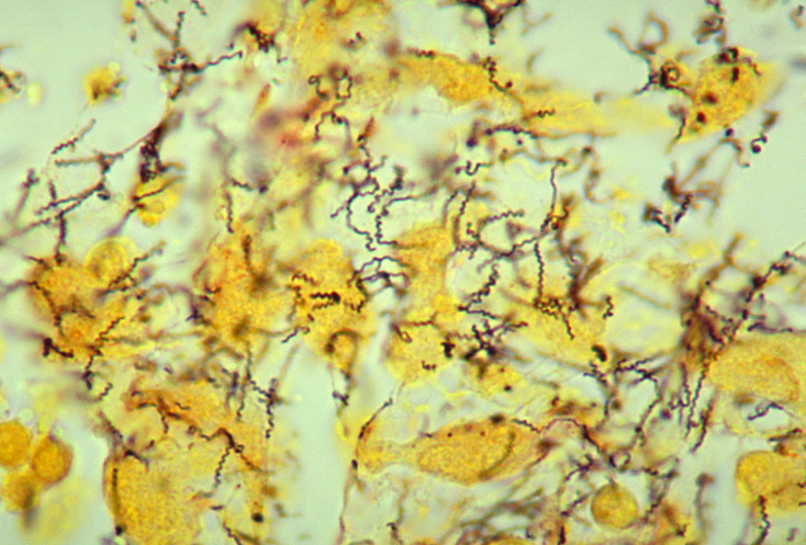
Treponema pallidum spirochetes in a sample using a modified Steiner silver stain
Image: “Treponema pallidum 01” by Dr. Edwin P. Ewing, Jr. License: Public Domain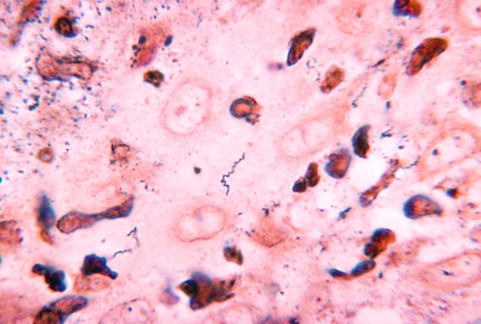
A tissue sample showing numerous thin, spiral-shaped Treponema carateum.
Image: “2900” by CDC. License: Public Domain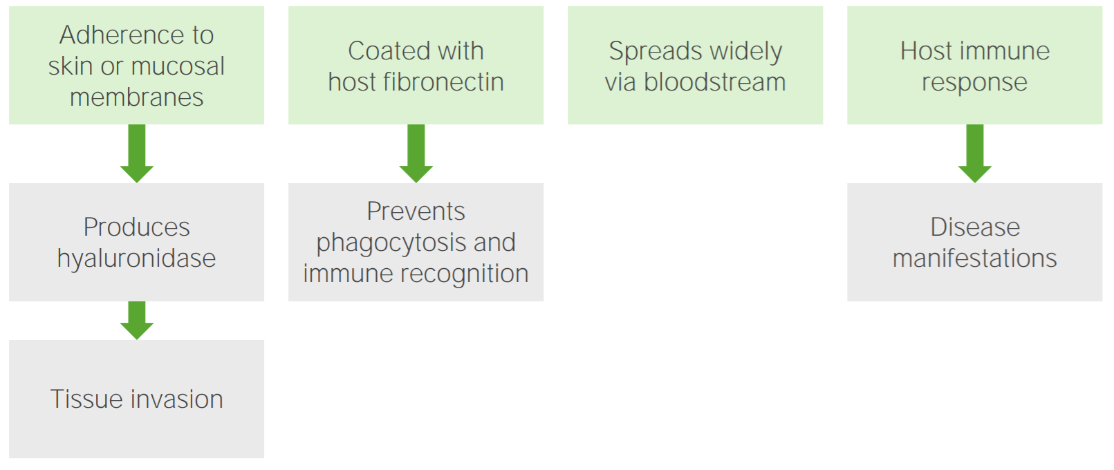
Pathogenesis of Treponema infections
The pathogen adheres to the skin or mucosal membranes, leading to the production of hyaluronidase which allows for tissue invasion. The pathogen coats itself in the host’s fibronectin, which prevents its phagocytosis and recognition by the immune system. Consequently, the pathogen disseminates through the bloodstream. A host immune response ultimately occurs, which causes disease manifestations.
The most common treponemal disease caused by T. pallidum pallidum:
| Stage | Primary syphilis Primary Syphilis Syphilis | Secondary syphilis Secondary Syphilis Syphilis | Tertiary syphilis Tertiary Syphilis Syphilis | Congenital syphilis Congenital syphilis Syphilis acquired in utero and manifested by any of several characteristic tooth (Hutchinson’s teeth) or bone malformations and by active mucocutaneous syphilis at birth or shortly thereafter. Ocular and neurologic changes may also occur. Syphilis |
|---|---|---|---|---|
| Transmission | Sexual contact | Skin Skin The skin, also referred to as the integumentary system, is the largest organ of the body. The skin is primarily composed of the epidermis (outer layer) and dermis (deep layer). The epidermis is primarily composed of keratinocytes that undergo rapid turnover, while the dermis contains dense layers of connective tissue. Skin: Structure and Functions contact with disseminated rash Rash Rocky Mountain Spotted Fever | Patients Patients Individuals participating in the health care system for the purpose of receiving therapeutic, diagnostic, or preventive procedures. Clinician–Patient Relationship are not contagious at this stage | Placental, during the first trimester |
| Symptoms | Localized disease:
|
Systemic disease:
|
Systemic disease:
|
|
| Diagnosis |
|
|||
| Management |
|
|||
| Prevention |
|
Mothers are tested and treated during pregnancy Pregnancy The status during which female mammals carry their developing young (embryos or fetuses) in utero before birth, beginning from fertilization to birth. Pregnancy: Diagnosis, Physiology, and Care. | ||
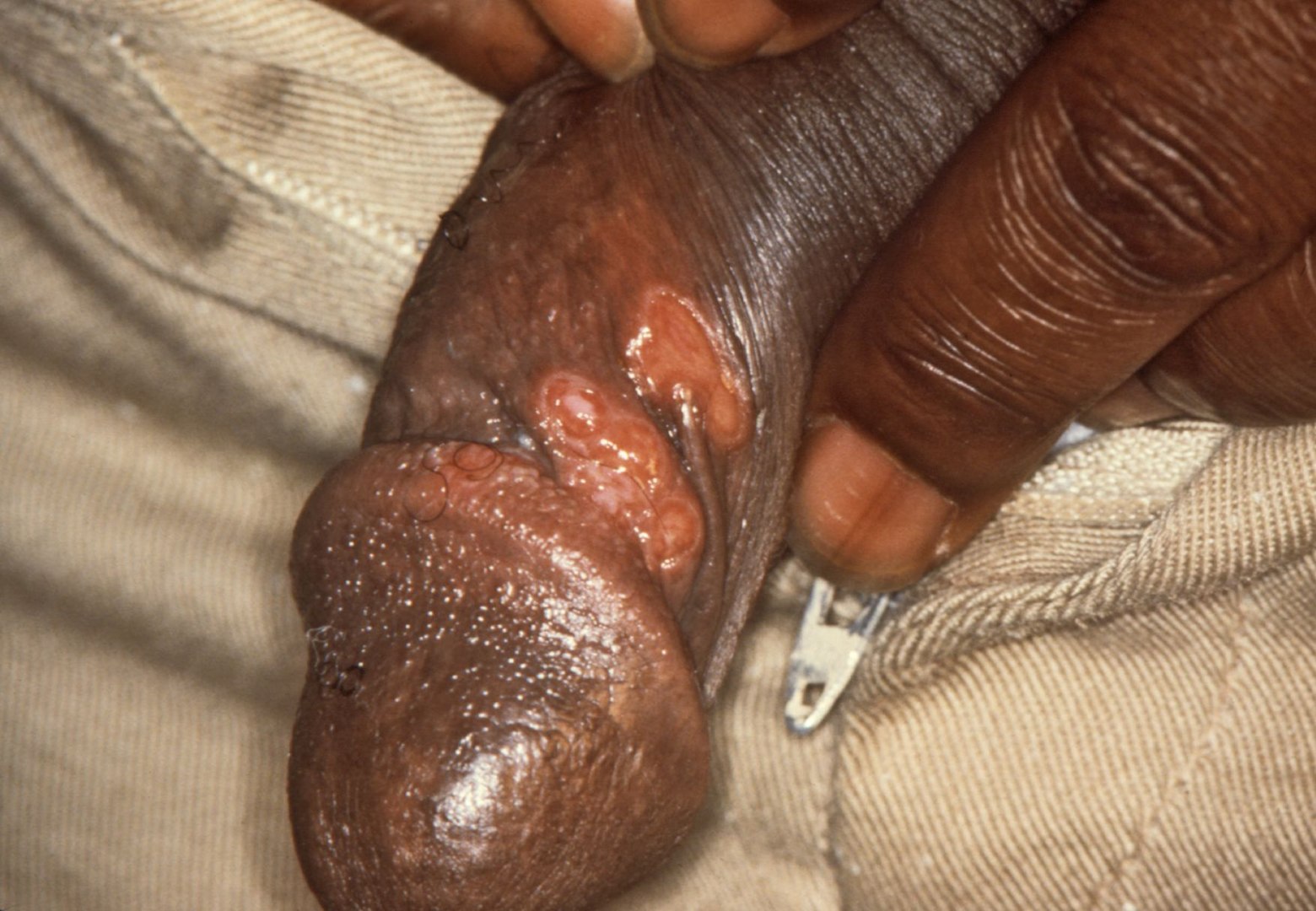
Primary, painless chancre of syphilis (Treponema pallidum pallidum infection)
Image: “Chancres on the penile shaft due to a primary syphilitic infection caused by Treponema pallidum 6803 lores” by M. Rein. License: Public DomainThe less-common species of Treponema and their diseases are summarized below:
| Disease | Yaws Yaws A systemic non-venereal infection of the tropics caused by treponema pallidum subspecies pertenue. Yaws, Bejel, and Pinta | Bejel Bejel Yaws, bejel, and pinta are endemic, nonvenereal treponematoses. The causative organisms are Treponema pallidum pertenue (yaws), T. pallidum endemicum (bejel), and T. carateum (pinta). These treponematoses are generally transmitted by direct skin-to-skin contact with infected skin lesions. Yaws, Bejel, and Pinta (endemic syphilis Syphilis Syphilis is a bacterial infection caused by the spirochete Treponema pallidum pallidum (T. p. pallidum), which is usually spread through sexual contact. Syphilis has 4 clinical stages: primary, secondary, latent, and tertiary. Syphilis) | Pinta Pinta An infectious disease of the skin caused by treponema carateum that occurs only in the Western hemisphere. Age of onset is between 10 and 20 years of age. This condition is characterized by marked changes in the skin color and is believed to be transmitted by direct person-to-person contact. Yaws, Bejel, and Pinta |
|---|---|---|---|
| Associated species | T. pallidum pertenue | T. pallidum endemicum | T. carateum |
| Transmission |
|
|
|
| Predominant locations | Tropical regions:
|
Desert regions:
|
Tropical regions: Central and South America |
| Primary demographic | Children | Children | Adults |
| Clinical manifestations | Primary phase: painless, yellow
papilloma
Papilloma
A circumscribed benign epithelial tumor projecting from the surrounding surface; more precisely, a benign epithelial neoplasm consisting of villous or arborescent outgrowths of fibrovascular stroma covered by neoplastic cells.
Cowden Syndrome lesion on lower extremities Secondary phase:
|
Primary phase: small, primary
papule
Papule
Elevated lesion < 1 cm in diameter
Generalized and Localized Rashes on the
oral mucosa
Oral mucosa
Lining of the oral cavity, including mucosa on the gums; the palate; the lip; the cheek; floor of the mouth; and other structures. The mucosa is generally a nonkeratinized stratified squamous epithelium covering muscle, bone, or glands but can show varying degree of keratinization at specific locations.
Stomatitis Secondary phase:
|
Primary phase:
Tertiary phase:
|
| Diagnosis | Since these treponemal species are morphologically indistinguishable, the diagnosis is based on:
|
||
| Management |
|
||
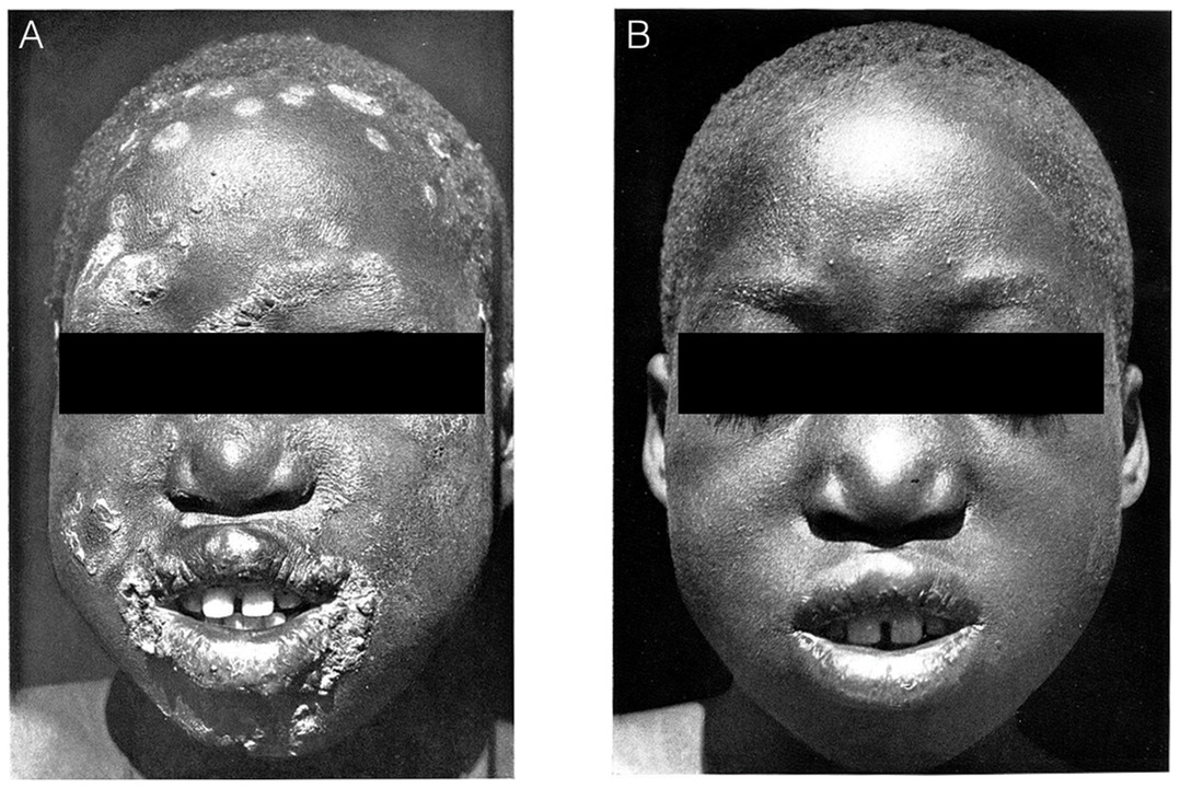
Cutaneous manifestation of yaws
(A) A patient with lesions on the face due to Treponema pallidum pertenue
(B) Resolution of the lesions after treatment with penicillin
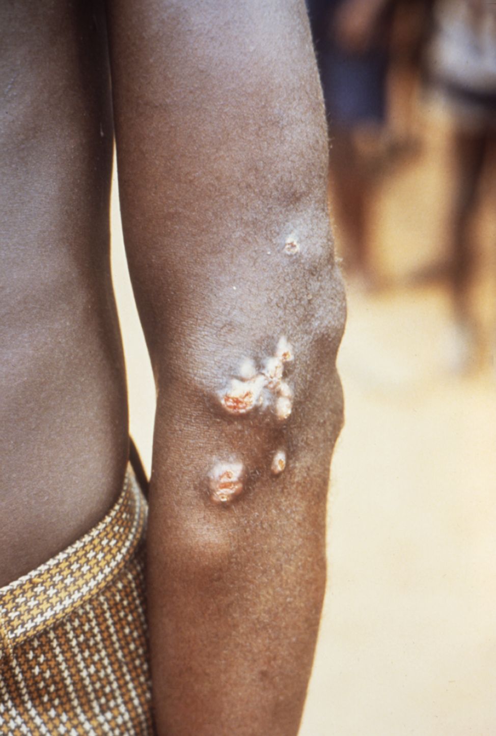
Cutaneous manifestation of yaws
Juxta-articular skin lesions and nodules on the elbow resulting from a Treponema pallidum pertenue infection
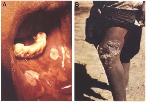
Mucocutaneous lesions of bejel (endemic syphilis)
A: An oral lesion of primary bejel resulting from Treponema pallidum endemicum infection
B: Chronic skin lesion of secondary bejel
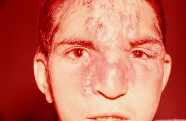
Gummatous lesions in late-stage bejel (Treponema pallidum endemicum infection)
Image: “Infiltration of skin due to endemic syphilis” by Alireza Abdolrasouli, Adam Croucher, Yahya Hemmati, and David Mabey. License: Public Domain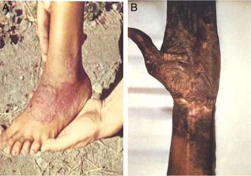
Cutaneous lesions of pinta (Treponema carateum infection)
(A) Erythematous plaque of early pinta
(B) Skin discoloration of late pinta
Spirochetes are gram negative Gram negative Bacteria which lose crystal violet stain but are stained pink when treated by gram’s method. Yersinia spp./Yersiniosis, spiral Spiral Computed tomography where there is continuous x-ray exposure to the patient while being transported in a spiral or helical pattern through the beam of irradiation. This provides improved three-dimensional contrast and spatial resolution compared to conventional computed tomography, where data is obtained and computed from individual sequential exposures. Computed Tomography (CT) shaped, and motile. The following table briefly compares some clinically relevant spirochetes:
| Organism | Treponema pallidum pallidum | Other T. pallidum subspecies | Treponema carateum | Borrelia Borrelia Borrelia are gram-negative microaerophilic spirochetes. Owing to their small size, they are not easily seen on Gram stain but can be visualized using dark-field microscopy, Giemsa, or Wright stain. Spirochetes are motile and move in a characteristic spinning fashion due to axial filaments in the periplasmic space. Borrelia burgdorfi | Borrelia recurrentis Borrelia recurrentis Borrelia | Leptospira interrogans Leptospira interrogans A genus of question mark-shaped bacteria spirochetes which is found in freshwater that is contaminated by animal urine. It causes leptospirosis. Leptospira/Leptospirosis |
|---|---|---|---|---|---|---|
| Micro |
|
|
|
|
|
|
| Virulence Virulence The degree of pathogenicity within a group or species of microorganisms or viruses as indicated by case fatality rates and/or the ability of the organism to invade the tissues of the host. The pathogenic capacity of an organism is determined by its virulence factors. Proteus |
|
|
|
|
Antigenic variation |
|
| Reservoir Reservoir Animate or inanimate sources which normally harbor disease-causing organisms and thus serve as potential sources of disease outbreaks. Reservoirs are distinguished from vectors (disease vectors) and carriers, which are agents of disease transmission rather than continuing sources of potential disease outbreaks. Humans may serve both as disease reservoirs and carriers. Escherichia coli | Humans | Humans | Humans |
|
Humans |
|
| Transmission | Sexual contact | P2P contact | P2P contact | Ixodes tick | Louse | Direct contact with animal tissue or fluids |
| Clinical | Syphilis Syphilis Syphilis is a bacterial infection caused by the spirochete Treponema pallidum pallidum (T. p. pallidum), which is usually spread through sexual contact. Syphilis has 4 clinical stages: primary, secondary, latent, and tertiary. Syphilis |
|
Pinta Pinta An infectious disease of the skin caused by treponema carateum that occurs only in the Western hemisphere. Age of onset is between 10 and 20 years of age. This condition is characterized by marked changes in the skin color and is believed to be transmitted by direct person-to-person contact. Yaws, Bejel, and Pinta | Lyme disease Lyme disease Lyme disease is a tick-borne infection caused by the gram-negative spirochete Borrelia burgdorferi. Lyme disease is transmitted by the black-legged Ixodes tick (known as a deer tick), which is only found in specific geographic regions. Patient presentation can vary depending on the stage of the disease and may include a characteristic erythema migrans rash. Lyme Disease | Relapsing fever Fever Fever is defined as a measured body temperature of at least 38°C (100.4°F). Fever is caused by circulating endogenous and/or exogenous pyrogens that increase levels of prostaglandin E2 in the hypothalamus. Fever is commonly associated with chills, rigors, sweating, and flushing of the skin. Fever |
|
| Diagnosis |
|
|
|
|
Blood-smear analysis |
|
| Management |
|
|
|
|
|
|
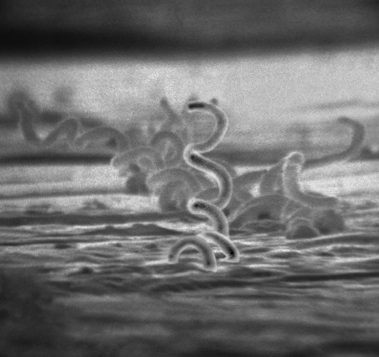
Visual comparison of spirochetes on electron micrograph: thick spirals of Treponema
Image: “Treponema pallidum” by Dr. David Cox. License: Public Domain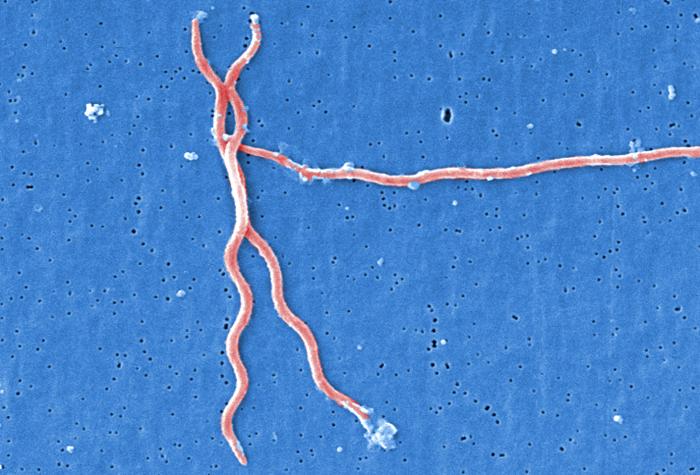
Visual comparison of spirochetes on electron micrograph: Borrelia, which are larger than Treponema
Image: “Lyme disease parasite Borrelia burgdorferi” by Claudia Molins. License: Public Domain
Visual comparison of spirochetes on electron micrograph: hooked ends of Leptospira
Image: “A filtration-based technique for simultaneous SEM and TEM sample preparation for the rapid detection of pathogens” by Beniac DR, Siemens CG, Wright CJ, Booth TF. License: CC BY 3.0, edited by Lecturio.