Achieve Mastery of Medical Concepts

Study for medical school and boards with Lecturio

Lentigo Maligna

Lentigo maligna is melanoma Melanoma Melanoma is a malignant tumor arising from melanocytes, the melanin-producing cells of the epidermis. These tumors are most common in fair-skinned individuals with a history of excessive sun exposure and sunburns. Melanoma in situ, a precancerous Precancerous Pathological conditions that tend eventually to become malignant. Barrett Esophagus lesion that may progress to an invasive melanoma Melanoma Melanoma is a malignant tumor arising from melanocytes, the melanin-producing cells of the epidermis. These tumors are most common in fair-skinned individuals with a history of excessive sun exposure and sunburns. Melanoma (specifically lentigo maligna melanoma Melanoma Melanoma is a malignant tumor arising from melanocytes, the melanin-producing cells of the epidermis. These tumors are most common in fair-skinned individuals with a history of excessive sun exposure and sunburns. Melanoma subtype). This condition typically occurs in sun-damaged areas (e.g., face and neck Neck The part of a human or animal body connecting the head to the rest of the body. Peritonsillar Abscess) of elderly patients Patients Individuals participating in the health care system for the purpose of receiving therapeutic, diagnostic, or preventive procedures. Clinician–Patient Relationship. Lentigo maligna presents as a brown macule Macule Nonpalpable lesion < 1 cm in diameter Generalized and Localized Rashes with color variegation and asymmetrical borders that grow slowly. The lesion should be biopsied to confirm a diagnosis and surgical excision with a safety margin is the 1st-line treatment.

Last updated: Sep 8, 2022

Editorial responsibility: Stanley Oiseth, Lindsay Jones, Evelin Maza

Overview

Definition

Lentigo maligna (also known as Hutchinson melanotic freckle) is melanoma Melanoma Melanoma is a malignant tumor arising from melanocytes, the melanin-producing cells of the epidermis. These tumors are most common in fair-skinned individuals with a history of excessive sun exposure and sunburns. Melanoma in situ. This type of precancerous Precancerous Pathological conditions that tend eventually to become malignant. Barrett Esophagus lesion may progress to lentigo maligna melanoma Melanoma Melanoma is a malignant tumor arising from melanocytes, the melanin-producing cells of the epidermis. These tumors are most common in fair-skinned individuals with a history of excessive sun exposure and sunburns. Melanoma.

Epidemiology

  • Peak incidence Incidence The number of new cases of a given disease during a given period in a specified population. It also is used for the rate at which new events occur in a defined population. It is differentiated from prevalence, which refers to all cases in the population at a given time. Measures of Disease Frequency between 65 and 80 years of age
  • Precursor to the 3rd most common melanoma Melanoma Melanoma is a malignant tumor arising from melanocytes, the melanin-producing cells of the epidermis. These tumors are most common in fair-skinned individuals with a history of excessive sun exposure and sunburns. Melanoma subtype (lentigo maligna melanoma Melanoma Melanoma is a malignant tumor arising from melanocytes, the melanin-producing cells of the epidermis. These tumors are most common in fair-skinned individuals with a history of excessive sun exposure and sunburns. Melanoma)
  • Women > men
  • Incidence Incidence The number of new cases of a given disease during a given period in a specified population. It also is used for the rate at which new events occur in a defined population. It is differentiated from prevalence, which refers to all cases in the population at a given time. Measures of Disease Frequency is 13.7 per 100,000.

Etiology

  • Genetic mutations Genetic Mutations Carcinogenesis:
    • Compared with other melanoma Melanoma Melanoma is a malignant tumor arising from melanocytes, the melanin-producing cells of the epidermis. These tumors are most common in fair-skinned individuals with a history of excessive sun exposure and sunburns. Melanoma subtypes, there is an increased likelihood of KIT mutations.
    • Other mutations include CCND1, MITF, NRAS, and p53.
  • Risk factors:
    • Cumulative (over time) ultraviolet radiation Radiation Emission or propagation of acoustic waves (sound), electromagnetic energy waves (such as light; radio waves; gamma rays; or x-rays), or a stream of subatomic particles (such as electrons; neutrons; protons; or alpha particles). Osteosarcoma
    • Genetic conditions:
      • Oculocutaneous albinism Oculocutaneous albinism Heterogeneous group of autosomal recessive disorders comprising at least four recognized types, all having in common varying degrees of hypopigmentation of the skin, hair, and eyes. The two most common are the tyrosinase-positive and tyrosinase-negative types. Albinism
      • Xeroderma pigmentosum
      • Porphyria cutanea tarda Porphyria cutanea tarda An autosomal dominant or acquired porphyria due to a deficiency of uroporphyrinogen decarboxylase in the liver. It is characterized by photosensitivity and cutaneous lesions with little or no neurologic symptoms. Type I is the acquired form and is strongly associated with liver diseases and hepatic toxicities caused by alcohol or estrogenic steroids. Type II is the familial form. Porphyrias
    • X-ray X-ray Penetrating electromagnetic radiation emitted when the inner orbital electrons of an atom are excited and release radiant energy. X-ray wavelengths range from 1 pm to 10 nm. Hard x-rays are the higher energy, shorter wavelength x-rays. Soft x-rays or grenz rays are less energetic and longer in wavelength. The short wavelength end of the x-ray spectrum overlaps the gamma rays wavelength range. The distinction between gamma rays and x-rays is based on their radiation source. Pulmonary Function Tests irradiation
    • Use of estrogen Estrogen Compounds that interact with estrogen receptors in target tissues to bring about the effects similar to those of estradiol. Estrogens stimulate the female reproductive organs, and the development of secondary female sex characteristics. Estrogenic chemicals include natural, synthetic, steroidal, or non-steroidal compounds. Ovaries: Anatomy/ progesterone Progesterone The major progestational steroid that is secreted primarily by the corpus luteum and the placenta. Progesterone acts on the uterus, the mammary glands and the brain. It is required in embryo implantation; pregnancy maintenance, and the development of mammary tissue for milk production. Progesterone, converted from pregnenolone, also serves as an intermediate in the biosynthesis of gonadal steroid hormones and adrenal corticosteroids. Gonadal Hormones 
    • Use of non-permanent hair dye

Related videos

Clinical Presentation

  • Majority occur on the head/ neck Neck The part of a human or animal body connecting the head to the rest of the body. Peritonsillar Abscess (mainly the cheek).
  • Variable Variable Variables represent information about something that can change. The design of the measurement scales, or of the methods for obtaining information, will determine the data gathered and the characteristics of that data. As a result, a variable can be qualitative or quantitative, and may be further classified into subgroups. Types of Variables color: 
    • Light brown to black (may have color variegation)
    • May have pink/white areas indicating inflammation Inflammation Inflammation is a complex set of responses to infection and injury involving leukocytes as the principal cellular mediators in the body’s defense against pathogenic organisms. Inflammation is also seen as a response to tissue injury in the process of wound healing. The 5 cardinal signs of inflammation are pain, heat, redness, swelling, and loss of function. Inflammation or regression Regression Corneal Abrasions, Erosion, and Ulcers
  • Asymmetric
  • Poorly defined borders
  • Smooth (nonpalpable)
  • Slow growth → speckles (as melanoma Melanoma Melanoma is a malignant tumor arising from melanocytes, the melanin-producing cells of the epidermis. These tumors are most common in fair-skinned individuals with a history of excessive sun exposure and sunburns. Melanoma enlarges)
  • Indicators of possible progression to invasive melanoma Melanoma Melanoma is a malignant tumor arising from melanocytes, the melanin-producing cells of the epidermis. These tumors are most common in fair-skinned individuals with a history of excessive sun exposure and sunburns. Melanoma:
    • Raised/palpable areas
    • Sharp borders
    • Darker pigmentation
  • Surrounding skin Skin The skin, also referred to as the integumentary system, is the largest organ of the body. The skin is primarily composed of the epidermis (outer layer) and dermis (deep layer). The epidermis is primarily composed of keratinocytes that undergo rapid turnover, while the dermis contains dense layers of connective tissue. Skin: Structure and Functions usually manifests solar skin Skin The skin, also referred to as the integumentary system, is the largest organ of the body. The skin is primarily composed of the epidermis (outer layer) and dermis (deep layer). The epidermis is primarily composed of keratinocytes that undergo rapid turnover, while the dermis contains dense layers of connective tissue. Skin: Structure and Functions damage:
    • Actinic keratoses
    • Solar elastosis
    • Solar lentigines
Lentigo maligna

Lentigo maligna: an asymmetric brown macule with color variegation present on the left cheek

Image: “Lentigo maligna” by kilbad. License: CC BY 3.0
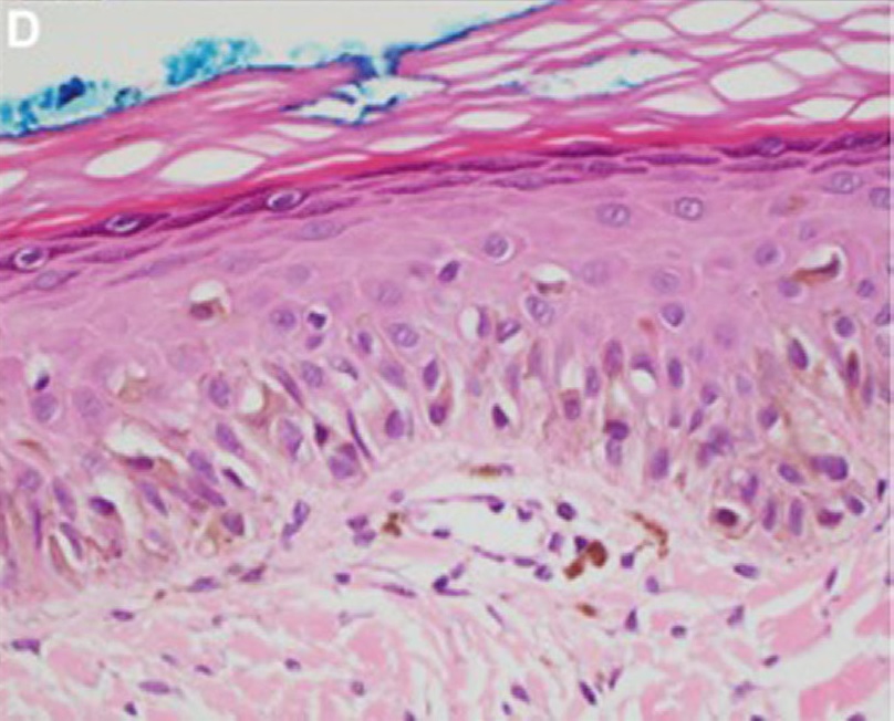
Diagnosis

History

  • Amount of sun exposure
  • Other skin Skin The skin, also referred to as the integumentary system, is the largest organ of the body. The skin is primarily composed of the epidermis (outer layer) and dermis (deep layer). The epidermis is primarily composed of keratinocytes that undergo rapid turnover, while the dermis contains dense layers of connective tissue. Skin: Structure and Functions cancers
  • Family history Family History Adult Health Maintenance of melanoma Melanoma Melanoma is a malignant tumor arising from melanocytes, the melanin-producing cells of the epidermis. These tumors are most common in fair-skinned individuals with a history of excessive sun exposure and sunburns. Melanoma
  • Hereditary diseases

Physical exam

  • Complete/thorough skin Skin The skin, also referred to as the integumentary system, is the largest organ of the body. The skin is primarily composed of the epidermis (outer layer) and dermis (deep layer). The epidermis is primarily composed of keratinocytes that undergo rapid turnover, while the dermis contains dense layers of connective tissue. Skin: Structure and Functions exam should be performed.
  • Synchronous malignant and premalignant lesions are not uncommon in individuals with extensive sun damage to the skin Skin The skin, also referred to as the integumentary system, is the largest organ of the body. The skin is primarily composed of the epidermis (outer layer) and dermis (deep layer). The epidermis is primarily composed of keratinocytes that undergo rapid turnover, while the dermis contains dense layers of connective tissue. Skin: Structure and Functions.

Dermoscopy Dermoscopy A noninvasive technique that enables direct microscopic examination of the surface and architecture of the skin. Seborrheic Keratosis

  • Asymmetric pigmented follicular openings (pseudonetwork)
  • Progression of findings:
    • Early stage: peppering of pigmentation around follicular ostia (blue-gray dots)
    • Coalescence of blue-gray dots into polygonal lines and rhomboidal structures
    • Late stage: homogeneous Homogeneous Imaging of the Spleen dark-brown to black blotches blocking the follicular ostia

Reflectance confocal microscopy

  • Availability of this technique is limited.
  • Requires specialized training
  • More sensitive, but less specific for lentigo maligna diagnosis than dermoscopy Dermoscopy A noninvasive technique that enables direct microscopic examination of the surface and architecture of the skin. Seborrheic Keratosis

Histology

  • Gold standard for lentigo maligna diagnosis
  • Sample obtained via:
  • Findings:
    • Replacement of basal keratinocytes Keratinocytes Epidermal cells which synthesize keratin and undergo characteristic changes as they move upward from the basal layers of the epidermis to the cornified (horny) layer of the skin. Successive stages of differentiation of the keratinocytes forming the epidermal layers are basal cell, spinous or prickle cell, and the granular cell. Skin: Structure and Functions by nests of atypical melanocytes Melanocytes Mammalian pigment cells that produce melanins, pigments found mainly in the epidermis, but also in the eyes and the hair, by a process called melanogenesis. Coloration can be altered by the number of melanocytes or the amount of pigment produced and stored in the organelles called melanosomes. The large non-mammalian melanin-containing cells are called melanophores. Skin: Structure and Functions
    • No basement membrane Basement membrane A darkly stained mat-like extracellular matrix (ecm) that separates cell layers, such as epithelium from endothelium or a layer of connective tissue. The ecm layer that supports an overlying epithelium or endothelium is called basal lamina. Basement membrane (bm) can be formed by the fusion of either two adjacent basal laminae or a basal lamina with an adjacent reticular lamina of connective tissue. Bm, composed mainly of type IV collagen; glycoprotein laminin; and proteoglycan, provides barriers as well as channels between interacting cell layers. Thin Basement Membrane Nephropathy (TBMN) invasion (if present, indicates progression to invasive melanoma Melanoma Melanoma is a malignant tumor arising from melanocytes, the melanin-producing cells of the epidermis. These tumors are most common in fair-skinned individuals with a history of excessive sun exposure and sunburns. Melanoma)
    • Changes of chronic sun damage:
Histopathology of lentigo maligna

Lentigo maligna:
A nest of atypical melanocytes can be noted. The basement membrane is intact. Therefore, this lesion is lentigo maligna (in situ).

Image: “Lentigo maligna” by Sheliza Halani et al. License: CC BY 4.0

Management and Prognosis

Management

Surgical excision:

  • 1st line treatment
  • Procedures:
    • Wide local excision:
      • Margins of 5–10 mm MM Multiple myeloma (MM) is a malignant condition of plasma cells (activated B lymphocytes) primarily seen in the elderly. Monoclonal proliferation of plasma cells results in cytokine-driven osteoclastic activity and excessive secretion of IgG antibodies. Multiple Myeloma should be obtained.
      • For small lesions with well-delineated borders
    • Staged excisions:
      • Involves histologic examination of the permanent sections
      • Excisions are performed until negative margins are obtained.
      • For large lesions (> 1 cm on the head/ neck Neck The part of a human or animal body connecting the head to the rest of the body. Peritonsillar Abscess and > 2 cm on trunk/extremities)
      • For lesions with ill-defined borders 
    • Mohs micrographic surgery:
      • Staged excision that involves examination of frozen sections
      • May not be as reliable as staged excision technique for lentigo maligna

Nonsurgical:

  • Reserved for:
    • Elderly frail patients Patients Individuals participating in the health care system for the purpose of receiving therapeutic, diagnostic, or preventive procedures. Clinician–Patient Relationship
    • Patients Patients Individuals participating in the health care system for the purpose of receiving therapeutic, diagnostic, or preventive procedures. Clinician–Patient Relationship refusing surgery
    • Large lesions in cosmetically sensitive areas where problematic reconstruction is expected
  • Includes:
    • Radiotherapy
    • Laser
    • Cryosurgery
    • Topical imiquimod Imiquimod A topically-applied aminoquinoline immune modulator that induces interferon production. It is used in the treatment of external genital and perianal warts, superficial carcinoma, basal cell; and actinic keratosis. Hypertrophic and Keloid Scars

Patient education:

  • Protection from ultraviolet radiation Radiation Emission or propagation of acoustic waves (sound), electromagnetic energy waves (such as light; radio waves; gamma rays; or x-rays), or a stream of subatomic particles (such as electrons; neutrons; protons; or alpha particles). Osteosarcoma (sunblock, clothing)
  • Self-examination of the skin Skin The skin, also referred to as the integumentary system, is the largest organ of the body. The skin is primarily composed of the epidermis (outer layer) and dermis (deep layer). The epidermis is primarily composed of keratinocytes that undergo rapid turnover, while the dermis contains dense layers of connective tissue. Skin: Structure and Functions

Prognosis Prognosis A prediction of the probable outcome of a disease based on a individual’s condition and the usual course of the disease as seen in similar situations. Non-Hodgkin Lymphomas

  • Up to 20% of presumed lentigo maligna biopsies are found to have invasive melanoma Melanoma Melanoma is a malignant tumor arising from melanocytes, the melanin-producing cells of the epidermis. These tumors are most common in fair-skinned individuals with a history of excessive sun exposure and sunburns. Melanoma.
  • Can take from < 10 to > 50 years for lentigo maligna to progress to lentigo maligna melanoma Melanoma Melanoma is a malignant tumor arising from melanocytes, the melanin-producing cells of the epidermis. These tumors are most common in fair-skinned individuals with a history of excessive sun exposure and sunburns. Melanoma.
  • 6%–9% recurrence rate after wide excision
  • In the absence of progression to lentigo maligna melanoma Melanoma Melanoma is a malignant tumor arising from melanocytes, the melanin-producing cells of the epidermis. These tumors are most common in fair-skinned individuals with a history of excessive sun exposure and sunburns. Melanoma, lentigo maligna does not shorten life expectancy Life expectancy Based on known statistical data, the number of years which any person of a given age may reasonably expected to live. Population Pyramids.
  • No disease-related deaths when complete excision is achieved

Differential Diagnosis

  • Solar lentigo: macular lesions with irregular margins and uniform brown pigmentation on sun-exposed skin Skin The skin, also referred to as the integumentary system, is the largest organ of the body. The skin is primarily composed of the epidermis (outer layer) and dermis (deep layer). The epidermis is primarily composed of keratinocytes that undergo rapid turnover, while the dermis contains dense layers of connective tissue. Skin: Structure and Functions similar to lentigo maligna. Biopsy Biopsy Removal and pathologic examination of specimens from the living body. Ewing Sarcoma of these lesions shows no nesting of atypical melanocytes Melanocytes Mammalian pigment cells that produce melanins, pigments found mainly in the epidermis, but also in the eyes and the hair, by a process called melanogenesis. Coloration can be altered by the number of melanocytes or the amount of pigment produced and stored in the organelles called melanosomes. The large non-mammalian melanin-containing cells are called melanophores. Skin: Structure and Functions. Solar lentigo is benign Benign Fibroadenoma and does not require treatment.
  • Melanoma Melanoma Melanoma is a malignant tumor arising from melanocytes, the melanin-producing cells of the epidermis. These tumors are most common in fair-skinned individuals with a history of excessive sun exposure and sunburns. Melanoma: a skin Skin The skin, also referred to as the integumentary system, is the largest organ of the body. The skin is primarily composed of the epidermis (outer layer) and dermis (deep layer). The epidermis is primarily composed of keratinocytes that undergo rapid turnover, while the dermis contains dense layers of connective tissue. Skin: Structure and Functions malignancy Malignancy Hemothorax derived from the malignant transformation Transformation Change brought about to an organism’s genetic composition by unidirectional transfer (transfection; transduction, genetic; conjugation, genetic, etc.) and incorporation of foreign DNA into prokaryotic or eukaryotic cells by recombination of part or all of that DNA into the cell’s genome. Bacteriology of melanocytes Melanocytes Mammalian pigment cells that produce melanins, pigments found mainly in the epidermis, but also in the eyes and the hair, by a process called melanogenesis. Coloration can be altered by the number of melanocytes or the amount of pigment produced and stored in the organelles called melanosomes. The large non-mammalian melanin-containing cells are called melanophores. Skin: Structure and Functions. The most common type of melanoma Melanoma Melanoma is a malignant tumor arising from melanocytes, the melanin-producing cells of the epidermis. These tumors are most common in fair-skinned individuals with a history of excessive sun exposure and sunburns. Melanoma is superficial spreading melanoma Melanoma Melanoma is a malignant tumor arising from melanocytes, the melanin-producing cells of the epidermis. These tumors are most common in fair-skinned individuals with a history of excessive sun exposure and sunburns. Melanoma, which usually presents as an irregular macule Macule Nonpalpable lesion < 1 cm in diameter Generalized and Localized Rashes/ patch Patch Nonpalpable lesion > 1 cm in diameter Generalized and Localized Rashes. On histopathologic examination, atypical melanocytes Melanocytes Mammalian pigment cells that produce melanins, pigments found mainly in the epidermis, but also in the eyes and the hair, by a process called melanogenesis. Coloration can be altered by the number of melanocytes or the amount of pigment produced and stored in the organelles called melanosomes. The large non-mammalian melanin-containing cells are called melanophores. Skin: Structure and Functions would be detected in the dermis Dermis A layer of vascularized connective tissue underneath the epidermis. The surface of the dermis contains innervated papillae. Embedded in or beneath the dermis are sweat glands; hair follicles; and sebaceous glands. Skin: Structure and Functions. Lentigo maligna can progress to lentigo maligna melanoma Melanoma Melanoma is a malignant tumor arising from melanocytes, the melanin-producing cells of the epidermis. These tumors are most common in fair-skinned individuals with a history of excessive sun exposure and sunburns. Melanoma. The mainstay of treatment is surgical excision.
  • Atypical nevus Nevus Nevi (singular nevus), also known as “moles,” are benign neoplasms of the skin. Nevus is a non-specific medical term because it encompasses both congenital and acquired lesions, hyper- and hypopigmented lesions, and raised or flat lesions. Nevus/Nevi: a benign Benign Fibroadenoma melanocytic neoplasm that mimics lentigo maligna in appearance as it may be asymmetric and > 6 mm MM Multiple myeloma (MM) is a malignant condition of plasma cells (activated B lymphocytes) primarily seen in the elderly. Monoclonal proliferation of plasma cells results in cytokine-driven osteoclastic activity and excessive secretion of IgG antibodies. Multiple Myeloma with color variegation.  Dermoscopic examination Dermoscopic Examination Scabies and biopsy Biopsy Removal and pathologic examination of specimens from the living body. Ewing Sarcoma are needed to differentiate. Does not require excision if the diagnosis is certain.
  • Seborrheic keratosis Seborrheic keratosis Seborrheic keratosis (SK) is the most common benign epithelial cutaneous neoplasm. The condition consists of immature keratinocytes. Seborrheic keratosis is the most common benign skin tumor in middle-aged and elderly adults and presents as a sharply demarcated, exophytic, skin lesion that may be tan or black and has a “stuck-on” appearance. Seborrheic Keratosis: a benign Benign Fibroadenoma neoplasm consisting of immature keratinocytes Keratinocytes Epidermal cells which synthesize keratin and undergo characteristic changes as they move upward from the basal layers of the epidermis to the cornified (horny) layer of the skin. Successive stages of differentiation of the keratinocytes forming the epidermal layers are basal cell, spinous or prickle cell, and the granular cell. Skin: Structure and Functions occurring commonly in the elderly. This condition is demarcated, waxy, and has a stuck-on appearance. A biopsy Biopsy Removal and pathologic examination of specimens from the living body. Ewing Sarcoma will show abnormal keratinocytes Keratinocytes Epidermal cells which synthesize keratin and undergo characteristic changes as they move upward from the basal layers of the epidermis to the cornified (horny) layer of the skin. Successive stages of differentiation of the keratinocytes forming the epidermal layers are basal cell, spinous or prickle cell, and the granular cell. Skin: Structure and Functions versus melanocytes Melanocytes Mammalian pigment cells that produce melanins, pigments found mainly in the epidermis, but also in the eyes and the hair, by a process called melanogenesis. Coloration can be altered by the number of melanocytes or the amount of pigment produced and stored in the organelles called melanosomes. The large non-mammalian melanin-containing cells are called melanophores. Skin: Structure and Functions. Dermoscopy Dermoscopy A noninvasive technique that enables direct microscopic examination of the surface and architecture of the skin. Seborrheic Keratosis would show cysts Cysts Any fluid-filled closed cavity or sac that is lined by an epithelium. Cysts can be of normal, abnormal, non-neoplastic, or neoplastic tissues. Fibrocystic Change and fissures. This neoplasm does not require treatment.
  • Actinic keratosis Actinic keratosis Actinic keratosis (AK) is a precancerous skin lesion that affects sun-exposed areas. The condition presents as small, non-tender macules/papules with a characteristic sandpaper-like texture that can become erythematous scaly plaques. Actinic Keratosis: a precancerous Precancerous Pathological conditions that tend eventually to become malignant. Barrett Esophagus lesion that affects sun-exposed areas (e.g., scalp and hands) in elderly patients Patients Individuals participating in the health care system for the purpose of receiving therapeutic, diagnostic, or preventive procedures. Clinician–Patient Relationship. This condition appears as a scaly, slightly elevated lesion that should be excised or treated topically to prevent invasive squamous cell carcinoma Squamous cell carcinoma Cutaneous squamous cell carcinoma (cSCC) is caused by malignant proliferation of atypical keratinocytes. This condition is the 2nd most common skin malignancy and usually affects sun-exposed areas of fair-skinned patients. The cancer presents as a firm, erythematous, keratotic plaque or papule. Squamous Cell Carcinoma (SCC) development. The only way to differentiate from lentigo maligna is by a biopsy Biopsy Removal and pathologic examination of specimens from the living body. Ewing Sarcoma that shows atypical keratinocytes Keratinocytes Epidermal cells which synthesize keratin and undergo characteristic changes as they move upward from the basal layers of the epidermis to the cornified (horny) layer of the skin. Successive stages of differentiation of the keratinocytes forming the epidermal layers are basal cell, spinous or prickle cell, and the granular cell. Skin: Structure and Functions in the basal layer.
  • Dermatofibroma: a common mesenchymal growth of the skin Skin The skin, also referred to as the integumentary system, is the largest organ of the body. The skin is primarily composed of the epidermis (outer layer) and dermis (deep layer). The epidermis is primarily composed of keratinocytes that undergo rapid turnover, while the dermis contains dense layers of connective tissue. Skin: Structure and Functions where skin Skin The skin, also referred to as the integumentary system, is the largest organ of the body. The skin is primarily composed of the epidermis (outer layer) and dermis (deep layer). The epidermis is primarily composed of keratinocytes that undergo rapid turnover, while the dermis contains dense layers of connective tissue. Skin: Structure and Functions fibroblasts Fibroblasts Connective tissue cells which secrete an extracellular matrix rich in collagen and other macromolecules. Sarcoidosis are the primary constituents. This condition usually presents as a firm, indurated, mobile nodule Nodule Chalazion measuring about 0.5–1 cm in size. Upon lateral compression Compression Blunt Chest Trauma, a dimple-like depression is seen in the overlying skin Skin The skin, also referred to as the integumentary system, is the largest organ of the body. The skin is primarily composed of the epidermis (outer layer) and dermis (deep layer). The epidermis is primarily composed of keratinocytes that undergo rapid turnover, while the dermis contains dense layers of connective tissue. Skin: Structure and Functions (“buttonhole” sign). Does not require treatment, but can be surgically excised if bothersome to the patient.

References

  1. Charifa A. (2020). Lentigo Maligna Melanoma. Retrieved February 24, 2021, from https://www.statpearls.com/articlelibrary/viewarticle/24188/
  2. Cohen L.M. (1995). Lentigo maligna and lentigo maligna melanoma. J Am Acad Dermatol. 1995 Dec;33(6):923-36; quiz 937-40.
  3. Sober A. J., Olbricht S., Hong A.M. (2019). Lentigo maligna: Clinical manifestations, diagnosis, and management. Retrieved February 24, 2021, from https://www.uptodate.com/contents/lentigo-maligna-clinical-manifestations-diagnosis-and-management

Create your free account or log in to continue reading!

Sign up now and get free access to Lecturio with concept pages, medical videos, and questions for your medical education.

User Reviews

Details