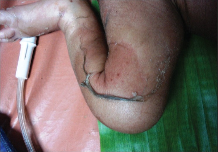
Advertisement

Advertisement

Advertisement

Advertisement

Staphylococcal Scalded Skin Syndrome (SSSS)

Staphylococcal scalded skin Skin The skin, also referred to as the integumentary system, is the largest organ of the body. The skin is primarily composed of the epidermis (outer layer) and dermis (deep layer). The epidermis is primarily composed of keratinocytes that undergo rapid turnover, while the dermis contains dense layers of connective tissue. Skin: Structure and Functions syndrome (SSSS), also known as Ritter disease and staphylococcal epidermal necrolysis, is a toxin-mediated condition caused by Staphylococcus aureus Staphylococcus aureus Potentially pathogenic bacteria found in nasal membranes, skin, hair follicles, and perineum of warm-blooded animals. They may cause a wide range of infections and intoxications. Brain Abscess. The exfoliative toxin produced disseminates and cleaves desmoglein Desmoglein A group of desmosomal cadherins with cytoplasmic tails that resemble those of classical cadherins. The Cell: Cell Junctions 1 in the epidermis Epidermis The external, nonvascular layer of the skin. It is made up, from within outward, of five layers of epithelium: (1) basal layer (stratum basale epidermidis); (2) spinous layer (stratum spinosum epidermidis); (3) granular layer (stratum granulosum epidermidis); (4) clear layer (stratum lucidum epidermidis); and (5) horny layer (stratum corneum epidermidis). Skin: Structure and Functions, causing separation and detachment of the skin Skin The skin, also referred to as the integumentary system, is the largest organ of the body. The skin is primarily composed of the epidermis (outer layer) and dermis (deep layer). The epidermis is primarily composed of keratinocytes that undergo rapid turnover, while the dermis contains dense layers of connective tissue. Skin: Structure and Functions. SSSS most commonly affects young children. Prodromal symptoms precede diffuse cutaneous erythema Erythema Redness of the skin produced by congestion of the capillaries. This condition may result from a variety of disease processes. Chalazion, tenderness, bullae Bullae Erythema Multiforme formation, and superficial desquamation. The mucous membranes are spared. The diagnosis is made clinically and can be confirmed with culture data (targeting possible primary infection Primary infection Herpes Simplex Virus 1 and 2 sites) and biopsy Biopsy Removal and pathologic examination of specimens from the living body. Ewing Sarcoma. However, cultures of bullae Bullae Erythema Multiforme are not useful. Antibiotics and supportive care should be initiated as soon as the diagnosis is suspected.

Last updated: May 17, 2024
Editorial responsibility: Stanley Oiseth, Lindsay Jones, Evelin Maza

Advertisement

Advertisement

Advertisement

Advertisement

Advertisement

Advertisement

Overview

Epidemiology

  • Primarily a disease of children
  • Incidence Incidence The number of new cases of a given disease during a given period in a specified population. It also is used for the rate at which new events occur in a defined population. It is differentiated from prevalence, which refers to all cases in the population at a given time. Measures of Disease Frequency (in the United States):
    • 8 cases per 1 million children
    • 1 case per 1 million adults
  • Demographics:
    • Most patients Patients Individuals participating in the health care system for the purpose of receiving therapeutic, diagnostic, or preventive procedures. Clinician–Patient Relationship are < 5 years of age.
    • Rare in adults
    • Data on sex Sex The totality of characteristics of reproductive structure, functions, phenotype, and genotype, differentiating the male from the female organism. Gender Dysphoria predominance are inconsistent (conflicting epidemiological studies epidemiological studies Epidemiological studies are used to establish associations between risk factors and health-related outcomes. These studies can take the form of observational or interventional studies. Epidemiological Studies)
  • Mortality Mortality All deaths reported in a given population. Measures of Health Status:
    • 1%–5% in children
    • 50%–60% in adults

Etiology

  • Causative organism: exfoliative toxin-producing strains of Staphylococcus aureus Staphylococcus aureus Potentially pathogenic bacteria found in nasal membranes, skin, hair follicles, and perineum of warm-blooded animals. They may cause a wide range of infections and intoxications. Brain Abscess:
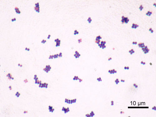
    • Gram-positive Gram-Positive Penicillins cocci Cocci Bacteriology in clusters
    • Coagulase-positive
    • Exfoliative toxin is produced in approximately 5% of S. aureus S. aureus Potentially pathogenic bacteria found in nasal membranes, skin, hair follicles, and perineum of warm-blooded animals. They may cause a wide range of infections and intoxications. Staphylococcus strains.
    • Most common strains: types 55 and 71
  • Transmission: contact with an asymptomatic or colonized carrier Carrier Vaccination
  • Common sites of outbreaks Outbreaks Sudden increase in the incidence of a disease. The concept includes epidemics and pandemics. Influenza Viruses/Influenza:
    • ICUs
    • Nurseries
  • Risk factors:
    • Children are more vulnerable because of:
      • Lack of immunity
      • Immature renal clearance Renal Clearance Pharmacokinetics and Pharmacodynamics (exotoxin cleared through the kidneys Kidneys The kidneys are a pair of bean-shaped organs located retroperitoneally against the posterior wall of the abdomen on either side of the spine. As part of the urinary tract, the kidneys are responsible for blood filtration and excretion of water-soluble waste in the urine. Kidneys: Anatomy)
    • Adults:
      • Immunodeficiency Immunodeficiency Chédiak-Higashi Syndrome
      • Renal failure Renal failure Conditions in which the kidneys perform below the normal level in the ability to remove wastes, concentrate urine, and maintain electrolyte balance; blood pressure; and calcium metabolism. Renal insufficiency can be classified by the degree of kidney damage (as measured by the level of proteinuria) and reduction in glomerular filtration rate. Crush Syndrome
      • Malignancy Malignancy Hemothorax
      • Diabetes Diabetes Diabetes mellitus (DM) is a metabolic disease characterized by hyperglycemia and dysfunction of the regulation of glucose metabolism by insulin. Type 1 DM is diagnosed mostly in children and young adults as the result of autoimmune destruction of β cells in the pancreas and the resulting lack of insulin. Type 2 DM has a significant association with obesity and is characterized by insulin resistance. Diabetes Mellitus
Microscopic image of staphylococcus aureus

Microscopic image of Staphylococcus aureus

Image: “Staphylococcus aureus Gram” by Y Tambe. License: CC BY-SA 3.0
Pathophysiology

  • Starts as a localized, staphylococcal infection:
    • Skin Skin The skin, also referred to as the integumentary system, is the largest organ of the body. The skin is primarily composed of the epidermis (outer layer) and dermis (deep layer). The epidermis is primarily composed of keratinocytes that undergo rapid turnover, while the dermis contains dense layers of connective tissue. Skin: Structure and Functions wounds
    • Conjunctivitis Conjunctivitis Conjunctivitis is a common inflammation of the bulbar and/or palpebral conjunctiva. It can be classified into infectious (mostly viral) and noninfectious conjunctivitis, which includes allergic causes. Patients commonly present with red eyes, increased tearing, burning, foreign body sensation, and photophobia. Conjunctivitis
    • Pharyngitis Pharyngitis Pharyngitis is an inflammation of the back of the throat (pharynx). Pharyngitis is usually caused by an upper respiratory tract infection, which is viral in most cases. It typically results in a sore throat and fever. Other symptoms may include a runny nose, cough, headache, and hoarseness. Pharyngitis
    • Pneumonia Pneumonia Pneumonia or pulmonary inflammation is an acute or chronic inflammation of lung tissue. Causes include infection with bacteria, viruses, or fungi. In more rare cases, pneumonia can also be caused through toxic triggers through inhalation of toxic substances, immunological processes, or in the course of radiotherapy. Pneumonia
    • The primary site of infection is not always evident.
  • Staphylococcus Staphylococcus Staphylococcus is a medically important genera of Gram-positive, aerobic cocci. These bacteria form clusters resembling grapes on culture plates. Staphylococci are ubiquitous for humans, and many strains compose the normal skin flora. Staphylococcus produces exotoxin → spread hematogenously
    • Two types of exotoxins Exotoxins Toxins produced, especially by bacterial or fungal cells, and released into the culture medium or environment. Bacteriology:
    • Exotoxin function:
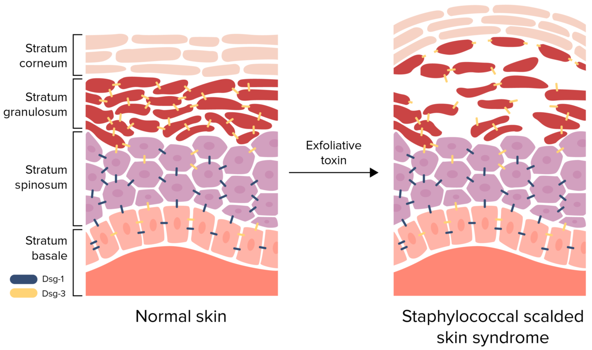
      • Cleaves desmoglein Desmoglein A group of desmosomal cadherins with cytoplasmic tails that resemble those of classical cadherins. The Cell: Cell Junctions (Dsg) 1 complex in the stratum granulosum Stratum granulosum Skin: Structure and Functions
      • Disrupts keratinocyte-to-keratinocyte adhesion Adhesion The process whereby platelets adhere to something other than platelets, e.g., collagen; basement membrane; microfibrils; or other ‘foreign’ surfaces. Coagulation Studies
      • Causes separation and detachment of the superficial epidermis Epidermis The external, nonvascular layer of the skin. It is made up, from within outward, of five layers of epithelium: (1) basal layer (stratum basale epidermidis); (2) spinous layer (stratum spinosum epidermidis); (3) granular layer (stratum granulosum epidermidis); (4) clear layer (stratum lucidum epidermidis); and (5) horny layer (stratum corneum epidermidis). Skin: Structure and Functions
Pathophysiology of staphylococcal scalded skin syndrome

Pathophysiology of staphylococcal scalded skin syndrome:
Exfoliative toxin cleaves desmoglein (Dsg) 1, disrupting the cell-to-cell adhesion of the stratum granulosum. This detachment of the superficial epidermis causes bullae formation and desquamation.
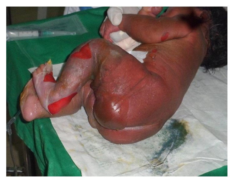
Image by Lecturio.

Clinical Presentation

  • Incubation Incubation The amount time between exposure to an infectious agent and becoming symptomatic. Rabies Virus period: 1–10 days
  • Prodromal symptoms:
    • Fever Fever Fever is defined as a measured body temperature of at least 38°C (100.4°F). Fever is caused by circulating endogenous and/or exogenous pyrogens that increase levels of prostaglandin E2 in the hypothalamus. Fever is commonly associated with chills, rigors, sweating, and flushing of the skin. Fever
    • Irritability
    • Malaise Malaise Tick-borne Encephalitis Virus
    • Poor feeding 
  • Sites of primary infection Primary infection Herpes Simplex Virus 1 and 2:
    • Infants: umbilical stump or diaper region
    • Older children: face
    • Frequently not evident
  • Cutaneous findings:
    • Early manifestations:
      • Erythematous macules on the face and flexural surfaces (e.g., axilla Axilla The axilla is a pyramid-shaped space located between the upper thorax and the arm. The axilla has a base, an apex, and 4 walls (anterior, medial, lateral, posterior). The base of the pyramid is made up of the axillary skin. The apex is the axillary inlet, located between the 1st rib, superior border of the scapula, and clavicle. Axilla and Brachial Plexus: Anatomy, inguinal folds, gluteal cleft)
      • Skin Skin The skin, also referred to as the integumentary system, is the largest organ of the body. The skin is primarily composed of the epidermis (outer layer) and dermis (deep layer). The epidermis is primarily composed of keratinocytes that undergo rapid turnover, while the dermis contains dense layers of connective tissue. Skin: Structure and Functions pain Pain An unpleasant sensation induced by noxious stimuli which are detected by nerve endings of nociceptive neurons. Pain: Types and Pathways
    • Erythema Erythema Redness of the skin produced by congestion of the capillaries. This condition may result from a variety of disease processes. Chalazion spreads diffusely within 24–48 hours.
      • Resembles an acute burn
      • Flaccid blisters develop, creating a wrinkled appearance.
      • Skin Skin The skin, also referred to as the integumentary system, is the largest organ of the body. The skin is primarily composed of the epidermis (outer layer) and dermis (deep layer). The epidermis is primarily composed of keratinocytes that undergo rapid turnover, while the dermis contains dense layers of connective tissue. Skin: Structure and Functions peeling and erosions Erosions Corneal Abrasions, Erosion, and Ulcers in areas of friction with red, moist skin Skin The skin, also referred to as the integumentary system, is the largest organ of the body. The skin is primarily composed of the epidermis (outer layer) and dermis (deep layer). The epidermis is primarily composed of keratinocytes that undergo rapid turnover, while the dermis contains dense layers of connective tissue. Skin: Structure and Functions underneath
      • Positive Nikolsky’s sign: extension Extension Examination of the Upper Limbs of skin Skin The skin, also referred to as the integumentary system, is the largest organ of the body. The skin is primarily composed of the epidermis (outer layer) and dermis (deep layer). The epidermis is primarily composed of keratinocytes that undergo rapid turnover, while the dermis contains dense layers of connective tissue. Skin: Structure and Functions blistering or sloughing by applying pressure
      • Fissures and crusting around the mouth, eyes, and nose Nose The nose is the human body’s primary organ of smell and functions as part of the upper respiratory system. The nose may be best known for inhaling oxygen and exhaling carbon dioxide, but it also contributes to other important functions, such as tasting. The anatomy of the nose can be divided into the external nose and the nasal cavity. Nose Anatomy (External & Internal) (called “SSSS sad face”)
      • Spares the mucosa
    • Widespread desquamation may take place within 36–72 hours.
    • Healing occurs within 2 weeks.
  • The loss of the skin Skin The skin, also referred to as the integumentary system, is the largest organ of the body. The skin is primarily composed of the epidermis (outer layer) and dermis (deep layer). The epidermis is primarily composed of keratinocytes that undergo rapid turnover, while the dermis contains dense layers of connective tissue. Skin: Structure and Functions barrier predisposes patients Patients Individuals participating in the health care system for the purpose of receiving therapeutic, diagnostic, or preventive procedures. Clinician–Patient Relationship to:
    • Dehydration Dehydration The condition that results from excessive loss of water from a living organism. Volume Depletion and Dehydration
    • Electrolyte imbalances
    • Sepsis Sepsis Systemic inflammatory response syndrome with a proven or suspected infectious etiology. When sepsis is associated with organ dysfunction distant from the site of infection, it is called severe sepsis. When sepsis is accompanied by hypotension despite adequate fluid infusion, it is called septic shock. Sepsis and Septic Shock
    • Hypothermia Hypothermia Hypothermia can be defined as a drop in the core body temperature below 35°C (95°F) and is classified into mild, moderate, severe, and profound forms based on the degree of temperature decrease. Hypothermia

Diagnosis and Management

Diagnosis

  • Usually diagnosed clinically
  • Cultures:
  • Biopsy Biopsy Removal and pathologic examination of specimens from the living body. Ewing Sarcoma:
    • Can be used for confirmation if the diagnosis is unclear.
    • Findings:
  • Supporting workup:
    • Leukocytosis Leukocytosis A transient increase in the number of leukocytes in a body fluid. West Nile Virus
    • ESR ESR Soft Tissue Abscess
    • Basic metabolic panel Basic Metabolic Panel Primary vs Secondary Headaches → assess for dehydration Dehydration The condition that results from excessive loss of water from a living organism. Volume Depletion and Dehydration and electrolyte imbalances
    • Chest radiography → rule out staphylococcal pneumonia Pneumonia Pneumonia or pulmonary inflammation is an acute or chronic inflammation of lung tissue. Causes include infection with bacteria, viruses, or fungi. In more rare cases, pneumonia can also be caused through toxic triggers through inhalation of toxic substances, immunological processes, or in the course of radiotherapy. Pneumonia as the originating source

Management

Management of SSSS includes aggressive treatment of primary infection Primary infection Herpes Simplex Virus 1 and 2 and supportive care.

  • Supportive measures:
    • Hospital admission:
      • Burn unit (if available) for severe cases
      • Intensive care unit
    • IV fluid hydration
    • Monitor and replace electrolytes Electrolytes Electrolytes are mineral salts that dissolve in water and dissociate into charged particles called ions, which can be either be positively (cations) or negatively (anions) charged. Electrolytes are distributed in the extracellular and intracellular compartments in different concentrations. Electrolytes are essential for various basic life-sustaining functions. Electrolytes
    • Gentle skin Skin The skin, also referred to as the integumentary system, is the largest organ of the body. The skin is primarily composed of the epidermis (outer layer) and dermis (deep layer). The epidermis is primarily composed of keratinocytes that undergo rapid turnover, while the dermis contains dense layers of connective tissue. Skin: Structure and Functions and wound care
    • Analgesia Analgesia Methods of pain relief that may be used with or in place of analgesics. Anesthesiology: History and Basic Concepts
  • IV antibiotics should be initiated upon suspicion of the diagnosis:
    • For coverage of methicillin-sensitive S. aureus S. aureus Potentially pathogenic bacteria found in nasal membranes, skin, hair follicles, and perineum of warm-blooded animals. They may cause a wide range of infections and intoxications. Staphylococcus (MSSA):
      • Oxacillin
      • Nafcillin
      • Cefazolin Cefazolin A semisynthetic cephalosporin analog with broad-spectrum antibiotic action due to inhibition of bacterial cell wall synthesis. It attains high serum levels and is excreted quickly via the urine. Cephalosporins
    • For coverage of methicillin-resistant S. aureus S. aureus Potentially pathogenic bacteria found in nasal membranes, skin, hair follicles, and perineum of warm-blooded animals. They may cause a wide range of infections and intoxications. Staphylococcus ( MRSA MRSA A strain of Staphylococcus aureus that is non-susceptible to the action of methicillin. The mechanism of resistance usually involves modification of normal or the presence of acquired penicillin binding proteins. Staphylococcus):
      • Vancomycin Vancomycin Antibacterial obtained from streptomyces orientalis. It is a glycopeptide related to ristocetin that inhibits bacterial cell wall assembly and is toxic to kidneys and the inner ear. Glycopeptides
      • Linezolid Linezolid An oxazolidinone and acetamide derived anti-bacterial agent and protein synthesis inhibitor that is used in the treatment of gram-positive bacterial infections of the skin and respiratory tract. Oxazolidinones
    • Clindamycin Clindamycin An antibacterial agent that is a semisynthetic analog of lincomycin. Lincosamides may be added in severe cases:
      • May ↓ toxin production
      • Evidence is lacking

Differential Diagnosis

  • Stevens–Johnson syndrome: a cutaneous, immune-mediated hypersensitivity reaction that is commonly triggered by medications: Stevens–Johnson syndrome runs on a spectrum with toxic epidermal necrolysis Toxic Epidermal Necrolysis Stevens-Johnson Syndrome and is characterized by keratinocyte necrosis Keratinocyte Necrosis Stevens-Johnson Syndrome and separation of the epidermis Epidermis The external, nonvascular layer of the skin. It is made up, from within outward, of five layers of epithelium: (1) basal layer (stratum basale epidermidis); (2) spinous layer (stratum spinosum epidermidis); (3) granular layer (stratum granulosum epidermidis); (4) clear layer (stratum lucidum epidermidis); and (5) horny layer (stratum corneum epidermidis). Skin: Structure and Functions from the dermis Dermis A layer of vascularized connective tissue underneath the epidermis. The surface of the dermis contains innervated papillae. Embedded in or beneath the dermis are sweat glands; hair follicles; and sebaceous glands. Skin: Structure and Functions. Patients Patients Individuals participating in the health care system for the purpose of receiving therapeutic, diagnostic, or preventive procedures. Clinician–Patient Relationship will present with a flu-like prodrome Prodrome Symptoms that appear 24–48 hours prior to migraine onset. Migraine Headache, followed by cutaneous bullae Bullae Erythema Multiforme and sloughing. Unlike SSSS, the mucous membranes are involved. Management is largely supportive, and withdrawal of the causative agent is required.
  • Bullous impetigo Bullous Impetigo Impetigo: a superficial skin Skin The skin, also referred to as the integumentary system, is the largest organ of the body. The skin is primarily composed of the epidermis (outer layer) and dermis (deep layer). The epidermis is primarily composed of keratinocytes that undergo rapid turnover, while the dermis contains dense layers of connective tissue. Skin: Structure and Functions infection caused by Streptococcus Streptococcus Streptococcus is one of the two medically important genera of gram-positive cocci, the other being Staphylococcus. Streptococci are identified as different species on blood agar on the basis of their hemolytic pattern and sensitivity to optochin and bacitracin. There are many pathogenic species of streptococci, including S. pyogenes, S. agalactiae, S. pneumoniae, and the viridans streptococci. Streptococcus or Staphylococcus Staphylococcus Staphylococcus is a medically important genera of Gram-positive, aerobic cocci. These bacteria form clusters resembling grapes on culture plates. Staphylococci are ubiquitous for humans, and many strains compose the normal skin flora. Staphylococcus: Unlike SSSS, the exotoxin remains localized and does not spread. Bullous impetigo Bullous Impetigo Impetigo is commonly seen in children and manifests as clusters of vesicles Vesicles Female Genitourinary Examination that quickly enlarge and form bullae Bullae Erythema Multiforme. After the bullae Bullae Erythema Multiforme burst, the exposed bases Bases Usually a hydroxide of lithium, sodium, potassium, rubidium or cesium, but also the carbonates of these metals, ammonia, and the amines. Acid-Base Balance become covered with a honey-colored crust Honey-Colored Crust Impetigo. The diagnosis is clinical, but cultures may be obtained from the lesions if the patient does not respond to antibiotic therapy.
  • Bullous pemphigoid Bullous pemphigoid Bullous pemphigoid and pemphigus vulgaris are two different blistering autoimmune diseases. In bullous pemphigoid, autoantibodies attack the hemidesmosomes, which connect epidermal keratinocytes to the basement membrane. This attack results in large, tense subepidermal blisters. Bullous Pemphigoid and Pemphigus Vulgaris: an autoimmune blistering disease caused by antibodies Antibodies Immunoglobulins (Igs), also known as antibodies, are glycoprotein molecules produced by plasma cells that act in immune responses by recognizing and binding particular antigens. The various Ig classes are IgG (the most abundant), IgM, IgE, IgD, and IgA, which differ in their biologic features, structure, target specificity, and distribution. Immunoglobulins: Types and Functions against hemidesmosomes: Patients Patients Individuals participating in the health care system for the purpose of receiving therapeutic, diagnostic, or preventive procedures. Clinician–Patient Relationship are usually elderly and present with pruritic, tense, bullous lesions and spared mucosal surfaces. Triggers include medications, trauma, skin Skin The skin, also referred to as the integumentary system, is the largest organ of the body. The skin is primarily composed of the epidermis (outer layer) and dermis (deep layer). The epidermis is primarily composed of keratinocytes that undergo rapid turnover, while the dermis contains dense layers of connective tissue. Skin: Structure and Functions conditions, and systemic disease. Biopsy Biopsy Removal and pathologic examination of specimens from the living body. Ewing Sarcoma with immunofluorescent staining is used for the diagnosis. Management includes steroids Steroids A group of polycyclic compounds closely related biochemically to terpenes. They include cholesterol, numerous hormones, precursors of certain vitamins, bile acids, alcohols (sterols), and certain natural drugs and poisons. Steroids have a common nucleus, a fused, reduced 17-carbon atom ring system, cyclopentanoperhydrophenanthrene. Most steroids also have two methyl groups and an aliphatic side-chain attached to the nucleus. Benign Liver Tumors, immunosuppressants Immunosuppressants Immunosuppressants are a class of drugs widely used in the management of autoimmune conditions and organ transplant rejection. The general effect is dampening of the immune response. Immunosuppressants, and antiinflammatory medications.
  • Pemphigus vulgaris Pemphigus vulgaris Bullous pemphigoid and pemphigus vulgaris are two different blistering autoimmune diseases. In pemphigus vulgaris, autoantibodies attack the desmosomal proteins, which connect the keratinocytes to one another. This attack results in a more severe, potentially fatal condition with fragile, flaccid blisters, usually with significant mucosal involvement. Bullous Pemphigoid and Pemphigus Vulgaris: an autoimmune disorder Autoimmune Disorder Septic Arthritis due to antibodies Antibodies Immunoglobulins (Igs), also known as antibodies, are glycoprotein molecules produced by plasma cells that act in immune responses by recognizing and binding particular antigens. The various Ig classes are IgG (the most abundant), IgM, IgE, IgD, and IgA, which differ in their biologic features, structure, target specificity, and distribution. Immunoglobulins: Types and Functions against desmoglein Desmoglein A group of desmosomal cadherins with cytoplasmic tails that resemble those of classical cadherins. The Cell: Cell Junctions 1 and 3, causing intraepidermal blistering and erosions Erosions Corneal Abrasions, Erosion, and Ulcers of the skin Skin The skin, also referred to as the integumentary system, is the largest organ of the body. The skin is primarily composed of the epidermis (outer layer) and dermis (deep layer). The epidermis is primarily composed of keratinocytes that undergo rapid turnover, while the dermis contains dense layers of connective tissue. Skin: Structure and Functions and mucous membranes: Patients Patients Individuals participating in the health care system for the purpose of receiving therapeutic, diagnostic, or preventive procedures. Clinician–Patient Relationship will have cutaneous bullae Bullae Erythema Multiforme appearing on normal-appearing skin Skin The skin, also referred to as the integumentary system, is the largest organ of the body. The skin is primarily composed of the epidermis (outer layer) and dermis (deep layer). The epidermis is primarily composed of keratinocytes that undergo rapid turnover, while the dermis contains dense layers of connective tissue. Skin: Structure and Functions and painful mucocutaneous erosions Erosions Corneal Abrasions, Erosion, and Ulcers. Diagnosis is by biopsy Biopsy Removal and pathologic examination of specimens from the living body. Ewing Sarcoma with immunofluorescent staining. Blistering occurs lower in the epidermis Epidermis The external, nonvascular layer of the skin. It is made up, from within outward, of five layers of epithelium: (1) basal layer (stratum basale epidermidis); (2) spinous layer (stratum spinosum epidermidis); (3) granular layer (stratum granulosum epidermidis); (4) clear layer (stratum lucidum epidermidis); and (5) horny layer (stratum corneum epidermidis). Skin: Structure and Functions than occurs with SSSS. Management includes corticosteroids Corticosteroids Chorioretinitis, immunosuppressants Immunosuppressants Immunosuppressants are a class of drugs widely used in the management of autoimmune conditions and organ transplant rejection. The general effect is dampening of the immune response. Immunosuppressants, and IV immunoglobulin Iv Immunoglobulin Dermatomyositis.
  • Exfoliative dermatitis Dermatitis Any inflammation of the skin. Atopic Dermatitis (Eczema): a generalized, symmetric, erythematous, scaling rash Rash Rocky Mountain Spotted Fever caused by an underlying cutaneous diseases ( psoriasis Psoriasis Psoriasis is a common T-cell-mediated inflammatory skin condition. The etiology is unknown, but is thought to be due to genetic inheritance and environmental triggers. There are 4 major subtypes, with the most common form being chronic plaque psoriasis. Psoriasis, atopic dermatitis Dermatitis Any inflammation of the skin. Atopic Dermatitis (Eczema)), medications, and malignancy Malignancy Hemothorax ( lymphoma Lymphoma A general term for various neoplastic diseases of the lymphoid tissue. Imaging of the Mediastinum): Unlike SSSS, bullae Bullae Erythema Multiforme are not typically seen and the Nikolsky sign Nikolsky Sign Dermatologic Examination is negative. Diagnosis is made clinically, and management focuses on treating the underlying cause, withdrawal of implicated medications, and supportive care.
  • Toxic shock syndrome Toxic Shock Syndrome Toxic shock syndrome (TSS) is an acute, multi-systemic disease caused by the toxin-producing bacteria, Staphylococcus aureus and Streptococcus pyogenes. Staphylococcal TSS is more common and associated with tampons and nasal packing. Toxic Shock Syndrome: a systemic syndrome caused by Staphylococcus Staphylococcus Staphylococcus is a medically important genera of Gram-positive, aerobic cocci. These bacteria form clusters resembling grapes on culture plates. Staphylococci are ubiquitous for humans, and many strains compose the normal skin flora. Staphylococcus or Streptococcus Streptococcus Streptococcus is one of the two medically important genera of gram-positive cocci, the other being Staphylococcus. Streptococci are identified as different species on blood agar on the basis of their hemolytic pattern and sensitivity to optochin and bacitracin. There are many pathogenic species of streptococci, including S. pyogenes, S. agalactiae, S. pneumoniae, and the viridans streptococci. Streptococcus endotoxins Endotoxins Toxins closely associated with the living cytoplasm or cell wall of certain microorganisms, which do not readily diffuse into the culture medium, but are released upon lysis of the cells. Bacteriology: Patients Patients Individuals participating in the health care system for the purpose of receiving therapeutic, diagnostic, or preventive procedures. Clinician–Patient Relationship present with fever Fever Fever is defined as a measured body temperature of at least 38°C (100.4°F). Fever is caused by circulating endogenous and/or exogenous pyrogens that increase levels of prostaglandin E2 in the hypothalamus. Fever is commonly associated with chills, rigors, sweating, and flushing of the skin. Fever, shock Shock Shock is a life-threatening condition associated with impaired circulation that results in tissue hypoxia. The different types of shock are based on the underlying cause: distributive (↑ cardiac output (CO), ↓ systemic vascular resistance (SVR)), cardiogenic (↓ CO, ↑ SVR), hypovolemic (↓ CO, ↑ SVR), obstructive (↓ CO), and mixed. Types of Shock, and multisystem organ dysfunction, which are not seen in SSSS. Cutaneous manifestations include a diffuse, erythematous rash Rash Rocky Mountain Spotted Fever and desquamation. Diagnosis is based on blood culture results, the clinical history, and the exam. Management includes hemodynamic support, fluid resuscitation Resuscitation The restoration to life or consciousness of one apparently dead. . Neonatal Respiratory Distress Syndrome, and antibiotics.
Comparison of common childhood rashes Rashes Rashes are a group of diseases that cause abnormal coloration and texture to the skin. The etiologies are numerous but can include irritation, allergens, infections, or inflammatory conditions. Rashes that present in only 1 area of the body are called localized rashes. Generalized rashes occur diffusely throughout the body. Generalized and Localized Rashes
Number Other names for the disease Etiology Description
1st disease 1st disease Measles (also known as rubeola) is caused by a single-stranded, linear, negative-sense RNA virus of the family Paramyxoviridae. It is highly contagious and spreads by respiratory droplets or direct-contact transmission from an infected person. Typically a disease of childhood, measles classically starts with cough, coryza, and conjunctivitis, followed by a maculopapular rash. Measles Virus
  • Measles Measles Measles (also known as rubeola) is caused by a single-stranded, linear, negative-sense RNA virus of the family Paramyxoviridae. It is highly contagious and spreads by respiratory droplets or direct-contact transmission from an infected person. Typically a disease of childhood, measles classically starts with cough, coryza, and conjunctivitis, followed by a maculopapular rash. Measles Virus
  • Rubeola Rubeola Measles (also known as rubeola) is caused by a single-stranded, linear, negative-sense RNA virus of the family Paramyxoviridae. It is highly contagious and spreads by respiratory droplets or direct-contact transmission from an infected person. Typically a disease of childhood, measles classically starts with cough, coryza, and conjunctivitis, followed by a maculopapular rash. Measles Virus
  • 14-day measles 14-day measles Measles (also known as rubeola) is caused by a single-stranded, linear, negative-sense RNA virus of the family Paramyxoviridae. It is highly contagious and spreads by respiratory droplets or direct-contact transmission from an infected person. Typically a disease of childhood, measles classically starts with cough, coryza, and conjunctivitis, followed by a maculopapular rash. Measles Virus
  • Morbilli Morbilli Measles (also known as rubeola) is caused by a single-stranded, linear, negative-sense RNA virus of the family Paramyxoviridae. It is highly contagious and spreads by respiratory droplets or direct-contact transmission from an infected person. Typically a disease of childhood, measles classically starts with cough, coryza, and conjunctivitis, followed by a maculopapular rash. Measles Virus
Measles Measles Measles (also known as rubeola) is caused by a single-stranded, linear, negative-sense RNA virus of the family Paramyxoviridae. It is highly contagious and spreads by respiratory droplets or direct-contact transmission from an infected person. Typically a disease of childhood, measles classically starts with cough, coryza, and conjunctivitis, followed by a maculopapular rash. Measles Virus morbillivirus Morbillivirus A genus of the family paramyxoviridae (subfamily paramyxovirinae) where the virions of most members have hemagglutinin but not neuraminidase activity. All members produce both cytoplasmic and intranuclear inclusion bodies. Measles virus is the type species. Measles Virus
  • Cough, coryza Coryza Inflammation of the nasal mucosa, the mucous membrane lining the nasal cavities. Rhinitis, conjunctivitis Conjunctivitis Conjunctivitis is a common inflammation of the bulbar and/or palpebral conjunctiva. It can be classified into infectious (mostly viral) and noninfectious conjunctivitis, which includes allergic causes. Patients commonly present with red eyes, increased tearing, burning, foreign body sensation, and photophobia. Conjunctivitis
  • Koplik’s spots (blue-white spots with a red halo) on the buccal membrane
  • Maculopapular Maculopapular Dermatologic Examination rash Rash Rocky Mountain Spotted Fever begins on the face and behind the ears → spreads to trunk/extremities
2nd disease
  • Scarlet fever Scarlet fever Infection with group a Streptococci that is characterized by tonsillitis and pharyngitis. An erythematous rash is commonly present. Scarlet Fever
  • Scarlatina Scarlatina Infection with group a Streptococci that is characterized by tonsillitis and pharyngitis. An erythematous rash is commonly present. Scarlet Fever
Streptococcus Streptococcus Streptococcus is one of the two medically important genera of gram-positive cocci, the other being Staphylococcus. Streptococci are identified as different species on blood agar on the basis of their hemolytic pattern and sensitivity to optochin and bacitracin. There are many pathogenic species of streptococci, including S. pyogenes, S. agalactiae, S. pneumoniae, and the viridans streptococci. Streptococcus pyogenes
3rd disease 3rd disease An acute infectious disease caused by the rubella virus. The virus enters the respiratory tract via airborne droplet and spreads to the lymphatic system. Rubella Virus
  • Rubella Rubella An acute infectious disease caused by the rubella virus. The virus enters the respiratory tract via airborne droplet and spreads to the lymphatic system. Rubella Virus
  • German measles German measles An acute infectious disease caused by the rubella virus. The virus enters the respiratory tract via airborne droplet and spreads to the lymphatic system. Rubella Virus
  • 3-day measles 3-day measles An acute infectious disease caused by the rubella virus. The virus enters the respiratory tract via airborne droplet and spreads to the lymphatic system. Rubella Virus
Rubella Rubella An acute infectious disease caused by the rubella virus. The virus enters the respiratory tract via airborne droplet and spreads to the lymphatic system. Rubella Virus virus Virus Viruses are infectious, obligate intracellular parasites composed of a nucleic acid core surrounded by a protein capsid. Viruses can be either naked (non-enveloped) or enveloped. The classification of viruses is complex and based on many factors, including type and structure of the nucleoid and capsid, the presence of an envelope, the replication cycle, and the host range. Virology
  • Asymptomatic in 50% of cases
  • Fine macular rash Rash Rocky Mountain Spotted Fever on the face (behind the ears) → spreads to the neck Neck The part of a human or animal body connecting the head to the rest of the body. Peritonsillar Abscess, trunk, and extremities (spares palms/soles)
  • Forscheimer spots: pinpoint red macules and petechiae Petechiae Primary Skin Lesions over the soft palate Soft palate A movable fold suspended from the posterior border of the hard palate. The uvula hangs from the middle of the lower border. Palate: Anatomy/ uvula Uvula A fleshy extension at the back of the soft palate that hangs above the opening of the throat. Peritonsillar Abscess
  • Generalized tender lymphadenopathy Lymphadenopathy Lymphadenopathy is lymph node enlargement (> 1 cm) and is benign and self-limited in most patients. Etiologies include malignancy, infection, and autoimmune disorders, as well as iatrogenic causes such as the use of certain medications. Generalized lymphadenopathy often indicates underlying systemic disease. Lymphadenopathy
4th disease
  • Staphylococcal scalded skin Skin The skin, also referred to as the integumentary system, is the largest organ of the body. The skin is primarily composed of the epidermis (outer layer) and dermis (deep layer). The epidermis is primarily composed of keratinocytes that undergo rapid turnover, while the dermis contains dense layers of connective tissue. Skin: Structure and Functions syndrome
  • Filatow–Dukes’ disease
  • Ritter’s disease
Due to Staphylococcus aureus Staphylococcus aureus Potentially pathogenic bacteria found in nasal membranes, skin, hair follicles, and perineum of warm-blooded animals. They may cause a wide range of infections and intoxications. Brain Abscess strains that make epidermolytic (exfoliative) toxin
  • Some believe that 4th disease is a misdiagnosis and is, thus, nonexistent.
  • The term was dropped in the 1960s and today is used only as medical trivia.
  • Begins with a diffuse erythematous rash Rash Rocky Mountain Spotted Fever that usually begins around the mouth → fluid-filled bullae Bullae Erythema Multiforme or cutaneous blisters → rupture and desquamate
  • Nikolsky sign Nikolsky Sign Dermatologic Examination: Applying pressure on the skin Skin The skin, also referred to as the integumentary system, is the largest organ of the body. The skin is primarily composed of the epidermis (outer layer) and dermis (deep layer). The epidermis is primarily composed of keratinocytes that undergo rapid turnover, while the dermis contains dense layers of connective tissue. Skin: Structure and Functions with a finger (stroking) results in sloughing of upper layers.
5th disease Erythema Erythema Redness of the skin produced by congestion of the capillaries. This condition may result from a variety of disease processes. Chalazion infectiosum Erythrovirus or parvovirus B19 Parvovirus B19 Primate erythroparvovirus 1 (generally referred to as parvovirus B19, B19 virus, or sometimes erythrovirus B19) ranks among the smallest DNA viruses. Parvovirus B19 is of the family Parvoviridae and genus Erythrovirus. In immunocompetent humans, parvovirus B19 classically results in erythema infectiosum (5th disease) or “slapped cheek syndrome.” Parvovirus B19 (primate erythroparvovirus Erythroparvovirus Parvovirus B19 1)
  • Facial erythema (“slapped-cheek rash”) that consist of red papules on the cheeks Cheeks The part of the face that is below the eye and to the side of the nose and mouth. Melasma
  • Begins on the face → spreads to the extremities → extends to trunk/buttocks
  • Initially confluent, then becomes net-like or reticular as it clears
6th disease
  • Exanthem Exanthem Diseases in which skin eruptions or rashes are a prominent manifestation. Classically, six such diseases were described with similar rashes; they were numbered in the order in which they were reported. Only the fourth (Duke’s disease), fifth (erythema infectiosum), and sixth (exanthema subitum) numeric designations survive as occasional synonyms in current terminology. Varicella-Zoster Virus/Chickenpox subitum
  • Roseola infantum Roseola infantum An acute, short-lived, viral disease of infants and young children characterized by a high fever at onset that drops to normal after 3-4 days and the concomitant appearance of a macular or maculopapular rash that appears first on the trunk and then spreads to other areas. It is the sixth of the classical exanthematous diseases and is caused by HHV-6. Human Herpesvirus 6 and 7
  • Rose rash Rash Rocky Mountain Spotted Fever of infants
  • 3-day fever Fever Fever is defined as a measured body temperature of at least 38°C (100.4°F). Fever is caused by circulating endogenous and/or exogenous pyrogens that increase levels of prostaglandin E2 in the hypothalamus. Fever is commonly associated with chills, rigors, sweating, and flushing of the skin. Fever
HHV-6B or HHV-7 HHV-7 Human herpesvirus (HHV)-6 and HHV-7 are similar double-stranded DNA viruses belonging to the Herpesviridae family. Human herpesviruses are ubiquitous and infections are commonly contracted during childhood. Human Herpesvirus 6 and 7
  • Sudden onset of high fever Fever Fever is defined as a measured body temperature of at least 38°C (100.4°F). Fever is caused by circulating endogenous and/or exogenous pyrogens that increase levels of prostaglandin E2 in the hypothalamus. Fever is commonly associated with chills, rigors, sweating, and flushing of the skin. Fever
  • Nagayama’s spots: papular spots on the soft palate Soft palate A movable fold suspended from the posterior border of the hard palate. The uvula hangs from the middle of the lower border. Palate: Anatomy/ uvula Uvula A fleshy extension at the back of the soft palate that hangs above the opening of the throat. Peritonsillar Abscess
  • Rash Rash Rocky Mountain Spotted Fever begins as fever Fever Fever is defined as a measured body temperature of at least 38°C (100.4°F). Fever is caused by circulating endogenous and/or exogenous pyrogens that increase levels of prostaglandin E2 in the hypothalamus. Fever is commonly associated with chills, rigors, sweating, and flushing of the skin. Fever resolves (the term “ exanthem Exanthem Diseases in which skin eruptions or rashes are a prominent manifestation. Classically, six such diseases were described with similar rashes; they were numbered in the order in which they were reported. Only the fourth (Duke’s disease), fifth (erythema infectiosum), and sixth (exanthema subitum) numeric designations survive as occasional synonyms in current terminology. Varicella-Zoster Virus/Chickenpox subitum” describes “surprise” of rash Rash Rocky Mountain Spotted Fever after the fever Fever Fever is defined as a measured body temperature of at least 38°C (100.4°F). Fever is caused by circulating endogenous and/or exogenous pyrogens that increase levels of prostaglandin E2 in the hypothalamus. Fever is commonly associated with chills, rigors, sweating, and flushing of the skin. Fever subsides)
  • Numerous rose-pink, almond-shaped macules on the trunk and neck Neck The part of a human or animal body connecting the head to the rest of the body. Peritonsillar Abscess → sometimes spreads to face/extremities

References

  1. Bukowski M, Wladyka B, Dubin G. (2010). Exfoliative toxins of Staphylococcus aureus. Toxins 2(5):1148–1165. https://pubmed.ncbi.nlm.nih.gov/22069631/
  2. McMahon P. (2020). Staphylococcal scalded skin syndrome. UpToDate. Retrieved February 1, 2021, from: https://www.uptodate.com/contents/staphylococcal-scalded-skin-syndrome
  3. Arora P, et al. (2011). Staphylococcal scalded skin syndrome in a preterm newborn presenting within first 24 h of life. BMJ Case Rep. https://pubmed.ncbi.nlm.nih.gov/22670002/
  4. Neubauer HC, et al. (2018). Variation in diagnostic test use and associated outcomes in staphylococcal scalded skin syndrome at children’s hospitals. Hosp Pediatr 8(9), 530–537. https://pubmed.ncbi.nlm.nih.gov/30139766/
  5. Staiman A, Hsu DY, Silverberg JI. (2018). Epidemiology of staphylococcal scalded skin syndrome in U.S. children. Br J Dermatol 178(3):704–708. https://pubmed.ncbi.nlm.nih.gov/29077993/
  6. Dhar AD. (2019). Staphylococcal scalded skin syndrome. MSD Manual Professional Version. Retrieved February 8, 2021, from https://www.msdmanuals.com/professional/dermatologic-disorders/bacterial-skin-infections/staphylococcal-scalded-skin-syndrome
  7. King RW, Carone HL, de Saint Victor PR. (2019). Staphylococcal scalded skin syndrome (SSSS). In Taylor JP III (Ed.). Medscape. Retrieved February 8, 2021, from https://emedicine.medscape.com/article/788199-overview

Create your free account or log in to continue reading!

Sign up now and get free access to Lecturio with concept pages, medical videos, and questions for your medical education.

User Reviews

It's Deal Week! 💸 Save 30% on all plans!

Details