The primary functions of the GI tract include the digestionDigestionDigestion refers to the process of the mechanical and chemical breakdown of food into smaller particles, which can then be absorbed and utilized by the body.Digestion and Absorption of food and the absorptionAbsorptionAbsorption involves the uptake of nutrient molecules and their transfer from the lumen of the GI tract across the enterocytes and into the interstitial space, where they can be taken up in the venous or lymphatic circulation.Digestion and Absorption of nutrients. Multiple organs in the GI system secrete various substances into the lumen to assist in digestionDigestionDigestion refers to the process of the mechanical and chemical breakdown of food into smaller particles, which can then be absorbed and utilized by the body.Digestion and Absorption and/or the regulation of GI function. The majority of digestive secretions come from the salivary glandsSalivary glandsThe salivary glands are exocrine glands positioned in and around the oral cavity. These glands are responsible for secreting saliva into the mouth, which aids in digestion. There are 3 major paired salivary glands: the sublingual, submandibular, and parotid glands.Salivary Glands: Anatomy, stomachStomachThe stomach is a muscular sac in the upper left portion of the abdomen that plays a critical role in digestion. The stomach develops from the foregut and connects the esophagus with the duodenum. Structurally, the stomach is C-shaped and forms a greater and lesser curvature and is divided grossly into regions: the cardia, fundus, body, and pylorus. Stomach: Anatomy, pancreasPancreasThe pancreas lies mostly posterior to the stomach and extends across the posterior abdominal wall from the duodenum on the right to the spleen on the left. This organ has both exocrine and endocrine tissue. Pancreas: Anatomy, and gallbladderGallbladderThe gallbladder is a pear-shaped sac, located directly beneath the liver, that sits on top of the superior part of the duodenum. The primary functions of the gallbladder include concentrating and storing up to 50 mL of bile. Gallbladder and Biliary Tract: Anatomy, although the intestines secrete fluids and mucus too, which are critical in protecting their inner walls.
DigestionDigestionDigestion refers to the process of the mechanical and chemical breakdown of food into smaller particles, which can then be absorbed and utilized by the body.Digestion and Absorption is divided into 3 phases, namely cephalic, gastric, and intestinal.
Cephalic phase
Begins when the brainBrainThe part of central nervous system that is contained within the skull (cranium). Arising from the neural tube, the embryonic brain is comprised of three major parts including prosencephalon (the forebrain); mesencephalon (the midbrain); and rhombencephalon (the hindbrain). The developed brain consists of cerebrum; cerebellum; and other structures in the brain stem.Nervous System: Anatomy, Structure, and Classification receives stimulatory sensorySensoryNeurons which conduct nerve impulses to the central nervous system.Nervous System: Histology inputs about food from the chemo- and mechanoreceptors located in the oral and nasal cavities
SensorySensoryNeurons which conduct nerve impulses to the central nervous system.Nervous System: Histology inputs capable of stimulating gastric activity include:
Seeing, smelling, tasting, or thinking about food
Chewing and swallowingSwallowingThe act of taking solids and liquids into the gastrointestinal tract through the mouth and throat.Gastrointestinal Motility
Induces higher brainBrainThe part of central nervous system that is contained within the skull (cranium). Arising from the neural tube, the embryonic brain is comprised of three major parts including prosencephalon (the forebrain); mesencephalon (the midbrain); and rhombencephalon (the hindbrain). The developed brain consists of cerebrum; cerebellum; and other structures in the brain stem.Nervous System: Anatomy, Structure, and Classification centers to stimulate the dorsal vagal complex (DVC)
The vagus nerveVagus nerveThe 10th cranial nerve. The vagus is a mixed nerve which contains somatic afferents (from skin in back of the ear and the external auditory meatus), visceral afferents (from the pharynx, larynx, thorax, and abdomen), parasympathetic efferents (to the thorax and abdomen), and efferents to striated muscle (of the larynx and pharynx).Pharynx: Anatomy (parasympathetic) releases acetylcholineAcetylcholineA neurotransmitter found at neuromuscular junctions, autonomic ganglia, parasympathetic effector junctions, a subset of sympathetic effector junctions, and at many sites in the central nervous system.Receptors and Neurotransmitters of the CNS (AChAChA neurotransmitter found at neuromuscular junctions, autonomic ganglia, parasympathetic effector junctions, a subset of sympathetic effector junctions, and at many sites in the central nervous system.Receptors and Neurotransmitters of the CNS), which leads to:
↑ Salivary secretions
↑ Gastric secretions (HClHCLHairy cell leukemia (HCL) is a rare, chronic, B-cell leukemia characterized by the accumulation of small mature B lymphocytes that have “hair-like projections” visible on microscopy. The abnormal cells accumulate in the peripheral blood, bone marrow (causing fibrosis), and red pulp of the spleen, leading to cytopenias.Hairy Cell Leukemia, pepsinogen) → chemical digestionDigestionDigestion refers to the process of the mechanical and chemical breakdown of food into smaller particles, which can then be absorbed and utilized by the body.Digestion and Absorption
Begins when swallowed food enters the stomachStomachThe stomach is a muscular sac in the upper left portion of the abdomen that plays a critical role in digestion. The stomach develops from the foregut and connects the esophagus with the duodenum. Structurally, the stomach is C-shaped and forms a greater and lesser curvature and is divided grossly into regions: the cardia, fundus, body, and pylorus. Stomach: Anatomy
Stimuli include:
Chemical stimuli: e.g., the presence of proteinsProteinsLinear polypeptides that are synthesized on ribosomes and may be further modified, crosslinked, cleaved, or assembled into complex proteins with several subunits. The specific sequence of amino acids determines the shape the polypeptide will take, during protein folding, and the function of the protein.Energy Homeostasis, peptides, and amino acidsAmino acidsOrganic compounds that generally contain an amino (-NH2) and a carboxyl (-COOH) group. Twenty alpha-amino acids are the subunits which are polymerized to form proteins.Basics of Amino Acids
Contained within the enteric nervous systemEnteric nervous systemTwo ganglionated neural plexuses in the gut wall which form one of the three major divisions of the autonomic nervous system. The enteric nervous system innervates the gastrointestinal tract, the pancreas, and the gallbladder. It contains sensory neurons, interneurons, and motor neurons. Thus the circuitry can autonomously sense the tension and the chemical environment in the gut and regulate blood vessel tone, motility, secretions, and fluid transport. The system is itself governed by the central nervous system and receives both parasympathetic and sympathetic innervation.Autonomic Nervous System: Anatomy (ENS)
SensorySensoryNeurons which conduct nerve impulses to the central nervous system.Nervous System: Histology signals travel to cell bodies in the ENS (located within the gut wall).
ENS coordinates the response → sends out a signal via the ENS efferents → AChAChA neurotransmitter found at neuromuscular junctions, autonomic ganglia, parasympathetic effector junctions, a subset of sympathetic effector junctions, and at many sites in the central nervous system.Receptors and Neurotransmitters of the CNS stimulates ↑ gastric secretions and GI motilityGI MotilityThe primary functions of the GI tract are digestion and absorption, which require coordinated contractions of the smooth muscles present in the GI tract. Peristaltic waves, segmentation contractions, and the migrating motor complex are all important contraction patterns that help to mix contents, get them in contact with the intestinal walls, and propel material down the tract at appropriate times and in appropriate amounts.Gastrointestinal Motility
Long reflexes (vago-vagal reflexes):
Response is coordinated in the brainBrainThe part of central nervous system that is contained within the skull (cranium). Arising from the neural tube, the embryonic brain is comprised of three major parts including prosencephalon (the forebrain); mesencephalon (the midbrain); and rhombencephalon (the hindbrain). The developed brain consists of cerebrum; cerebellum; and other structures in the brain stem.Nervous System: Anatomy, Structure, and Classification.
SensorySensoryNeurons which conduct nerve impulses to the central nervous system.Nervous System: Histology signals are transmitted to vagus nerveVagus nerveThe 10th cranial nerve. The vagus is a mixed nerve which contains somatic afferents (from skin in back of the ear and the external auditory meatus), visceral afferents (from the pharynx, larynx, thorax, and abdomen), parasympathetic efferents (to the thorax and abdomen), and efferents to striated muscle (of the larynx and pharynx).Pharynx: Anatomy afferents → travels to the DVC in the medulla
DVC coordinates a response → sends out a signal via vagal nerve efferents to the stomachStomachThe stomach is a muscular sac in the upper left portion of the abdomen that plays a critical role in digestion. The stomach develops from the foregut and connects the esophagus with the duodenum. Structurally, the stomach is C-shaped and forms a greater and lesser curvature and is divided grossly into regions: the cardia, fundus, body, and pylorus. Stomach: Anatomy → AChAChA neurotransmitter found at neuromuscular junctions, autonomic ganglia, parasympathetic effector junctions, a subset of sympathetic effector junctions, and at many sites in the central nervous system.Receptors and Neurotransmitters of the CNS stimulates gastric secretions and motilityMotilityThe motor activity of the gastrointestinal tract.Gastrointestinal Motility
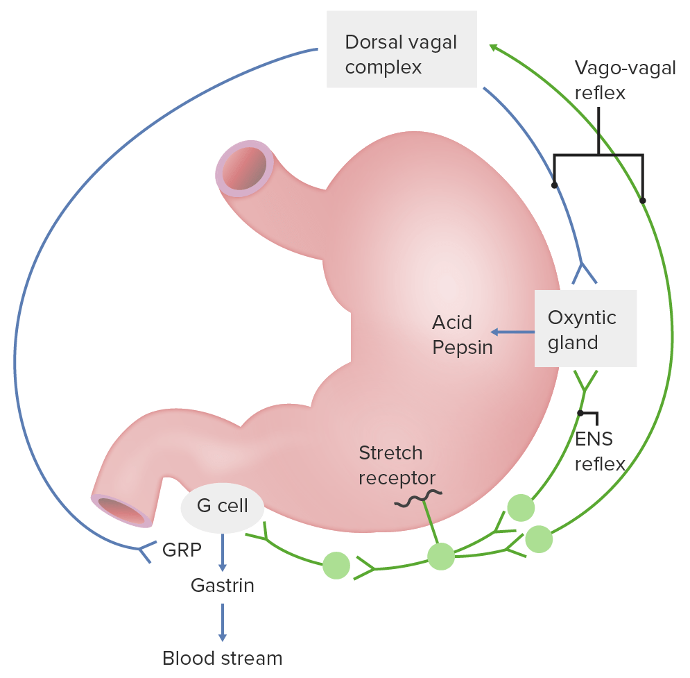
Neural regulation of gastric secretions: The enteric nervous system (ENS) reflex (also known as the short reflex) and the vaso-vagal reflex (also known as the long reflex) are shown. GRP: gastrin-releasing peptide
Image by Lecturio.
Intestinal phase
Begins when food leaves the stomachStomachThe stomach is a muscular sac in the upper left portion of the abdomen that plays a critical role in digestion. The stomach develops from the foregut and connects the esophagus with the duodenum. Structurally, the stomach is C-shaped and forms a greater and lesser curvature and is divided grossly into regions: the cardia, fundus, body, and pylorus. Stomach: Anatomy and enters the duodenumDuodenumThe shortest and widest portion of the small intestine adjacent to the pylorus of the stomach. It is named for having the length equal to about the width of 12 fingers.Small Intestine: Anatomy
DuodenumDuodenumThe shortest and widest portion of the small intestine adjacent to the pylorus of the stomach. It is named for having the length equal to about the width of 12 fingers.Small Intestine: Anatomy:
Modulates gastric activity via hormonesHormonesHormones are messenger molecules that are synthesized in one part of the body and move through the bloodstream to exert specific regulatory effects on another part of the body. Hormones play critical roles in coordinating cellular activities throughout the body in response to the constant changes in both the internal and external environments. Hormones: Overview and Types and neural reflexes:
Initially, the signals stimulate gastric activity.
Soon, the signals inhibit gastric activity.
Secretes signaling moleculesSignaling moleculesSecond Messengers (e.g., cholecystokinin) that stimulate secretions from the pancreasPancreasThe pancreas lies mostly posterior to the stomach and extends across the posterior abdominal wall from the duodenum on the right to the spleen on the left. This organ has both exocrine and endocrine tissue. Pancreas: Anatomy and gallbladderGallbladderThe gallbladder is a pear-shaped sac, located directly beneath the liver, that sits on top of the superior part of the duodenum. The primary functions of the gallbladder include concentrating and storing up to 50 mL of bile. Gallbladder and Biliary Tract: Anatomy
AbsorptionAbsorptionAbsorption involves the uptake of nutrient molecules and their transfer from the lumen of the GI tract across the enterocytes and into the interstitial space, where they can be taken up in the venous or lymphatic circulation.Digestion and Absorption of nutrients begins.
Phases of digestion with their functional components
DigestionDigestionDigestion refers to the process of the mechanical and chemical breakdown of food into smaller particles, which can then be absorbed and utilized by the body.Digestion and Absorption of carbohydratesCarbohydratesA class of organic compounds composed of carbon, hydrogen, and oxygen in a ratio of cn(H2O)n. The largest class of organic compounds, including starch; glycogen; cellulose; polysaccharides; and simple monosaccharides.Basics of Carbohydrates and lipidsLipidsLipids are a diverse group of hydrophobic organic molecules, which include fats, oils, sterols, and waxes.Fatty Acids and Lipids
Salivary glandsSalivary glandsThe salivary glands are exocrine glands positioned in and around the oral cavity. These glands are responsible for secreting saliva into the mouth, which aids in digestion. There are 3 major paired salivary glands: the sublingual, submandibular, and parotid glands.Salivary Glands: Anatomy
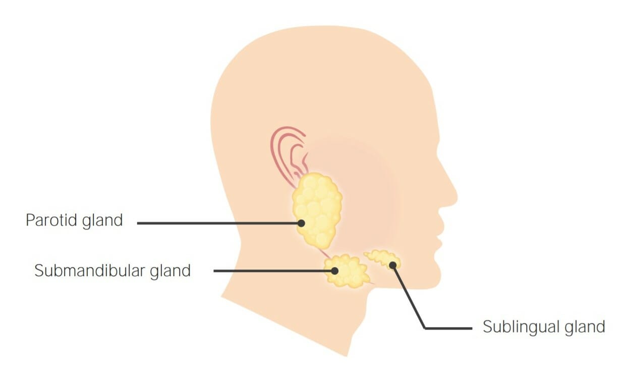
There are 3 primary salivary glandsSalivary glandsThe salivary glands are exocrine glands positioned in and around the oral cavity. These glands are responsible for secreting saliva into the mouth, which aids in digestion. There are 3 major paired salivary glands: the sublingual, submandibular, and parotid glands.Salivary Glands: Anatomy (all with a tubuloacinar structure), which together produce a combination of serous and mucous secretions.
Submandibular glands: both serous and mucous secretions
Sublingual glands: mainly mucous secretions, with a small serous component
Location of the 3 primary salivary glands: Parotid, submandibular, and sublingual glands
Image by Lecturio.
Constituents of salivaSalivaThe clear, viscous fluid secreted by the salivary glands and mucous glands of the mouth. It contains mucins, water, organic salts, and ptyalin.Salivary Glands: Anatomy
SalivaSalivaThe clear, viscous fluid secreted by the salivary glands and mucous glands of the mouth. It contains mucins, water, organic salts, and ptyalin.Salivary Glands: Anatomy consists of:
Water
Mucus
ElectrolytesElectrolytesElectrolytes are mineral salts that dissolve in water and dissociate into charged particles called ions, which can be either be positively (cations) or negatively (anions) charged. Electrolytes are distributed in the extracellular and intracellular compartments in different concentrations. Electrolytes are essential for various basic life-sustaining functions.Electrolytes:
K+: assists in the reabsorption of Na+ and water
HCO3–: buffersBuffersA chemical system that functions to control the levels of specific ions in solution. When the level of hydrogen ion in solution is controlled the system is called a ph buffer.Acid-Base Balance acid
EnzymesEnzymesEnzymes are complex protein biocatalysts that accelerate chemical reactions without being consumed by them. Due to the body’s constant metabolic needs, the absence of enzymes would make life unsustainable, as reactions would occur too slowly without these molecules. Basics of Enzymes:
Muramidase: lysozyme (an enzyme that can destroy cell walls of certain bacteriaBacteriaBacteria are prokaryotic single-celled microorganisms that are metabolically active and divide by binary fission. Some of these organisms play a significant role in the pathogenesis of diseases. Bacteriology)
Lactoferrin: binds ironIronA metallic element with atomic symbol fe, atomic number 26, and atomic weight 55. 85. It is an essential constituent of hemoglobins; cytochromes; and iron-binding proteins. It plays a role in cellular redox reactions and in the transport of oxygen.Trace Elements, which helps prevent bacterial growth
IgAIgARepresents 15-20% of the human serum immunoglobulins, mostly as the 4-chain polymer in humans or dimer in other mammals. Secretory iga is the main immunoglobulin in secretions.Immunoglobulins: Types and Functions: immune mediator (secreted antibody)
SalivaSalivaThe clear, viscous fluid secreted by the salivary glands and mucous glands of the mouth. It contains mucins, water, organic salts, and ptyalin.Salivary Glands: Anatomy production
Overview:
SalivaSalivaThe clear, viscous fluid secreted by the salivary glands and mucous glands of the mouth. It contains mucins, water, organic salts, and ptyalin.Salivary Glands: Anatomy is produced by acinar cells in the salivary glandsSalivary glandsThe salivary glands are exocrine glands positioned in and around the oral cavity. These glands are responsible for secreting saliva into the mouth, which aids in digestion. There are 3 major paired salivary glands: the sublingual, submandibular, and parotid glands.Salivary Glands: Anatomy as a filtrate.
Modified by ductal cells as it moves through the ducts
Salivary glandsSalivary glandsThe salivary glands are exocrine glands positioned in and around the oral cavity. These glands are responsible for secreting saliva into the mouth, which aids in digestion. There are 3 major paired salivary glands: the sublingual, submandibular, and parotid glands.Salivary Glands: Anatomy secrete approximately 1‒1.5 L of salivaSalivaThe clear, viscous fluid secreted by the salivary glands and mucous glands of the mouth. It contains mucins, water, organic salts, and ptyalin.Salivary Glands: Anatomy daily.
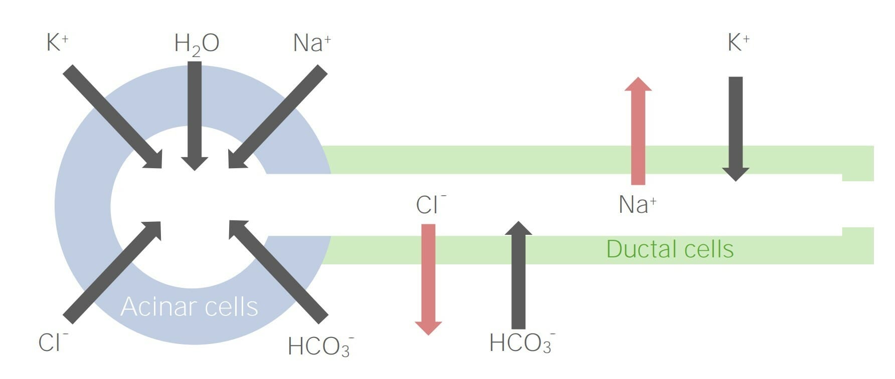
Diagram detailing the ionic secretion by acinar cells and their movement across ductal cells
Image by Lecturio.
Acinar cells:
Acinar cells secrete a filtrate containing Na+, K+, Cl–, HCO3–, water, and other substances.
Na+ moves paracellularly to the acini lumen down its concentration gradient → Na+ is secreted in salivaSalivaThe clear, viscous fluid secreted by the salivary glands and mucous glands of the mouth. It contains mucins, water, organic salts, and ptyalin.Salivary Glands: Anatomy
Brought into acinar cells via the Na+/Cl– cotransporter on the basolateral membrane → Na+ is transported to the interstitial space with H+
CO2 is produced during metabolism → CO2 combines with H2O → carbonic acid (H2CO3) → splits into H+ and HCO3–
H+ and Na+ are transported into the interstitial space via the H+/Na+ cotransporter on the basolateral membrane.
HCO3– (from metabolism) and Cl– (from the Na+/Cl– basolateral cotransporter) are secreted via the HCO3–/Cl– cotransporter on the apical (lumenal) membrane
Produce and secrete other substances (e.g., mucin, enzymesEnzymesEnzymes are complex protein biocatalysts that accelerate chemical reactions without being consumed by them. Due to the body’s constant metabolic needs, the absence of enzymes would make life unsustainable, as reactions would occur too slowly without these molecules. Basics of Enzymes) into the acini
Diagram showing ion secretion by acinar cells: In the acinar cells, Na+, K+, Cl–, and HCO3– are filtered or secreted into the salivary fluid (note: Na+ and Cl– are later reabsorbed by ductal cells).
Image by Lecturio.
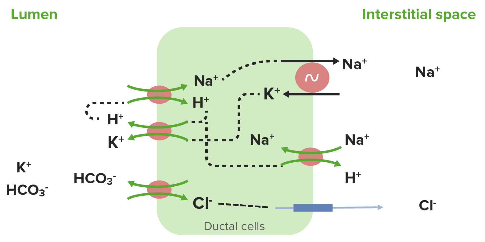
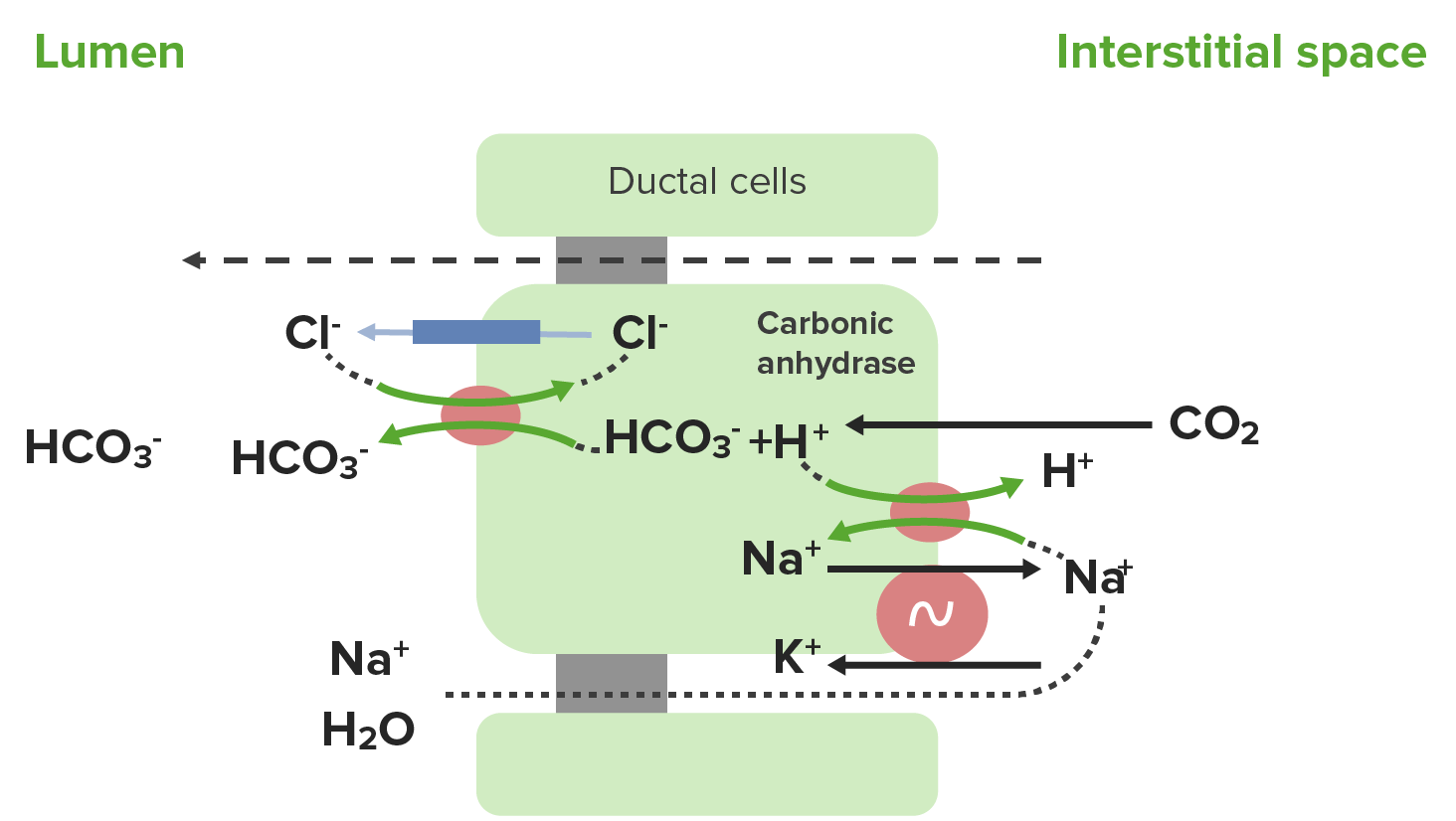
Ductal cells:
Ductal cells modify the filtrate as it moves through the ducts, ultimately reabsorbing Na+ and Cl– and secreting more K+ and HCO3–.
H+ (left over from the carbonic anhydraseCarbonic anhydraseA family of zinc-containing enzymes that catalyze the reversible hydration of carbon dioxide. They play an important role in the transport of carbon dioxide from the tissues to the lung.Carbonic Anhydrase Inhibitors reaction) is removed across the basolateral membrane via the H+/Na+ countertransporter (Na+ is moving down its concentration gradient into the cell).
As water is not reabsorbed (but some ions are), the resulting salivaSalivaThe clear, viscous fluid secreted by the salivary glands and mucous glands of the mouth. It contains mucins, water, organic salts, and ptyalin.Salivary Glands: Anatomy is hypotonicHypotonicSolutions that have a lesser osmotic pressure than a reference solution such as blood, plasma, or interstitial fluid.Renal Sodium and Water Regulation.
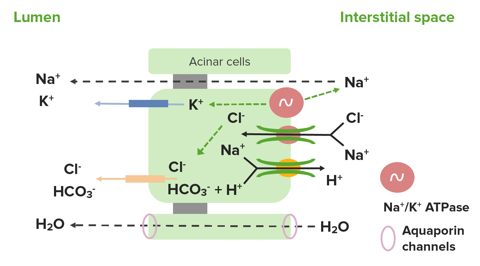
Diagram showing ion transport by ductal cells: The Na+/H+ cotransporter reabsorbs Na+ and H+ from the salivary fluid. The Na+ is then pumped across the basolateral membrane by Na+/K+ ATPase, and K+ is brought into the cell. Next, H+ is recycled back into the lumen along with K+ via an H+/K+ cotransporter. The H+ is then used to reabsorb more Na+, whereas K+ remains in the saliva and is excreted. Chloride is reabsorbed, whereas HCO3– is excreted via the Cl–/HCO3– exchanger on the apical membrane.
Image by Lecturio.
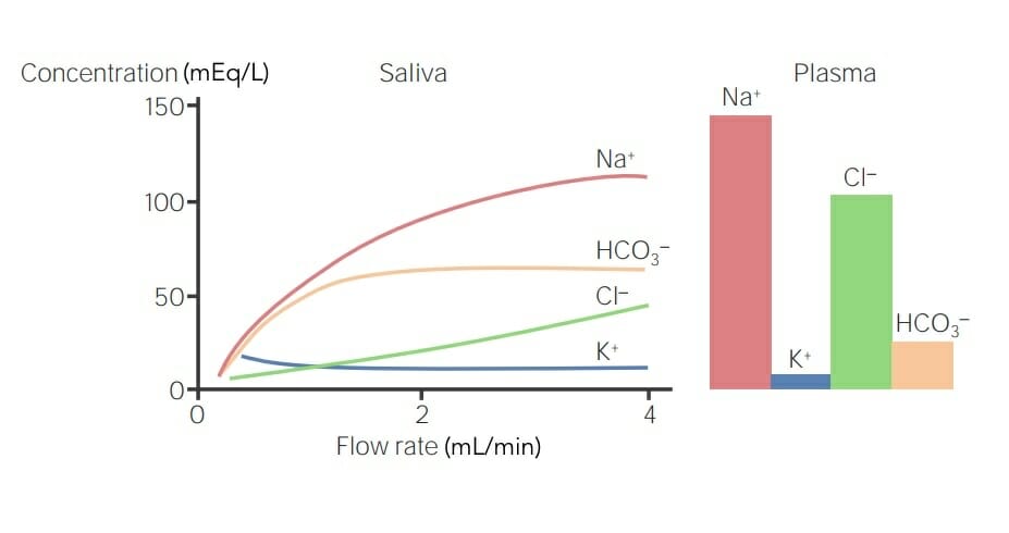
Salivary secretion of ions and their plasma levels: If the concentration of the ion in plasma is higher than that in saliva, the ion is reabsorbed. If the concentration of the ion in plasma is lower than that in saliva, the ion is secreted. The faster the saliva flow, the less time there is for the secretion or reabsorption of ions, thereby affecting their salivary concentrations. Higher concentrations of Na+ and Cl– are present in the plasma than in the saliva and are thus are reabsorbed, whereas K+ and HCO3– are present at lower concentrations in the plasma than in saliva, and are thus secreted.
Image by Lecturio.
Control and regulation of salivary secretions
Involved in the cephalic phase of digestionDigestionDigestion refers to the process of the mechanical and chemical breakdown of food into smaller particles, which can then be absorbed and utilized by the body.Digestion and Absorption:
SensorySensoryNeurons which conduct nerve impulses to the central nervous system.Nervous System: Histology input capable of stimulating salivation:
Thinking about food
Seeing, smelling, or tasting food
Induces higher brainBrainThe part of central nervous system that is contained within the skull (cranium). Arising from the neural tube, the embryonic brain is comprised of three major parts including prosencephalon (the forebrain); mesencephalon (the midbrain); and rhombencephalon (the hindbrain). The developed brain consists of cerebrum; cerebellum; and other structures in the brain stem.Nervous System: Anatomy, Structure, and Classification centers to stimulate the ANSANSThe ans is a component of the peripheral nervous system that uses both afferent (sensory) and efferent (effector) neurons, which control the functioning of the internal organs and involuntary processes via connections with the CNS. The ans consists of the sympathetic and parasympathetic nervous systems.Autonomic Nervous System: Anatomy (primarily the parasympathetics)
AChAChA neurotransmitter found at neuromuscular junctions, autonomic ganglia, parasympathetic effector junctions, a subset of sympathetic effector junctions, and at many sites in the central nervous system.Receptors and Neurotransmitters of the CNS: increases salivary secretions
Vasoactive intestinal peptideVasoactive intestinal peptideA highly basic, 28 amino acid neuropeptide released from intestinal mucosa. It has a wide range of biological actions affecting the cardiovascular, gastrointestinal, and respiratory systems and is neuroprotective. It binds special receptors.Gastrointestinal Neural and Hormonal Signaling (VIPVIPA highly basic, 28 amino acid neuropeptide released from intestinal mucosa. It has a wide range of biological actions affecting the cardiovascular, gastrointestinal, and respiratory systems and is neuroprotective. It binds special receptors.Gastrointestinal Neural and Hormonal Signaling): increases blood flowBlood flowBlood flow refers to the movement of a certain volume of blood through the vasculature over a given unit of time (e.g., mL per minute).Vascular Resistance, Flow, and Mean Arterial Pressure to salivary glandsSalivary glandsThe salivary glands are exocrine glands positioned in and around the oral cavity. These glands are responsible for secreting saliva into the mouth, which aids in digestion. There are 3 major paired salivary glands: the sublingual, submandibular, and parotid glands.Salivary Glands: Anatomy
Sympathetic stimulation: increases salivary secretions to a lesser degree via norepinephrineNorepinephrinePrecursor of epinephrine that is secreted by the adrenal medulla and is a widespread central and autonomic neurotransmitter. Norepinephrine is the principal transmitter of most postganglionic sympathetic fibers, and of the diffuse projection system in the brain that arises from the locus ceruleus.Receptors and Neurotransmitters of the CNS release
Clinical relevance of salivary secretions
XerostomiaXerostomiaDecreased salivary flow.Sjögren Syndrome, or dry mouth, is the clinical term used to identify impaired salivary secretionSecretionCoagulation Studies, which occurs commonly as part of Sjögren syndromeSjögren SyndromeRheumatoid Arthritis, as an adverse effect of some medications (such as antidepressants, antihypertensivesAntihypertensivesThe 1st-line medication classes for hypertension include thiazide-like diuretics, angiotensin-converting enzyme inhibitors (ACEis), angiotensin II receptor blockers (ARBs), and calcium channel blockers (CCBS). Contraindications, adverse effects, and drug-to-drug interactions are agent specific.Hypertension Drugs, or anticholinergicsAnticholinergicsAnticholinergic drugs block the effect of the neurotransmitter acetylcholine at the muscarinic receptors in the central and peripheral nervous systems. Anticholinergic agents inhibit the parasympathetic nervous system, resulting in effects on the smooth muscle in the respiratory tract, vascular system, urinary tract, GI tract, and pupils of the eyes. Anticholinergic Drugs), and in individuals undergoing radiationRadiationEmission or propagation of acoustic waves (sound), electromagnetic energy waves (such as light; radio waves; gamma rays; or x-rays), or a stream of subatomic particles (such as electrons; neutrons; protons; or alpha particles).Osteosarcoma therapy for head and neckNeckThe part of a human or animal body connecting the head to the rest of the body.Peritonsillar Abscess cancers.
Functions of the stomachStomachThe stomach is a muscular sac in the upper left portion of the abdomen that plays a critical role in digestion. The stomach develops from the foregut and connects the esophagus with the duodenum. Structurally, the stomach is C-shaped and forms a greater and lesser curvature and is divided grossly into regions: the cardia, fundus, body, and pylorus. Stomach: Anatomy
Protecting the rest of the GI system by killing most microbes
Preparation of chymeChymeSmall Intestine: Anatomy for the small intestineSmall intestineThe small intestine is the longest part of the GI tract, extending from the pyloric orifice of the stomach to the ileocecal junction. The small intestine is the major organ responsible for chemical digestion and absorption of nutrients. It is divided into 3 segments: the duodenum, the jejunum, and the ileum. Small Intestine: Anatomy via:
Mechanical breakdown
Chemical digestionDigestionDigestion refers to the process of the mechanical and chemical breakdown of food into smaller particles, which can then be absorbed and utilized by the body.Digestion and Absorption by acid and pepsinogen
AbsorptionAbsorptionAbsorption involves the uptake of nutrient molecules and their transfer from the lumen of the GI tract across the enterocytes and into the interstitial space, where they can be taken up in the venous or lymphatic circulation.Digestion and Absorption of lipophilic substances
Storage and gradual release of material into the duodenumDuodenumThe shortest and widest portion of the small intestine adjacent to the pylorus of the stomach. It is named for having the length equal to about the width of 12 fingers.Small Intestine: Anatomy (regulates food entering the small intestineSmall intestineThe small intestine is the longest part of the GI tract, extending from the pyloric orifice of the stomach to the ileocecal junction. The small intestine is the major organ responsible for chemical digestion and absorption of nutrients. It is divided into 3 segments: the duodenum, the jejunum, and the ileum. Small Intestine: Anatomy)
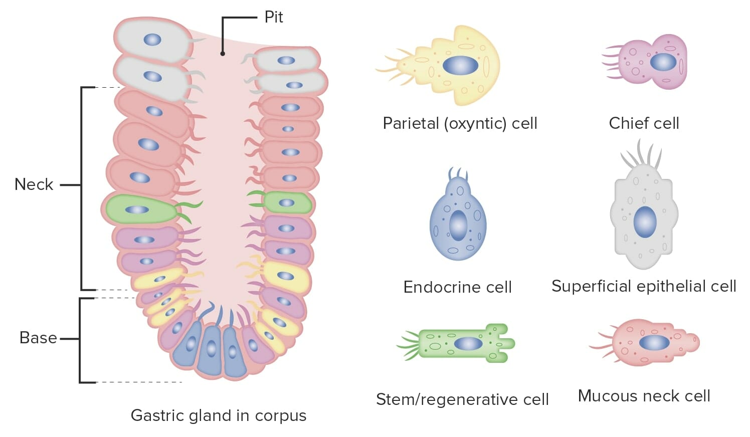
Gastric oxyntic glands: cells and their secretions
Gastric oxyntic glands are found below (and emptying into) the gastric pits. The glands contain numerous cell types, including:
Secrete bicarbonateBicarbonateInorganic salts that contain the -HCO3 radical. They are an important factor in determining the ph of the blood and the concentration of bicarbonate ions is regulated by the kidney. Levels in the blood are an index of the alkali reserve or buffering capacity.Electrolytes and insoluble mucus:
Form a protective barrier against the acidic environment of the stomachStomachThe stomach is a muscular sac in the upper left portion of the abdomen that plays a critical role in digestion. The stomach develops from the foregut and connects the esophagus with the duodenum. Structurally, the stomach is C-shaped and forms a greater and lesser curvature and is divided grossly into regions: the cardia, fundus, body, and pylorus. Stomach: Anatomy
Concentrate bicarbonateBicarbonateInorganic salts that contain the -HCO3 radical. They are an important factor in determining the ph of the blood and the concentration of bicarbonate ions is regulated by the kidney. Levels in the blood are an index of the alkali reserve or buffering capacity.Electrolytes in the mucus
Mucous neckNeckThe part of a human or animal body connecting the head to the rest of the body.Peritonsillar Abscess cells:
Located in the neckNeckThe part of a human or animal body connecting the head to the rest of the body.Peritonsillar Abscess of the glands where they join the gastric pits
Secrete soluble mucus
Stem cells:
Found between the pits and the entrance of the glands
StomachStomachThe stomach is a muscular sac in the upper left portion of the abdomen that plays a critical role in digestion. The stomach develops from the foregut and connects the esophagus with the duodenum. Structurally, the stomach is C-shaped and forms a greater and lesser curvature and is divided grossly into regions: the cardia, fundus, body, and pylorus. Stomach: Anatomy epithelial cells are replaced every 3‒6 days.
Parietal cellsParietal cellsRounded or pyramidal cells of the gastric glands. They secrete hydrochloric acid and produce gastric intrinsic factor, a glycoprotein that binds vitamin B12.Stomach: Anatomy:
Located in the lower-middle region of the glands
Secrete:
HClHCLHairy cell leukemia (HCL) is a rare, chronic, B-cell leukemia characterized by the accumulation of small mature B lymphocytes that have “hair-like projections” visible on microscopy. The abnormal cells accumulate in the peripheral blood, bone marrow (causing fibrosis), and red pulp of the spleen, leading to cytopenias.Hairy Cell Leukemia
Intrinsic factorIntrinsic factorA glycoprotein secreted by the cells of the gastric glands that is required for the absorption of vitamin B 12 (cyanocobalamin). Deficiency of intrinsic factor leads to vitamin B12 deficiency and anemia, pernicious.Gastritis: important for vitamin B12Vitamin B12A cobalt-containing coordination compound produced by intestinal microorganisms and found also in soil and water. Higher plants do not concentrate vitamin B 12 from the soil and so are a poor source of the substance as compared with animal tissues. Intrinsic factor is important for the assimilation of vitamin B 12.Folate and Vitamin B12absorptionAbsorptionAbsorption involves the uptake of nutrient molecules and their transfer from the lumen of the GI tract across the enterocytes and into the interstitial space, where they can be taken up in the venous or lymphatic circulation.Digestion and Absorption
Chief cellsChief cellsEpithelial cells that line the basal half of the gastric glands. Chief cells synthesize and export an inactive enzyme pepsinogen which is converted into the highly proteolytic enzyme pepsin in the acid environment of the stomach.Stomach: Anatomy:
Most numerous glandular cells
Located in the lower-middle region of the glands
Secrete:
Pepsinogen → converted to its active form pepsinPepsinPepsin breaks down proteins into proteoses, peptones, and large polypeptides.Proteins and Peptidesby HClHCLHairy cell leukemia (HCL) is a rare, chronic, B-cell leukemia characterized by the accumulation of small mature B lymphocytes that have “hair-like projections” visible on microscopy. The abnormal cells accumulate in the peripheral blood, bone marrow (causing fibrosis), and red pulp of the spleen, leading to cytopenias.Hairy Cell Leukemia→ breaks down proteinsProteinsLinear polypeptides that are synthesized on ribosomes and may be further modified, crosslinked, cleaved, or assembled into complex proteins with several subunits. The specific sequence of amino acids determines the shape the polypeptide will take, during protein folding, and the function of the protein.Energy Homeostasis
Gastric lipaseGastric lipaseDigestion and Absorption → breaks down fatsFatsThe glyceryl esters of a fatty acid, or of a mixture of fatty acids. They are generally odorless, colorless, and tasteless if pure, but they may be flavored according to origin. Fats are insoluble in water, soluble in most organic solvents. They occur in animal and vegetable tissue and are generally obtained by boiling or by extraction under pressure. They are important in the diet (dietary fats) as a source of energy.Energy Homeostasis
Enteroendocrine cells:
Located in the base of the glands
D cells secrete somatostatin:
Inhibition of many secretions
Released in response to H+ (the natural way to “turn off” acid production)
G cells secrete gastrin:
Stimulates parietal cellsParietal cellsRounded or pyramidal cells of the gastric glands. They secrete hydrochloric acid and produce gastric intrinsic factor, a glycoprotein that binds vitamin B12.Stomach: Anatomy to secrete HClHCLHairy cell leukemia (HCL) is a rare, chronic, B-cell leukemia characterized by the accumulation of small mature B lymphocytes that have “hair-like projections” visible on microscopy. The abnormal cells accumulate in the peripheral blood, bone marrow (causing fibrosis), and red pulp of the spleen, leading to cytopenias.Hairy Cell Leukemia
Has trophic/growth effects on the GI mucosa
Released in response to proteinsProteinsLinear polypeptides that are synthesized on ribosomes and may be further modified, crosslinked, cleaved, or assembled into complex proteins with several subunits. The specific sequence of amino acids determines the shape the polypeptide will take, during protein folding, and the function of the protein.Energy Homeostasis, peptides, and amino acidsAmino acidsOrganic compounds that generally contain an amino (-NH2) and a carboxyl (-COOH) group. Twenty alpha-amino acids are the subunits which are polymerized to form proteins.Basics of Amino Acids
Enterochromaffin-like (ECL) cells secrete histamine (which stimulates parietal cellsParietal cellsRounded or pyramidal cells of the gastric glands. They secrete hydrochloric acid and produce gastric intrinsic factor, a glycoprotein that binds vitamin B12.Stomach: Anatomy to secrete HClHCLHairy cell leukemia (HCL) is a rare, chronic, B-cell leukemia characterized by the accumulation of small mature B lymphocytes that have “hair-like projections” visible on microscopy. The abnormal cells accumulate in the peripheral blood, bone marrow (causing fibrosis), and red pulp of the spleen, leading to cytopenias.Hairy Cell Leukemia).
Other chemical messengers secreted by enteroendocrine cells:
Substance P
VIPVIPA highly basic, 28 amino acid neuropeptide released from intestinal mucosa. It has a wide range of biological actions affecting the cardiovascular, gastrointestinal, and respiratory systems and is neuroprotective. It binds special receptors.Gastrointestinal Neural and Hormonal Signaling
Secretin
Neuropeptide Y
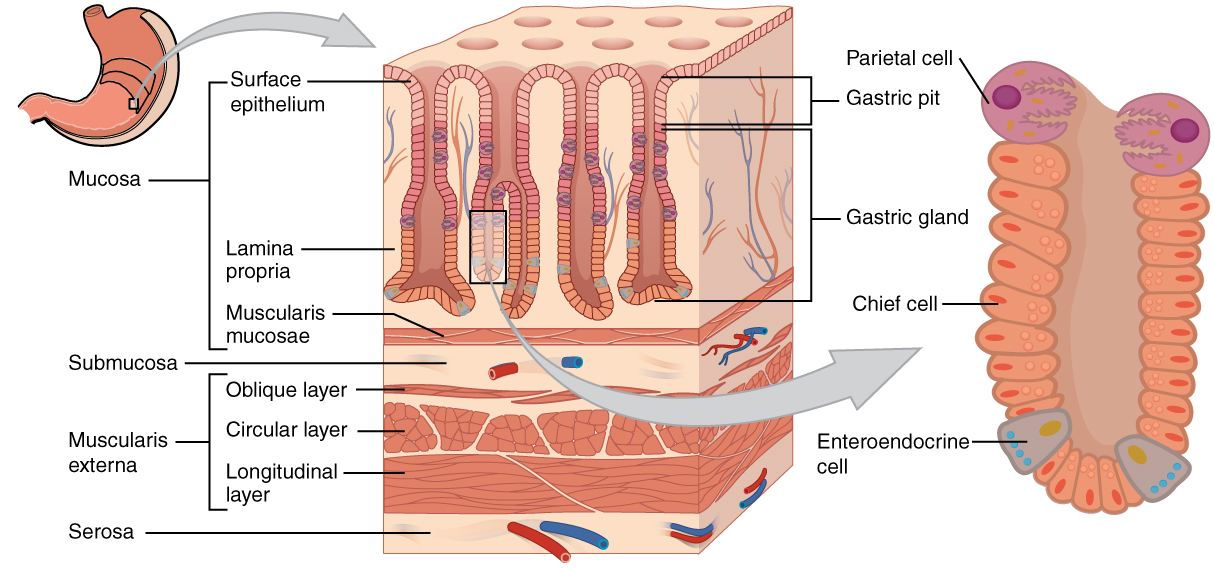
Layers of the stomach wall: In the epithelium, gastric pits lead to gastric glands that secrete a variety of substances to aid in digestion.
Image: “The stomach wall is adapted for the functions of the stomach” by OpenStax College. License: CC BY 4.0
Structure of a gastric gland with its different cell types
Image by Lecturio.
Production and secretionSecretionCoagulation Studies of acid in parietal cellsParietal cellsRounded or pyramidal cells of the gastric glands. They secrete hydrochloric acid and produce gastric intrinsic factor, a glycoprotein that binds vitamin B12.Stomach: Anatomy
Normal metabolism produces CO2 → combines with H2O → H2CO3 → splits into H+ + HCO3–
H+ is pumped out to the lumen in exchange for K+ by H+/K+ ATPase:
1 K+ molecule is brought into the cell.
K+ leaves the cell through the basolateral membrane down its concentration gradient through a K+ channel.
HCO3– is exchanged with Cl– across the basolateral membrane:
HCO3– is moved into the interstitial space.
Cl– is brought into the cell → moves into the lumen through its own channel
End result:
H+ and Cl– are secreted into the lumen.
K+ and HCO3– are moved into the interstitial space.
Ion movement in parietal cells: Carbonic acid dissociates into H+ and HCO3–. The H+ is exchanged for K+ in the apical membrane by H+/K+ ATPase. An HCO3– is exchanged for Cl– in the basolateral membrane; Cl– is then moved into the lumen.
Image by Lecturio.
Control and regulation of gastric secretions
Gastric secretions are heavily influenced by parasympathetic signaling via the vagus nerveVagus nerveThe 10th cranial nerve. The vagus is a mixed nerve which contains somatic afferents (from skin in back of the ear and the external auditory meatus), visceral afferents (from the pharynx, larynx, thorax, and abdomen), parasympathetic efferents (to the thorax and abdomen), and efferents to striated muscle (of the larynx and pharynx).Pharynx: Anatomy (cranial nerve X), which releases AChAChA neurotransmitter found at neuromuscular junctions, autonomic ganglia, parasympathetic effector junctions, a subset of sympathetic effector junctions, and at many sites in the central nervous system.Receptors and Neurotransmitters of the CNS that induces acid production by several pathways.
Production of secretions:
40% in the cephalic phase
50% in the gastric phase
10% in the intestinal phase
HClHCLHairy cell leukemia (HCL) is a rare, chronic, B-cell leukemia characterized by the accumulation of small mature B lymphocytes that have “hair-like projections” visible on microscopy. The abnormal cells accumulate in the peripheral blood, bone marrow (causing fibrosis), and red pulp of the spleen, leading to cytopenias.Hairy Cell LeukemiasecretionSecretionCoagulation Studies from parietal cellsParietal cellsRounded or pyramidal cells of the gastric glands. They secrete hydrochloric acid and produce gastric intrinsic factor, a glycoprotein that binds vitamin B12.Stomach: Anatomy is stimulated by:
AChAChA neurotransmitter found at neuromuscular junctions, autonomic ganglia, parasympathetic effector junctions, a subset of sympathetic effector junctions, and at many sites in the central nervous system.Receptors and Neurotransmitters of the CNS from the vagus and myenteric nerves (long and short reflex pathways, respectively)
Gastrin from G cells in the stomachStomachThe stomach is a muscular sac in the upper left portion of the abdomen that plays a critical role in digestion. The stomach develops from the foregut and connects the esophagus with the duodenum. Structurally, the stomach is C-shaped and forms a greater and lesser curvature and is divided grossly into regions: the cardia, fundus, body, and pylorus. Stomach: Anatomy
Histamine from ECL cells in the stomachStomachThe stomach is a muscular sac in the upper left portion of the abdomen that plays a critical role in digestion. The stomach develops from the foregut and connects the esophagus with the duodenum. Structurally, the stomach is C-shaped and forms a greater and lesser curvature and is divided grossly into regions: the cardia, fundus, body, and pylorus. Stomach: Anatomy
HClHCLHairy cell leukemia (HCL) is a rare, chronic, B-cell leukemia characterized by the accumulation of small mature B lymphocytes that have “hair-like projections” visible on microscopy. The abnormal cells accumulate in the peripheral blood, bone marrow (causing fibrosis), and red pulp of the spleen, leading to cytopenias.Hairy Cell LeukemiasecretionSecretionCoagulation Studies is inhibited by:
Somatostatin
ProstaglandinsProstaglandinsA group of compounds derived from unsaturated 20-carbon fatty acids, primarily arachidonic acid, via the cyclooxygenase pathway. They are extremely potent mediators of a diverse group of physiological processes.Eicosanoids
Acid-stimulation pathways
Direct pathwayDirect PathwayHuntington Disease(direct activation of parietal cellsParietal cellsRounded or pyramidal cells of the gastric glands. They secrete hydrochloric acid and produce gastric intrinsic factor, a glycoprotein that binds vitamin B12.Stomach: Anatomy):
AChAChA neurotransmitter found at neuromuscular junctions, autonomic ganglia, parasympathetic effector junctions, a subset of sympathetic effector junctions, and at many sites in the central nervous system.Receptors and Neurotransmitters of the CNS stimulates the muscarinic (M3) receptorsReceptorsReceptors are proteins located either on the surface of or within a cell that can bind to signaling molecules known as ligands (e.g., hormones) and cause some type of response within the cell.Receptors of parietal cellsParietal cellsRounded or pyramidal cells of the gastric glands. They secrete hydrochloric acid and produce gastric intrinsic factor, a glycoprotein that binds vitamin B12.Stomach: Anatomy.
Activates Gq (a G protein)
Gq activates phospholipase CPhospholipase CA subclass of phospholipases that hydrolyze the phosphoester bond found in the third position of glycerophospholipids. Although the singular term phospholipase C specifically refers to an enzyme that catalyzes the hydrolysis of phosphatidylcholine, it is commonly used in the literature to refer to broad variety of enzymes that specifically catalyze the hydrolysis of phosphatidylinositols.Pseudomonas (PLC).
PLC cleaves phosphatidylinositol-4,5-bisphosphate (PIP2) to produce:
Inositol trisphosphateInositol trisphosphateIntracellular messenger formed by the action of phospholipase C on phosphatidylinositol 4, 5-bisphosphate, which is one of the phospholipids that make up the cell membrane. Inositol 1, 4, 5-trisphosphate is released into the cytoplasm where it releases calcium ions from internal stores within the cell’s endoplasmic reticulum. These calcium ions stimulate the activity of B kinase or calmodulin.Second Messengers (IP3) → calciumCalciumA basic element found in nearly all tissues. It is a member of the alkaline earth family of metals with the atomic symbol ca, atomic number 20, and atomic weight 40. Calcium is the most abundant mineral in the body and combines with phosphorus to form calcium phosphate in the bones and teeth. It is essential for the normal functioning of nerves and muscles and plays a role in blood coagulation (as factor IV) and in many enzymatic processes.Electrolytes (CaCACondylomata acuminata are a clinical manifestation of genital HPV infection. Condylomata acuminata are described as raised, pearly, flesh-colored, papular, cauliflower-like lesions seen in the anogenital region that may cause itching, pain, or bleeding.Condylomata Acuminata (Genital Warts)2+) release from the ER
Both CaCACondylomata acuminata are a clinical manifestation of genital HPV infection. Condylomata acuminata are described as raised, pearly, flesh-colored, papular, cauliflower-like lesions seen in the anogenital region that may cause itching, pain, or bleeding.Condylomata Acuminata (Genital Warts)2+ and PKC activate the H+/K+ ATPase to secrete H+.
Gastrin pathway:
AChAChA neurotransmitter found at neuromuscular junctions, autonomic ganglia, parasympathetic effector junctions, a subset of sympathetic effector junctions, and at many sites in the central nervous system.Receptors and Neurotransmitters of the CNS stimulates G cells to release gastrin.
Gastrin activates cholecystokinin B receptorsReceptorsReceptors are proteins located either on the surface of or within a cell that can bind to signaling molecules known as ligands (e.g., hormones) and cause some type of response within the cell.Receptors on parietal cellsParietal cellsRounded or pyramidal cells of the gastric glands. They secrete hydrochloric acid and produce gastric intrinsic factor, a glycoprotein that binds vitamin B12.Stomach: Anatomy.
Cholecystokinin B activates PLC → cleaves PIP2 into IP3 + DAGDAGSecond Messengers → ↑ CaCACondylomata acuminata are a clinical manifestation of genital HPV infection. Condylomata acuminata are described as raised, pearly, flesh-colored, papular, cauliflower-like lesions seen in the anogenital region that may cause itching, pain, or bleeding.Condylomata Acuminata (Genital Warts)2+ + PKC → ↑ H+/K+ ATPase activity
Histamine pathway:
AChAChA neurotransmitter found at neuromuscular junctions, autonomic ganglia, parasympathetic effector junctions, a subset of sympathetic effector junctions, and at many sites in the central nervous system.Receptors and Neurotransmitters of the CNS stimulates ECL cells to release histamine.
Histamine activates H2receptorsReceptorsReceptors are proteins located either on the surface of or within a cell that can bind to signaling molecules known as ligands (e.g., hormones) and cause some type of response within the cell.Receptors on parietal cellsParietal cellsRounded or pyramidal cells of the gastric glands. They secrete hydrochloric acid and produce gastric intrinsic factor, a glycoprotein that binds vitamin B12.Stomach: Anatomy.
H2receptorsReceptorsReceptors are proteins located either on the surface of or within a cell that can bind to signaling molecules known as ligands (e.g., hormones) and cause some type of response within the cell.Receptors activate Gs (a G protein).
Gs activates adenylate cyclase (AC).
AC converts ATP to cAMPcAMPAn adenine nucleotide containing one phosphate group which is esterified to both the 3′- and 5′-positions of the sugar moiety. It is a second messenger and a key intracellular regulator, functioning as a mediator of activity for a number of hormones, including epinephrine, glucagon, and acth.Phosphodiesterase Inhibitors.
cAMPcAMPAn adenine nucleotide containing one phosphate group which is esterified to both the 3′- and 5′-positions of the sugar moiety. It is a second messenger and a key intracellular regulator, functioning as a mediator of activity for a number of hormones, including epinephrine, glucagon, and acth.Phosphodiesterase Inhibitors phosphorylates/activates PKA.
PKA stimulates H+/K+ ATPase to secrete H+.
The effects of these pathways are potentiating/synergistic in nature → AChAChA neurotransmitter found at neuromuscular junctions, autonomic ganglia, parasympathetic effector junctions, a subset of sympathetic effector junctions, and at many sites in the central nervous system.Receptors and Neurotransmitters of the CNS + gastrin + histamine activity simultaneously results in greater HClHCLHairy cell leukemia (HCL) is a rare, chronic, B-cell leukemia characterized by the accumulation of small mature B lymphocytes that have “hair-like projections” visible on microscopy. The abnormal cells accumulate in the peripheral blood, bone marrow (causing fibrosis), and red pulp of the spleen, leading to cytopenias.Hairy Cell LeukemiasecretionSecretionCoagulation Studies than the sum of HClHCLHairy cell leukemia (HCL) is a rare, chronic, B-cell leukemia characterized by the accumulation of small mature B lymphocytes that have “hair-like projections” visible on microscopy. The abnormal cells accumulate in the peripheral blood, bone marrow (causing fibrosis), and red pulp of the spleen, leading to cytopenias.Hairy Cell LeukemiasecretionSecretionCoagulation Studies if they each acted alone
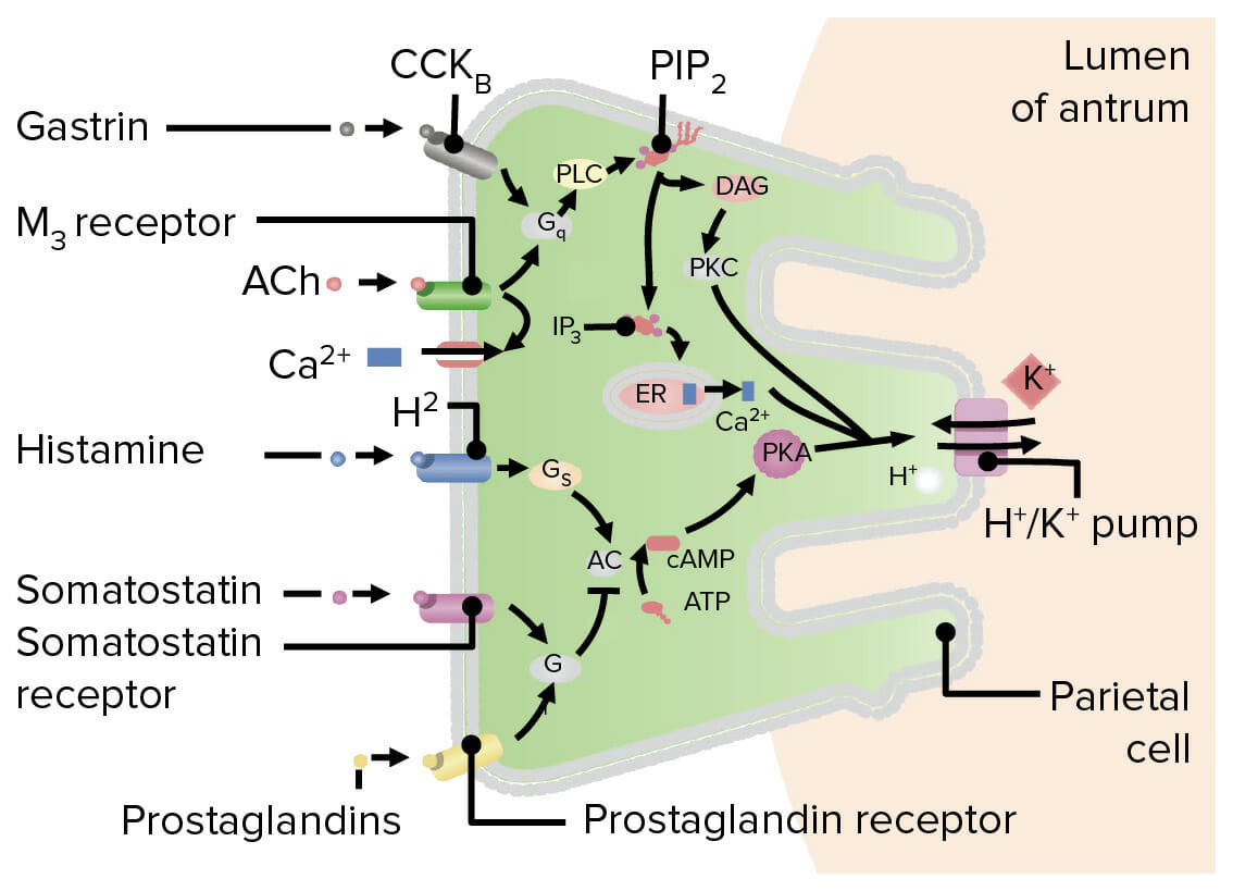
Diagram showing stimulation and inhibition pathways for acid secretion in a parietal cell: Gastrin and acetylcholine activate Gq, a G protein, which cleaves phosphatidylinositol-4,5-bisphosphate (PIP2) into inositol trisphosphate (IP3) and diacylglycerol (DAG). Inositol trisphosphate causes calcium (Ca+2) release from the ER, while DAG activates protein kinase C (PKC) via phosphorylation. Both Ca+2 and PKC stimulate the H+/K+ pump to secrete acid. Histamine activates Gs, which activates adenylate cyclase (AC), resulting in an increase of intracellular cAMP. Next, cAMP activates protein kinase A (PKA), which stimulates the H+/K+ pump to secrete acid. Somatostatin and prostaglandins inhibit acid production by inducing Gi, which inhibits AC. CCK: cholecystokinin
PLC: phospholipase C
Ach: acetylcholine
Image by Lecturio.
Diagram detailing the direct and indirect pathways of acid-release stimulation by acetylcholine (ACh): The direct pathway involves stimulation of the muscarinic (M3) receptors on parietal cells. The indirect pathways involve the stimulation of enterochromaffin-like (ECL) cells, inducing the release of histamine, and the stimulation of G cells inducing them to secrete gastrin. Both histamine and gastrin then stimulate parietal cells to secrete acid. ENS: enteric nervous system CCK: cholecystokinin
Image by Lecturio.
Acid-inhibition pathways
Somatostatin and prostaglandinsProstaglandinsA group of compounds derived from unsaturated 20-carbon fatty acids, primarily arachidonic acid, via the cyclooxygenase pathway. They are extremely potent mediators of a diverse group of physiological processes.Eicosanoids:
Somatostatin (released from D cells in the stomachStomachThe stomach is a muscular sac in the upper left portion of the abdomen that plays a critical role in digestion. The stomach develops from the foregut and connects the esophagus with the duodenum. Structurally, the stomach is C-shaped and forms a greater and lesser curvature and is divided grossly into regions: the cardia, fundus, body, and pylorus. Stomach: Anatomy) and prostaglandinsProstaglandinsA group of compounds derived from unsaturated 20-carbon fatty acids, primarily arachidonic acid, via the cyclooxygenase pathway. They are extremely potent mediators of a diverse group of physiological processes.Eicosanoids activate the inhibitory Gi protein.
Gi inhibits AC → cAMPcAMPAn adenine nucleotide containing one phosphate group which is esterified to both the 3′- and 5′-positions of the sugar moiety. It is a second messenger and a key intracellular regulator, functioning as a mediator of activity for a number of hormones, including epinephrine, glucagon, and acth.Phosphodiesterase Inhibitors levels fall → ↓ activation of PKA → ↓ H+/K+ ATPase activity
Clinical relevance of gastric secretions
GERDGERDGastroesophageal reflux disease (GERD) occurs when the stomach acid frequently flows back into the esophagus. This backwash (acid reflux) can irritate the lining of the esophagus, causing symptoms such as retrosternal burning pain (heartburn). Gastroesophageal Reflux Disease (GERD): occurs when the stomachStomachThe stomach is a muscular sac in the upper left portion of the abdomen that plays a critical role in digestion. The stomach develops from the foregut and connects the esophagus with the duodenum. Structurally, the stomach is C-shaped and forms a greater and lesser curvature and is divided grossly into regions: the cardia, fundus, body, and pylorus. Stomach: Anatomy acid frequently flows back into the esophagusEsophagusThe esophagus is a muscular tube-shaped organ of around 25 centimeters in length that connects the pharynx to the stomach. The organ extends from approximately the 6th cervical vertebra to the 11th thoracic vertebra and can be divided grossly into 3 parts: the cervical part, the thoracic part, and the abdominal part. Esophagus: Anatomy. The backwash (acid reflux) can irritate the lining of the esophagusEsophagusThe esophagus is a muscular tube-shaped organ of around 25 centimeters in length that connects the pharynx to the stomach. The organ extends from approximately the 6th cervical vertebra to the 11th thoracic vertebra and can be divided grossly into 3 parts: the cervical part, the thoracic part, and the abdominal part. Esophagus: Anatomy, causing symptoms such as retrosternal burning painPainAn unpleasant sensation induced by noxious stimuli which are detected by nerve endings of nociceptive neurons.Pain: Types and Pathways (heartburnHeartburnSubsternal pain or burning sensation, usually associated with regurgitation of gastric juice into the esophagus.Gastroesophageal Reflux Disease (GERD)) and may eventually lead to inflammationInflammationInflammation is a complex set of responses to infection and injury involving leukocytes as the principal cellular mediators in the body’s defense against pathogenic organisms. Inflammation is also seen as a response to tissue injury in the process of wound healing. The 5 cardinal signs of inflammation are pain, heat, redness, swelling, and loss of function. Inflammation (esophagitisEsophagitisEsophagitis is the inflammation or irritation of the esophagus. The major types of esophagitis are medication-induced, infectious, eosinophilic, corrosive, and acid reflux. Patients typically present with odynophagia, dysphagia, and retrosternal chest pain. Esophagitis), metaplasiaMetaplasiaA condition in which there is a change of one adult cell type to another similar adult cell type.Cellular Adaptation (Barrett esophagusEsophagusThe esophagus is a muscular tube-shaped organ of around 25 centimeters in length that connects the pharynx to the stomach. The organ extends from approximately the 6th cervical vertebra to the 11th thoracic vertebra and can be divided grossly into 3 parts: the cervical part, the thoracic part, and the abdominal part. Esophagus: Anatomy), and progression to esophageal cancerEsophageal cancerEsophageal cancer is 1 of the most common causes of cancer-related deaths worldwide. Nearly all esophageal cancers are either adenocarcinoma (commonly affecting the distal esophagus) or squamous cell carcinoma (affecting the proximal two-thirds of the esophagus). Esophageal Cancer. Uncomplicated GERDGERDGastroesophageal reflux disease (GERD) occurs when the stomach acid frequently flows back into the esophagus. This backwash (acid reflux) can irritate the lining of the esophagus, causing symptoms such as retrosternal burning pain (heartburn). Gastroesophageal Reflux Disease (GERD) can be managed with lifestyle changes and over-the-counter medications.
Barrett esophagusEsophagusThe esophagus is a muscular tube-shaped organ of around 25 centimeters in length that connects the pharynx to the stomach. The organ extends from approximately the 6th cervical vertebra to the 11th thoracic vertebra and can be divided grossly into 3 parts: the cervical part, the thoracic part, and the abdominal part. Esophagus: Anatomy: a condition characterized by metaplastic changes in the normal stratified squamous epitheliumStratified squamous epitheliumSurface Epithelium: Histology of the esophagusEsophagusThe esophagus is a muscular tube-shaped organ of around 25 centimeters in length that connects the pharynx to the stomach. The organ extends from approximately the 6th cervical vertebra to the 11th thoracic vertebra and can be divided grossly into 3 parts: the cervical part, the thoracic part, and the abdominal part. Esophagus: Anatomy to columnar epitheliumEpitheliumThe epithelium is a complex of specialized cellular organizations arranged into sheets and lining cavities and covering the surfaces of the body. The cells exhibit polarity, having an apical and a basal pole. Structures important for the epithelial integrity and function involve the basement membrane, the semipermeable sheet on which the cells rest, and interdigitations, as well as cellular junctions. Surface Epithelium: Histology. The change is a consequence of chronic GERDGERDGastroesophageal reflux disease (GERD) occurs when the stomach acid frequently flows back into the esophagus. This backwash (acid reflux) can irritate the lining of the esophagus, causing symptoms such as retrosternal burning pain (heartburn). Gastroesophageal Reflux Disease (GERD) and is considered premalignant.
Medications to reducegastric acidGastric acidHydrochloric acid present in gastric juice.Gastroesophageal Reflux Disease (GERD) secretions: Drugs include proton pumpPumpACES and RUSH: Resuscitation Ultrasound Protocols inhibitors (PPIs) and H2receptorReceptorReceptors are proteins located either on the surface of or within a cell that can bind to signaling molecules known as ligands (e.g., hormones) and cause some type of response within the cell.Receptors antagonists and are most commonly indicated in treating peptic ulcerPeptic ulcerPeptic ulcer disease (PUD) refers to the full-thickness ulcerations of duodenal or gastric mucosa. The ulcerations form when exposure to acid and digestive enzymes overcomes mucosal defense mechanisms. The most common etiologies include Helicobacter pylori (H. pylori) infection and prolonged use of non-steroidal anti-inflammatory drugs (NSAIDs). Peptic Ulcer Disease disease (PUDPUDPeptic ulcer disease (PUD) refers to the full-thickness ulcerations of duodenal or gastric mucosa. The ulcerations form when exposure to acid and digestive enzymes overcomes mucosal defense mechanisms. The most common etiologies include Helicobacter pylori (H. pylori) infection and prolonged use of non-steroidal anti-inflammatory drugs (NSAIDs). Peptic Ulcer Disease), GERDGERDGastroesophageal reflux disease (GERD) occurs when the stomach acid frequently flows back into the esophagus. This backwash (acid reflux) can irritate the lining of the esophagus, causing symptoms such as retrosternal burning pain (heartburn). Gastroesophageal Reflux Disease (GERD), and dyspepsiaDyspepsiaImpaired digestion, especially after eating.Lactose Intolerance. The mechanism of action of PPIs in reducing gastric acidGastric acidHydrochloric acid present in gastric juice.Gastroesophageal Reflux Disease (GERD) is by inhibiting the H+/K+ ATPase in parietal cellsParietal cellsRounded or pyramidal cells of the gastric glands. They secrete hydrochloric acid and produce gastric intrinsic factor, a glycoprotein that binds vitamin B12.Stomach: Anatomy, whereas that of H2 blockers is by inhibiting the stimulatory effects of histamine on parietal cellsParietal cellsRounded or pyramidal cells of the gastric glands. They secrete hydrochloric acid and produce gastric intrinsic factor, a glycoprotein that binds vitamin B12.Stomach: Anatomy.
Zollinger-Ellison syndromeZollinger-ellison syndromeA syndrome that is characterized by the triad of severe peptic ulcer, hypersecretion of gastric acid, and gastrin-producing tumors of the pancreas or other tissue (gastrinoma). This syndrome may be sporadic or be associated with multiple endocrine neoplasia type 1.Esophagitis (ZES): a gastrin-secreting tumorTumorInflammation (often malignant) arising from the pancreasPancreasThe pancreas lies mostly posterior to the stomach and extends across the posterior abdominal wall from the duodenum on the right to the spleen on the left. This organ has both exocrine and endocrine tissue. Pancreas: Anatomy, stomachStomachThe stomach is a muscular sac in the upper left portion of the abdomen that plays a critical role in digestion. The stomach develops from the foregut and connects the esophagus with the duodenum. Structurally, the stomach is C-shaped and forms a greater and lesser curvature and is divided grossly into regions: the cardia, fundus, body, and pylorus. Stomach: Anatomy, duodenumDuodenumThe shortest and widest portion of the small intestine adjacent to the pylorus of the stomach. It is named for having the length equal to about the width of 12 fingers.Small Intestine: Anatomy, jejunumJejunumThe middle portion of the small intestine, between duodenum and ileum. It represents about 2/5 of the remaining portion of the small intestine below duodenum.Small Intestine: Anatomy, and/or lymph nodesLymph NodesThey are oval or bean shaped bodies (1 – 30 mm in diameter) located along the lymphatic system.Lymphatic Drainage System: Anatomy, which is characterized by recurrent/refractory peptic ulcers, gastroesophageal reflux, and diarrheaDiarrheaDiarrhea is defined as ≥ 3 watery or loose stools in a 24-hour period. There are a multitude of etiologies, which can be classified based on the underlying mechanism of disease. The duration of symptoms (acute or chronic) and characteristics of the stools (e.g., watery, bloody, steatorrheic, mucoid) can help guide further diagnostic evaluation. Diarrhea. Diagnosis is based on elevated fasting serum gastrin levels. Treatment is with surgical resection of the tumorTumorInflammation and/or symptomatic management.
Exocrine (85% by massMassThree-dimensional lesion that occupies a space within the breastImaging of the Breast):
Release pancreatic enzymesEnzymesEnzymes are complex protein biocatalysts that accelerate chemical reactions without being consumed by them. Due to the body’s constant metabolic needs, the absence of enzymes would make life unsustainable, as reactions would occur too slowly without these molecules. Basics of Enzymes into the duodenumDuodenumThe shortest and widest portion of the small intestine adjacent to the pylorus of the stomach. It is named for having the length equal to about the width of 12 fingers.Small Intestine: Anatomy
Arranged as clusters of acini draining into a ductal system → main and accessory pancreatic ducts → duodenumDuodenumThe shortest and widest portion of the small intestine adjacent to the pylorus of the stomach. It is named for having the length equal to about the width of 12 fingers.Small Intestine: Anatomy
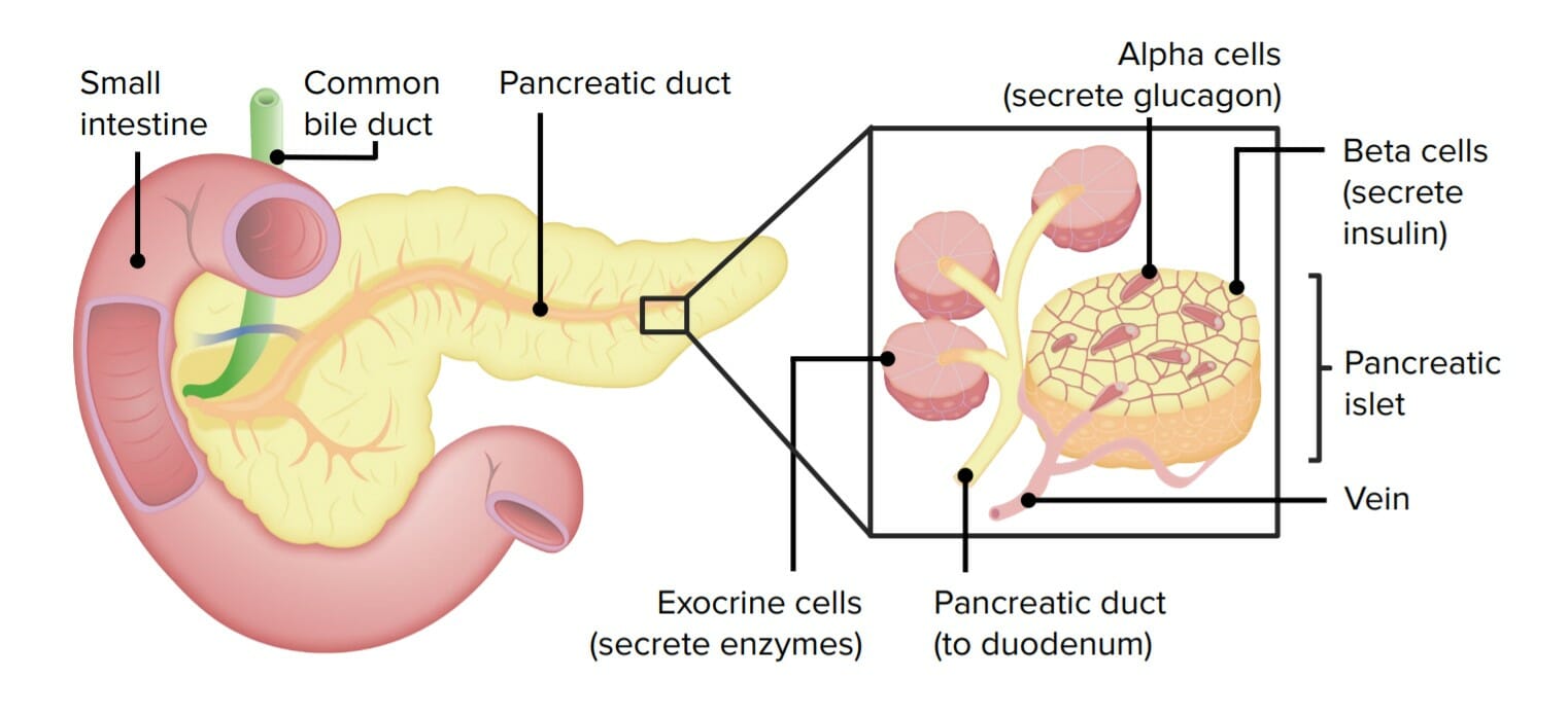
Endocrine:
Releases hormonesHormonesHormones are messenger molecules that are synthesized in one part of the body and move through the bloodstream to exert specific regulatory effects on another part of the body. Hormones play critical roles in coordinating cellular activities throughout the body in response to the constant changes in both the internal and external environments. Hormones: Overview and Types into the bloodstream
β cells: secretionSecretionCoagulation Studies of insulinInsulinInsulin is a peptide hormone that is produced by the beta cells of the pancreas. Insulin plays a role in metabolic functions such as glucose uptake, glycolysis, glycogenesis, lipogenesis, and protein synthesis. Exogenous insulin may be needed for individuals with diabetes mellitus, in whom there is a deficiency in endogenous insulin or increased insulin resistance. Insulin
A drawing of the pancreas identifying its 2 major tissue components: the endocrine pancreas (islets of Langerhans or pancreatic islets) and exocrine pancreas (exocrine cells or pancreatic acini)
Image by Lecturio.
Exocrine pancreasExocrine pancreasThe major component (about 80%) of the pancreas composed of acinar functional units of tubular and spherical cells. The acinar cells synthesize and secrete several digestive enzymes such as trypsinogen; lipase; amylase; and ribonuclease. Secretion from the exocrine pancreas drains into the pancreatic ductal system and empties into the duodenum.Pancreas: Anatomy secretions
The exocrine pancreasExocrine pancreasThe major component (about 80%) of the pancreas composed of acinar functional units of tubular and spherical cells. The acinar cells synthesize and secrete several digestive enzymes such as trypsinogen; lipase; amylase; and ribonuclease. Secretion from the exocrine pancreas drains into the pancreatic ductal system and empties into the duodenum.Pancreas: Anatomy secretes a mixture known as pancreatic juicePancreatic JuiceThe fluid containing digestive enzymes secreted by the pancreas in response to food in the duodenum.Pancreas: Anatomy, which contains water, enzymesEnzymesEnzymes are complex protein biocatalysts that accelerate chemical reactions without being consumed by them. Due to the body’s constant metabolic needs, the absence of enzymes would make life unsustainable, as reactions would occur too slowly without these molecules. Basics of Enzymes, zymogens (inactive proteinsProteinsLinear polypeptides that are synthesized on ribosomes and may be further modified, crosslinked, cleaved, or assembled into complex proteins with several subunits. The specific sequence of amino acids determines the shape the polypeptide will take, during protein folding, and the function of the protein.Energy Homeostasis), HCO3–, and electrolytesElectrolytesElectrolytes are mineral salts that dissolve in water and dissociate into charged particles called ions, which can be either be positively (cations) or negatively (anions) charged. Electrolytes are distributed in the extracellular and intracellular compartments in different concentrations. Electrolytes are essential for various basic life-sustaining functions.Electrolytes:
Buffer (neutralizes acid from the stomachStomachThe stomach is a muscular sac in the upper left portion of the abdomen that plays a critical role in digestion. The stomach develops from the foregut and connects the esophagus with the duodenum. Structurally, the stomach is C-shaped and forms a greater and lesser curvature and is divided grossly into regions: the cardia, fundus, body, and pylorus. Stomach: Anatomy): HCO3–
For carbohydrate digestionDigestionDigestion refers to the process of the mechanical and chemical breakdown of food into smaller particles, which can then be absorbed and utilized by the body.Digestion and Absorption: pancreatic amylaseAmylaseA group of amylolytic enzymes that cleave starch, glycogen, and related alpha-1, 4-glucans.Digestion and Absorption
For lipid digestionDigestionDigestion refers to the process of the mechanical and chemical breakdown of food into smaller particles, which can then be absorbed and utilized by the body.Digestion and Absorption:
Pancreatic lipaseLipaseAn enzyme of the hydrolase class that catalyzes the reaction of triacylglycerol and water to yield diacylglycerol and a fatty acid anion. It is produced by glands on the tongue and by the pancreas and initiates the digestion of dietary fats.Malabsorption and Maldigestion
Phospholipase A2
CholesterolCholesterolThe principal sterol of all higher animals, distributed in body tissues, especially the brain and spinal cord, and in animal fats and oils.Cholesterol Metabolism esterase
For protein and peptide digestionDigestionDigestion refers to the process of the mechanical and chemical breakdown of food into smaller particles, which can then be absorbed and utilized by the body.Digestion and Absorption (secreted primarily as zymogens):
TrypsinogenTrypsinogenThe inactive proenzyme of trypsin secreted by the pancreas, activated in the duodenum via cleavage by enteropeptidase.Pancreatic Parameters → activated by enteropeptidaseEnteropeptidaseA specialized proteolytic enzyme secreted by intestinal cells. It converts trypsinogen into its active form trypsin by removing the n-terminal peptide.Digestion and Absorption (formerly enterokinaseEnterokinaseA specialized proteolytic enzyme secreted by intestinal cells. It converts trypsinogen into its active form trypsin by removing the N-terminal peptide.Digestion and Absorption) totrypsinTrypsinA serine endopeptidase that is formed from trypsinogen in the pancreas. It is converted into its active form by enteropeptidase in the small intestine. It catalyzes hydrolysis of the carboxyl group of either arginine or lysine.Proteins and Peptides
Chymotrypsinogen → activated by trypsinTrypsinA serine endopeptidase that is formed from trypsinogen in the pancreas. It is converted into its active form by enteropeptidase in the small intestine. It catalyzes hydrolysis of the carboxyl group of either arginine or lysine.Proteins and Peptides to chymotrypsinChymotrypsinA serine endopeptidase secreted by the pancreas as its zymogen, chymotrypsinogen and carried in the pancreatic juice to the duodenum where it is activated by trypsin. It selectively cleaves aromatic amino acids on the carboxyl side.Pancreatic Parameters
Procarboxypeptidase → activated by trypsinTrypsinA serine endopeptidase that is formed from trypsinogen in the pancreas. It is converted into its active form by enteropeptidase in the small intestine. It catalyzes hydrolysis of the carboxyl group of either arginine or lysine.Proteins and Peptides to carboxypeptidaseCarboxypeptidaseEnzymes that act at a free c-terminus of a polypeptide to liberate a single amino acid residue.Pancreatic Parameters
Proelastase → activated by trypsinTrypsinA serine endopeptidase that is formed from trypsinogen in the pancreas. It is converted into its active form by enteropeptidase in the small intestine. It catalyzes hydrolysis of the carboxyl group of either arginine or lysine.Proteins and PeptideselastaseElastaseA protease of broad specificity, obtained from dried pancreas. Molecular weight is approximately 25, 000. The enzyme breaks down elastin, the specific protein of elastic fibers, and digests other proteins such as fibrin, hemoglobin, and albumin.Proteins and Peptides
For nucleotide digestionDigestionDigestion refers to the process of the mechanical and chemical breakdown of food into smaller particles, which can then be absorbed and utilized by the body.Digestion and Absorption:
RibonucleaseRibonucleaseEnzymes that catalyze the hydrolysis of ester bonds within RNA.Interferons (RNAse)
Ion concentrations in pancreatic juicePancreatic JuiceThe fluid containing digestive enzymes secreted by the pancreas in response to food in the duodenum.Pancreas: Anatomy
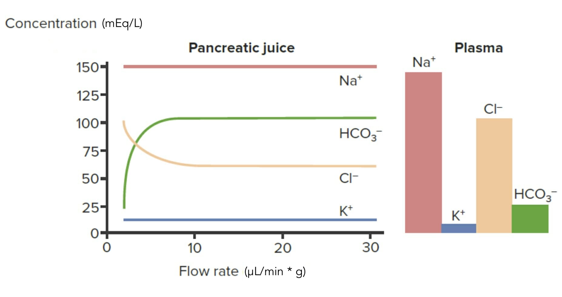
HCO3–:
Actively secreted: concentration in pancreatic juicePancreatic JuiceThe fluid containing digestive enzymes secreted by the pancreas in response to food in the duodenum.Pancreas: Anatomy > that in plasmaPlasmaThe residual portion of blood that is left after removal of blood cells by centrifugation without prior blood coagulation.Transfusion Products
Actively reabsorbed: concentration in pancreatic juicePancreatic JuiceThe fluid containing digestive enzymes secreted by the pancreas in response to food in the duodenum.Pancreas: Anatomy < that in plasmaPlasmaThe residual portion of blood that is left after removal of blood cells by centrifugation without prior blood coagulation.Transfusion Products
Reabsorption drops as flow rateFlow ratemaximum flow the ventilator will deliver a set tidal volume in liters per minuteInvasive Mechanical Ventilation increases.
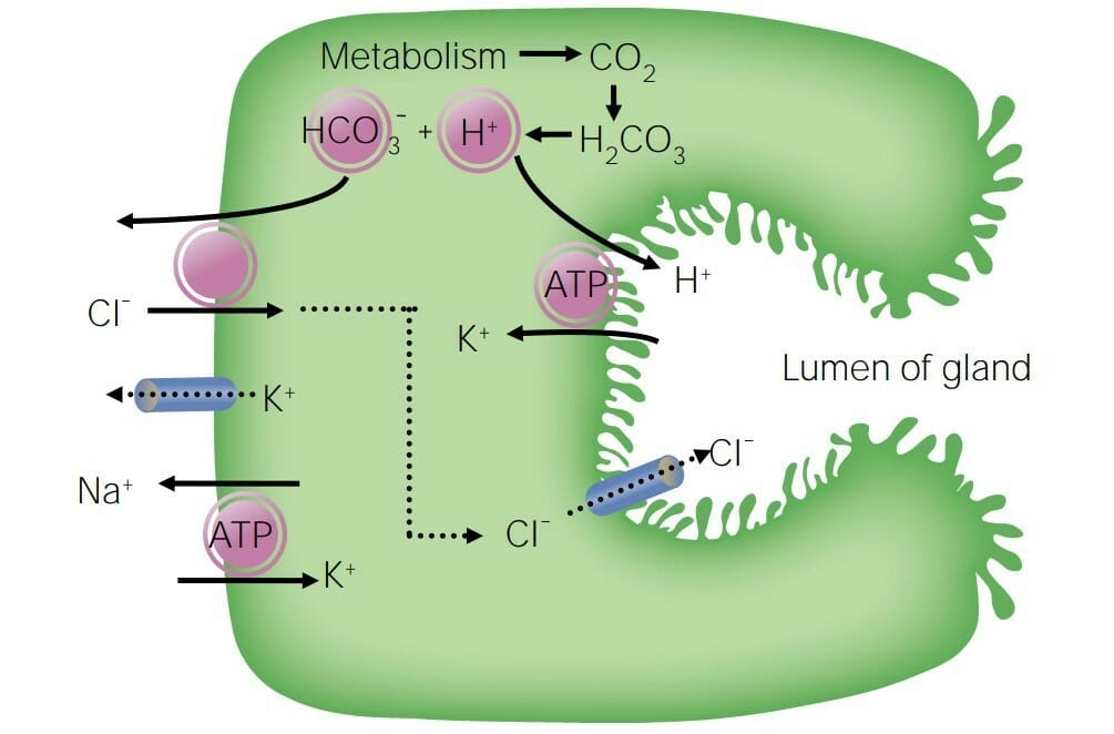
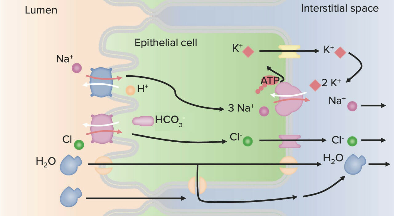
CO2 enters cells → combines with water to form H2CO3 → splits into H+ and HCO3–
H+ is moved back across the basolateral membrane into the interstitial space via the H+/Na+ exchanger.
HCO3– is secreted across the apical membrane into the lumen via the HCO3–/Cl– exchanger.
Cl– can be recycled back into the lumen through the Cl– channel.
Na+ is removed from the cell to across the basolateral membrane via the Na+/K+ ATPase exchanger.
Na+ and K+ are neither actively secreted nor reabsorbed:
Concentration in pancreatic juicePancreatic JuiceThe fluid containing digestive enzymes secreted by the pancreas in response to food in the duodenum.Pancreas: Anatomy is similar to that in the plasmaPlasmaThe residual portion of blood that is left after removal of blood cells by centrifugation without prior blood coagulation.Transfusion Products.
Some Na+ moves paracellularly into the lumen.
Water follows Na+ into the lumen.
Pancreatic secretion of ions and their plasma levels: Note that as the flow rate (x-axis) increases, HCO3– concentration in the pancreatic secretion (y-axis) increases above plasma levels, indicating secretion. The opposite happens to Cl–, where its concentration drops lower than plasma levels, indicating reabsorption.
Image by Lecturio.
Secretion of HCO3– by pancreatic ductal cells: CO2 enters the cells, combines with water to form carbonic acid (H2CO3), and then splits into H+ and HCO3–. The H+ is moved back across the basolateral membrane into the interstitial space via an H+/Na+ exchanger, whereas HCO3– is secreted across the apical membrane into the lumen via an HCO3–/Cl– exchanger. The Cl– can be recycled back into the lumen through a Cl– channel. Next, Na+ is removed from the cell across the basolateral membrane via Na+/K+ ATPase. Some of the Na+ moves paracellularly into the lumen, bringing water with it.
Image by Lecturio.
Control and regulation
Percentage of secretions produced:
25% in the cephalic phase, stimulated primarily by AChAChA neurotransmitter found at neuromuscular junctions, autonomic ganglia, parasympathetic effector junctions, a subset of sympathetic effector junctions, and at many sites in the central nervous system.Receptors and Neurotransmitters of the CNS released from the vagus nerveVagus nerveThe 10th cranial nerve. The vagus is a mixed nerve which contains somatic afferents (from skin in back of the ear and the external auditory meatus), visceral afferents (from the pharynx, larynx, thorax, and abdomen), parasympathetic efferents (to the thorax and abdomen), and efferents to striated muscle (of the larynx and pharynx).Pharynx: Anatomy
10% in the gastric phase, stimulated primarily by vago-vagal reflexes
65% in the intestinal phase, stimulated by secretin and cholecystokinin, both of which are hormonesHormonesHormones are messenger molecules that are synthesized in one part of the body and move through the bloodstream to exert specific regulatory effects on another part of the body. Hormones play critical roles in coordinating cellular activities throughout the body in response to the constant changes in both the internal and external environments. Hormones: Overview and Types released in the duodenumDuodenumThe shortest and widest portion of the small intestine adjacent to the pylorus of the stomach. It is named for having the length equal to about the width of 12 fingers.Small Intestine: Anatomy
Release of secretin and cholecystokinin from the duodenumDuodenumThe shortest and widest portion of the small intestine adjacent to the pylorus of the stomach. It is named for having the length equal to about the width of 12 fingers.Small Intestine: Anatomy:
Acidic content in the stomachStomachThe stomach is a muscular sac in the upper left portion of the abdomen that plays a critical role in digestion. The stomach develops from the foregut and connects the esophagus with the duodenum. Structurally, the stomach is C-shaped and forms a greater and lesser curvature and is divided grossly into regions: the cardia, fundus, body, and pylorus. Stomach: Anatomy induces the release of secretin.
Amino acidsAmino acidsOrganic compounds that generally contain an amino (-NH2) and a carboxyl (-COOH) group. Twenty alpha-amino acids are the subunits which are polymerized to form proteins.Basics of Amino Acids and fatsFatsThe glyceryl esters of a fatty acid, or of a mixture of fatty acids. They are generally odorless, colorless, and tasteless if pure, but they may be flavored according to origin. Fats are insoluble in water, soluble in most organic solvents. They occur in animal and vegetable tissue and are generally obtained by boiling or by extraction under pressure. They are important in the diet (dietary fats) as a source of energy.Energy Homeostasis induce the release of cholecystokinin.
Secretin and cholecystokinin enter the bloodstream and are transported to the pancreasPancreasThe pancreas lies mostly posterior to the stomach and extends across the posterior abdominal wall from the duodenum on the right to the spleen on the left. This organ has both exocrine and endocrine tissue. Pancreas: Anatomy.
Neural stimulation:
Direct stimulation by the vagus nerveVagus nerveThe 10th cranial nerve. The vagus is a mixed nerve which contains somatic afferents (from skin in back of the ear and the external auditory meatus), visceral afferents (from the pharynx, larynx, thorax, and abdomen), parasympathetic efferents (to the thorax and abdomen), and efferents to striated muscle (of the larynx and pharynx).Pharynx: Anatomy (AChAChA neurotransmitter found at neuromuscular junctions, autonomic ganglia, parasympathetic effector junctions, a subset of sympathetic effector junctions, and at many sites in the central nervous system.Receptors and Neurotransmitters of the CNS)
Stimulation via other neurotransmitters:
VIPVIPA highly basic, 28 amino acid neuropeptide released from intestinal mucosa. It has a wide range of biological actions affecting the cardiovascular, gastrointestinal, and respiratory systems and is neuroprotective. It binds special receptors.Gastrointestinal Neural and Hormonal Signaling
Gastrin-releasing peptidegastrin-releasing peptideNeuropeptide and gut hormone that helps regulate gastric acid secretion and motor function. Once released from nerves in the antrum of the stomach, the neuropeptide stimulates release of gastrin from the gastrin-secreting cells.Gastrointestinal Neural and Hormonal Signaling (GRP)
↑ In intracellular cAMPcAMPAn adenine nucleotide containing one phosphate group which is esterified to both the 3′- and 5′-positions of the sugar moiety. It is a second messenger and a key intracellular regulator, functioning as a mediator of activity for a number of hormones, including epinephrine, glucagon, and acth.Phosphodiesterase Inhibitors, which is caused by:
Secretin
VIPVIPA highly basic, 28 amino acid neuropeptide released from intestinal mucosa. It has a wide range of biological actions affecting the cardiovascular, gastrointestinal, and respiratory systems and is neuroprotective. It binds special receptors.Gastrointestinal Neural and Hormonal Signaling
↑ In intracellular CaCACondylomata acuminata are a clinical manifestation of genital HPV infection. Condylomata acuminata are described as raised, pearly, flesh-colored, papular, cauliflower-like lesions seen in the anogenital region that may cause itching, pain, or bleeding.Condylomata Acuminata (Genital Warts)2+:
AChAChA neurotransmitter found at neuromuscular junctions, autonomic ganglia, parasympathetic effector junctions, a subset of sympathetic effector junctions, and at many sites in the central nervous system.Receptors and Neurotransmitters of the CNS
Cholecystokinin
GRP
Both pathways result in phosphorylationPhosphorylationThe introduction of a phosphoryl group into a compound through the formation of an ester bond between the compound and a phosphorus moiety.Post-translational Protein Processing of structural and regulatory proteinsRegulatory proteinsProteins and Peptides → induce docking and fusion of secretory granules → proteinsProteinsLinear polypeptides that are synthesized on ribosomes and may be further modified, crosslinked, cleaved, or assembled into complex proteins with several subunits. The specific sequence of amino acids determines the shape the polypeptide will take, during protein folding, and the function of the protein.Energy Homeostasis are secreted into acini
Diagram of an exocrine pancreatic cell and its secretion-stimulation pathways: Note how vasoactive intestinal peptide (VIP) and secretin increase the concentration of intracellular cAMP, whereas gastrin-releasing peptide (GRP), acetylcholine (ACh), and cholecystokinin (CCK) increase intracellular Ca2+ concentrations. Both pathways result in phosphorylation of the structural and regulatory proteins, ultimately inducing docking and fusion of secretory granules that contain enzymes.
Image by Lecturio.
Clinical relevance of pancreatic secretions
Acute pancreatitisPancreatitisInflammation of the pancreas. Pancreatitis is classified as acute unless there are computed tomographic or endoscopic retrograde cholangiopancreatographic findings of chronic pancreatitis. The two most common forms of acute pancreatitis are alcoholic pancreatitis and gallstone pancreatitis.Acute Pancreatitis: occurs when pancreatic enzymesEnzymesEnzymes are complex protein biocatalysts that accelerate chemical reactions without being consumed by them. Due to the body’s constant metabolic needs, the absence of enzymes would make life unsustainable, as reactions would occur too slowly without these molecules. Basics of Enzymes are retained within the pancreasPancreasThe pancreas lies mostly posterior to the stomach and extends across the posterior abdominal wall from the duodenum on the right to the spleen on the left. This organ has both exocrine and endocrine tissue. Pancreas: Anatomy, resulting in self-digestion. Acute pancreatitisPancreatitisInflammation of the pancreas. Pancreatitis is classified as acute unless there are computed tomographic or endoscopic retrograde cholangiopancreatographic findings of chronic pancreatitis. The two most common forms of acute pancreatitis are alcoholic pancreatitis and gallstone pancreatitis.Acute Pancreatitis can occur due to obstruction (e.g., gallstonesGallstonesCholelithiasis (gallstones) is the presence of stones in the gallbladder. Most gallstones are cholesterol stones, while the rest are composed of bilirubin (pigment stones) and other mixed components. Patients are commonly asymptomatic but may present with biliary colic (intermittent pain in the right upper quadrant).Cholelithiasis, cancer in the head of the pancreasPancreasThe pancreas lies mostly posterior to the stomach and extends across the posterior abdominal wall from the duodenum on the right to the spleen on the left. This organ has both exocrine and endocrine tissue. Pancreas: Anatomy) and can be seen in cases of cysticCysticFibrocystic ChangefibrosisFibrosisAny pathological condition where fibrous connective tissue invades any organ, usually as a consequence of inflammation or other injury.Bronchiolitis Obliterans.
CysticCysticFibrocystic ChangefibrosisFibrosisAny pathological condition where fibrous connective tissue invades any organ, usually as a consequence of inflammation or other injury.Bronchiolitis Obliterans: an autosomal recessiveAutosomal recessiveAutosomal inheritance, both dominant and recessive, refers to the transmission of genes from the 22 autosomal chromosomes. Autosomal recessive diseases are only expressed when 2 copies of the recessive allele are inherited.Autosomal Recessive and Autosomal Dominant Inheritance disorder caused by mutations in the CFTRgeneGeneA category of nucleic acid sequences that function as units of heredity and which code for the basic instructions for the development, reproduction, and maintenance of organisms.Basic Terms of Genetics. Mutations lead to dysfunction of the Cl–channelsChannelsThe Cell: Cell Membrane, resulting in the formation of hyperviscous mucus. Hyperviscous mucus obstructs the flowFlowBlood flows through the heart, arteries, capillaries, and veins in a closed, continuous circuit. Flow is the movement of volume per unit of time. Flow is affected by the pressure gradient and the resistance fluid encounters between 2 points. Vascular resistance is the opposition to flow, which is caused primarily by blood friction against vessel walls.Vascular Resistance, Flow, and Mean Arterial Pressure of pancreatic juicePancreatic JuiceThe fluid containing digestive enzymes secreted by the pancreas in response to food in the duodenum.Pancreas: Anatomy and bileBileAn emulsifying agent produced in the liver and secreted into the duodenum. Its composition includes bile acids and salts; cholesterol; and electrolytes. It aids digestion of fats in the duodenum.Gallbladder and Biliary Tract: Anatomy in the GI tract, resulting in maldigestionMaldigestionMalabsorption involves many disorders in which there is an inability of the gut to absorb nutrients from dietary intake, potentially including water and/or electrolytes. A closely related term, maldigestion is the inability to break down large molecules of food into their smaller constituents. Malabsorption and maldigestion can affect macronutrients (fats, proteins, and carbohydrates), micronutrients (vitamins and minerals), or both. Malabsorption and Maldigestion, malabsorptionMalabsorptionGeneral term for a group of malnutrition syndromes caused by failure of normal intestinal absorption of nutrients.Malabsorption and Maldigestion, and progressive liverLiverThe liver is the largest gland in the human body. The liver is found in the superior right quadrant of the abdomen and weighs approximately 1.5 kilograms. Its main functions are detoxification, metabolism, nutrient storage (e.g., iron and vitamins), synthesis of coagulation factors, formation of bile, filtration, and storage of blood. Liver: Anatomy and pancreatic disease. Common presentations include chronic respiratory infectionsInfectionsInvasion of the host organism by microorganisms or their toxins or by parasites that can cause pathological conditions or diseases.Chronic Granulomatous Disease, failure to thriveFailure to ThriveFailure to thrive (FTT), or faltering growth, describes suboptimal weight gain and growth in children. The majority of cases are due to inadequate caloric intake; however, genetic, infectious, and oncological etiologies are also common. Failure to Thrive, and pancreatic insufficiency (due to loss of exocrine function).
BileBileAn emulsifying agent produced in the liver and secreted into the duodenum. Its composition includes bile acids and salts; cholesterol; and electrolytes. It aids digestion of fats in the duodenum.Gallbladder and Biliary Tract: Anatomy
Synthesized by hepatocytesHepatocytesThe main structural component of the liver. They are specialized epithelial cells that are organized into interconnected plates called lobules.Liver: Anatomy
Transported to the gallbladderGallbladderThe gallbladder is a pear-shaped sac, located directly beneath the liver, that sits on top of the superior part of the duodenum. The primary functions of the gallbladder include concentrating and storing up to 50 mL of bile. Gallbladder and Biliary Tract: Anatomy (via the hepatic and cysticCysticFibrocystic Change ducts) for storage
Primary function: emulsification (rather than digestionDigestionDigestion refers to the process of the mechanical and chemical breakdown of food into smaller particles, which can then be absorbed and utilized by the body.Digestion and Absorption) of fatsFatsThe glyceryl esters of a fatty acid, or of a mixture of fatty acids. They are generally odorless, colorless, and tasteless if pure, but they may be flavored according to origin. Fats are insoluble in water, soluble in most organic solvents. They occur in animal and vegetable tissue and are generally obtained by boiling or by extraction under pressure. They are important in the diet (dietary fats) as a source of energy.Energy Homeostasis
Structure of the hepatobiliary tree
Image by Lecturio.
Components of biliary secretions (i.e., bileBileAn emulsifying agent produced in the liver and secreted into the duodenum. Its composition includes bile acids and salts; cholesterol; and electrolytes. It aids digestion of fats in the duodenum.Gallbladder and Biliary Tract: Anatomy)
BileBileAn emulsifying agent produced in the liver and secreted into the duodenum. Its composition includes bile acids and salts; cholesterol; and electrolytes. It aids digestion of fats in the duodenum.Gallbladder and Biliary Tract: Anatomy salts
CholesterolCholesterolThe principal sterol of all higher animals, distributed in body tissues, especially the brain and spinal cord, and in animal fats and oils.Cholesterol Metabolism
LecithinLecithinA complex mixture of phospholipids; glycolipids; and triglycerides; with substantial amounts of phosphatidylcholines; phosphatidylethanolamines; and phosphatidylinositols, which are sometimes loosely termed as 1, 2-diacyl-3-phosphocholines. Lecithin is a component of the cell membrane and commercially extracted from soybeans and egg yolk. The emulsifying and surfactant properties are useful in food additives and for forming organogels (gels).Fatty Acids and Lipids
BileBileAn emulsifying agent produced in the liver and secreted into the duodenum. Its composition includes bile acids and salts; cholesterol; and electrolytes. It aids digestion of fats in the duodenum.Gallbladder and Biliary Tract: Anatomy concentration in the gallbladderGallbladderThe gallbladder is a pear-shaped sac, located directly beneath the liver, that sits on top of the superior part of the duodenum. The primary functions of the gallbladder include concentrating and storing up to 50 mL of bile. Gallbladder and Biliary Tract: Anatomy
The primary function of the gallbladderGallbladderThe gallbladder is a pear-shaped sac, located directly beneath the liver, that sits on top of the superior part of the duodenum. The primary functions of the gallbladder include concentrating and storing up to 50 mL of bile. Gallbladder and Biliary Tract: Anatomy is the storage and concentration of bileBileAn emulsifying agent produced in the liver and secreted into the duodenum. Its composition includes bile acids and salts; cholesterol; and electrolytes. It aids digestion of fats in the duodenum.Gallbladder and Biliary Tract: Anatomy. BileBileAn emulsifying agent produced in the liver and secreted into the duodenum. Its composition includes bile acids and salts; cholesterol; and electrolytes. It aids digestion of fats in the duodenum.Gallbladder and Biliary Tract: Anatomy can be concentrated in the gallbladderGallbladderThe gallbladder is a pear-shaped sac, located directly beneath the liver, that sits on top of the superior part of the duodenum. The primary functions of the gallbladder include concentrating and storing up to 50 mL of bile. Gallbladder and Biliary Tract: Anatomy by about 5%‒20% through dehydrationDehydrationThe condition that results from excessive loss of water from a living organism.Volume Depletion and Dehydration:
Na+:
Reabsorbed from the lumen in exchange for H+ via the Na+/H+ exchanger in the apical membrane
Na+ within the cell is then pumped across the basolateral membrane into the interstitial space in exchange for K+ via the Na+/K+ ATPase.
Cl–:
Reabsorbed from the lumen in exchange for HCO3– via the HCO3–/Cl– exchanger in the apical membrane
Cl– within the cell moves to the interstitial space through the Cl–channelsChannelsThe Cell: Cell Membrane in the basolateral membrane.
Water follows NaCl:
Moves from the gallbladderGallbladderThe gallbladder is a pear-shaped sac, located directly beneath the liver, that sits on top of the superior part of the duodenum. The primary functions of the gallbladder include concentrating and storing up to 50 mL of bile. Gallbladder and Biliary Tract: Anatomy lumen into the interstitial space
Mechanism of biliary concentration: Na+ is exchanged for H+ in the apical membrane and later exchanged for K+ in the basolateral membrane. Next, Cl– is exchanged for HCO3– in the apical membrane and moves to the interstitial space through its own channels. The concentration gradient that is produced induces the transcellular and paracellular movement of water.
Image by Lecturio.
Control and regulation
BileBileAn emulsifying agent produced in the liver and secreted into the duodenum. Its composition includes bile acids and salts; cholesterol; and electrolytes. It aids digestion of fats in the duodenum.Gallbladder and Biliary Tract: Anatomy is continuously produced by the liverLiverThe liver is the largest gland in the human body. The liver is found in the superior right quadrant of the abdomen and weighs approximately 1.5 kilograms. Its main functions are detoxification, metabolism, nutrient storage (e.g., iron and vitamins), synthesis of coagulation factors, formation of bile, filtration, and storage of blood. Liver: Anatomy; thus, regulation is via release from the gallbladderGallbladderThe gallbladder is a pear-shaped sac, located directly beneath the liver, that sits on top of the superior part of the duodenum. The primary functions of the gallbladder include concentrating and storing up to 50 mL of bile. Gallbladder and Biliary Tract: Anatomy.
The primary mediator is cholecystokinin, which has 2 main effects:
GallbladderGallbladderThe gallbladder is a pear-shaped sac, located directly beneath the liver, that sits on top of the superior part of the duodenum. The primary functions of the gallbladder include concentrating and storing up to 50 mL of bile. Gallbladder and Biliary Tract: Anatomy contraction
Relaxation of the sphincter of Oddi
Other regulators:
Vagus nerveVagus nerveThe 10th cranial nerve. The vagus is a mixed nerve which contains somatic afferents (from skin in back of the ear and the external auditory meatus), visceral afferents (from the pharynx, larynx, thorax, and abdomen), parasympathetic efferents (to the thorax and abdomen), and efferents to striated muscle (of the larynx and pharynx).Pharynx: Anatomy: weak stimulator of gallbladderGallbladderThe gallbladder is a pear-shaped sac, located directly beneath the liver, that sits on top of the superior part of the duodenum. The primary functions of the gallbladder include concentrating and storing up to 50 mL of bile. Gallbladder and Biliary Tract: Anatomy contraction
Inhibitors of bileBileAn emulsifying agent produced in the liver and secreted into the duodenum. Its composition includes bile acids and salts; cholesterol; and electrolytes. It aids digestion of fats in the duodenum.Gallbladder and Biliary Tract: Anatomy release:
Somatostatin
NorepinephrineNorepinephrinePrecursor of epinephrine that is secreted by the adrenal medulla and is a widespread central and autonomic neurotransmitter. Norepinephrine is the principal transmitter of most postganglionic sympathetic fibers, and of the diffuse projection system in the brain that arises from the locus ceruleus.Receptors and Neurotransmitters of the CNS
Intestines secrete about 1‒2 L of intestinal juice per day.
Secretions released into the lumen:
Mucus
Serous fluid
Functions of intestinal juice:
Protection of the intestinal walls
Regulation of GI function
Minimal secretions for digestive functions (i.e., secretions contain very few enzymesEnzymesEnzymes are complex protein biocatalysts that accelerate chemical reactions without being consumed by them. Due to the body’s constant metabolic needs, the absence of enzymes would make life unsustainable, as reactions would occur too slowly without these molecules. Basics of Enzymes)
GIPGIPA gastrointestinal peptide hormone of about 43-amino acids. It is found to be a potent stimulator of insulin secretion and a relatively poor inhibitor of gatsric acid secretion.Gastrointestinal Neural and Hormonal Signaling
Freiburghaus, A. U., Roduner, J. B., & Hadorn, H. B. (2021). Activation of human pancreatic proteolytic enzymes: The role of enteropeptidase and trypsin. JPGN Reports, 2(4), e138. https://doi.org/10.1097/PG9.0000000000000138