Achieve Mastery of Medical Concepts

Study for medical school and boards with Lecturio

Antepartum Hemorrhage

Antepartum hemorrhage is defined as vaginal bleeding that occurs after 20 weeks of gestation and is unrelated to labor. The most important causes are placental abruption (most common), placenta previa Placenta Previa Abnormal placentation in which the placenta implants in the lower segment of the uterus (the zone of dilation) and may cover part or all of the opening of the cervix. It is often associated with serious antepartum bleeding and premature labor. Placental Abnormalities (2nd most common), vasa previa, and uterine rupture. Placental abruption and uterine rupture are diagnosed clinically. Placenta previa Placenta Previa Abnormal placentation in which the placenta implants in the lower segment of the uterus (the zone of dilation) and may cover part or all of the opening of the cervix. It is often associated with serious antepartum bleeding and premature labor. Placental Abnormalities and vasa previa are usually diagnosed as part of routine screening Screening Preoperative Care on a midtrimester ultrasound, and digital exams in these women must be avoided. All of these conditions can lead to maternal and/or fetal hemorrhage, necessitating emergency cesarean delivery Cesarean Delivery Cesarean delivery (CD) is the operative delivery of ≥ 1 infants through a surgical incision in the maternal abdomen and uterus. Cesarean deliveries may be indicated for a number of either maternal or fetal reasons, most commonly including fetal intolerance to labor, arrest of labor, a history of prior uterine surgery, fetal malpresentation, and placental abnormalities. Cesarean Delivery (CD) and maternal and/or fetal resuscitation Resuscitation The restoration to life or consciousness of one apparently dead. . Neonatal Respiratory Distress Syndrome.

Last updated: Sep 29, 2022

Editorial responsibility: Stanley Oiseth, Lindsay Jones, Evelin Maza

Overview

Definition

Antepartum hemorrhage is defined as vaginal bleeding that occurs after 20 weeks of gestation and is unrelated to labor.

Epidemiology and etiology

  • Incidence Incidence The number of new cases of a given disease during a given period in a specified population. It also is used for the rate at which new events occur in a defined population. It is differentiated from prevalence, which refers to all cases in the population at a given time. Measures of Disease Frequency: occurs in approximately 5% of pregnancies 
  • Most common etiologies:
    • Placental abruption: 30%
    • Placenta previa Placenta Previa Abnormal placentation in which the placenta implants in the lower segment of the uterus (the zone of dilation) and may cover part or all of the opening of the cervix. It is often associated with serious antepartum bleeding and premature labor. Placental Abnormalities: 20%
  • The etiology is often not determined.
Table: Causes of antepartum bleeding by location
Anatomic location Causes of antepartum bleeding
Placenta Placenta A highly vascularized mammalian fetal-maternal organ and major site of transport of oxygen, nutrients, and fetal waste products. It includes a fetal portion (chorionic villi) derived from trophoblasts and a maternal portion (decidua) derived from the uterine endometrium. The placenta produces an array of steroid, protein and peptide hormones (placental hormones). Placenta, Umbilical Cord, and Amniotic Cavity
  • Placental abruption*
  • Placenta previa Placenta Previa Abnormal placentation in which the placenta implants in the lower segment of the uterus (the zone of dilation) and may cover part or all of the opening of the cervix. It is often associated with serious antepartum bleeding and premature labor. Placental Abnormalities*
  • Vasa previa*
Uterus Uterus The uterus, cervix, and fallopian tubes are part of the internal female reproductive system. The uterus has a thick wall made of smooth muscle (the myometrium) and an inner mucosal layer (the endometrium). The most inferior portion of the uterus is the cervix, which connects the uterine cavity to the vagina. Uterus, Cervix, and Fallopian Tubes: Anatomy Uterine rupture*
Cervix Cervix The uterus, cervix, and fallopian tubes are part of the internal female reproductive system. The most inferior portion of the uterus is the cervix, which connects the uterine cavity to the vagina. Externally, the cervix is lined by stratified squamous cells; however, the cervical canal is lined by columnar epithelium. Uterus, Cervix, and Fallopian Tubes: Anatomy
  • “Bloody show”: a small amount of bloody mucus discharge that often precedes labor as the cervix Cervix The uterus, cervix, and fallopian tubes are part of the internal female reproductive system. The most inferior portion of the uterus is the cervix, which connects the uterine cavity to the vagina. Externally, the cervix is lined by stratified squamous cells; however, the cervical canal is lined by columnar epithelium. Uterus, Cervix, and Fallopian Tubes: Anatomy ripens
  • Cervicitis Cervicitis Inflammation of the uterine cervix. Gonorrhea
    • STIs STIs Sexually transmitted infections (STIs) or sexually transmitted diseases (STDs) are infections that spread either by vaginal intercourse, anal sex, or oral sex. Symptoms and signs may include vaginal discharge, penile discharge, dysuria, skin lesions (e.g., warts, ulcers) on or around the genitals, and pelvic pain. Some infections can lead to infertility and chronic debilitating disease. Sexually Transmitted Infections (STIs): gonorrhea Gonorrhea Gonorrhea is a sexually transmitted infection (STI) caused by the gram-negative bacteria Neisseria gonorrhoeae (N. gonorrhoeae). Gonorrhea may be asymptomatic but commonly manifests as cervicitis or urethritis with less common presentations such as proctitis, conjunctivitis, or pharyngitis. Gonorrhea, chlamydia Chlamydia Chlamydiae are obligate intracellular gram-negative bacteria. They lack a peptidoglycan layer and are best visualized using Giemsa stain. The family of Chlamydiaceae comprises 3 pathogens that can infect humans: Chlamydia trachomatis, Chlamydia psittaci, and Chlamydia pneumoniae. Chlamydia
    • Secondary to vaginitis: candidiasis Candidiasis Candida is a genus of dimorphic, opportunistic fungi. Candida albicans is part of the normal human flora and is the most common cause of candidiasis. The clinical presentation varies and can include localized mucocutaneous infections (e.g., oropharyngeal, esophageal, intertriginous, and vulvovaginal candidiasis) and invasive disease (e.g., candidemia, intraabdominal abscess, pericarditis, and meningitis). Candida/Candidiasis, bacterial vaginosis Bacterial vaginosis Polymicrobial, nonspecific vaginitis associated with positive cultures of gardnerella vaginalis and other anaerobic organisms and a decrease in lactobacilli. It remains unclear whether the initial pathogenic event is caused by the growth of anaerobes or a primary decrease in lactobacilli. Vulvovaginitis
  • Cervical ectropion Ectropion The turning outward (eversion) of the edge of the eyelid, resulting in the exposure of the palpebral conjunctiva. Sjögren’s Syndrome: exposed cervical columnar epithelium Epithelium The epithelium is a complex of specialized cellular organizations arranged into sheets and lining cavities and covering the surfaces of the body. The cells exhibit polarity, having an apical and a basal pole. Structures important for the epithelial integrity and function involve the basement membrane, the semipermeable sheet on which the cells rest, and interdigitations, as well as cellular junctions. Surface Epithelium: Histology that is prone to light bleeding when touched
  • Polyps
  • Carcinoma
Vagina Vagina The vagina is the female genital canal, extending from the vulva externally to the cervix uteri internally. The structures have sexual, reproductive, and urinary functions and a rich blood supply, mainly arising from the internal iliac artery. Vagina, Vulva, and Pelvic Floor: Anatomy and vulva Vulva The vulva is the external genitalia of the female and includes the mons pubis, labia majora, labia minora, clitoris, vestibule, vestibular bulb, and greater vestibular glands. Vagina, Vulva, and Pelvic Floor: Anatomy
*Clinically important obstetric cause.

Placental Abruption

Overview

Placental abruption is the complete or partial premature Premature Childbirth before 37 weeks of pregnancy (259 days from the first day of the mother’s last menstrual period, or 245 days after fertilization). Necrotizing Enterocolitis detachment of a normally implanted placenta Placenta A highly vascularized mammalian fetal-maternal organ and major site of transport of oxygen, nutrients, and fetal waste products. It includes a fetal portion (chorionic villi) derived from trophoblasts and a maternal portion (decidua) derived from the uterine endometrium. The placenta produces an array of steroid, protein and peptide hormones (placental hormones). Placenta, Umbilical Cord, and Amniotic Cavity before the birth of the infant.

Clinical relevance:

  • With a detached placenta Placenta A highly vascularized mammalian fetal-maternal organ and major site of transport of oxygen, nutrients, and fetal waste products. It includes a fetal portion (chorionic villi) derived from trophoblasts and a maternal portion (decidua) derived from the uterine endometrium. The placenta produces an array of steroid, protein and peptide hormones (placental hormones). Placenta, Umbilical Cord, and Amniotic Cavity:
    • The infant is no longer able to get oxygen.
    • Maternal and fetal hemorrhage occurs through the placenta Placenta A highly vascularized mammalian fetal-maternal organ and major site of transport of oxygen, nutrients, and fetal waste products. It includes a fetal portion (chorionic villi) derived from trophoblasts and a maternal portion (decidua) derived from the uterine endometrium. The placenta produces an array of steroid, protein and peptide hormones (placental hormones). Placenta, Umbilical Cord, and Amniotic Cavity.
  • An obstetric emergency that usually requires immediate delivery

Epidemiology

  • Incidence Incidence The number of new cases of a given disease during a given period in a specified population. It also is used for the rate at which new events occur in a defined population. It is differentiated from prevalence, which refers to all cases in the population at a given time. Measures of Disease Frequency: approximately 2–10 per 1000 births
  • Age: more common in women < 20 and > 35 years of age
  • Gestational age Gestational age The age of the conceptus, beginning from the time of fertilization. In clinical obstetrics, the gestational age is often estimated as the time from the last day of the last menstruation which is about 2 weeks before ovulation and fertilization. Pregnancy: Diagnosis, Physiology, and Care at abruption:
    • Term: 60%
    • 32–36 weeks: 25%
    • < 32 weeks: 15%

Risk factors

  • Previous abruption (strongest risk factor)
  • Hypertension Hypertension Hypertension, or high blood pressure, is a common disease that manifests as elevated systemic arterial pressures. Hypertension is most often asymptomatic and is found incidentally as part of a routine physical examination or during triage for an unrelated medical encounter. Hypertension
    • Preeclampsia Preeclampsia A complication of pregnancy, characterized by a complex of symptoms including maternal hypertension and proteinuria with or without pathological edema. Symptoms may range between mild and severe. Pre-eclampsia usually occurs after the 20th week of gestation, but may develop before this time in the presence of trophoblastic disease. Hypertensive Pregnancy Disorders
    • Poorly controlled chronic hypertension Hypertension Hypertension, or high blood pressure, is a common disease that manifests as elevated systemic arterial pressures. Hypertension is most often asymptomatic and is found incidentally as part of a routine physical examination or during triage for an unrelated medical encounter. Hypertension
  • Trauma to the abdomen
  • Cocaine Cocaine An alkaloid ester extracted from the leaves of plants including coca. It is a local anesthetic and vasoconstrictor and is clinically used for that purpose, particularly in the eye, ear, nose, and throat. It also has powerful central nervous system effects similar to the amphetamines and is a drug of abuse. Cocaine, like amphetamines, acts by multiple mechanisms on brain catecholaminergic neurons; the mechanism of its reinforcing effects is thought to involve inhibition of dopamine uptake. Local Anesthetics or tobacco use
  • Quick decompression of the uterus Uterus The uterus, cervix, and fallopian tubes are part of the internal female reproductive system. The uterus has a thick wall made of smooth muscle (the myometrium) and an inner mucosal layer (the endometrium). The most inferior portion of the uterus is the cervix, which connects the uterine cavity to the vagina. Uterus, Cervix, and Fallopian Tubes: Anatomy
    • Rupture of membranes in a pregnancy Pregnancy The status during which female mammals carry their developing young (embryos or fetuses) in utero before birth, beginning from fertilization to birth. Pregnancy: Diagnosis, Physiology, and Care with polyhydramnios Polyhydramnios Polyhydramnios is a pathological excess of amniotic fluid. Common causes of polyhydramnios include fetal anomalies, gestational diabetes, multiple gestations, and congenital infections. Patients are often asymptomatic but may present with dyspnea, extremity swelling, or abdominal distention. Polyhydramnios
    • Delivery of the 1st infant in a multiple gestation
  • Fetal growth restriction Fetal growth restriction Fetal growth restriction (FGR), also known as intrauterine fetal growth restriction (IUGR), is an estimated fetal weight (EFW) or abdominal circumference < 10th percentile for gestational age. The term small for gestational age (SGA) is sometimes erroneously used interchangeably with FGR. Fetal Growth Restriction (may suggest a small chronic partial abruption)
  • Uterine anomalies 

Etiology

  • Unknown, but likely related to: 
    • Chronic placental disease processes 
    • Abnormalities in the development of placental vasculature
  • Direct abdominal trauma (less common)

Pathophysiology

  • Rupture of maternal vessels in the decidua basalis → accumulation of blood splits the decidua → a thin layer of decidua is separated off with its placental attachment
  • Thrombin Thrombin An enzyme formed from prothrombin that converts fibrinogen to fibrin. Hemostasis is a potent uterotonic agent → clotting in the decidua triggers Triggers Hereditary Angioedema (C1 Esterase Inhibitor Deficiency) uterine contractions
  • Complete (or nearly complete) abruption:
    • Caused by high-pressure arterial hemorrhage in the center of the placenta Placenta A highly vascularized mammalian fetal-maternal organ and major site of transport of oxygen, nutrients, and fetal waste products. It includes a fetal portion (chorionic villi) derived from trophoblasts and a maternal portion (decidua) derived from the uterine endometrium. The placenta produces an array of steroid, protein and peptide hormones (placental hormones). Placenta, Umbilical Cord, and Amniotic Cavity
    • Symptoms develop rapidly → life-threatening for fetus and mother
  • Partial abruption:
    • Caused by low-pressure venous hemorrhage, usually at the periphery of the placenta Placenta A highly vascularized mammalian fetal-maternal organ and major site of transport of oxygen, nutrients, and fetal waste products. It includes a fetal portion (chorionic villi) derived from trophoblasts and a maternal portion (decidua) derived from the uterine endometrium. The placenta produces an array of steroid, protein and peptide hormones (placental hormones). Placenta, Umbilical Cord, and Amniotic Cavity
    • Often self-limited, with only a small area of separation
    • Symptoms develop slowly → can lead to “chronic abruption” throughout the remainder of pregnancy Pregnancy The status during which female mammals carry their developing young (embryos or fetuses) in utero before birth, beginning from fertilization to birth. Pregnancy: Diagnosis, Physiology, and Care

Clinical presentation

Bleeding from placental separation may be either external with visible vaginal bleeding (80%), or concealed (blood pools behind the placenta Placenta A highly vascularized mammalian fetal-maternal organ and major site of transport of oxygen, nutrients, and fetal waste products. It includes a fetal portion (chorionic villi) derived from trophoblasts and a maternal portion (decidua) derived from the uterine endometrium. The placenta produces an array of steroid, protein and peptide hormones (placental hormones). Placenta, Umbilical Cord, and Amniotic Cavity) without vaginal bleeding (20%).

  • Painful bleeding:
    • Abdominal and/or back pain Pain An unpleasant sensation induced by noxious stimuli which are detected by nerve endings of nociceptive neurons. Pain: Types and Pathways
    • Bleeding can range from mild to life-threatening.
  • Contractions (often hypertonic Hypertonic Solutions that have a greater osmotic pressure than a reference solution such as blood, plasma, or interstitial fluid. Renal Sodium and Water Regulation or high-frequency)
  • Uterine tenderness
  • Fetal distress/decreased fetal movement
  • Small, partial abruptions may be less dramatic and present with:
    • Smaller amounts of bleeding over time (chronic abruptions)
    • Oligohydramnios Oligohydramnios Oligohydramnios refers to amniotic fluid volume less than expected for the current gestational age. Oligohydramnios is diagnosed by ultrasound and defined as an amniotic fluid index (AFI) of ‰¤ 5 cm or a single deep pocket (SDP) of < 2 cm in the 2nd or 3rd trimester. Oligohydramnios
    • Intrauterine fetal growth restriction Fetal growth restriction Fetal growth restriction (FGR), also known as intrauterine fetal growth restriction (IUGR), is an estimated fetal weight (EFW) or abdominal circumference < 10th percentile for gestational age. The term small for gestational age (SGA) is sometimes erroneously used interchangeably with FGR. Fetal Growth Restriction
Placental abruption

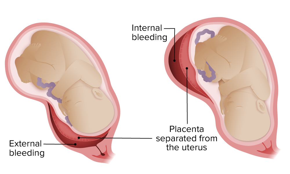
Placental abruption: External versus concealed bleeding

Image by Lecturio.

Diagnosis

The diagnosis of placental abruption is usually clinical, based on the history, exam, and fetal monitoring Fetal monitoring The primary goals of antepartum testing and monitoring are to assess fetal well-being, identify treatable situations that may cause complications, and evaluate for chromosomal abnormalities. These tests are divided into screening tests (which include cell-free DNA testing, serum analyte testing, and nuchal translucency measurements), and diagnostic tests, which provide a definitive diagnosis of aneuploidy and include chorionic villus sampling (CVS) and amniocentesis. Antepartum Testing and Monitoring.

  • Fetal nonstress test (NST):
    • Measures fetal heart rate Heart rate The number of times the heart ventricles contract per unit of time, usually per minute. Cardiac Physiology and uterine contractions (tocometry)
    • Used to assess fetal status
    • Signs of fetal distress:
      • Fetal bradycardia Bradycardia Bradyarrhythmia is a rhythm in which the heart rate is less than 60/min. Bradyarrhythmia can be physiologic, without symptoms or hemodynamic change. Pathologic bradyarrhythmia results in reduced cardiac output and hemodynamic instability causing syncope, dizziness, or dyspnea. Bradyarrhythmias
      • Heart rate Heart rate The number of times the heart ventricles contract per unit of time, usually per minute. Cardiac Physiology variability
      • Late decelerations
    • Tocometry: frequent and/or hypertonic Hypertonic Solutions that have a greater osmotic pressure than a reference solution such as blood, plasma, or interstitial fluid. Renal Sodium and Water Regulation contractions
  • Ultrasonography:
    • Exclude placenta previa Placenta Previa Abnormal placentation in which the placenta implants in the lower segment of the uterus (the zone of dilation) and may cover part or all of the opening of the cervix. It is often associated with serious antepartum bleeding and premature labor. Placental Abnormalities
    • May show a retroplacental hematoma Hematoma A collection of blood outside the blood vessels. Hematoma can be localized in an organ, space, or tissue. Intussusception
    • Low sensitivity, but decent specificity 
  • Laboratory: helpful in managing acute resuscitation Resuscitation The restoration to life or consciousness of one apparently dead. . Neonatal Respiratory Distress Syndrome, rather than diagnosis of abruption itself
    • Type and cross (to prepare for potential transfusion)
    • CBC → evaluate degree of anemia Anemia Anemia is a condition in which individuals have low Hb levels, which can arise from various causes. Anemia is accompanied by a reduced number of RBCs and may manifest with fatigue, shortness of breath, pallor, and weakness. Subtypes are classified by the size of RBCs, chronicity, and etiology. Anemia: Overview and Types
    • Coagulation panel:
      • PT/INR, aPTT
      • Fibrinogen Fibrinogen Plasma glycoprotein clotted by thrombin, composed of a dimer of three non-identical pairs of polypeptide chains (alpha, beta, gamma) held together by disulfide bonds. Fibrinogen clotting is a sol-gel change involving complex molecular arrangements: whereas fibrinogen is cleaved by thrombin to form polypeptides a and b, the proteolytic action of other enzymes yields different fibrinogen degradation products. Hemostasis → evaluate for DIC DIC Disseminated intravascular coagulation (DIC) is a condition characterized by systemic bodywide activation of the coagulation cascade. This cascade results in both widespread microvascular thrombi contributing to multiple organ dysfunction and consumption of clotting factors and platelets, leading to hemorrhage. Disseminated Intravascular Coagulation
    • Kleihauer-Betke test Kleihauer-Betke Test Hemolytic Disease of the Fetus and Newborn: can detect fetal RBCs RBCs Erythrocytes, or red blood cells (RBCs), are the most abundant cells in the blood. While erythrocytes in the fetus are initially produced in the yolk sac then the liver, the bone marrow eventually becomes the main site of production. Erythrocytes: Histology in maternal circulation Circulation The movement of the blood as it is pumped through the cardiovascular system. ABCDE Assessment, indicating fetomaternal hemorrhage → potential for isoimmunization

Management

  • Severe abruption:
    • Individuals with DIC DIC Disseminated intravascular coagulation (DIC) is a condition characterized by systemic bodywide activation of the coagulation cascade. This cascade results in both widespread microvascular thrombi contributing to multiple organ dysfunction and consumption of clotting factors and platelets, leading to hemorrhage. Disseminated Intravascular Coagulation, hypovolemic shock Hypovolemic Shock Types of Shock, and/or nonreassuring fetal status
    • Step 1: stabilization
      • Assess and manage (as needed) the airway Airway ABCDE Assessment and breathing (e.g., give supplemental O2)
      • Establish 2 large-bore IV lines.
      • Initiate IV fluids IV fluids Intravenous fluids are one of the most common interventions administered in medicine to approximate physiologic bodily fluids. Intravenous fluids are divided into 2 categories: crystalloid and colloid solutions. Intravenous fluids have a wide variety of indications, including intravascular volume expansion, electrolyte manipulation, and maintenance fluids. Intravenous Fluids.
      • Transfuse blood products as needed.
    • Step 2: emergent cesarean delivery Cesarean Delivery Cesarean delivery (CD) is the operative delivery of ≥ 1 infants through a surgical incision in the maternal abdomen and uterus. Cesarean deliveries may be indicated for a number of either maternal or fetal reasons, most commonly including fetal intolerance to labor, arrest of labor, a history of prior uterine surgery, fetal malpresentation, and placental abnormalities. Cesarean Delivery
  • Mild or chronic abruption:
    • Monitor and observe the mother to ensure the abruption remains stable.
    • Frequent assessments of fetal well-being
    • Corticosteroids Corticosteroids Chorioretinitis ( betamethasone Betamethasone A glucocorticoid given orally, parenterally, by local injection, by inhalation, or applied topically in the management of various disorders in which corticosteroids are indicated. Its lack of mineralocorticoid properties makes betamethasone particularly suitable for treating cerebral edema and congenital adrenal hyperplasia. Glucocorticoids) for fetal lung maturity in premature infants Premature infants A human infant born before 37 weeks of gestation. Sudden Infant Death Syndrome (SIDS)
    • Delivery depends on gestational age Gestational age The age of the conceptus, beginning from the time of fertilization. In clinical obstetrics, the gestational age is often estimated as the time from the last day of the last menstruation which is about 2 weeks before ovulation and fertilization. Pregnancy: Diagnosis, Physiology, and Care at presentation. Deliver if:
      • Presenting at full term
      • Decompensation of mother or infant
  • Give anti-D immunoglobulin (to prevent isoimmunization) to:
    • All Rh-negative mothers who remain pregnant
    • Postpartum Rh-negative mothers with Rh-positive infants

Complications

  • Maternal complications:
    • DIC DIC Disseminated intravascular coagulation (DIC) is a condition characterized by systemic bodywide activation of the coagulation cascade. This cascade results in both widespread microvascular thrombi contributing to multiple organ dysfunction and consumption of clotting factors and platelets, leading to hemorrhage. Disseminated Intravascular Coagulation 
    • Renal failure Renal failure Conditions in which the kidneys perform below the normal level in the ability to remove wastes, concentrate urine, and maintain electrolyte balance; blood pressure; and calcium metabolism. Renal insufficiency can be classified by the degree of kidney damage (as measured by the level of proteinuria) and reduction in glomerular filtration rate. Crush Syndrome
    • ARDS
    • Peripartum hysterectomy
    • Isoimmunization: Rh-negative mothers develop anti-Rho(D) antibodies Antibodies Immunoglobulins (Igs), also known as antibodies, are glycoprotein molecules produced by plasma cells that act in immune responses by recognizing and binding particular antigens. The various Ig classes are IgG (the most abundant), IgM, IgE, IgD, and IgA, which differ in their biologic features, structure, target specificity, and distribution. Immunoglobulins: Types and Functions that may attack a future Rh-positive fetus.
    • Complications associated with blood transfusions Blood transfusions The introduction of whole blood or blood component directly into the bloodstream. Transfusion Products
    • Increased risk of cardiovascular disease later in life
  • Fetal complications:

Placenta Previa

Overview

Definitions:

  • Placenta previa Placenta Previa Abnormal placentation in which the placenta implants in the lower segment of the uterus (the zone of dilation) and may cover part or all of the opening of the cervix. It is often associated with serious antepartum bleeding and premature labor. Placental Abnormalities: The placenta Placenta A highly vascularized mammalian fetal-maternal organ and major site of transport of oxygen, nutrients, and fetal waste products. It includes a fetal portion (chorionic villi) derived from trophoblasts and a maternal portion (decidua) derived from the uterine endometrium. The placenta produces an array of steroid, protein and peptide hormones (placental hormones). Placenta, Umbilical Cord, and Amniotic Cavity implants in the lower uterine segment and covers the internal cervical os.
  • Low-lying placenta Placenta A highly vascularized mammalian fetal-maternal organ and major site of transport of oxygen, nutrients, and fetal waste products. It includes a fetal portion (chorionic villi) derived from trophoblasts and a maternal portion (decidua) derived from the uterine endometrium. The placenta produces an array of steroid, protein and peptide hormones (placental hormones). Placenta, Umbilical Cord, and Amniotic Cavity: the placental edge is < 2 cm from (but not covering) the internal os Internal os Uterus, Cervix, and Fallopian Tubes: Anatomy (managed in the same way as placenta previa Placenta Previa Abnormal placentation in which the placenta implants in the lower segment of the uterus (the zone of dilation) and may cover part or all of the opening of the cervix. It is often associated with serious antepartum bleeding and premature labor. Placental Abnormalities)
  • “Marginal” and “partial” placenta previa Placenta Previa Abnormal placentation in which the placenta implants in the lower segment of the uterus (the zone of dilation) and may cover part or all of the opening of the cervix. It is often associated with serious antepartum bleeding and premature labor. Placental Abnormalities are older terms that should be avoided.

Clinical relevance: 

  • As the cervix Cervix The uterus, cervix, and fallopian tubes are part of the internal female reproductive system. The most inferior portion of the uterus is the cervix, which connects the uterine cavity to the vagina. Externally, the cervix is lined by stratified squamous cells; however, the cervical canal is lined by columnar epithelium. Uterus, Cervix, and Fallopian Tubes: Anatomy dilates beneath the placenta Placenta A highly vascularized mammalian fetal-maternal organ and major site of transport of oxygen, nutrients, and fetal waste products. It includes a fetal portion (chorionic villi) derived from trophoblasts and a maternal portion (decidua) derived from the uterine endometrium. The placenta produces an array of steroid, protein and peptide hormones (placental hormones). Placenta, Umbilical Cord, and Amniotic Cavity, shearing forces Shearing forces Vascular Resistance, Flow, and Mean Arterial Pressure result in placental detachment over and around the internal os Internal os Uterus, Cervix, and Fallopian Tubes: Anatomy → bleeding (both maternal and fetal blood loss)
  • Never perform a digital cervix Cervix The uterus, cervix, and fallopian tubes are part of the internal female reproductive system. The most inferior portion of the uterus is the cervix, which connects the uterine cavity to the vagina. Externally, the cervix is lined by stratified squamous cells; however, the cervical canal is lined by columnar epithelium. Uterus, Cervix, and Fallopian Tubes: Anatomy exam on a woman with placenta previa Placenta Previa Abnormal placentation in which the placenta implants in the lower segment of the uterus (the zone of dilation) and may cover part or all of the opening of the cervix. It is often associated with serious antepartum bleeding and premature labor. Placental Abnormalities; you will stick your finger into the placenta Placenta A highly vascularized mammalian fetal-maternal organ and major site of transport of oxygen, nutrients, and fetal waste products. It includes a fetal portion (chorionic villi) derived from trophoblasts and a maternal portion (decidua) derived from the uterine endometrium. The placenta produces an array of steroid, protein and peptide hormones (placental hormones). Placenta, Umbilical Cord, and Amniotic Cavity and cause severe hemorrhage.
Placenta previa

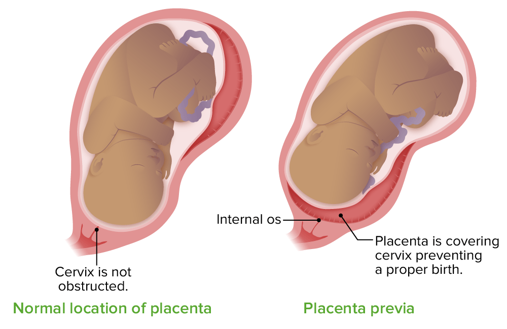
Location of the placenta in placenta previa

Image by Lecturio.

Epidemiology

  • Prevalence Prevalence The total number of cases of a given disease in a specified population at a designated time. It is differentiated from incidence, which refers to the number of new cases in the population at a given time. Measures of Disease Frequency: 4 per 1000 births
  • In up to 2% of women, a midtrimester ultrasound will show placenta previa Placenta Previa Abnormal placentation in which the placenta implants in the lower segment of the uterus (the zone of dilation) and may cover part or all of the opening of the cervix. It is often associated with serious antepartum bleeding and premature labor. Placental Abnormalities.
  • Approximately 90% of placenta Placenta A highly vascularized mammalian fetal-maternal organ and major site of transport of oxygen, nutrients, and fetal waste products. It includes a fetal portion (chorionic villi) derived from trophoblasts and a maternal portion (decidua) derived from the uterine endometrium. The placenta produces an array of steroid, protein and peptide hormones (placental hormones). Placenta, Umbilical Cord, and Amniotic Cavity previas identified at < 20 weeks resolve by delivery.
    • The lower uterine segment lengthens from 0.5 cm at 20 weeks to over 5 cm at term.
    • This lengthening “moves” the placenta Placenta A highly vascularized mammalian fetal-maternal organ and major site of transport of oxygen, nutrients, and fetal waste products. It includes a fetal portion (chorionic villi) derived from trophoblasts and a maternal portion (decidua) derived from the uterine endometrium. The placenta produces an array of steroid, protein and peptide hormones (placental hormones). Placenta, Umbilical Cord, and Amniotic Cavity away from the os.
  • Complete placenta previa Placenta Previa Abnormal placentation in which the placenta implants in the lower segment of the uterus (the zone of dilation) and may cover part or all of the opening of the cervix. It is often associated with serious antepartum bleeding and premature labor. Placental Abnormalities is much less likely to resolve when ≥ 2.5 cm covers the internal os Internal os Uterus, Cervix, and Fallopian Tubes: Anatomy in the midtrimester.

Risk factors

  • Most important risk factors:
    • Previous placenta previa Placenta Previa Abnormal placentation in which the placenta implants in the lower segment of the uterus (the zone of dilation) and may cover part or all of the opening of the cervix. It is often associated with serious antepartum bleeding and premature labor. Placental Abnormalities
    • Previous CD (risk increases with each additional CD)
    • Multiple gestation
  • Other risk factors:
    • Previous curettage Curettage A scraping, usually of the interior of a cavity or tract, for removal of new growth or other abnormal tissue, or to obtain material for tissue diagnosis. It is performed with a curet (curette), a spoon-shaped instrument designed for that purpose. Benign Bone Tumors
    • Previous or recurrent abortions
    • Infertility Infertility Infertility is the inability to conceive in the context of regular intercourse. The most common causes of infertility in women are related to ovulatory dysfunction or tubal obstruction, whereas, in men, abnormal sperm is a common cause. Infertility treatment
    • Advanced maternal age (> 35 years old)
    • Multiparity
    • Uterine structural anomaly 
    • Smoking Smoking Willful or deliberate act of inhaling and exhaling smoke from burning substances or agents held by hand. Interstitial Lung Diseases 
    • Cocaine Cocaine An alkaloid ester extracted from the leaves of plants including coca. It is a local anesthetic and vasoconstrictor and is clinically used for that purpose, particularly in the eye, ear, nose, and throat. It also has powerful central nervous system effects similar to the amphetamines and is a drug of abuse. Cocaine, like amphetamines, acts by multiple mechanisms on brain catecholaminergic neurons; the mechanism of its reinforcing effects is thought to involve inhibition of dopamine uptake. Local Anesthetics use

Clinical presentation

  • Painless vaginal bleeding
  • Fetal distress
  • Usually asymptomatic until cervical dilation Cervical dilation A measurement of the diameter of the cervical canal, reported in centimeters Normal and Abnormal Labor begins after 30 weeks

Diagnosis

  • Transvaginal ultrasonography:
    • Shows placenta Placenta A highly vascularized mammalian fetal-maternal organ and major site of transport of oxygen, nutrients, and fetal waste products. It includes a fetal portion (chorionic villi) derived from trophoblasts and a maternal portion (decidua) derived from the uterine endometrium. The placenta produces an array of steroid, protein and peptide hormones (placental hormones). Placenta, Umbilical Cord, and Amniotic Cavity near or covering the cervical os
    • Usually diagnosed on routine midtrimester ultrasound
    • Assessing placental location is a recommended part of routine obstetric care.
  • Assess fetal well-being with a NST or a period of fetal monitoring Fetal monitoring The primary goals of antepartum testing and monitoring are to assess fetal well-being, identify treatable situations that may cause complications, and evaluate for chromosomal abnormalities. These tests are divided into screening tests (which include cell-free DNA testing, serum analyte testing, and nuchal translucency measurements), and diagnostic tests, which provide a definitive diagnosis of aneuploidy and include chorionic villus sampling (CVS) and amniocentesis. Antepartum Testing and Monitoring
  • Avoid digital exams on a woman with placenta previa Placenta Previa Abnormal placentation in which the placenta implants in the lower segment of the uterus (the zone of dilation) and may cover part or all of the opening of the cervix. It is often associated with serious antepartum bleeding and premature labor. Placental Abnormalities.
Placenta previa on ultrasound

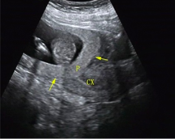
Transabdominal ultrasound showing placenta previa
P = placenta
CX = cervix

Image: “Placenta previa” by Haiyan Yu et al. License: CC BY 4.0

Management

  • All individuals with placenta previa Placenta Previa Abnormal placentation in which the placenta implants in the lower segment of the uterus (the zone of dilation) and may cover part or all of the opening of the cervix. It is often associated with serious antepartum bleeding and premature labor. Placental Abnormalities need to deliver via CD.
  • Asymptomatic antenatal management:
    • Pelvic rest (nothing in the vagina Vagina The vagina is the female genital canal, extending from the vulva externally to the cervix uteri internally. The structures have sexual, reproductive, and urinary functions and a rich blood supply, mainly arising from the internal iliac artery. Vagina, Vulva, and Pelvic Floor: Anatomy, no sexual activity that may lead to orgasm)
    • Avoid strenuous exercise Strenuous exercise Physical activity which is usually regular and done with the intention of improving or maintaining physical fitness or health. Contrast with physical exertion which is concerned largely with the physiologic and metabolic response to energy expenditure. Cardiovascular Response to Exercise and heavy lifting
    • Monitor placental location with serial ultrasounds
    • Plan to deliver at 36‒38 weeks, prior to the onset of labor.
    • When exam is required, visual assessment with a speculum only
  • If bleeding:
    • Assess hemodynamic stability of mother → resuscitate as needed 
    • Continuous fetal monitoring Fetal monitoring The primary goals of antepartum testing and monitoring are to assess fetal well-being, identify treatable situations that may cause complications, and evaluate for chromosomal abnormalities. These tests are divided into screening tests (which include cell-free DNA testing, serum analyte testing, and nuchal translucency measurements), and diagnostic tests, which provide a definitive diagnosis of aneuploidy and include chorionic villus sampling (CVS) and amniocentesis. Antepartum Testing and Monitoring 
    • Corticosteroids Corticosteroids Chorioretinitis for fetal lung maturity (if no emergent delivery)
    • Anti-D immunoglobulin for Rh-negative mothers
    • Indications for delivery (regardless of gestational age Gestational age The age of the conceptus, beginning from the time of fertilization. In clinical obstetrics, the gestational age is often estimated as the time from the last day of the last menstruation which is about 2 weeks before ovulation and fertilization. Pregnancy: Diagnosis, Physiology, and Care):
      • Active labor
      • Nonreassuring fetal monitoring Fetal monitoring The primary goals of antepartum testing and monitoring are to assess fetal well-being, identify treatable situations that may cause complications, and evaluate for chromosomal abnormalities. These tests are divided into screening tests (which include cell-free DNA testing, serum analyte testing, and nuchal translucency measurements), and diagnostic tests, which provide a definitive diagnosis of aneuploidy and include chorionic villus sampling (CVS) and amniocentesis. Antepartum Testing and Monitoring
      • Continued bleeding and/or hemodynamic instability
    • If bleeding is mild and resolves, observation may be an option.

Vasa Previa

Overview

Definitions:

  • Vasa previa: Fetal vessels run across the internal cervical os.
  • Velamentous cord: portion of the umbilical cord Umbilical cord The flexible rope-like structure that connects a developing fetus to the placenta in mammals. The cord contains blood vessels which carry oxygen and nutrients from the mother to the fetus and waste products away from the fetus. Placenta, Umbilical Cord, and Amniotic Cavity lacks the protective Wharton’s jelly near the placental insertion site, making the cord prone to rupture:
    • Known as “membranous vessels” because they are attached to the chorion Chorion The outermost extraembryonic membrane surrounding the developing embryo. In reptiles and birds, it adheres to the shell and allows exchange of gases between the egg and its environment. In mammals, the chorion evolves into the fetal contribution of the placenta. Placenta, Umbilical Cord, and Amniotic Cavity
    • Length of unprotected cord may be several centimeters long

Clinical significance:
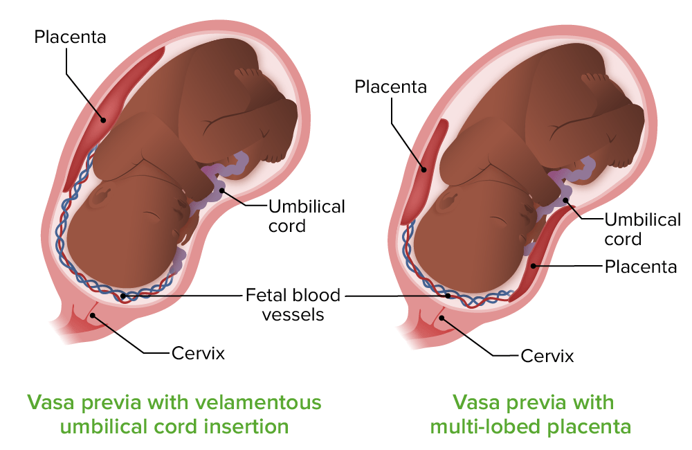
  • A digital exam may rupture these vessels, causing fetal exsanguination within minutes.
  • Rupture of membranes may rupture the vessels.
  • These vessels may also become compressed by the fetal head, leading to fetal distress.
Vasa previa

Vasa previa:
On the left, the vessels in the umbilical cord run directly over the internal cervical os (vasa previa) and lack the protective Wharton jelly (velamentous cord insertion). On the right, the vasa previa vessels run to an accessory lobe of the placenta; the vessels also lack Wharton jelly.

Image by Lecturio.

Epidemiology

  • Prevalence Prevalence The total number of cases of a given disease in a specified population at a designated time. It is differentiated from incidence, which refers to the number of new cases in the population at a given time. Measures of Disease Frequency
    • Velamentous cord:
      • 1% of singleton gestations
      • Up to 15% of monochorionic twin gestations
    • Vasa previa: 
      • 1 in 1500 deliveries
      • As high as 1 in 200 in pregnancies conceived by in vitro fertilization Fertilization To undergo fertilization, the sperm enters the uterus, travels towards the ampulla of the fallopian tube, and encounters the oocyte. The zona pellucida (the outer layer of the oocyte) deteriorates along with the zygote, which travels towards the uterus and eventually forms a blastocyst, allowing for implantation to occur. Fertilization and First Week ( IVF IVF An assisted reproductive technique that includes the direct handling and manipulation of oocytes and sperm to achieve fertilization in vitro. Infertility)
  • Risk factors: 
    • Accessory (succenturiate) placental lobes
    • Multiple gestation
    • Placenta previa Placenta Previa Abnormal placentation in which the placenta implants in the lower segment of the uterus (the zone of dilation) and may cover part or all of the opening of the cervix. It is often associated with serious antepartum bleeding and premature labor. Placental Abnormalities
    • Velamentous cord insertion Velamentous cord insertion The last few centimeters of the umbilical cord entering the placenta lack the protective Wharton’s jelly. The vessels are “exposed” and covered only by the fetal membranes Placental Abnormalities
    • IVF IVF An assisted reproductive technique that includes the direct handling and manipulation of oocytes and sperm to achieve fertilization in vitro. Infertility

Clinical presentation

  • Painless vaginal bleeding (often after rupture of membranes)
  • Fetal distress:
    • Blood loss from the fetus (much lower blood volumes)
    • Fetal heart abnormalities, including a sinusoidal pattern (indicates severe fetal anemia Anemia Anemia is a condition in which individuals have low Hb levels, which can arise from various causes. Anemia is accompanied by a reduced number of RBCs and may manifest with fatigue, shortness of breath, pallor, and weakness. Subtypes are classified by the size of RBCs, chronicity, and etiology. Anemia: Overview and Types)
    • Fetal death due to exsanguination (can occur within minutes)
  • Unlike placenta previa Placenta Previa Abnormal placentation in which the placenta implants in the lower segment of the uterus (the zone of dilation) and may cover part or all of the opening of the cervix. It is often associated with serious antepartum bleeding and premature labor. Placental Abnormalities, vasa previa usually persists throughout pregnancy Pregnancy The status during which female mammals carry their developing young (embryos or fetuses) in utero before birth, beginning from fertilization to birth. Pregnancy: Diagnosis, Physiology, and Care.
  • Women are often asymptomatic until cervical dilation Cervical dilation A measurement of the diameter of the cervical canal, reported in centimeters Normal and Abnormal Labor or disruption due to mechanical trauma.

Diagnosis

  • Transvaginal ultrasound Transvaginal Ultrasound Obstetric Imaging with Doppler Doppler Ultrasonography applying the doppler effect, with frequency-shifted ultrasound reflections produced by moving targets (usually red blood cells) in the bloodstream along the ultrasound axis in direct proportion to the velocity of movement of the targets, to determine both direction and velocity of blood flow. Ultrasound (Sonography)
    • Demonstrates fetal vessels traversing the internal cervical os
    • Assessing the umbilical cord Umbilical cord The flexible rope-like structure that connects a developing fetus to the placenta in mammals. The cord contains blood vessels which carry oxygen and nutrients from the mother to the fetus and waste products away from the fetus. Placenta, Umbilical Cord, and Amniotic Cavity vessels and insertion site at around 20 weeks is a recommended element of routine prenatal care Prenatal care Prenatal care is a systematic and periodic assessment of pregnant women during gestation to assure the best health outcome for the mother and her fetus. Prenatal care prevents and identifies maternal and fetal problems that adversely affect the pregnancy outcome. Prenatal Care.
  • Avoid digital exam on a woman with vasa previa.
Doppler ultrasound of vasa previa

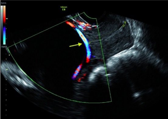
Transvaginal ultrasound showing a vasa previa:
The color Doppler shows blood flowing through the vasa previa vessel over the internal cervical os. The cervical canal is marked with the yellow asterisks on each end.

Image: “Doppler ultrasound of vasa previa” by Division of Reproductive Endocrinology & Infertility, Beth Israel Deaconess Medical Center/Boston IVF, Harvard Medical School, Boston, MA, USA. License: CC BY 3.0

Management

  • Delivery is by CD.
  • Asymptomatic antenatal management:
    • Pelvic rest
    • Corticosteroids Corticosteroids Chorioretinitis for fetal lung maturity (typically between 28‒32 weeks)
    • Inpatient observation with daily fetal monitoring Fetal monitoring The primary goals of antepartum testing and monitoring are to assess fetal well-being, identify treatable situations that may cause complications, and evaluate for chromosomal abnormalities. These tests are divided into screening tests (which include cell-free DNA testing, serum analyte testing, and nuchal translucency measurements), and diagnostic tests, which provide a definitive diagnosis of aneuploidy and include chorionic villus sampling (CVS) and amniocentesis. Antepartum Testing and Monitoring starting around 30‒34 weeks
    • Plan to deliver at 34‒37 weeks, prior to the onset of labor.
    • When exam is required, visual assessment with a speculum only
  • Indications for emergent CD:
    • Labor
    • Rupture of membranes
    • Nonreassuring fetal monitoring Fetal monitoring The primary goals of antepartum testing and monitoring are to assess fetal well-being, identify treatable situations that may cause complications, and evaluate for chromosomal abnormalities. These tests are divided into screening tests (which include cell-free DNA testing, serum analyte testing, and nuchal translucency measurements), and diagnostic tests, which provide a definitive diagnosis of aneuploidy and include chorionic villus sampling (CVS) and amniocentesis. Antepartum Testing and Monitoring
    • Vaginal bleeding with fetal tachycardia Tachycardia Abnormally rapid heartbeat, usually with a heart rate above 100 beats per minute for adults. Tachycardia accompanied by disturbance in the cardiac depolarization (cardiac arrhythmia) is called tachyarrhythmia. Sepsis in Children

Uterine Rupture

Definition

Uterine rupture is a clinically significant disruption in all layers of the uterus Uterus The uterus, cervix, and fallopian tubes are part of the internal female reproductive system. The uterus has a thick wall made of smooth muscle (the myometrium) and an inner mucosal layer (the endometrium). The most inferior portion of the uterus is the cervix, which connects the uterine cavity to the vagina. Uterus, Cervix, and Fallopian Tubes: Anatomy, usually through a previous uterine scar Scar Dermatologic Examination.
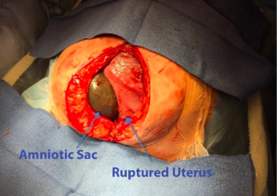
Epidemiology

  • Incidence Incidence The number of new cases of a given disease during a given period in a specified population. It also is used for the rate at which new events occur in a defined population. It is differentiated from prevalence, which refers to all cases in the population at a given time. Measures of Disease Frequency in individuals with a prior CD: 
    • Women attempting a trial of labor after cesarean (TOLAC): 0.5%
    • Women with a prior CD delivering via planned, repeat CD Repeat CD Extraction of the fetus by abdominal hysterotomy anytime following a previous cesarean. Cesarean Delivery: 0.03%
  • Incidence Incidence The number of new cases of a given disease during a given period in a specified population. It also is used for the rate at which new events occur in a defined population. It is differentiated from prevalence, which refers to all cases in the population at a given time. Measures of Disease Frequency in individuals without a prior uterine scar Scar Dermatologic Examination
    • Rare
    • Estimated at 1 per 20,000 pregnancies
  • Perinatal death Perinatal death The death of a fetus of gestational age 28 weeks or more, or the death of a live-born infant less than 28 days of age. Congenital TORCH Infections rate associated with uterine rupture: 5%–25%

Risk factors

  • Prior uterine incision:
    • Prior CD
    • Classical (high vertical or fundal) uterine incision (highest risk)
    • Myomectomy of large intramural fibroids Fibroids A benign tumor derived from smooth muscle tissue, also known as a fibroid tumor. They rarely occur outside of the uterus and the gastrointestinal tract but can occur in the skin and subcutaneous tissue, probably arising from the smooth muscle of small blood vessels in these tissues. Infertility
  • TOLAC
  • Induction and augmentation of labor (excessive oxytocin stimulation)

Clinical presentation

  • Abdominal pain Abdominal Pain Acute Abdomen (often severe and with sudden onset)
  • Vaginal bleeding (may be light if a majority of blood is lost intraabdominally)
  • Nonreassuring fetal heart rate Heart rate The number of times the heart ventricles contract per unit of time, usually per minute. Cardiac Physiology patterns:
    • Bradycardia Bradycardia Bradyarrhythmia is a rhythm in which the heart rate is less than 60/min. Bradyarrhythmia can be physiologic, without symptoms or hemodynamic change. Pathologic bradyarrhythmia results in reduced cardiac output and hemodynamic instability causing syncope, dizziness, or dyspnea. Bradyarrhythmias
    • Late decelerations
    • Minimal or absent fetal heart rate Heart rate The number of times the heart ventricles contract per unit of time, usually per minute. Cardiac Physiology variability
  • Loss of uterine tone
  • Sudden loss of station of the presenting fetal part
  • Maternal hemodynamic instability
  • May be diagnosed postpartum after a TOLAC with a postpartum hemorrhage Postpartum hemorrhage Postpartum hemorrhage is one of the most common and deadly obstetric complications. Since 2017, postpartum hemorrhage has been defined as blood loss greater than 1,000 mL for both cesarean and vaginal deliveries, or excessive blood loss with signs of hemodynamic instability. Postpartum Hemorrhage that does not respond to uterotonic agents

Diagnosis

  • The diagnosis is clinical, based on presentation and risk factors.
  • Diagnosis is confirmed on laparotomy Laparotomy Incision into the side of the abdomen between the ribs and pelvis. Laparotomy and Laparoscopy.

Management

  • Significant fetal and maternal distress usually necessitate immediate action:
    • Immediate emergency CD
    • Concurrent maternal stabilization with IV fluids IV fluids Intravenous fluids are one of the most common interventions administered in medicine to approximate physiologic bodily fluids. Intravenous fluids are divided into 2 categories: crystalloid and colloid solutions. Intravenous fluids have a wide variety of indications, including intravascular volume expansion, electrolyte manipulation, and maintenance fluids. Intravenous Fluids and blood products, as needed
  • Surgical repair of the uterine rupture, following delivery of the infant and maternal stabilization
  • Hysterectomy (last resort in cases of persistent hemorrhage and/or nonrepairable uterus Uterus The uterus, cervix, and fallopian tubes are part of the internal female reproductive system. The uterus has a thick wall made of smooth muscle (the myometrium) and an inner mucosal layer (the endometrium). The most inferior portion of the uterus is the cervix, which connects the uterine cavity to the vagina. Uterus, Cervix, and Fallopian Tubes: Anatomy)
Uterine rupture photograph

Uterine rupture:
This woman has undergone a laparotomy through a vertical midline incision. Within the abdominal cavity, a bulging amniotic sac is noted through a large defect in the anterior uterine wall.

Image: “Silent uterine rupture” by Department of Obstetrics and Gynecology, Michigan State University/Sparrow Hospital, Lansing, MI 48912, USA. License: CC BY 3.0

Comparison of Diagnoses

Table: Comparison of causes of antepartum bleeding
Condition Typical presentation Important risk factors Typical management
Placental abruption
  • Painful bleeding
  • Contractions
  • Nonreassuring fetal status
  • Hypertension Hypertension Hypertension, or high blood pressure, is a common disease that manifests as elevated systemic arterial pressures. Hypertension is most often asymptomatic and is found incidentally as part of a routine physical examination or during triage for an unrelated medical encounter. Hypertension
  • Rapid uterine decompression
  • Trauma
  • Cocaine Cocaine An alkaloid ester extracted from the leaves of plants including coca. It is a local anesthetic and vasoconstrictor and is clinically used for that purpose, particularly in the eye, ear, nose, and throat. It also has powerful central nervous system effects similar to the amphetamines and is a drug of abuse. Cocaine, like amphetamines, acts by multiple mechanisms on brain catecholaminergic neurons; the mechanism of its reinforcing effects is thought to involve inhibition of dopamine uptake. Local Anesthetics or tobacco use
Emergent delivery
Placenta previa Placenta Previa Abnormal placentation in which the placenta implants in the lower segment of the uterus (the zone of dilation) and may cover part or all of the opening of the cervix. It is often associated with serious antepartum bleeding and premature labor. Placental Abnormalities Painless bleeding OR asymptomatic
  • Prior placenta previa Placenta Previa Abnormal placentation in which the placenta implants in the lower segment of the uterus (the zone of dilation) and may cover part or all of the opening of the cervix. It is often associated with serious antepartum bleeding and premature labor. Placental Abnormalities
  • Prior cesarean delivery Cesarean Delivery Cesarean delivery (CD) is the operative delivery of ≥ 1 infants through a surgical incision in the maternal abdomen and uterus. Cesarean deliveries may be indicated for a number of either maternal or fetal reasons, most commonly including fetal intolerance to labor, arrest of labor, a history of prior uterine surgery, fetal malpresentation, and placental abnormalities. Cesarean Delivery
  • Multiple gestation
  • No digital exams
  • Pelvic rest
  • Deliver with bleeding, or at 36‒38 weeks.
Vasa previa Painless bleeding OR asymptomatic
  • Accessory placental lobe
  • Current placenta previa Placenta Previa Abnormal placentation in which the placenta implants in the lower segment of the uterus (the zone of dilation) and may cover part or all of the opening of the cervix. It is often associated with serious antepartum bleeding and premature labor. Placental Abnormalities
  • Multiple gestation
  • Velamentous cord
  • No digital exams
  • Pelvic rest
  • Deliver with bleeding, or at 34–37 weeks.
Uterine rupture
  • Painful bleeding
  • Loss of fetal station Fetal station Refers to how high (or low) the presenting fetal part is compared to the maternal ischial spine, reported in centimeters Normal and Abnormal Labor
  • Nonreassuring fetal status
  • Prior uterine incision (especially vertical or fundal incisions)
  • TOLAC
  • Induction of labor
  • Emergent delivery
  • Surgical repair of rupture

References

  1. Ananth, C.V., Kinzler, W.L. (2021). Placental abruption: Pathophysiology, clinical features, diagnosis, and consequences. In: Barss, V.A. (Ed.), UpToDate. Retrieved March 12, 2021, from https://www.uptodate.com/contents/placental-abruption-pathophysiology-clinical-features-diagnosis-and-consequences
  2. Deering, S. (2018). Abruptio placentae. Medscape. Retrieved March 12, 2021, from https://emedicine.medscape.com/article/252810-overview 
  3. Lockwood, C.J. (2019). Placenta previa: Epidemiology, clinical features, diagnosis, morbidity, and mortality. UpToDate. Retrieved March 11, 2021, from https://www.uptodate.com/contents/placenta-previa-epidemiology-clinical-features-diagnosis-morbidity-and-mortality
  4. Bakker, R. (2018). Placenta previa. Medscape. Retrieved March 12, 2021, from https://emedicine.medscape.com/article/262063-overview#a4 
  5. Anderson-Bagg, F. (2020). Placenta previa. StatPearls. Retrieved March 11, 2021, from https://www.statpearls.com/articlelibrary/viewarticle/27262/ 
  6. Lockwood, C.J. (2020). Velamentous umbilical cord insertion and vasa previa. UpToDate. Retrieved March 12, 2021, from https://www.uptodate.com/contents/velamentous-umbilical-cord-insertion-and-vasa-previa 
  7. Landon, M.B. (2020). Uterine rupture: after previous cesarean delivery. UpToDate. Retrieved March 12, 2021, from https://www.uptodate.com/contents/uterine-rupture-after-previous-cesarean-delivery

Create your free account or log in to continue reading!

Sign up now and get free access to Lecturio with concept pages, medical videos, and questions for your medical education.

User Reviews

Details